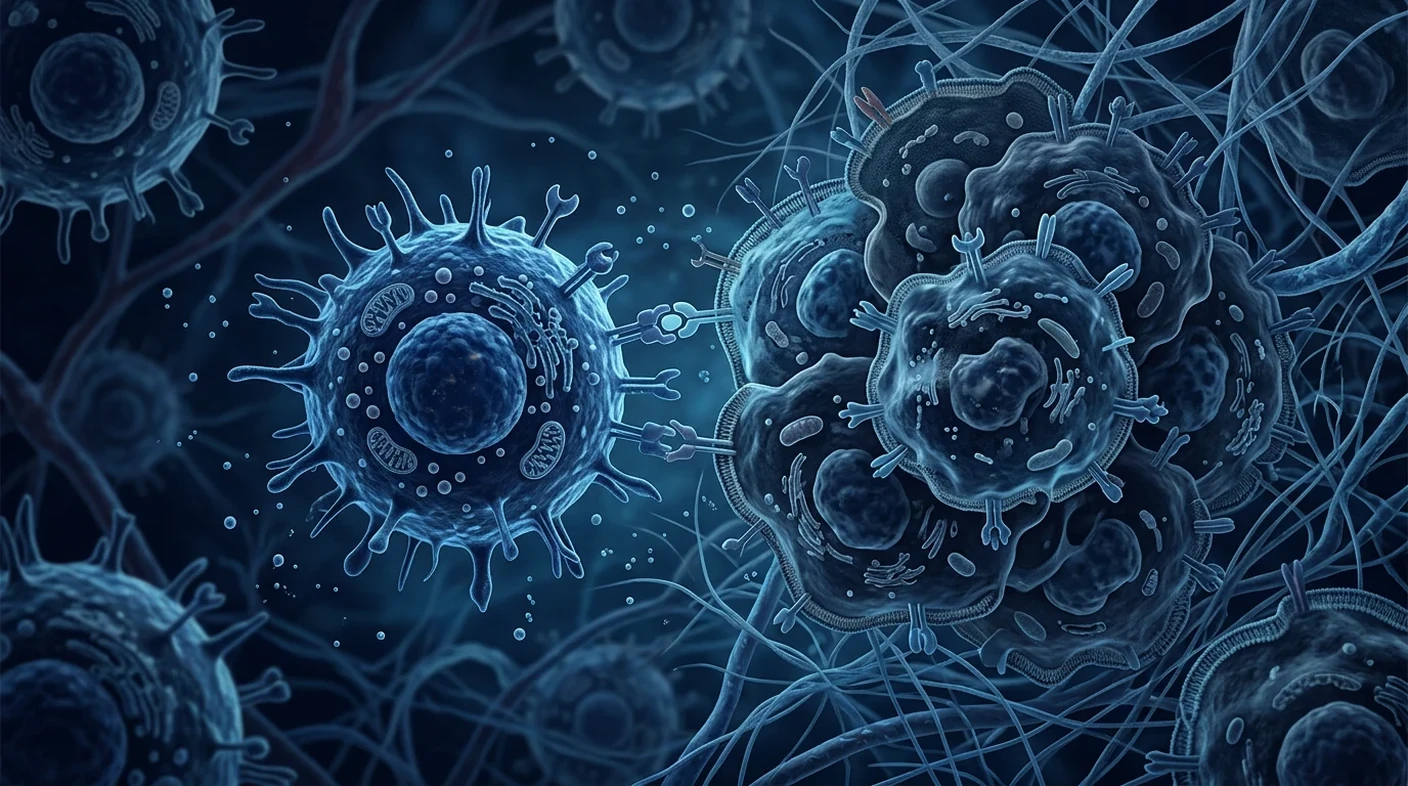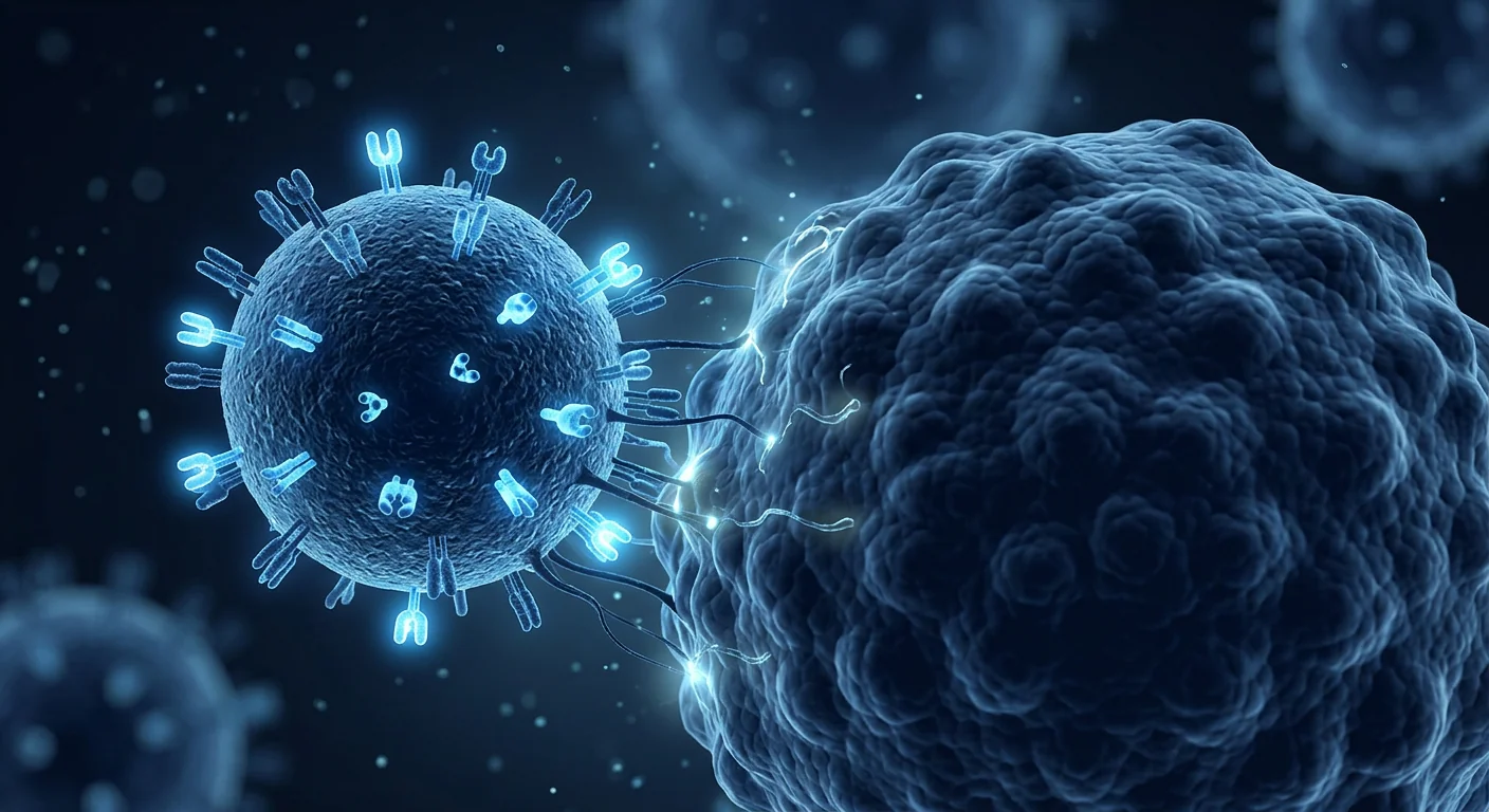
An NK cell activity test is a laboratory blood test that measures how well a person's natural killer cells perform a function — usually killing target cells or releasing interferon-gamma — rather than simply counting how many NK cells are present. It is a functional immune assay, and in most countries it is used for research, immunodeficiency work-up, and monitoring, not as a stand-alone cancer test.
Counting is not the same as testing function. A standard lymphocyte panel reports the number of CD3-negative, CD56-positive cells in a millilitre of blood. That number says nothing about whether those cells can recognise a stressed cell, form a synapse and kill it.
Function is what the biology actually depends on. Natural killer cells were defined by their ability to kill certain tumour cell lines without prior sensitisation [1], and their clinical relevance rests on that cytotoxic and cytokine-producing capacity [2].
The gap between plausible and proven is where most confusion sits. Functional NK testing is scientifically real and methodologically well described. What is not established is that a single NK activity number from a commercial panel can screen a healthy person for cancer, grade their "immune age", or justify an unproven cell therapy.
The one-sentence version
NK cell activity testing measures a genuine biological property, but a single result in one person is influenced by age, recent infection, stress, sleep, medication and the assay platform itself — so it should be read as one contextual data point, never as a diagnosis.
What is an NK cell activity test?
An NK cell activity test quantifies the functional output of natural killer cells from a fresh blood sample, most often as target-cell killing, degranulation, or interferon-gamma production after standardised stimulation. The output is a percentage, an index, or a cytokine concentration — units that differ between laboratories and are not interchangeable.
Natural killer cells are innate lymphocytes that act without the antigen-specific priming that T cells require. They patrol for cells that have lost normal HLA class I display or that express stress ligands, and they respond with cytotoxic granule release and cytokine secretion [2].
The population is not uniform. CD56bright cells are predominantly cytokine producers, while CD56dim CD16+ cells are the main cytotoxic subset [3]. Deep phenotyping has since shown that human NK compartments are extraordinarily diverse between individuals [4]. A single activity figure compresses all of that into one number.
How is NK cell activity measured in the laboratory?
NK cell function is measured by four main approaches: chromium-release or fluorescent cytotoxicity assays, degranulation marker assays, cytokine-release assays, and multiparameter flow cytometry of receptor expression. Each answers a slightly different question.
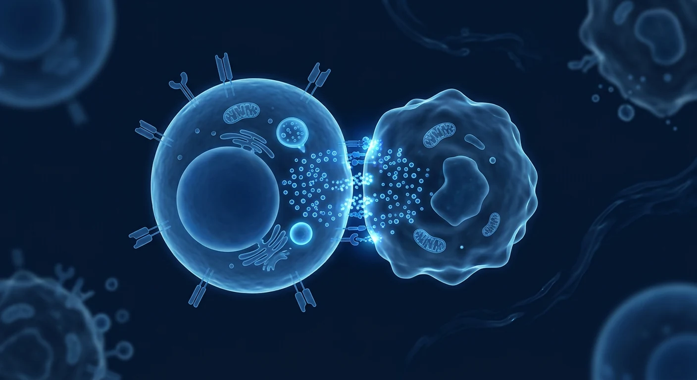
- Cytotoxicity assays. Effector cells are incubated with a standard target cell line (classically K562) at defined effector-to-target ratios, and killing is quantified by isotope or fluorescent dye release. This is the closest measure to "can these cells kill".
- Degranulation assays. Surface CD107a appears when cytotoxic granules fuse with the membrane, making it a validated single-cell marker of NK activation [6].
- Cytokine-release assays. Whole blood is stimulated and secreted interferon-gamma is measured by immunoassay. This is the format used by several commercially available "NK activity" panels [8].
- Flow cytometric functional panels. Standardised protocols combine degranulation, cytokine staining and receptor phenotyping to characterise NK function in clinical immunology laboratories [7].
Two practical points matter more than patients expect. First, results are platform-specific: a percentage from a K562 cytotoxicity assay cannot be compared with an interferon-gamma concentration from a whole-blood stimulation kit. Second, these are live-cell assays, so sample handling time, temperature and transport directly influence the result.
What does a low NK cell activity result mean?
A low NK cell activity result means the sample performed below the laboratory's reference range on that specific assay on that specific day — it is not, by itself, a diagnosis of immune deficiency or cancer. Interpretation requires clinical context, repeat testing, and awareness of what else was happening in the body when blood was drawn.
Genuine NK cell deficiency exists and is clinically important. Classical and functional NK cell deficiencies present with characteristic severe herpesvirus infections and are diagnosed through defined immunological criteria, not through a single commercial activity number [11]. Study of these rare patients has clarified what NK cells actually do in human health [12].
Conversely, transiently reduced activity is common and usually unremarkable. Acute viral infection redistributes and activates NK cells [13], and ageing shifts subset composition and function [14]. Neither finding makes an individual result a disease marker.
Does low NK cell activity predict cancer?
Population-level research has linked lower natural cytotoxic activity with higher subsequent cancer incidence, but that association does not make NK activity testing a validated individual cancer screening test. This distinction is the single most misused point in NK activity marketing.
The most cited evidence is an 11-year Japanese cohort study in which baseline peripheral-blood natural cytotoxic activity was associated with cancer incidence across a general population [9]. It is a well-conducted epidemiological observation about groups, not a validated predictive tool for a named individual.
Related work reinforces biological plausibility without closing the gap. A systematic review and meta-analysis found that NK-cell infiltration within tumour tissue was associated with improved overall survival across solid cancers [10] — a finding about tumour tissue, not about a circulating activity number. Interferon-gamma-based NK activity has been reported as a supportive diagnostic marker in gastric cancer, explicitly as an adjunct rather than a replacement for diagnostic pathways [8].
What follows practically: an NK activity result must never be used to rule out cancer, to postpone colonoscopy, mammography, HPV testing, low-dose CT or any other guideline-recommended screening, or to justify treatment in an asymptomatic person. The same caution applies to other blood-based immune or tumour-associated assays, as discussed in our article on EDIM compared with liquid biopsy and standard screening.
NK cell activity versus NK cell count: which is more informative?
Count and activity answer different questions, and a normal count can coexist with reduced function — which is precisely why functional assays exist. The comparison below summarises what each measurement can and cannot support.
| Measurement | What it reports | Main limitation |
|---|---|---|
| NK cell count (CD3−CD56+) | Number of circulating NK cells | Says nothing about killing capacity |
| Cytotoxicity assay | Target-cell killing at set ratios | Live-cell assay; handling-sensitive |
| CD107a degranulation [6] | Granule release per cell | Degranulation is not always killing |
| Interferon-gamma release | Cytokine output after stimulation | Reflects stimulation conditions too |
| Receptor phenotyping [4] | Subset and receptor composition | Highly variable between individuals |
For most clinical questions, the useful comparison is not "which number is higher" but "which measurement was validated for the question being asked".
Why do NK activity results vary so much between tests?
NK cell activity varies because it is a live biological function shaped by genetics, environment, recent exposures and pre-analytical handling — repeat testing under identical conditions is the only way to see a real trend.

Mass cytometry work has shown that human NK-cell diversity is driven by both genetic and environmental determinants, with environmental influence substantial [5]. On top of that baseline diversity, the following shift individual results:
- Recent or ongoing infection, which activates and redistributes NK cells [13]
- Age, which alters subset balance and functional output [14]
- Medication, particularly corticosteroids and immunosuppressants
- Acute physical or psychological stress and sleep loss
- Time from venepuncture to assay, temperature and transport conditions
- Assay platform, target cell line, and effector-to-target ratio
The practical consequence: a single result is weak evidence. If NK activity is being tracked, it should be the same assay, the same laboratory, similar time of day, and away from acute illness.
When is NK activity testing genuinely useful?
NK functional testing has a defined role in immunodeficiency evaluation, in research, and in monitoring cell-therapy products — and a much weaker role as a general wellness or cancer-screening panel.
- Suspected NK cell deficiency. In patients with severe, recurrent or unusual herpesvirus infections, functional testing forms part of a specialist immunological work-up [11].
- Research and clinical trials. Functional readouts are standard endpoints in immunology studies using validated protocols [7].
- Cell-therapy product testing. Potency assays for manufactured NK products are a regulatory requirement, not a patient blood test — the distinction is covered in our article on NK cell manufacturing and quality control.
- Longitudinal monitoring in a defined clinical context, where the same assay is repeated to observe a trend rather than to produce a one-off verdict.
Outside those settings, a stand-alone NK activity number rarely changes correct management.
How does NK activity testing relate to NK cell therapy?
A patient's own NK activity result does not establish eligibility for, or predict response to, NK cell therapy — the two are separate questions with separate evidence bases. Adoptive NK-cell therapy remains largely investigational outside defined transplant contexts [15][16].
Using a low activity result as the rationale for selling an infusion is a marketing inference, not a clinical one. Product characteristics — source, expansion method, potency, dose, and trial registration — determine what is being given, as explained in our NK cell therapy patient guide and the comparison of autologous and allogeneic NK cell sources.
If a programme in Bangkok or elsewhere offers an activity test and an infusion as one package, the reasonable question is what decision the test result could actually change, and what happens if the number is normal.
What should patients in Bangkok ask before ordering an NK activity test?
Before paying for NK cell activity testing in Bangkok, patients should establish which assay is used, what clinical question it answers, and what will be done differently depending on the result.
- Which specific assay is performed, and in which accredited laboratory?
- What is the reference range, and how was it established for this population?
- What clinical decision would a low result change, and what would a normal result change?
- How soon after collection is the sample processed?
- Is the test being offered as part of a treatment package, and what is the financial relationship?
- Which guideline-recommended screening tests remain necessary regardless of the result?
A laboratory that can answer these questions in writing is behaving appropriately. A programme that presents the number as a cancer risk score is overstating what the assay supports.
Frequently asked questions
In short: NK cell activity testing measures a real immune function, varies substantially with day-to-day physiology and assay method, and cannot replace guideline-recommended cancer screening or diagnose disease on its own.
Can an NK cell activity test detect cancer?
No. Population studies have associated lower natural cytotoxic activity with higher cancer incidence at group level [9], but no NK activity assay is validated as an individual cancer detection or screening test. Standard screening remains necessary.
What is a normal NK cell activity level?
There is no universal number. Each laboratory defines a reference range for its own assay, target cell line and stimulation conditions, so results from different providers cannot be compared directly.
Can I raise my NK cell activity with supplements?
Many products claim this, and the supporting evidence is generally small, short, heterogeneous, and measured on surrogate laboratory endpoints rather than health outcomes. An assay change is not proof of clinical benefit.
Does stress or poor sleep lower NK activity?
Acute stress and sleep deprivation are associated with measurable short-term changes in NK function, which is one reason a single reading should not be over-interpreted. Repeat testing under stable conditions is more informative.
Is NK cell activity testing the same as an EDIM test?
No. They measure different things: NK activity assays measure immune-cell function, while the EDIM test measures Apo10 and TKTL1 epitopes within monocytes. Both are adjunctive and neither is a stand-alone cancer diagnosis — see our EDIM blood test patient guide.
Should I get NK activity testing if I feel healthy?
For most asymptomatic people it will not change correct medical management. It is more defensible when there is a specific clinical question, such as evaluation of unusual recurrent infections, under specialist supervision.
Limitations and medical disclaimer
The limitations of NK activity testing are methodological, not merely cautionary. Assay platforms are not standardised across laboratories; reference ranges are population- and method-specific; results are sensitive to pre-analytical handling; intra-individual variation over days to weeks can be substantial; and outcome evidence linking a single measurement to individual prognosis or treatment benefit is limited.
This article is educational and does not describe a VELAR diagnostic or treatment protocol. It is not medical advice and does not replace assessment by a qualified physician. NK cell activity testing must not be used to exclude cancer or to delay or replace guideline-recommended screening, diagnosis or treatment. Patients considering any cell therapy should discuss evidence, eligibility, registration status, monitoring and documented risks with a qualified specialist.
References
- Kiessling R, Klein E, Wigzell H. "Natural" killer cells in the mouse. I. Cytotoxic cells with specificity for mouse Moloney leukemia cells. European Journal of Immunology. 1975;5(2):112-117. doi:10.1002/eji.1830050208 ↩
- Vivier E, Tomasello E, Baratin M, Walzer T, Ugolini S. Functions of natural killer cells. Nature Immunology. 2008;9(5):503-510. doi:10.1038/ni1582 ↩
- Cooper MA, Fehniger TA, Caligiuri MA. The biology of human natural killer-cell subsets. Trends in Immunology. 2001;22(11):633-640. doi:10.1016/S1471-4906(01)02060-9 ↩
- Freud AG, Mundy-Bosse BL, Yu J, Caligiuri MA. The broad spectrum of human natural killer cell diversity. Immunity. 2017;47(5):820-833. doi:10.1016/j.immuni.2017.10.008 ↩
- Horowitz A, Strauss-Albee DM, Leipold M, et al. Genetic and environmental determinants of human NK cell diversity revealed by mass cytometry. Science Translational Medicine. 2013;5(208):208ra145. doi:10.1126/scitranslmed.3006702 ↩
- Alter G, Malenfant JM, Altfeld M. CD107a as a functional marker for the identification of natural killer cell activity. Journal of Immunological Methods. 2004;294(1-2):15-22. doi:10.1016/j.jim.2004.08.008 ↩
- Bryceson YT, Fauriat C, Nunes JM, et al. Functional analysis of human NK cells by flow cytometry. Methods in Molecular Biology. 2010;612:335-352. doi:10.1007/978-1-60761-362-6_23 ↩
- Lee J, Park KH, Ryu JH, et al. Natural killer cell activity for IFN-gamma production as a supportive diagnostic marker for gastric cancer. Oncotarget. 2017;8(41):70431-70440. doi:10.18632/oncotarget.19712 ↩
- Imai K, Matsuyama S, Miyake S, Suga K, Nakachi K. Natural cytotoxic activity of peripheral-blood lymphocytes and cancer incidence: an 11-year follow-up study of a general population. The Lancet. 2000;356(9244):1795-1799. doi:10.1016/S0140-6736(00)03231-1 ↩
- Nersesian S, Schwartz SL, Grantham SR, et al. NK cell infiltration is associated with improved overall survival in solid cancers: a systematic review and meta-analysis. Translational Oncology. 2021;14(1):100930. doi:10.1016/j.tranon.2020.100930 ↩
- Orange JS. Natural killer cell deficiency. Journal of Allergy and Clinical Immunology. 2013;132(3):515-525. doi:10.1016/j.jaci.2013.07.020 ↩
- Mace EM, Orange JS. Emerging insights into human health and NK cell biology from the study of NK cell deficiencies. Immunological Reviews. 2018;287(1):202-225. doi:10.1111/imr.12725 ↩
- Björkström NK, Strunz B, Ljunggren HG. Natural killer cells in antiviral immunity. Nature Reviews Immunology. 2022;22(2):112-123. doi:10.1038/s41577-021-00558-3 ↩
- Solana R, Campos C, Pera A, Tarazona R. Shaping of NK cell subsets by aging. Current Opinion in Immunology. 2014;29:56-61. doi:10.1016/j.coi.2014.04.002 ↩
- Shimasaki N, Jain A, Campana D. NK cells for cancer immunotherapy. Nature Reviews Drug Discovery. 2020;19:200-218. doi:10.1038/s41573-019-0052-1 ↩
- Myers JA, Miller JS. Exploring the NK cell platform for cancer immunotherapy. Nature Reviews Clinical Oncology. 2021;18:85-100. doi:10.1038/s41571-020-0426-7 ↩
NK 细胞活性检测是一项实验室血液检测,衡量的是受检者自然杀伤细胞的“功能表现”——通常是杀伤靶细胞或释放干扰素-γ的能力——而不仅仅是清点血液里有多少个 NK 细胞。它属于功能性免疫检测;在多数国家,它用于科研、免疫缺陷评估与随访监测,而不是作为独立的癌症检测手段。
计数与测功能并不是同一件事。常规淋巴细胞分型报告的是每毫升血中 CD3 阴性、CD56 阳性细胞的数量。这个数字并不能说明这些细胞是否还能识别异常细胞、形成杀伤突触并完成杀伤。
生物学真正依赖的是功能。自然杀伤细胞最初被定义的依据,正是它们无需事先致敏即可杀伤某些肿瘤细胞系的能力 [1];其临床意义也建立在这种细胞毒性与细胞因子分泌能力之上 [2]。
“合理”与“已证实”之间的差距,正是大部分误解所在。NK 功能检测在方法学上真实且有充分文献描述。尚未确立的是:商业化套餐中的单一 NK 活性数值,能否为健康人筛查癌症、评定所谓“免疫年龄”,或用来支持一项尚未获证的细胞治疗。
一句话版本
NK 细胞活性检测测量的是一项真实的生物学性质,但单次结果会受年龄、近期感染、压力、睡眠、用药以及检测平台本身影响——因此只能当作一个需要结合临床情境解读的数据点,绝不能当作诊断。
什么是 NK 细胞活性检测?
NK 细胞活性检测用新鲜血液样本量化自然杀伤细胞的功能输出,常见形式是靶细胞杀伤率、脱颗粒比例,或标准化刺激后的干扰素-γ分泌量。结果可能是百分比、指数或细胞因子浓度——这些单位在不同实验室之间并不相同,也不可互换。
自然杀伤细胞属于先天淋巴细胞,不需要 T 细胞所依赖的抗原特异性预致敏。它们巡查那些 HLA I 类分子表达丢失或表达应激配体的细胞,并以细胞毒颗粒释放与细胞因子分泌作出反应 [2]。
这一细胞群并不均一。CD56bright 亚群以分泌细胞因子为主,而 CD56dim CD16+ 亚群才是主要的细胞毒性亚群 [3]。此后的深度表型研究进一步显示,人类 NK 细胞组成在个体之间差异极大 [4]。单一活性数值把这一切压缩成了一个数字。
实验室如何测量 NK 细胞活性?
NK 功能测量主要有四条路径:铬释放或荧光法细胞毒性试验、脱颗粒标志物检测、细胞因子释放检测,以及受体表达的多参数流式细胞术。它们回答的问题略有不同。
- 细胞毒性试验。将效应细胞与标准靶细胞系(经典为 K562)按设定的效靶比共培养,通过同位素或荧光染料释放量化杀伤。这是最接近“这些细胞还能不能杀”的测量。
- 脱颗粒检测。细胞毒颗粒与细胞膜融合时,表面出现 CD107a,因此它是经过验证的单细胞 NK 活化标志 [6]。
- 细胞因子释放检测。刺激全血后用免疫测定法检测分泌的干扰素-γ。多个商业化“NK 活性”套餐采用这一格式 [8]。
- 流式功能组合。标准化方案把脱颗粒、细胞因子染色与受体表型结合,用于临床免疫实验室刻画 NK 功能 [7]。
有两点比患者通常预想的更重要。第一,结果与平台绑定:K562 细胞毒性试验的百分比,无法与全血刺激试剂盒的干扰素-γ浓度相比较。第二,这些都是活细胞试验,样本处理时间、温度与运输条件会直接影响结果。
NK 细胞活性偏低意味着什么?
活性偏低只意味着该样本在那一天、该项特定检测上低于该实验室的参考范围——它本身既不是免疫缺陷的诊断,也不是癌症的诊断。解读需要临床情境、复查,以及了解抽血当时身体还发生了什么。
真正的 NK 细胞缺陷确实存在且具临床意义。经典型与功能型 NK 细胞缺陷会表现为特征性的严重疱疹病毒感染,其诊断依据是明确的免疫学标准,而不是某个商业活性数值 [11]。对这些罕见患者的研究,恰恰澄清了 NK 细胞在人体健康中的实际作用 [12]。
反过来说,短暂性活性下降很常见,通常并无特殊意义。急性病毒感染会使 NK 细胞重新分布并被活化 [13],衰老会改变亚群构成与功能输出 [14]。这两种情形都不会让单次结果成为疾病标志。
NK 细胞活性低能预测癌症吗?
人群层面的研究确实把较低的天然细胞毒活性与日后较高的癌症发生率联系起来,但这并不等于 NK 活性检测已成为经验证的个体癌症筛查工具。这一区分是 NK 活性营销中最常被滥用的一点。
最常被引用的证据,是日本一项 11 年随访的队列研究:基线外周血天然细胞毒活性与一般人群的癌症发生率相关 [9]。这是一项针对群体的、实施良好的流行病学观察,而不是针对某个具体个人的验证性预测工具。
相关研究增强了生物学合理性,但并未弥合这一差距。一项系统评价与荟萃分析发现,肿瘤组织内的 NK 细胞浸润与多种实体瘤的总生存改善相关 [10]——这是关于肿瘤组织的发现,而非关于循环活性数值。基于干扰素-γ的 NK 活性曾被报道可作为胃癌的“支持性”诊断标志,并被明确定位为辅助手段,而非替代诊断流程 [8]。
由此得出的实际结论是:NK 活性结果绝不可用于排除癌症,也不可用于推迟结肠镜、乳腺 X 线摄影、HPV 检测、低剂量 CT 或任何指南推荐的筛查,更不能用于为无症状者提供治疗依据。同样的谨慎也适用于其他血液学免疫或肿瘤相关检测,参见我们关于 EDIM 与液体活检及常规筛查比较 的文章。
NK 细胞活性与 NK 细胞计数:哪一个更有价值?
计数与活性回答的是不同问题;计数正常与功能下降完全可以并存——这正是功能性检测存在的理由。下表总结了各项测量能支持与不能支持的结论。
| 测量项目 | 报告内容 | 主要局限 |
|---|---|---|
| NK 细胞计数(CD3−CD56+) | 循环 NK 细胞数量 | 无法说明杀伤能力 |
| 细胞毒性试验 | 设定效靶比下的靶细胞杀伤 | 活细胞试验,对处理条件敏感 |
| CD107a 脱颗粒 [6] | 单细胞颗粒释放 | 脱颗粒不等于完成杀伤 |
| 干扰素-γ释放 | 刺激后的细胞因子输出 | 同时反映刺激条件 |
| 受体表型 [4] | 亚群与受体构成 | 个体间差异极大 |
对大多数临床问题而言,有用的比较不是“哪个数字更高”,而是“哪项测量针对所问的问题做过验证”。
为什么两次检测的 NK 活性结果差别很大?
NK 细胞活性之所以波动,是因为它是一项活的生物功能,受遗传、环境、近期暴露与分析前处理共同塑造——只有在相同条件下复查,才能看出真实趋势。
质谱流式研究显示,人类 NK 细胞多样性由遗传与环境因素共同决定,且环境影响相当可观 [5]。在这种基线差异之上,以下因素还会改变个体结果:
- 近期或正在进行的感染,会活化并重新分布 NK 细胞 [13]
- 年龄,会改变亚群平衡与功能输出 [14]
- 用药,尤其是糖皮质激素与免疫抑制剂
- 急性身体或心理压力以及睡眠不足
- 从采血到检测的时间、温度与运输条件
- 检测平台、靶细胞系与效靶比
实际后果是:单次结果证据力很弱。若要追踪 NK 活性,应使用同一检测、同一实验室、相近的时间点,并避开急性疾病期。
NK 活性检测在什么情况下真正有用?
NK 功能检测在免疫缺陷评估、科研以及细胞治疗产品监测中具有明确作用;作为一般健康体检或癌症筛查套餐,其作用则弱得多。
- 怀疑 NK 细胞缺陷。对于严重、反复或异常疱疹病毒感染的患者,功能检测是专科免疫学评估的组成部分 [11]。
- 科研与临床试验。功能读出是免疫学研究的标准终点,并有经过验证的方案 [7]。
- 细胞治疗产品检测。制备 NK 产品的效力检测是监管要求,而不是患者的血液检查——这一区别见我们关于 NK 细胞生产与质量控制 的文章。
- 在明确临床情境下的纵向监测,即重复同一检测以观察趋势,而非用一次结果下结论。
在上述场景之外,单独的 NK 活性数值很少会改变正确的处置方案。
NK 活性检测与 NK 细胞治疗是什么关系?
患者自身的 NK 活性结果既不能确立 NK 细胞治疗的适应性,也不能预测疗效——这是两个各有证据基础的独立问题。在特定移植情境之外,回输式 NK 细胞治疗在很大程度上仍属研究性质 [15][16]。
把活性偏低当作售卖输注的理由,是营销式推论而非临床推论。真正决定“给了什么”的是产品属性——来源、扩增方式、效力、剂量与试验注册情况,详见我们的 NK 细胞治疗患者指南 以及 自体与异体 NK 细胞来源 的比较。
如果曼谷或其他地方的某个项目把活性检测与输注打包出售,合理的问题是:这个结果究竟能改变什么决定?如果数值正常,又会怎样?
曼谷的患者在做 NK 活性检测前应问什么?
在曼谷付费做 NK 细胞活性检测之前,患者应弄清使用的是哪种检测、它回答什么临床问题,以及不同结果会带来哪些不同处置。
- 具体采用哪种检测,由哪家获认可的实验室执行?
- 参考范围是多少,针对本地人群是如何建立的?
- 结果偏低会改变什么临床决定?结果正常又会改变什么?
- 采样后多久开始处理样本?
- 该检测是否属于某个治疗套餐,其中存在何种经济关联?
- 无论结果如何,仍然必须做哪些指南推荐的筛查?
能够以书面形式回答这些问题的实验室,行为是恰当的。把该数值呈现为“癌症风险评分”的项目,则夸大了这项检测所能支持的结论。
常见问题
简而言之:NK 细胞活性检测测量的是真实的免疫功能,但会随日常生理状态与检测方法明显波动,无法替代指南推荐的癌症筛查,也不能单独诊断疾病。
NK 细胞活性检测能查出癌症吗?
不能。人群研究在群体层面把较低的天然细胞毒活性与较高的癌症发生率联系起来 [9],但没有任何 NK 活性检测被验证为个体的癌症检出或筛查工具。常规筛查仍然必要。
NK 细胞活性的正常值是多少?
没有通用数值。每家实验室会针对自己的检测、靶细胞系与刺激条件设定参考范围,因此不同机构的结果不能直接比较。
补充剂能提高 NK 细胞活性吗?
很多产品这样宣称,但支持性证据通常样本小、周期短、异质性高,且测量的是实验室替代指标而非健康结局。检测数值变化并不等于临床获益。
压力或睡眠不足会降低 NK 活性吗?
急性压力与睡眠剥夺与 NK 功能的短期可测变化相关,这也是单次读数不宜过度解读的原因之一。在状态稳定时复查更有意义。
NK 细胞活性检测与 EDIM 检测一样吗?
不一样。二者测量对象不同:NK 活性检测测的是免疫细胞功能,而 EDIM 检测测的是单核细胞内的 Apo10 与 TKTL1 表位。两者都属辅助性质,都不是独立的癌症诊断——参见我们的 EDIM 血液检测患者指南。
身体健康的人需要做 NK 活性检测吗?
对多数无症状人群而言,它不会改变正确的医疗处置。当存在具体临床问题时(例如评估异常的反复感染),在专科医师指导下进行更有依据。
局限与医疗免责声明
NK 活性检测的局限是方法学层面的,而不只是提醒性的表述。各实验室的检测平台尚未标准化;参考范围与人群、方法绑定;结果对分析前处理敏感;个体在数天至数周内的自身波动可能相当大;将单次测量与个体预后或治疗获益联系起来的结局证据仍然有限。
本文为科普内容,并未描述 VELAR 的任何诊断或治疗方案。它不构成医疗建议,也不能替代合格医师的评估。NK 细胞活性检测不得用于排除癌症,或用于推迟、替代指南推荐的筛查、诊断与治疗。考虑任何细胞治疗的患者,应与合格专科医师讨论证据、适应性、注册状态、监测安排与已记录的风险。
References
- Kiessling R, Klein E, Wigzell H. "Natural" killer cells in the mouse. I. Cytotoxic cells with specificity for mouse Moloney leukemia cells. European Journal of Immunology. 1975;5(2):112-117. doi:10.1002/eji.1830050208
- Vivier E, Tomasello E, Baratin M, Walzer T, Ugolini S. Functions of natural killer cells. Nature Immunology. 2008;9(5):503-510. doi:10.1038/ni1582
- Cooper MA, Fehniger TA, Caligiuri MA. The biology of human natural killer-cell subsets. Trends in Immunology. 2001;22(11):633-640. doi:10.1016/S1471-4906(01)02060-9
- Freud AG, Mundy-Bosse BL, Yu J, Caligiuri MA. The broad spectrum of human natural killer cell diversity. Immunity. 2017;47(5):820-833. doi:10.1016/j.immuni.2017.10.008
- Horowitz A, Strauss-Albee DM, Leipold M, et al. Genetic and environmental determinants of human NK cell diversity revealed by mass cytometry. Science Translational Medicine. 2013;5(208):208ra145. doi:10.1126/scitranslmed.3006702
- Alter G, Malenfant JM, Altfeld M. CD107a as a functional marker for the identification of natural killer cell activity. Journal of Immunological Methods. 2004;294(1-2):15-22. doi:10.1016/j.jim.2004.08.008
- Bryceson YT, Fauriat C, Nunes JM, et al. Functional analysis of human NK cells by flow cytometry. Methods in Molecular Biology. 2010;612:335-352. doi:10.1007/978-1-60761-362-6_23
- Lee J, Park KH, Ryu JH, et al. Natural killer cell activity for IFN-gamma production as a supportive diagnostic marker for gastric cancer. Oncotarget. 2017;8(41):70431-70440. doi:10.18632/oncotarget.19712
- Imai K, Matsuyama S, Miyake S, Suga K, Nakachi K. Natural cytotoxic activity of peripheral-blood lymphocytes and cancer incidence: an 11-year follow-up study of a general population. The Lancet. 2000;356(9244):1795-1799. doi:10.1016/S0140-6736(00)03231-1
- Nersesian S, Schwartz SL, Grantham SR, et al. NK cell infiltration is associated with improved overall survival in solid cancers: a systematic review and meta-analysis. Translational Oncology. 2021;14(1):100930. doi:10.1016/j.tranon.2020.100930
- Orange JS. Natural killer cell deficiency. Journal of Allergy and Clinical Immunology. 2013;132(3):515-525. doi:10.1016/j.jaci.2013.07.020
- Mace EM, Orange JS. Emerging insights into human health and NK cell biology from the study of NK cell deficiencies. Immunological Reviews. 2018;287(1):202-225. doi:10.1111/imr.12725
- Björkström NK, Strunz B, Ljunggren HG. Natural killer cells in antiviral immunity. Nature Reviews Immunology. 2022;22(2):112-123. doi:10.1038/s41577-021-00558-3
- Solana R, Campos C, Pera A, Tarazona R. Shaping of NK cell subsets by aging. Current Opinion in Immunology. 2014;29:56-61. doi:10.1016/j.coi.2014.04.002
- Shimasaki N, Jain A, Campana D. NK cells for cancer immunotherapy. Nature Reviews Drug Discovery. 2020;19:200-218. doi:10.1038/s41573-019-0052-1
- Myers JA, Miller JS. Exploring the NK cell platform for cancer immunotherapy. Nature Reviews Clinical Oncology. 2021;18:85-100. doi:10.1038/s41571-020-0426-7
اختبار نشاط الخلايا القاتلة الطبيعية هو تحليل دم مخبري يقيس مدى أداء هذه الخلايا لوظيفة محددة — عادةً قتل خلايا هدف أو إفراز الإنترفيرون غاما — وليس مجرد عدّ كم خلية موجودة. فهو اختبار مناعي وظيفي، ويُستخدم في معظم البلدان للبحث وتقييم نقص المناعة والمتابعة، لا كاختبار مستقل للسرطان.
العدّ ليس قياساً للوظيفة. فتقرير اللمفاويات المعتاد يذكر عدد الخلايا السالبة لـ CD3 والموجبة لـ CD56 في كل مليلتر دم. وهذا الرقم لا يقول شيئاً عمّا إذا كانت تلك الخلايا قادرة على التعرّف على خلية مضطربة وتكوين مشبك مناعي وقتلها.
الوظيفة هي ما تعتمد عليه البيولوجيا فعلاً. فقد عُرِّفت هذه الخلايا أصلاً بقدرتها على قتل بعض سلالات الخلايا الورمية دون تحسيس مسبق [1]، وتقوم أهميتها السريرية على تلك القدرة السامة للخلايا والمنتجة للسيتوكينات [2].
والفجوة بين «المعقول» و«المُثبت» هي موضع معظم الالتباس. فالاختبار الوظيفي حقيقي ومُوصَّف منهجياً بدقة. أما غير الثابت فهو أن رقم نشاط واحد من حزمة تجارية يمكنه فحص شخص سليم بحثاً عن سرطان، أو تحديد «عمره المناعي»، أو تبرير علاج خلوي غير متحقق منه.
الخلاصة في سطر
يقيس اختبار نشاط الخلايا القاتلة الطبيعية خاصية بيولوجية حقيقية، لكن النتيجة الواحدة تتأثر بالعمر والعدوى الحديثة والتوتر والنوم والأدوية ومنصة الاختبار نفسها — فتُقرأ كنقطة بيانات واحدة في سياقها، لا كتشخيص.
ما هو اختبار نشاط الخلايا القاتلة الطبيعية؟
يقيس هذا الاختبار المخرَج الوظيفي للخلايا القاتلة الطبيعية من عيّنة دم طازجة، غالباً كنسبة قتل لخلايا هدف، أو تحرّر حبيبات، أو إنتاج إنترفيرون غاما بعد تحفيز معياري. والناتج نسبة مئوية أو مؤشر أو تركيز سيتوكين — ووحدات تختلف بين المختبرات ولا يمكن تبادلها.
هذه الخلايا لمفاويات فطرية تعمل دون التحسيس النوعي للمستضد الذي تحتاجه الخلايا التائية. فهي تراقب الخلايا التي فقدت عرض HLA من الصنف الأول أو التي تُظهر ربائط إجهاد، وتستجيب بتحرير حبيبات سامة وإفراز سيتوكينات [2].
وهذه المجموعة ليست متماثلة. فالخلايا CD56bright منتِجة للسيتوكينات في الأساس، بينما CD56dim CD16+ هي المجموعة السامة الرئيسية [3]. وقد أظهر التنميط العميق منذ ذلك الحين تنوعاً هائلاً بين الأفراد [4]. ورقم نشاط واحد يضغط كل ذلك في قيمة واحدة.
كيف يُقاس نشاط هذه الخلايا في المختبر؟
تُقاس الوظيفة بأربع مقاربات رئيسية: اختبارات السمية الخلوية بتحرّر الكروم أو بالفلورسنت، واختبارات علامات تحرّر الحبيبات، واختبارات إفراز السيتوكينات، وقياس التدفق الخلوي متعدد المعايير لتعبير المستقبلات. وكل منها يجيب سؤالاً مختلفاً قليلاً.
- اختبارات السمية الخلوية. تُحتضن الخلايا المؤثِّرة مع سلالة خلايا هدف معيارية (تقليدياً K562) بنسب مؤثِّر إلى هدف محددة، ويُقاس القتل بتحرّر نظير مشع أو صبغة فلورية. وهذا أقرب قياس لسؤال «هل تستطيع هذه الخلايا القتل».
- اختبارات تحرّر الحبيبات. يظهر CD107a على السطح عند اندماج الحبيبات السامة بالغشاء، فيكون علامة مفردة الخلية مُتحقَّقاً منها للتفعيل [6].
- اختبارات إفراز السيتوكينات. يُحفَّز الدم الكامل ويُقاس الإنترفيرون غاما المُفرز بمقايسة مناعية. وهذا هو الشكل المستخدم في عدة حزم تجارية لـ«نشاط» هذه الخلايا [8].
- لوحات التدفق الوظيفية. تجمع بروتوكولات معيارية بين تحرّر الحبيبات وتلوين السيتوكينات وتنميط المستقبلات لتوصيف الوظيفة في مختبرات المناعة السريرية [7].
وهناك نقطتان عمليتان أهم مما يتوقع المرضى. أولاً، النتائج مرتبطة بالمنصة: فنسبة من اختبار سمية على K562 لا تُقارن بتركيز إنترفيرون غاما من حزمة تحفيز دم كامل. وثانياً، هذه اختبارات على خلايا حيّة، فزمن معالجة العيّنة ودرجة الحرارة وظروف النقل تؤثر مباشرة في النتيجة.
ماذا تعني نتيجة نشاط منخفضة؟
النتيجة المنخفضة تعني أن العيّنة أدّت دون المدى المرجعي لذلك المختبر في ذلك الاختبار بعينه وفي ذلك اليوم — وليست بحد ذاتها تشخيصاً لنقص مناعة أو لسرطان. فالتفسير يحتاج سياقاً سريرياً وإعادة اختبار ووعياً بما كان يجري في الجسم لحظة السحب.
ونقص الخلايا القاتلة الطبيعية الحقيقي موجود ومهم سريرياً. فأشكاله الكلاسيكية والوظيفية تتظاهر بعدوى شديدة مميزة بفيروسات الهربس، وتُشخَّص بمعايير مناعية محددة لا برقم نشاط تجاري واحد [11]. وقد أوضحت دراسة هؤلاء المرضى النادرين ما تفعله هذه الخلايا فعلاً في صحة الإنسان [12].
وبالمقابل، الانخفاض العابر شائع وغالباً غير ذي دلالة. فالعدوى الفيروسية الحادة تُعيد توزيع هذه الخلايا وتفعّلها [13]، والتقدّم في العمر يغيّر تركيب المجموعات الفرعية ووظيفتها [14]. ولا يجعل أيٌّ من ذلك النتيجة الواحدة واسماً لمرض.
هل يتنبأ انخفاض النشاط بالسرطان؟
ربطت أبحاث على مستوى السكان بين انخفاض السمية الخلوية الطبيعية وارتفاع حدوث السرطان لاحقاً، لكن ذلك لا يجعل الاختبار أداة فحص فردية مُتحقَّقاً منها للسرطان. وهذا التمييز هو أكثر نقطة تُستغل في تسويق هذه الاختبارات.
وأكثر الأدلة استشهاداً دراسة أترابية يابانية على 11 عاماً، ارتبط فيها النشاط السام الطبيعي في الدم المحيطي عند خط الأساس بحدوث السرطان في عموم السكان [9]. وهي ملاحظة وبائية جيدة التنفيذ عن مجموعات، لا أداة تنبؤية مُتحقَّقاً منها لفرد بعينه.
والأعمال ذات الصلة تعزّز المعقولية دون إغلاق الفجوة. فقد وجد استعراض منهجي وتحليل تلوي أن تسلّل هذه الخلايا داخل نسيج الورم ارتبط بتحسن البقاء الكلي في الأورام الصلبة [10] — وهي نتيجة عن نسيج الورم لا عن رقم نشاط في الدم. كما أُبلغ عن النشاط المعتمد على الإنترفيرون غاما كواسم تشخيصي «مساند» في سرطان المعدة، وبوصفه إضافةً لا بديلاً عن المسارات التشخيصية [8].
وعملياً: لا يجوز أبداً استخدام نتيجة النشاط لنفي السرطان، ولا لتأخير تنظير القولون أو تصوير الثدي أو اختبار HPV أو التصوير المقطعي منخفض الجرعة أو أي فحص موصى به في الأدلة، ولا لتبرير علاج شخص بلا أعراض. وينطبق الحذر ذاته على اختبارات مناعية أو ورمية أخرى في الدم، كما نوضح في مقالنا عن مقارنة EDIM بالخزعة السائلة والفحوص المعتادة.
النشاط مقابل العدد: أيّهما أكثر إفادة؟
يجيب العدد والنشاط سؤالين مختلفين، وقد يتزامن عدد طبيعي مع وظيفة منخفضة — ولهذا بالضبط توجد الاختبارات الوظيفية. ويلخّص الجدول ما يدعمه كل قياس وما لا يدعمه.
| القياس | ما يُبلغ عنه | القيد الرئيسي |
|---|---|---|
| عدد الخلايا (CD3−CD56+) | عدد الخلايا الدائرة | لا يقول شيئاً عن قدرة القتل |
| اختبار السمية الخلوية | قتل خلايا الهدف بنسب محددة | اختبار خلايا حيّة، حسّاس للمناولة |
| تحرّر الحبيبات CD107a [6] | تحرّر الحبيبات لكل خلية | تحرّر الحبيبات ليس دائماً قتلاً |
| إفراز الإنترفيرون غاما | مخرَج السيتوكين بعد التحفيز | يعكس ظروف التحفيز أيضاً |
| تنميط المستقبلات [4] | تركيب المجموعات والمستقبلات | تباين كبير بين الأفراد |
ولمعظم الأسئلة السريرية، المقارنة المفيدة ليست «أي رقم أعلى» بل «أي قياس تحقَّقت صلاحيته للسؤال المطروح».
لماذا تتفاوت النتائج كثيراً بين اختبار وآخر؟
يتفاوت النشاط لأنه وظيفة حيوية حيّة تتشكّل بالوراثة والبيئة والتعرّضات الحديثة والمناولة قبل التحليل — وإعادة الاختبار بالشروط ذاتها هي الطريقة الوحيدة لرؤية اتجاه حقيقي.
أظهرت أعمال قياس التدفق الكتلي أن تنوع هذه الخلايا في الإنسان تحدّده عوامل وراثية وبيئية معاً، وأن أثر البيئة كبير [5]. وفوق هذا التباين الأساسي، تُغيّر العوامل الآتية النتيجة الفردية:
- عدوى حديثة أو مستمرة، تفعّل الخلايا وتعيد توزيعها [13]
- العمر، فيغيّر توازن المجموعات والمخرَج الوظيفي [14]
- الأدوية، خصوصاً الستيروئيدات القشرية ومثبطات المناعة
- التوتر الجسدي أو النفسي الحاد وقلة النوم
- الزمن من سحب الدم إلى التحليل ودرجة الحرارة وظروف النقل
- منصة الاختبار وسلالة خلايا الهدف ونسبة المؤثِّر إلى الهدف
والنتيجة العملية: النتيجة الواحدة دليل ضعيف. فإن كان النشاط يُتابَع، فليكن بالاختبار ذاته والمختبر ذاته وتوقيت يومي مشابه وبعيداً عن المرض الحاد.
متى يكون الاختبار مفيداً فعلاً؟
للاختبار الوظيفي دور محدد في تقييم نقص المناعة وفي البحث وفي مراقبة منتجات العلاج الخلوي — ودور أضعف بكثير كحزمة عافية عامة أو فحص للسرطان.
- الاشتباه بنقص هذه الخلايا. في المرضى ذوي العدوى الهربسية الشديدة أو المتكررة أو غير المعتادة، يكون الاختبار جزءاً من تقييم مناعي تخصصي [11].
- البحث والتجارب السريرية. المخرَجات الوظيفية نقاط نهاية معتادة في دراسات المناعة ببروتوكولات مُتحقَّق منها [7].
- اختبار منتجات العلاج الخلوي. اختبارات الفعالية للمنتجات المُصنَّعة مطلبٌ تنظيمي لا تحليل دم للمريض — والفرق موضّح في مقالنا عن تصنيع الخلايا القاتلة الطبيعية وضبط جودتها.
- المتابعة الطولية في سياق سريري محدد، حيث يُعاد الاختبار ذاته لرصد اتجاه لا لإصدار حكم من قياس واحد.
وخارج هذه السياقات، يندر أن يغيّر رقم نشاط منفرد التدبير الصحيح.
ما علاقة الاختبار بالعلاج بالخلايا القاتلة الطبيعية؟
نتيجة نشاط المريض لا تُثبت أهليته للعلاج بهذه الخلايا ولا تتنبأ باستجابته — فهما سؤالان منفصلان بقاعدتي أدلة منفصلتين. فالعلاج بالنقل التبنّي يبقى تجريبياً في معظمه خارج سياقات زرع محددة [15][16].
واستخدام نتيجة منخفضة كمبرر لبيع تسريب هو استدلال تسويقي لا سريري. فخصائص المنتج — المصدر وطريقة التوسيع والفعالية والجرعة وتسجيل التجربة — هي ما يحدد ما يُعطى فعلاً، كما نوضح في دليل المريض للعلاج بالخلايا القاتلة الطبيعية وفي مقارنة المصادر الذاتية والغيرية.
وإذا عرض برنامج في بانكوك أو غيرها اختباراً وتسريباً في حزمة واحدة، فالسؤال المعقول: أي قرار يمكن لنتيجة الاختبار أن تغيّره فعلاً، وماذا يحدث إن كان الرقم طبيعياً؟
ماذا يسأل المرضى في بانكوك قبل طلب الاختبار؟
قبل الدفع مقابل اختبار نشاط الخلايا القاتلة الطبيعية في بانكوك، ينبغي تحديد الاختبار المستخدم والسؤال السريري الذي يجيبه وما سيُفعل بشكل مختلف بحسب النتيجة.
- أي اختبار يُجرى تحديداً، وفي أي مختبر معتمد؟
- ما المدى المرجعي، وكيف حُدّد لهذه المجموعة السكانية؟
- أي قرار سريري ستغيّره نتيجة منخفضة، وأي قرار ستغيّره نتيجة طبيعية؟
- بعد كم من الوقت من السحب تُعالَج العيّنة؟
- هل يُعرض الاختبار داخل حزمة علاجية، وما العلاقة المالية؟
- أي فحوص موصى بها في الأدلة تبقى ضرورية بغض النظر عن النتيجة؟
والمختبر الذي يجيب هذه الأسئلة كتابةً يتصرف بشكل ملائم. أما البرنامج الذي يقدّم الرقم كدرجة لخطر السرطان فهو يبالغ في ما يدعمه الاختبار.
الأسئلة الشائعة
باختصار: يقيس الاختبار وظيفة مناعية حقيقية، ويتفاوت كثيراً بحسب الفسيولوجيا اليومية وطريقة القياس، ولا يمكنه أن يحل محل الفحوص الموصى بها ولا أن يشخّص مرضاً بمفرده.
هل يكشف الاختبار السرطان؟
لا. ربطت دراسات سكانية انخفاض السمية الطبيعية بارتفاع حدوث السرطان على مستوى المجموعات [9]، لكن لا يوجد اختبار نشاط مُتحقَّق منه ككشف أو فحص فردي للسرطان. وتبقى الفحوص المعتادة ضرورية.
ما المستوى الطبيعي للنشاط؟
لا يوجد رقم عالمي. فكل مختبر يحدد مدىً مرجعياً لاختباره وسلالة الهدف وظروف التحفيز لديه، فلا تُقارن نتائج مزوّدين مختلفين مباشرة.
هل ترفع المكمّلات الغذائية النشاط؟
تدّعي منتجات كثيرة ذلك، والأدلة المساندة عادةً صغيرة وقصيرة ومتباينة وتقيس نقاط نهاية مخبرية بديلة لا نتائج صحية. وتغيّر رقم الاختبار ليس إثباتاً لفائدة سريرية.
هل يخفض التوتر أو قلة النوم النشاط؟
يرتبط التوتر الحاد والحرمان من النوم بتغيّرات قصيرة الأمد قابلة للقياس، وهذا أحد أسباب عدم المبالغة في تفسير قراءة واحدة. وإعادة الاختبار في ظروف مستقرة أكثر إفادة.
هل هذا الاختبار نفسه اختبار EDIM؟
لا. فهما يقيسان أشياء مختلفة: اختبارات النشاط تقيس وظيفة الخلايا المناعية، بينما يقيس EDIM حاتمات Apo10 وTKTL1 داخل الوحيدات. وكلاهما مساند ولا أحدهما تشخيص مستقل للسرطان — راجع دليل المريض لاختبار EDIM.
هل أحتاج الاختبار وأنا بصحة جيدة؟
لمعظم من لا أعراض لديهم، لن يغيّر التدبير الطبي الصحيح. ويكون أكثر تبريراً عند وجود سؤال سريري محدد، كتقييم عدوى متكررة غير معتادة، تحت إشراف تخصصي.
القيود وإخلاء المسؤولية الطبية
قيود هذا الاختبار منهجية لا مجرد تحفّظ لفظي. فمنصات القياس غير موحّدة بين المختبرات؛ والمديات المرجعية مرتبطة بالسكان والطريقة؛ والنتائج حسّاسة للمناولة قبل التحليل؛ والتباين داخل الفرد نفسه خلال أيام إلى أسابيع قد يكون كبيراً؛ والأدلة التي تربط قياساً واحداً بإنذار فردي أو بفائدة علاجية محدودة.
هذا المقال تعليمي ولا يصف إجراءً تشخيصياً أو علاجياً في VELAR. وهو ليس مشورة طبية ولا يحل محل تقييم طبيب مؤهل. ولا يجوز استخدام اختبار نشاط الخلايا القاتلة الطبيعية لنفي السرطان أو لتأخير أو استبدال الفحص والتشخيص والعلاج الموصى بها في الأدلة. وعلى من يدرس أي علاج خلوي مناقشة الأدلة والأهلية وحالة التسجيل والرصد والمخاطر الموثقة مع اختصاصي مؤهل.
References
- Kiessling R, Klein E, Wigzell H. "Natural" killer cells in the mouse. I. Cytotoxic cells with specificity for mouse Moloney leukemia cells. European Journal of Immunology. 1975;5(2):112-117. doi:10.1002/eji.1830050208
- Vivier E, Tomasello E, Baratin M, Walzer T, Ugolini S. Functions of natural killer cells. Nature Immunology. 2008;9(5):503-510. doi:10.1038/ni1582
- Cooper MA, Fehniger TA, Caligiuri MA. The biology of human natural killer-cell subsets. Trends in Immunology. 2001;22(11):633-640. doi:10.1016/S1471-4906(01)02060-9
- Freud AG, Mundy-Bosse BL, Yu J, Caligiuri MA. The broad spectrum of human natural killer cell diversity. Immunity. 2017;47(5):820-833. doi:10.1016/j.immuni.2017.10.008
- Horowitz A, Strauss-Albee DM, Leipold M, et al. Genetic and environmental determinants of human NK cell diversity revealed by mass cytometry. Science Translational Medicine. 2013;5(208):208ra145. doi:10.1126/scitranslmed.3006702
- Alter G, Malenfant JM, Altfeld M. CD107a as a functional marker for the identification of natural killer cell activity. Journal of Immunological Methods. 2004;294(1-2):15-22. doi:10.1016/j.jim.2004.08.008
- Bryceson YT, Fauriat C, Nunes JM, et al. Functional analysis of human NK cells by flow cytometry. Methods in Molecular Biology. 2010;612:335-352. doi:10.1007/978-1-60761-362-6_23
- Lee J, Park KH, Ryu JH, et al. Natural killer cell activity for IFN-gamma production as a supportive diagnostic marker for gastric cancer. Oncotarget. 2017;8(41):70431-70440. doi:10.18632/oncotarget.19712
- Imai K, Matsuyama S, Miyake S, Suga K, Nakachi K. Natural cytotoxic activity of peripheral-blood lymphocytes and cancer incidence: an 11-year follow-up study of a general population. The Lancet. 2000;356(9244):1795-1799. doi:10.1016/S0140-6736(00)03231-1
- Nersesian S, Schwartz SL, Grantham SR, et al. NK cell infiltration is associated with improved overall survival in solid cancers: a systematic review and meta-analysis. Translational Oncology. 2021;14(1):100930. doi:10.1016/j.tranon.2020.100930
- Orange JS. Natural killer cell deficiency. Journal of Allergy and Clinical Immunology. 2013;132(3):515-525. doi:10.1016/j.jaci.2013.07.020
- Mace EM, Orange JS. Emerging insights into human health and NK cell biology from the study of NK cell deficiencies. Immunological Reviews. 2018;287(1):202-225. doi:10.1111/imr.12725
- Björkström NK, Strunz B, Ljunggren HG. Natural killer cells in antiviral immunity. Nature Reviews Immunology. 2022;22(2):112-123. doi:10.1038/s41577-021-00558-3
- Solana R, Campos C, Pera A, Tarazona R. Shaping of NK cell subsets by aging. Current Opinion in Immunology. 2014;29:56-61. doi:10.1016/j.coi.2014.04.002
- Shimasaki N, Jain A, Campana D. NK cells for cancer immunotherapy. Nature Reviews Drug Discovery. 2020;19:200-218. doi:10.1038/s41573-019-0052-1
- Myers JA, Miller JS. Exploring the NK cell platform for cancer immunotherapy. Nature Reviews Clinical Oncology. 2021;18:85-100. doi:10.1038/s41571-020-0426-7