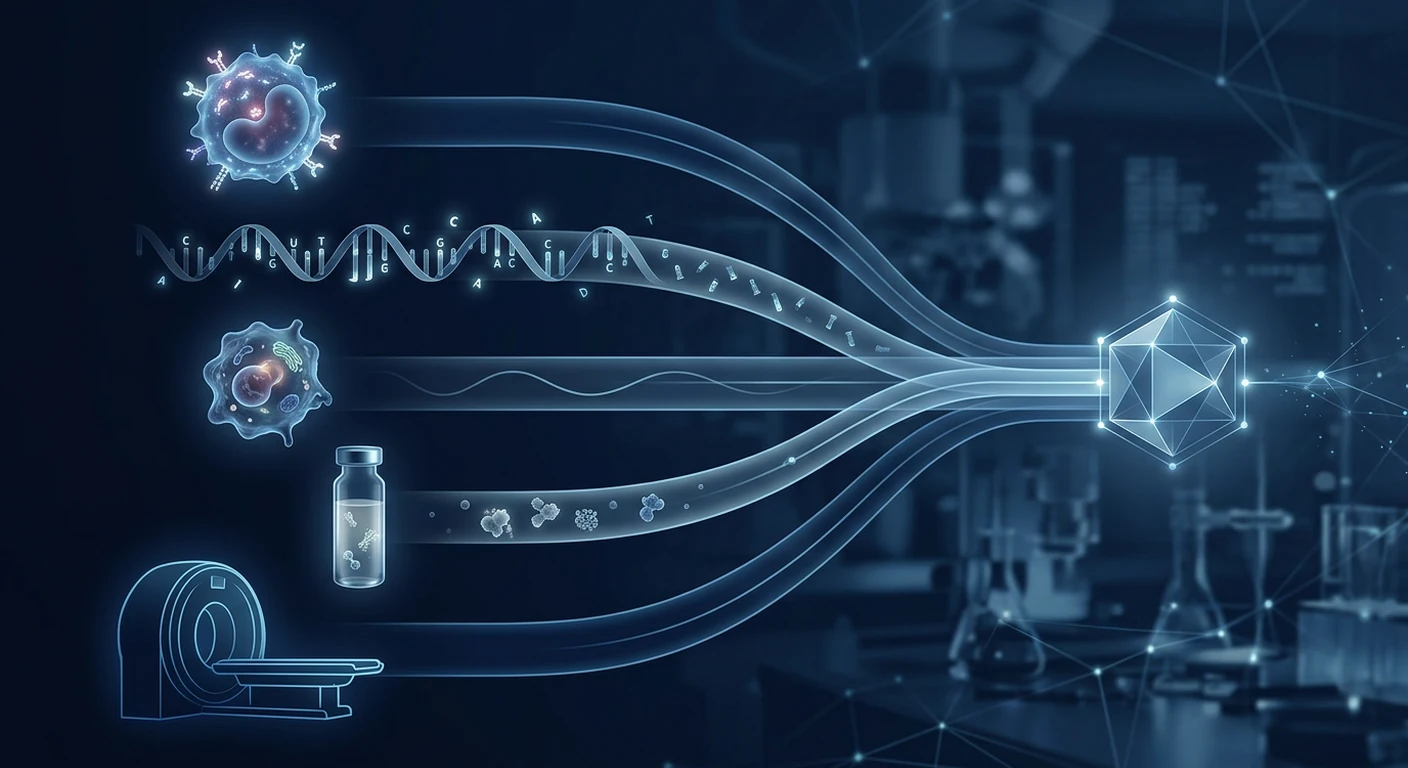
EDIM, circulating tumour DNA (ctDNA), circulating tumour cells (CTCs), conventional serum tumour markers, imaging, and guideline screening do not answer the same clinical question. EDIM looks inside activated monocytes for tumour-associated epitopes; ctDNA assays measure tumour-derived DNA fragments; CTC tests seek intact malignant cells; serum markers quantify soluble proteins; imaging localises structural abnormalities; and screening programmes apply validated tests to defined populations. Treating these tools as interchangeable creates false reassurance and unnecessary alarm.
The practical conclusion is simple: EDIM is investigational and must not replace organ-specific cancer screening, diagnostic imaging, pathology, or clinician-directed follow-up. A positive result does not establish cancer, its site, stage, or histology. A negative result does not rule cancer out. This comparison explains where each method may contribute, what evidence supports it, and where important uncertainty remains.
What EDIM measures
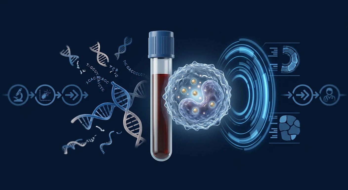
Epitope detection in monocytes (EDIM) is a flow-cytometry method that measures tumour-associated material inside activated CD14-positive/CD16-positive monocytes. The proposed mechanism is that these innate immune cells engulf tumour cells or fragments in tissues, return to circulation, and carry intracellular epitopes that can then be detected in a blood sample. Commercial implementations commonly measure Apo10, an epitope associated with abnormal DNaseX expression and apoptosis, and TKTL1, a protein linked by proponents to altered glucose metabolism.[1]
This is biologically different from detecting a molecule freely circulating in plasma. EDIM uses an immune cell as an indirect “collector” of tissue material. That concept could theoretically amplify a signal when little tumour material reaches plasma, but it also introduces dependencies: monocyte recruitment, phagocytosis, trafficking, marker expression, sample handling, antibody performance, gating strategy, and the chosen score threshold can all affect results.
Published EDIM evidence includes small studies in selected cancers. A study of cholangiocarcinoma, pancreatic cancer and colorectal cancer reported separation from healthy controls, but included only 62 cancer cases and did not establish performance in an unselected screening population.[2] Paediatric studies included 29 patients with rhabdomyosarcoma and a neuroblastoma pilot involving 38 patients and 37 controls.[3][4] These hypothesis-supporting results require larger, independent, prospectively designed studies that include benign disease, inflammatory conditions, other cancers, and people without symptoms.
Evidence checkpoint
Diagnostic accuracy measured in case-control cohorts often looks better than performance in real-world screening, where cancer prevalence is low and benign abnormalities are common. EDIM publications also include author or commercial links to the technology; this does not invalidate results, but independent replication is essential.
EDIM vs ctDNA liquid biopsy
ctDNA assays examine short tumour-derived DNA fragments within cell-free DNA in plasma. Depending on the assay, they search for mutations, copy-number changes, fragmentation patterns, or methylation signatures. In patients with a known cancer, ctDNA can help identify actionable alterations, estimate molecular response, or detect minimal residual disease in selected settings. For early detection, the central problem is abundance: small or biologically quiet tumours may shed extremely little DNA.[5][6]
EDIM measures proteins carried by monocytes, whereas ctDNA measures nucleic acid shed by tumour cells. ctDNA can provide genomic information that may point to a therapy or tumour origin. Standard Apo10/TKTL1 EDIM does not identify a mutation, histologic subtype, or anatomical site. Conversely, EDIM's immune-cell capture concept is not constrained in exactly the same way as direct plasma shedding, although its true sensitivity in small, presymptomatic tumours is not yet established.
Multi-analyte and methylation-based blood tests have shown promising specificity and tissue-of-origin prediction in validation cohorts.[7][8] Yet even these more extensively studied tests are not universal replacements for established screening. Their clinical value depends not only on sensitivity and specificity, but also on whether a positive signal can be localised efficiently, whether follow-up causes harm, whether interval cancers are reduced, and ultimately whether testing lowers cancer mortality.
EDIM vs circulating tumour cells
CTC testing seeks intact tumour cells that have entered the bloodstream. These cells can be counted, phenotyped, or molecularly characterised. CTCs offer rich biological information because an intact cell contains DNA, RNA, proteins, and morphology. However, they are rare, technically difficult to capture, and heterogeneous. Epithelial-marker capture may miss cells that have changed phenotype, while size-based methods may collect non-malignant cells.
CTC assays are best established as prognostic or disease-monitoring tools in certain metastatic cancers, not as population-wide early detection tests. EDIM does not need to recover an intact malignant cell; it detects selected cargo inside phagocytic immune cells. That may make laboratory capture easier, but also means the detected epitope is an indirect signal rather than a tumour cell available for full characterisation. Neither method, by itself, replaces tissue pathology when a definitive diagnosis is required.[9]
EDIM vs conventional serum tumour markers
Conventional tumour markers—such as PSA, CEA, CA 19-9, CA-125, AFP and hCG—are soluble substances measured in serum. Their appropriate uses vary sharply. AFP and hCG have recognised roles in germ-cell tumour management; PSA can support an individualised prostate-screening decision; CEA is commonly used after colorectal cancer treatment; and CA-125 may help evaluate an adnexal mass or monitor known ovarian cancer. Most are not sufficiently specific or sensitive for indiscriminate pan-cancer screening.[10]
Benign disease can elevate serum markers: inflammation, smoking, biliary obstruction, menstruation, pregnancy, liver disease, and benign prostatic enlargement are examples. Some cancers never produce the expected marker. Therefore a value is interpreted in clinical context and often as a trend, not as a binary cancer verdict. Breast-cancer guidance, for example, has long cautioned against using circulating markers alone for early detection.[11]
EDIM differs by measuring intracellular epitopes in monocytes and by aiming for a cross-tumour biological signal rather than an organ-associated secreted protein. But “pan-cancer” breadth does not automatically mean better screening. A broad positive result without localisation can trigger serial scans, invasive procedures, cost, radiation exposure, and anxiety. Comparative studies against representative benign and high-risk populations—not just healthy controls—are needed.
EDIM vs imaging and diagnostic work-up
Imaging answers an anatomical question: is there a structural lesion, where is it, and how extensive is it? Ultrasound, mammography, low-dose CT, diagnostic CT, MRI, PET/CT, and endoscopy each have specific indications. Imaging may detect lesions unrelated to cancer and can miss microscopic disease. Blood biomarkers may signal biology before a mass is visible, but they generally cannot define surgical anatomy or prove malignancy.
A positive EDIM or other blood-based signal therefore creates a second question rather than completing a diagnosis: where should clinicians look, and how aggressively? Follow-up should be individualised by symptoms, examination, age, sex, family history, prior cancer, exposures, and standard screening status. Whole-body imaging is not automatically benign; incidental findings can lead to cascades of repeat tests and biopsies.
Pathology remains the usual diagnostic reference standard. Imaging can identify and stage a suspicious lesion, but tissue examination determines tumour type and often supplies molecular information used for treatment. In limited circumstances clinicians diagnose without biopsy because the location or risk makes sampling inappropriate, but a non-localising EDIM score should never be represented as histologic proof.
EDIM vs organ-specific guideline screening
Guideline screening is designed for people without symptoms who meet defined age or risk criteria. It is not simply “looking for cancer.” A screening programme balances benefits, false positives, overdiagnosis, procedure complications, testing intervals, and mortality outcomes. Recommendations vary by country and should be personalised with a clinician.
- Colorectal cancer: stool-based tests and visual examinations such as colonoscopy are supported for eligible adults; abnormal non-colonoscopy tests require colonoscopic follow-up.[12]
- Lung cancer: annual low-dose CT is recommended by several guidelines for selected adults with substantial smoking exposure; a blood test is not a substitute.[13]
- Breast cancer: mammography remains the principal population screening method for average-risk women in the recommended age range.[14]
- Cervical cancer: HPV testing, cytology, or guideline-approved combinations prevent cancer by detecting viral risk and precancerous change.[15]
- Prostate cancer: PSA screening is a preference-sensitive decision in appropriate age groups because potential mortality benefit must be weighed against false positives and overtreatment.[16]
EDIM has not demonstrated that replacing any of these programmes preserves or improves mortality benefit. A person with a negative EDIM result should still complete screening for which they are eligible. Symptoms such as a new lump, unexplained bleeding, persistent cough, progressive swallowing difficulty, neurological change, or unexplained weight loss require clinical assessment regardless of any screening result.
Practical comparison table
| Method | What it measures | Best-supported role | Main limitation | Can it diagnose/localise alone? |
|---|---|---|---|---|
| EDIM | Apo10/TKTL1 or other epitopes inside activated monocytes | Investigational adjunct; research on detection/monitoring | Small selected cohorts; limited independent screening validation | No |
| ctDNA/cfDNA | Tumour-derived mutations, methylation, fragmentation or copy-number patterns | Molecular profiling and monitoring in selected cancers; emerging early detection | Low shedding in early disease; clonal haematopoiesis; cost | Usually no; some tests predict origin |
| CTCs | Intact rare tumour cells in blood | Prognosis and monitoring in selected metastatic cancers | Rarity, heterogeneity and capture bias | No |
| Serum tumour markers | Soluble proteins or hormones | Risk assessment, monitoring or recurrence surveillance in defined contexts | Benign elevations and marker-negative cancers | No |
| Imaging/endoscopy | Anatomy, tissue density, metabolism or mucosal lesions | Screening in defined programmes; localisation, staging and procedure planning | Incidental findings, radiation for some tests, false negatives | Localises; usually cannot prove histology |
| Guideline screening | Organ-specific risk through validated tests | Population-level early detection/prevention in eligible groups | Organ-specific; interval cancers and overdiagnosis still occur | Screening result requires diagnostic follow-up |
How to interpret a positive or negative EDIM result
A positive EDIM result is a reason for clinical review, not a cancer diagnosis. First confirm the exact assay, markers, threshold, laboratory accreditation, pre-analytical conditions, and whether repeat testing is appropriate. A clinician should review symptoms, examination, family history, exposures, prior imaging, and whether routine screening is current. Further tests should follow a reasoned pathway rather than an automatic maximal scan.
A negative EDIM result cannot provide an all-clear. The tumour may not express the measured epitopes; monocyte uptake may be insufficient; disease may be too small; biological and technical variability may lower the score; or the person's cancer type may not be represented in published studies. Continue recommended screening and investigate concerning symptoms promptly.
Limitations and unanswered questions
- Generalisability: current EDIM studies are small and enriched with known cancer cases rather than true screening cohorts.
- Comparator choice: healthy controls do not reflect benign inflammatory disease, premalignant lesions, or high-risk clinic populations.
- Clinical utility: evidence that testing changes management beneficially or reduces late-stage cancer and mortality is not established.
- Localisation: standard Apo10/TKTL1 results do not identify the organ requiring investigation.
- Reproducibility: thresholds, flow-cytometry gating, specimen timing, and laboratory procedures need transparent external validation.
- Conflicts and independence: some foundational publications have commercial or intellectual-property connections; independent multicentre studies matter.
- Downstream harms: false positives can produce anxiety, radiation, biopsies and incidental diagnoses; false negatives can delay care.
Frequently asked questions
Is EDIM the same as a liquid biopsy?
It is often described as a form of liquid biopsy, but it is distinct from the common ctDNA meaning of that term. EDIM examines tumour-associated epitopes inside monocytes; ctDNA assays examine tumour-derived DNA in plasma. The sample is blood in both cases, but the analyte, laboratory method, and evidence base differ.
Can EDIM replace mammography, colonoscopy, HPV testing, PSA discussion, or lung CT?
No. EDIM has not been shown to preserve the benefits of these organ-specific screening pathways. Continue guideline-recommended screening based on age, anatomy, risk, and shared decision-making even if an EDIM result is negative.
Does a positive EDIM score mean I have cancer?
No. It indicates that the measured monocyte-associated signal crossed the assay threshold. Diagnosis requires clinical correlation and usually targeted imaging, endoscopy, and/or pathology. The positive predictive value also depends strongly on how common cancer is in the tested population.
Which blood test is best for early cancer detection?
There is no single best blood test for every person and cancer. Established serum markers have narrow uses; ctDNA multi-cancer tests are evolving; CTC tests are mainly used in selected known cancers; and EDIM remains investigational. Eligibility for proven organ-specific screening should be addressed first.
Can these tests be combined?
Potentially, because they measure different biology, but more tests can also increase false-positive findings. Combination strategies need prospective evidence showing that the added information improves outcomes enough to justify cost and downstream procedures.
What should I ask before purchasing EDIM or another multi-cancer blood test?
Ask who should be tested, which population validated the assay, sensitivity by cancer type and stage, specificity in benign disease, laboratory accreditation, conflicts of interest, the exact follow-up plan, total downstream cost, and what happens after a negative result. Avoid providers who describe screening as diagnosis or advise abandoning standard care.
Bottom line
EDIM offers an interesting immune-cell-based approach to detecting tumour-associated biology, but its published evidence remains much smaller and less independently validated than the evidence supporting standard screening and diagnostic pathways. ctDNA and CTC technologies provide different molecular or cellular information; serum markers have defined but limited roles; imaging localises abnormalities; and pathology generally establishes diagnosis.
Use EDIM, if chosen after informed discussion, only as an adjunct. Do not use it to cancel a mammogram, stool test, colonoscopy, HPV test, PSA conversation, low-dose lung CT, diagnostic scan, or biopsy. The safest interpretation is integrated: risk profile, symptoms, established screening, imaging, laboratory evidence, and specialist judgement together.
References
- Coy JF. EDIM-TKTL1/Apo10 Blood Test: An Innate Immune System Based Liquid Biopsy for the Early Detection, Characterization and Targeted Treatment of Cancer. International Journal of Molecular Sciences. 2017;18(4):878. doi:10.3390/ijms18040878 ↩
- Saman S, Stagno MJ, Warmann SW, et al.. Biomarkers Apo10 and TKTL1: Epitope-detection in monocytes (EDIM) as a new diagnostic approach for cholangiocellular, pancreatic and colorectal carcinoma. Cancer Biomarkers. 2020;27(1):129–137. doi:10.3233/CBM-190414 ↩
- Urla C, Stagno MJ, Schmidt A, et al.. Epitope Detection in Monocytes (EDIM) As a New Method of Liquid Biopsy in Pediatric Rhabdomyosarcoma. Biomedicines. 2022;10(8):1812. doi:10.3390/biomedicines10081812 ↩
- Stagno MJ, Schmidt A, Bochem J, et al.. Epitope detection in monocytes (EDIM) for liquid biopsy including identification of GD2 in childhood neuroblastoma—a pilot study. British Journal of Cancer. 2022;127:1324–1332. doi:10.1038/s41416-022-01855-x ↩
- Wan JCM, Massie C, Garcia-Corbacho J, et al.. Liquid biopsies come of age: towards implementation of circulating tumour DNA. Nature Reviews Cancer. 2017;17:223–238. doi:10.1038/nrc.2017.7 ↩
- Heitzer E, Haque IS, Roberts CES, Speicher MR. Current and future perspectives of liquid biopsies in genomics-driven oncology. Nature Reviews Genetics. 2019;20:71–88. doi:10.1038/s41576-018-0071-5 ↩
- Cohen JD, Li L, Wang Y, et al.. Detection and localization of surgically resectable cancers with a multi-analyte blood test. Science. 2018;359:926–930. doi:10.1126/science.aar3247 ↩
- Klein EA, Richards D, Cohn A, et al.. Clinical validation of a targeted methylation-based multi-cancer early detection test using an independent validation set. Annals of Oncology. 2021;32:1167–1177. doi:10.1016/j.annonc.2021.05.806 ↩
- Pantel K, Alix-Panabières C. Liquid biopsy and minimal residual disease — latest advances and implications for cure. Nature Reviews Clinical Oncology. 2019;16:409–424. doi:10.1038/s41571-019-0187-3 ↩
- Sturgeon CM, Duffy MJ, Stenman UH, et al.. National Academy of Clinical Biochemistry laboratory medicine practice guidelines for use of tumor markers in testicular, prostate, colorectal, breast, and ovarian cancers. Clinical Chemistry. 2008;54:11–79. doi:10.1373/clinchem.2008.105601 ↩
- Duffy MJ. Serum tumor markers in breast cancer: are they of clinical value?. Clinical Chemistry. 2006;52:345–351. doi:10.1373/clinchem.2005.059832 ↩
- Davidson KW, Barry MJ, Mangione CM, et al.. Screening for Colorectal Cancer: US Preventive Services Task Force Recommendation Statement. JAMA. 2021;325:1965–1977. doi:10.1001/jama.2021.6238 ↩
- Krist AH, Davidson KW, Mangione CM, et al.. Screening for Lung Cancer: US Preventive Services Task Force Recommendation Statement. JAMA. 2021;325:962–970. doi:10.1001/jama.2021.1117 ↩
- US Preventive Services Task Force. Screening for Breast Cancer: US Preventive Services Task Force Recommendation Statement. JAMA. 2024;331:1918–1930. doi:10.1001/jama.2024.5534 ↩
- Curry SJ, Krist AH, Owens DK, et al.. Screening for Cervical Cancer: US Preventive Services Task Force Recommendation Statement. JAMA. 2018;320:674–686. doi:10.1001/jama.2018.10897 ↩
- Grossman DC, Curry SJ, Owens DK, et al.. Screening for Prostate Cancer: US Preventive Services Task Force Recommendation Statement. JAMA. 2018;319:1901–1913. doi:10.1001/jama.2018.3710 ↩
EDIM、循环肿瘤DNA(ctDNA)、循环肿瘤细胞(CTC)、传统血清肿瘤标志物、影像学和指南推荐的筛查,并不回答同一个临床问题。EDIM检测活化单核细胞内的肿瘤相关表位;ctDNA检测血浆中的肿瘤来源DNA片段;CTC寻找完整恶性细胞;血清标志物测量可溶性蛋白;影像学定位结构异常;筛查项目则把经过验证的检查用于明确人群。把它们当作可互换工具,可能带来虚假安心或不必要恐慌。
核心结论是:EDIM仍属研究性工具,不能替代器官特异性癌症筛查、诊断影像、病理检查或医生安排的随访。阳性不能证明癌症、部位、分期或组织学类型;阴性也不能排除癌症。
EDIM测量什么
单核细胞表位检测(EDIM)是一种流式细胞术方法,测量活化CD14阳性/CD16阳性单核细胞内的肿瘤相关物质。其假设机制是:先天免疫细胞在组织中吞噬肿瘤细胞或碎片,返回血液后携带可检测的细胞内表位。常见商业检测测量与异常凋亡相关的Apo10和与改变的葡萄糖代谢相关的TKTL1。[1]
EDIM不是直接检测血浆中游离分子,而是把免疫细胞当作间接“收集器”。这一概念理论上可能放大微弱信号,但结果也取决于单核细胞募集、吞噬、转运、标志物表达、样本处理、抗体性能、流式门控和阈值。
现有研究规模较小且癌种经过筛选:一项胆管癌、胰腺癌和结直肠癌研究共62例癌症患者;横纹肌肉瘤研究29例;神经母细胞瘤试点研究为38例患者和37名对照。[2][3][4]这些结果支持进一步研究,但尚不能代表普通无症状人群筛查。需要大型、独立、前瞻性研究,并纳入良性疾病、炎症、高风险者和其他癌种。
证据提醒
病例—对照研究的准确率常高于真实筛查表现,因为真实人群癌症患病率低、良性异常多。部分EDIM基础论文与技术存在作者或商业关联;这不等于结果无效,但使独立重复验证更加重要。
EDIM与ctDNA液体活检
ctDNA检测分析血浆游离DNA中的肿瘤来源片段。不同平台寻找突变、拷贝数变化、片段模式或甲基化特征。在已确诊癌症中,ctDNA可在特定情境支持靶点选择、疗效监测或微小残留病评估。早期检测的主要难点是小肿瘤可能释放极少DNA。[5][6]
EDIM测量单核细胞携带的蛋白表位;ctDNA测量核酸。ctDNA可能提供可指导治疗或提示组织来源的基因组信息,标准Apo10/TKTL1 EDIM则不能确定突变、组织学亚型或解剖位置。EDIM不完全依赖肿瘤向血浆直接释放DNA,但其对小型、无症状肿瘤的真实敏感度尚未确定。
多分析物和甲基化多癌早检在验证队列显示出有希望的特异度及组织来源预测。[7][8]但临床价值还取决于阳性后能否有效定位、随访伤害、晚期癌比例及死亡率,而不仅是实验室准确率。
EDIM与循环肿瘤细胞
CTC检测寻找进入血流的完整肿瘤细胞。完整细胞含DNA、RNA、蛋白和形态信息,可计数和分子表征;但其数量极少、异质性高且难捕获。CTC最成熟的用途是在某些转移性癌症中提供预后或监测信息,而非普通人群早筛。
EDIM无需捕获完整恶性细胞,而检测吞噬细胞内的特定物质。这可能简化捕获,却只能提供间接信号,不能像完整细胞那样全面表征。需要明确诊断时,两者都不能单独取代组织病理。[9]
EDIM与传统血清肿瘤标志物
PSA、CEA、CA19-9、CA-125、AFP和hCG等是血清中的可溶性蛋白或激素。用途差异很大:AFP和hCG用于生殖细胞肿瘤管理;PSA可支持个体化前列腺筛查决策;CEA常用于结直肠癌治疗后监测;CA-125可辅助评估附件肿块或监测已知卵巢癌。大多数不适合无差别泛癌筛查。[10]
炎症、吸烟、胆道梗阻、月经、妊娠、肝病和前列腺良性增生都可能升高某些指标,部分癌症又不会产生预期标志物。因此必须结合背景和趋势解释,不能把单一数值当作癌症裁决。乳腺癌文献也长期警告不要单独使用循环标志物做早期检测。[11]
EDIM与影像及病理
影像学回答解剖问题:是否存在病灶、位于何处、范围多大。超声、乳腺摄影、低剂量CT、诊断CT、MRI、PET/CT和内镜各有适应证。影像可能发现与癌症无关的偶发异常,也可能漏掉微小疾病;血液标志物可能在肿块可见前提示生物学变化,却通常不能规划手术或证明恶性。
因此,EDIM或其他血液信号阳性只是提出第二个问题,而不是完成诊断:应到哪里寻找、检查多积极?随访应综合症状、体检、年龄、性别、家族史、既往癌症、暴露和标准筛查状态。全身影像并非无害,偶发发现可能引发重复扫描和活检。
病理通常仍是诊断参考标准。影像定位和分期,组织检查确定肿瘤类型并可提供治疗所需分子资料。非定位性的EDIM分数绝不能表述为组织学证据。
EDIM与器官特异性指南筛查
指南筛查面向符合年龄或风险条件且无症状的人群。它综合收益、假阳性、过度诊断、操作并发症、间隔和死亡结局;各国建议不同,应与医生个体化决定。
- 结直肠癌:符合条件者可使用粪便检测或结肠镜等检查;非结肠镜筛查异常通常需结肠镜随访。[12]
- 肺癌:有显著吸烟暴露的部分成人使用年度低剂量CT;血液检查不能替代。[13]
- 乳腺癌:乳腺摄影仍是平均风险女性主要人群筛查方式。[14]
- 宫颈癌:HPV检测、细胞学或认可组合通过发现病毒风险和癌前病变预防癌症。[15]
- 前列腺癌:PSA筛查需共同决策,权衡潜在收益与假阳性、过度诊断和过度治疗。[16]
没有证据表明用EDIM替代这些项目能保持或改善死亡率收益。EDIM阴性者仍应完成适合自己的标准筛查。新肿块、不明出血、持续咳嗽、吞咽困难、神经变化或不明原因体重下降,无论筛查结果如何都需就医。
实用比较表
| 方法 | 测量对象 | 证据较充分的用途 | 主要局限 | 能否单独诊断/定位 |
|---|---|---|---|---|
| EDIM | 活化单核细胞内Apo10/TKTL1等表位 | 研究性辅助检测及监测 | 小型选择性队列,独立筛查验证有限 | 不能 |
| ctDNA/cfDNA | 突变、甲基化、片段或拷贝数 | 特定癌症分子分型和监测;早检研发 | 早期释放少、克隆性造血、费用 | 通常不能;部分预测来源 |
| CTC | 完整稀有肿瘤细胞 | 部分转移癌预后与监测 | 稀少、异质、捕获偏倚 | 不能 |
| 血清标志物 | 可溶性蛋白或激素 | 明确情境下的评估、监测或复发随访 | 良性升高和标志物阴性癌症 | 不能 |
| 影像/内镜 | 解剖、密度、代谢或黏膜病变 | 特定筛查、定位、分期及手术规划 | 偶发发现、部分有辐射、假阴性 | 能定位;通常不能证明组织学 |
| 指南筛查 | 经验证的器官特异性风险信号 | 合格人群早检/预防 | 器官特异,仍有间期癌和过度诊断 | 异常需诊断随访 |
如何解释EDIM阳性或阴性
阳性是临床复核的理由,不是癌症诊断。应确认具体平台、标志物、阈值、实验室资质、样本条件及是否需要复测。医生应评估症状、体检、家族史、暴露、既往影像和常规筛查,按逻辑选择后续检查,而非自动进行最大范围扫描。
阴性不是“全身无癌证明”。肿瘤可能不表达目标表位,单核细胞摄取不足,疾病太小,技术或生物变异降低分数,或该癌种没有充分研究。仍需继续指南筛查并及时评估警示症状。
局限和未解决问题
- 当前研究小且富集已知病例,不是真实筛查队列。
- 健康对照不能代表炎症、良性疾病、癌前病变和高风险门诊。
- 尚未证实EDIM改善管理、减少晚期癌症或降低死亡率。
- Apo10/TKTL1结果不能指出需要检查的器官。
- 阈值、门控、采样时机和实验室流程需要透明外部验证。
- 部分基础研究有商业或知识产权关系,需要独立多中心复制。
- 假阳性可带来焦虑、辐射、活检和偶发诊断;假阴性可能延误治疗。
常见问题
EDIM与液体活检相同吗?
常被归入液体活检,但不同于通常所指的ctDNA。EDIM检测单核细胞内表位,ctDNA检测血浆肿瘤DNA;样本都是血液,但分析物、实验方法和证据基础不同。
EDIM能替代乳腺摄影、结肠镜、HPV检查、PSA讨论或肺CT吗?
不能。它尚未证明能保留这些筛查路径的收益。即使EDIM阴性,也应按年龄、解剖、风险和共同决策完成指南筛查。
EDIM阳性是否意味着患癌?
不是。它只说明单核细胞相关信号超过阈值。诊断通常需要临床关联、靶向影像、内镜及/或病理;阳性预测值还强烈取决于受检人群癌症患病率。
哪种血液早癌检测最好?
没有适合所有人和所有癌症的单一最佳检测。血清标志物用途较窄;ctDNA多癌早检仍在发展;CTC主要用于部分已知癌症;EDIM仍属研究性。首先应处理已证实有效的器官特异性筛查。
这些检测可以组合吗?
理论上可以,因为测量不同生物学信号;但检测越多也可能增加假阳性。组合策略需前瞻性证据证明额外信息改善结局并值得其成本和后续操作。
购买EDIM前应问什么?
询问适用人群、验证队列、各癌种和分期敏感度、良性病中的特异度、实验室资质、利益冲突、阳性后的明确路径、总成本及阴性后的安排。警惕把筛查说成诊断或建议放弃标准医疗的机构。
结论
EDIM是一种有研究价值的免疫细胞检测思路,但发表证据远少于标准筛查和诊断路径,独立验证也不足。ctDNA和CTC提供不同的分子或细胞信息;血清标志物有明确但有限的用途;影像负责定位;病理通常确诊。
若充分知情后选择EDIM,应仅把它作为辅助。不要因此取消乳腺摄影、粪便检测、结肠镜、HPV检查、PSA讨论、低剂量肺CT、诊断扫描或活检。最安全的判断来自风险、症状、标准筛查、影像、实验室证据和专科医生意见的整合。
参考文献
- Coy JF. EDIM-TKTL1/Apo10 Blood Test: An Innate Immune System Based Liquid Biopsy for the Early Detection, Characterization and Targeted Treatment of Cancer. International Journal of Molecular Sciences. 2017;18(4):878. doi:10.3390/ijms18040878 ↩
- Saman S, Stagno MJ, Warmann SW, et al.. Biomarkers Apo10 and TKTL1: Epitope-detection in monocytes (EDIM) as a new diagnostic approach for cholangiocellular, pancreatic and colorectal carcinoma. Cancer Biomarkers. 2020;27(1):129–137. doi:10.3233/CBM-190414 ↩
- Urla C, Stagno MJ, Schmidt A, et al.. Epitope Detection in Monocytes (EDIM) As a New Method of Liquid Biopsy in Pediatric Rhabdomyosarcoma. Biomedicines. 2022;10(8):1812. doi:10.3390/biomedicines10081812 ↩
- Stagno MJ, Schmidt A, Bochem J, et al.. Epitope detection in monocytes (EDIM) for liquid biopsy including identification of GD2 in childhood neuroblastoma—a pilot study. British Journal of Cancer. 2022;127:1324–1332. doi:10.1038/s41416-022-01855-x ↩
- Wan JCM, Massie C, Garcia-Corbacho J, et al.. Liquid biopsies come of age: towards implementation of circulating tumour DNA. Nature Reviews Cancer. 2017;17:223–238. doi:10.1038/nrc.2017.7 ↩
- Heitzer E, Haque IS, Roberts CES, Speicher MR. Current and future perspectives of liquid biopsies in genomics-driven oncology. Nature Reviews Genetics. 2019;20:71–88. doi:10.1038/s41576-018-0071-5 ↩
- Cohen JD, Li L, Wang Y, et al.. Detection and localization of surgically resectable cancers with a multi-analyte blood test. Science. 2018;359:926–930. doi:10.1126/science.aar3247 ↩
- Klein EA, Richards D, Cohn A, et al.. Clinical validation of a targeted methylation-based multi-cancer early detection test using an independent validation set. Annals of Oncology. 2021;32:1167–1177. doi:10.1016/j.annonc.2021.05.806 ↩
- Pantel K, Alix-Panabières C. Liquid biopsy and minimal residual disease — latest advances and implications for cure. Nature Reviews Clinical Oncology. 2019;16:409–424. doi:10.1038/s41571-019-0187-3 ↩
- Sturgeon CM, Duffy MJ, Stenman UH, et al.. National Academy of Clinical Biochemistry laboratory medicine practice guidelines for use of tumor markers in testicular, prostate, colorectal, breast, and ovarian cancers. Clinical Chemistry. 2008;54:11–79. doi:10.1373/clinchem.2008.105601 ↩
- Duffy MJ. Serum tumor markers in breast cancer: are they of clinical value?. Clinical Chemistry. 2006;52:345–351. doi:10.1373/clinchem.2005.059832 ↩
- Davidson KW, Barry MJ, Mangione CM, et al.. Screening for Colorectal Cancer: US Preventive Services Task Force Recommendation Statement. JAMA. 2021;325:1965–1977. doi:10.1001/jama.2021.6238 ↩
- Krist AH, Davidson KW, Mangione CM, et al.. Screening for Lung Cancer: US Preventive Services Task Force Recommendation Statement. JAMA. 2021;325:962–970. doi:10.1001/jama.2021.1117 ↩
- US Preventive Services Task Force. Screening for Breast Cancer: US Preventive Services Task Force Recommendation Statement. JAMA. 2024;331:1918–1930. doi:10.1001/jama.2024.5534 ↩
- Curry SJ, Krist AH, Owens DK, et al.. Screening for Cervical Cancer: US Preventive Services Task Force Recommendation Statement. JAMA. 2018;320:674–686. doi:10.1001/jama.2018.10897 ↩
- Grossman DC, Curry SJ, Owens DK, et al.. Screening for Prostate Cancer: US Preventive Services Task Force Recommendation Statement. JAMA. 2018;319:1901–1913. doi:10.1001/jama.2018.3710 ↩
لا تجيب تقنية EDIM، والحمض النووي الورمي المتداول (ctDNA)، والخلايا الورمية المتداولة (CTCs)، وواسمات الأورام المصلية التقليدية، والتصوير، وبرامج التحري الإرشادية عن السؤال السريري نفسه. تفحص EDIM حواتم مرتبطة بالورم داخل الوحيدات المنشطة؛ وتقيس اختبارات ctDNA شظايا DNA ورمية في البلازما؛ وتبحث اختبارات CTC عن خلايا خبيثة سليمة؛ وتقيس الواسمات المصلية بروتينات ذائبة؛ ويحدد التصوير موضع الشذوذ البنيوي؛ أما برامج التحري فتطبق اختبارات مثبتة على فئات محددة.
الخلاصة العملية: تظل EDIM تقنية استقصائية، ولا ينبغي أن تستبدل التحري الخاص بكل عضو أو التصوير التشخيصي أو علم الأمراض أو المتابعة التي يوجهها الطبيب. النتيجة الإيجابية لا تثبت وجود السرطان أو موقعه أو مرحلته أو نوعه النسيجي، والسلبية لا تستبعده.
ما الذي تقيسه EDIM؟
كشف الحواتم في الوحيدات هو أسلوب قياس بالتدفق الخلوي يقيس مواد مرتبطة بالورم داخل وحيدات CD14+/CD16+ المنشطة. تفترض الآلية أن خلايا المناعة الفطرية تبتلع خلايا الورم أو شظاياه في الأنسجة ثم تعود إلى الدم حاملة حواتم داخلية قابلة للكشف. تقيس التطبيقات الشائعة Apo10 المرتبط بالتعبير غير الطبيعي لـ DNaseX والاستماتة، وTKTL1 الذي يربطه مؤيدو الاختبار باستقلاب الغلوكوز المتغير.[1]
يختلف ذلك بيولوجياً عن قياس جزيء حر في البلازما؛ فالخلية المناعية تعمل «جامعاً» غير مباشر. قد يضخم هذا المفهوم إشارة ضعيفة نظرياً، لكنه يعتمد أيضاً على تجنيد الوحيدات والبلعمة والحركة والتعبير عن الواسم ومعالجة العينة والأجسام المضادة وبوابة التدفق والعتبة.
الأدلة المنشورة صغيرة ومنتقاة. ضمت دراسة سرطان القنوات الصفراوية والبنكرياس والقولون والمستقيم 62 حالة سرطان فقط؛ وضمت دراسة الساركومة العضلية المخططة 29 مريضاً؛ وشملت دراسة الورم الأرومي العصبي التجريبية 38 مريضاً و37 شاهداً.[2][3][4] تدعم النتائج مزيداً من البحث لكنها لا تثبت الأداء في سكان بلا أعراض. نحتاج دراسات كبيرة مستقلة مستقبلية تضم الأمراض الحميدة والالتهاب والفئات عالية الخطورة وسرطانات أخرى.
نقطة تدقيق الأدلة
غالباً تبدو الدقة أعلى في دراسات الحالات والشواهد منها في التحري الواقعي حيث انتشار السرطان منخفض والشذوذات الحميدة شائعة. لبعض المنشورات الأساسية روابط تجارية أو فكرية بالتقنية؛ لا يبطل ذلك النتائج لكنه يجعل التكرار المستقل ضرورياً.
EDIM مقابل خزعة ctDNA السائلة
تحلل اختبارات ctDNA شظايا DNA مشتقة من الورم ضمن DNA الخالي من الخلايا في البلازما. تبحث المنصات عن طفرات أو تغيرات عدد النسخ أو أنماط التجزؤ أو المثيلة. لدى مريض بسرطان معروف قد تدعم اختيار علاج موجّه أو مراقبة الاستجابة أو كشف المرض المتبقي في سياقات محددة. أما في الكشف المبكر فالمشكلة أن الورم الصغير قد يطرح كمية ضئيلة جداً من DNA.[5][6]
تقيس EDIM حواتم بروتينية تحملها الوحيدات، بينما يقيس ctDNA حمضاً نووياً. وقد يوفر ctDNA معلومات جينومية تشير إلى علاج أو منشأ نسيجي، بينما لا يحدد EDIM القياسي طفرة أو نوعاً نسيجياً أو موقعاً تشريحياً. ولا يعتمد مفهوم EDIM بالطريقة نفسها على طرح DNA مباشرة، لكن حساسيته الحقيقية للأورام الصغيرة قبل الأعراض غير مثبتة.
أظهرت اختبارات متعددة التحاليل والمثيلة نتائج واعدة في النوعية وتوقع منشأ النسيج ضمن مجموعات التحقق.[7][8] لكن المنفعة تعتمد أيضاً على إمكان تحديد موضع الإشارة، وأضرار المتابعة، وتقليل السرطان المتأخر والوفيات، لا على الدقة المختبرية وحدها.
EDIM مقابل الخلايا الورمية المتداولة
تبحث اختبارات CTC عن خلايا ورمية كاملة دخلت مجرى الدم. تحمل الخلية الكاملة DNA وRNA وبروتينات وشكلاً خلوياً، لكنها نادرة وغير متجانسة وصعبة الالتقاط. أفضل أدوارها المثبتة هي الإنذار والمراقبة في بعض السرطانات النقيلية، لا التحري المبكر الشامل.
لا تحتاج EDIM إلى استعادة خلية خبيثة كاملة، بل تكشف حمولة منتقاة داخل خلية بلعمية. قد يسهل ذلك الالتقاط، لكنه يوفر إشارة غير مباشرة لا خلية قابلة للتوصيف الشامل. ولا يستبدل أي منهما علم الأمراض النسيجي عند طلب تشخيص نهائي.[9]
EDIM مقابل واسمات الأورام المصلية
PSA وCEA وCA19-9 وCA-125 وAFP وhCG مواد ذائبة تقاس في المصل. تختلف استعمالاتها: يساعد AFP وhCG في تدبير أورام الخلايا الجنسية؛ وقد يدعم PSA قراراً فردياً حول تحري البروستاتا؛ ويستخدم CEA بعد علاج سرطان القولون؛ وقد يفيد CA-125 في تقييم كتلة ملحقية أو مراقبة سرطان مبيض معروف. معظمها غير مناسب لتحرٍ شامل عشوائي.[10]
قد ترفع الالتهابات والتدخين وانسداد القنوات الصفراوية والحيض والحمل ومرض الكبد وتضخم البروستاتا الحميد بعض الواسمات، وقد لا يفرز بعض السرطان الواسم المتوقع. لذلك تفسر القيم في سياق وغالباً كاتجاه زمني، لا حكماً ثنائياً. وحذرت أدلة سرطان الثدي من استخدام الواسمات المتداولة منفردة للكشف المبكر.[11]
تستهدف EDIM إشارة بيولوجية عابرة للأورام بدلاً من بروتين يفرزه عضو، لكن الاتساع «الشامل» لا يعني تلقائياً تحرياً أفضل. نتيجة واسعة بلا تحديد موضع قد تؤدي إلى صور متكررة وخزعات وقلق وتعرض للإشعاع.
EDIM مقابل التصوير والتشخيص
يجيب التصوير عن سؤال تشريحي: هل توجد آفة، وأين، وما امتدادها؟ للموجات فوق الصوتية وتصوير الثدي وCT منخفض الجرعة وCT التشخيصي وMRI وPET/CT والتنظير استطبابات مختلفة. قد يكشف التصوير موجودات عرضية غير سرطانية أو يفوّت مرضاً مجهرياً. وقد تسبق الإشارة الدموية ظهور كتلة، لكنها لا تحدد عادة تشريح الجراحة ولا تثبت الخباثة.
لذلك تطرح نتيجة EDIM الإيجابية سؤالاً ثانياً ولا تكمل التشخيص: أين نبحث وبأي شدة؟ يجب تخصيص المتابعة وفق الأعراض والفحص والعمر والجنس والتاريخ العائلي والسرطان السابق والتعرضات وحالة التحري. التصوير الكامل ليس بلا ضرر؛ فقد تقود الموجودات العرضية إلى سلسلة فحوص وخزعات.
يبقى علم الأمراض المرجع التشخيصي المعتاد. يحدد التصوير موضع الآفة ومرحلتها، ويحدد فحص النسيج النوع الورمي ويوفر معلومات علاجية. لا يجوز تقديم درجة EDIM غير المحددة للموقع كدليل نسيجي.
EDIM مقابل التحري الإرشادي الخاص بالأعضاء
صمم التحري الإرشادي للأشخاص بلا أعراض ممن يستوفون شروط العمر أو الخطورة. وهو يوازن الفائدة والإيجابيات الكاذبة وفرط التشخيص ومضاعفات الإجراءات والفواصل والوفيات، وتختلف التوصيات بين الدول.
- القولون والمستقيم: اختبارات البراز أو تنظير القولون للبالغين المؤهلين، وتحتاج النتيجة غير الطبيعية في اختبار غير تنظيري إلى تنظير متابعة.[12]
- الرئة: CT سنوي منخفض الجرعة لفئات منتقاة ذات تعرض تدخيني كبير؛ لا يستبدله فحص دم.[13]
- الثدي: يبقى تصوير الثدي الوسيلة السكانية الرئيسية لمتوسطات الخطورة ضمن العمر الموصى به.[14]
- عنق الرحم: اختبار HPV أو الخلايا أو التركيبات المعتمدة تكشف الخطر الفيروسي والتغير السابق للسرطان.[15]
- البروستاتا: قرار PSA تشاركي بسبب موازنة الفائدة المحتملة مع الإيجابيات الكاذبة وفرط العلاج.[16]
لم تثبت EDIM أن استبدال أي برنامج منها يحفظ أو يحسن فائدة الوفيات. يجب مواصلة التحري المناسب حتى مع نتيجة EDIM سلبية. والكتلة الجديدة أو النزف غير المفسر أو السعال المستمر أو صعوبة البلع أو التغير العصبي أو فقدان الوزن تستلزم تقييماً مهما كانت نتيجة التحري.
جدول مقارنة عملي
| الطريقة | ما تقيسه | الدور الأفضل دعماً | القيد الرئيسي | هل تشخص/تحدد وحدها؟ |
|---|---|---|---|---|
| EDIM | حواتم داخل الوحيدات المنشطة | مساعد استقصائي وبحوث الكشف/المراقبة | مجموعات صغيرة منتقاة وتحقق مستقل محدود | لا |
| ctDNA/cfDNA | طفرات ومثيلة وتجزؤ وعدد نسخ | التنميط والمراقبة في سرطانات محددة؛ كشف مبكر ناشئ | طرح منخفض مبكراً وتكون دموي نسيلي وكلفة | غالباً لا؛ بعض الاختبارات تتوقع المنشأ |
| CTC | خلايا ورمية كاملة نادرة | الإنذار والمراقبة في بعض السرطان النقيلي | الندرة وعدم التجانس وانحياز الالتقاط | لا |
| الواسمات المصلية | بروتينات أو هرمونات ذائبة | التقييم والمراقبة في سياقات محددة | ارتفاعات حميدة وسرطان بلا واسم | لا |
| التصوير/التنظير | التشريح أو الكثافة أو الاستقلاب أو المخاطية | تحرٍ محدد وتحديد موضع ومرحلة | موجودات عرضية وإشعاع وفوات | يحدد الموضع؛ لا يثبت النسيج عادة |
| التحري الإرشادي | خطر خاص بعضو باختبار مثبت | الكشف/الوقاية في المؤهلين | خاص بعضو مع سرطان فاصل وفرط تشخيص | الخلل يحتاج متابعة تشخيصية |
تفسير نتيجة EDIM
الإيجابية سبب لمراجعة سريرية وليست تشخيصاً. ينبغي تأكيد المنصة والواسمات والعتبة واعتماد المختبر وظروف العينة والحاجة إلى التكرار. يراجع الطبيب الأعراض والفحص والتاريخ العائلي والتعرضات والتصوير والتحري، ثم يختار مساراً منطقياً لا تصويراً أقصى تلقائياً.
السلبية ليست شهادة خلو من السرطان. قد لا يعبر الورم عن الحواتم، أو يكون ابتلاع الوحيدات غير كافٍ، أو المرض صغيراً، أو تخفض المتغيرات التقنية الدرجة، أو لم يدرس نوع السرطان. استمر في التحري الموصى به وافحص أعراض الإنذار سريعاً.
القيود والأسئلة المفتوحة
- الدراسات صغيرة وغنية بحالات معروفة وليست مجموعات تحرٍ حقيقية.
- الأصحاء لا يمثلون الالتهاب والمرض الحميد والآفات السابقة للسرطان.
- لم تثبت منفعة في تقليل السرطان المتأخر أو الوفيات.
- Apo10/TKTL1 لا يحدد العضو.
- تحتاج العتبات والبوابات وتوقيت العينة والإجراءات إلى تحقق خارجي.
- توجد روابط تجارية في بعض الأعمال الأساسية؛ الدراسات المستقلة مهمة.
- الإيجابية الكاذبة تسبب القلق والإشعاع والخزعات، والسلبية الكاذبة تؤخر الرعاية.
أسئلة شائعة
هل EDIM هي نفسها الخزعة السائلة؟
توصف كشكل منها لكنها تختلف عن معنى ctDNA الشائع. تفحص EDIM الحواتم داخل الوحيدات، بينما يفحص ctDNA حمضاً نووياً في البلازما؛ تختلف المادة والطريقة وقاعدة الأدلة.
هل تستبدل تصوير الثدي أو تنظير القولون أو HPV أو نقاش PSA أو CT الرئة؟
لا. لم تثبت قدرتها على حفظ فوائد هذه المسارات. واصل التحري وفق العمر والتشريح والخطر والقرار المشترك حتى بعد نتيجة سلبية.
هل الإيجابية تعني السرطان؟
لا؛ تعني أن الإشارة تجاوزت عتبة الاختبار. يتطلب التشخيص ربطاً سريرياً وتصويراً موجهاً وتنظيراً و/أو مرضيات، وتتأثر القيمة التنبؤية بانتشار السرطان في الفئة المختبرة.
ما أفضل فحص دم للكشف المبكر؟
لا يوجد فحص واحد أفضل لكل شخص وكل سرطان. للواسمات أدوار ضيقة، وتتطور اختبارات ctDNA، وتستخدم CTC أساساً في سرطان معروف، وEDIM استقصائية. عالج أهلية التحري المثبت أولاً.
هل يمكن جمع الاختبارات؟
ربما لأنها تقيس بيولوجيا مختلفة، لكن زيادة الاختبارات قد تزيد الإيجابيات الكاذبة. يلزم دليل مستقبلي يثبت تحسن النتائج بما يبرر الكلفة والإجراءات.
ماذا أسأل قبل شراء الاختبار؟
اسأل عن الفئة المناسبة، ومجموعة التحقق، والحساسية حسب النوع والمرحلة، والنوعية في المرض الحميد، واعتماد المختبر، وتعارض المصالح، وخطة المتابعة والكلفة وما يحدث بعد السلبية. تجنب من يصف التحري كتشخيص أو ينصح بترك الرعاية القياسية.
الخلاصة
تقدم EDIM فكرة مناعية مثيرة للاهتمام، لكن أدلتها المنشورة أصغر وأقل تحققاً مستقلاً من مسارات التحري والتشخيص القياسية. يوفر ctDNA وCTC معلومات مختلفة؛ وللواسمات أدوار محدودة؛ ويحدد التصوير الموضع؛ ويثبت علم الأمراض التشخيص عادة.
إذا اختيرت EDIM بعد نقاش واعٍ، فلتكن أداة مساعدة فقط. لا تلغِ تصوير الثدي أو اختبار البراز أو تنظير القولون أو HPV أو نقاش PSA أو CT الرئة أو التصوير التشخيصي أو الخزعة. التفسير الأكثر أماناً يدمج الخطورة والأعراض والتحري والتصوير والمختبر ورأي الاختصاصي.
المراجع
- Coy JF. EDIM-TKTL1/Apo10 Blood Test: An Innate Immune System Based Liquid Biopsy for the Early Detection, Characterization and Targeted Treatment of Cancer. International Journal of Molecular Sciences. 2017;18(4):878. doi:10.3390/ijms18040878 ↩
- Saman S, Stagno MJ, Warmann SW, et al.. Biomarkers Apo10 and TKTL1: Epitope-detection in monocytes (EDIM) as a new diagnostic approach for cholangiocellular, pancreatic and colorectal carcinoma. Cancer Biomarkers. 2020;27(1):129–137. doi:10.3233/CBM-190414 ↩
- Urla C, Stagno MJ, Schmidt A, et al.. Epitope Detection in Monocytes (EDIM) As a New Method of Liquid Biopsy in Pediatric Rhabdomyosarcoma. Biomedicines. 2022;10(8):1812. doi:10.3390/biomedicines10081812 ↩
- Stagno MJ, Schmidt A, Bochem J, et al.. Epitope detection in monocytes (EDIM) for liquid biopsy including identification of GD2 in childhood neuroblastoma—a pilot study. British Journal of Cancer. 2022;127:1324–1332. doi:10.1038/s41416-022-01855-x ↩
- Wan JCM, Massie C, Garcia-Corbacho J, et al.. Liquid biopsies come of age: towards implementation of circulating tumour DNA. Nature Reviews Cancer. 2017;17:223–238. doi:10.1038/nrc.2017.7 ↩
- Heitzer E, Haque IS, Roberts CES, Speicher MR. Current and future perspectives of liquid biopsies in genomics-driven oncology. Nature Reviews Genetics. 2019;20:71–88. doi:10.1038/s41576-018-0071-5 ↩
- Cohen JD, Li L, Wang Y, et al.. Detection and localization of surgically resectable cancers with a multi-analyte blood test. Science. 2018;359:926–930. doi:10.1126/science.aar3247 ↩
- Klein EA, Richards D, Cohn A, et al.. Clinical validation of a targeted methylation-based multi-cancer early detection test using an independent validation set. Annals of Oncology. 2021;32:1167–1177. doi:10.1016/j.annonc.2021.05.806 ↩
- Pantel K, Alix-Panabières C. Liquid biopsy and minimal residual disease — latest advances and implications for cure. Nature Reviews Clinical Oncology. 2019;16:409–424. doi:10.1038/s41571-019-0187-3 ↩
- Sturgeon CM, Duffy MJ, Stenman UH, et al.. National Academy of Clinical Biochemistry laboratory medicine practice guidelines for use of tumor markers in testicular, prostate, colorectal, breast, and ovarian cancers. Clinical Chemistry. 2008;54:11–79. doi:10.1373/clinchem.2008.105601 ↩
- Duffy MJ. Serum tumor markers in breast cancer: are they of clinical value?. Clinical Chemistry. 2006;52:345–351. doi:10.1373/clinchem.2005.059832 ↩
- Davidson KW, Barry MJ, Mangione CM, et al.. Screening for Colorectal Cancer: US Preventive Services Task Force Recommendation Statement. JAMA. 2021;325:1965–1977. doi:10.1001/jama.2021.6238 ↩
- Krist AH, Davidson KW, Mangione CM, et al.. Screening for Lung Cancer: US Preventive Services Task Force Recommendation Statement. JAMA. 2021;325:962–970. doi:10.1001/jama.2021.1117 ↩
- US Preventive Services Task Force. Screening for Breast Cancer: US Preventive Services Task Force Recommendation Statement. JAMA. 2024;331:1918–1930. doi:10.1001/jama.2024.5534 ↩
- Curry SJ, Krist AH, Owens DK, et al.. Screening for Cervical Cancer: US Preventive Services Task Force Recommendation Statement. JAMA. 2018;320:674–686. doi:10.1001/jama.2018.10897 ↩
- Grossman DC, Curry SJ, Owens DK, et al.. Screening for Prostate Cancer: US Preventive Services Task Force Recommendation Statement. JAMA. 2018;319:1901–1913. doi:10.1001/jama.2018.3710 ↩