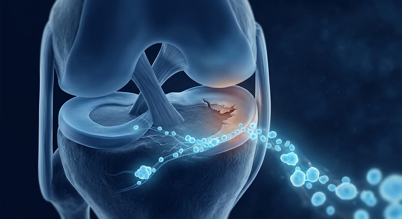
The EDIM blood test is an investigational cancer-risk assessment that examines immune cells for two tumour-associated biomarkers, Apo10 and TKTL1. EDIM stands for epitope detection in monocytes. The laboratory uses flow cytometry to measure how many circulating monocytes contain epitopes associated with abnormal apoptosis and altered glucose metabolism. The result can identify a signal that deserves clinical attention, but it cannot show where a tumour is, prove that cancer is present, or exclude cancer when the score is low.
Most importantly, EDIM does not diagnose cancer and does not replace guideline-recommended screening. Mammography, cervical screening, colorectal stool testing or colonoscopy, low-dose CT for eligible smokers, prostate discussions, skin examination, and other age- and risk-appropriate pathways remain necessary. If a symptom, examination, scan, or standard test is concerning, it must be investigated regardless of an EDIM result.
This patient guide explains the biological idea, sample journey, interpretation, uncertainty, and sensible next steps. It is designed to support a physician conversation, not to promote a single blood number as an answer to a complex clinical question.
What does EDIM stand for, and what is it measuring?
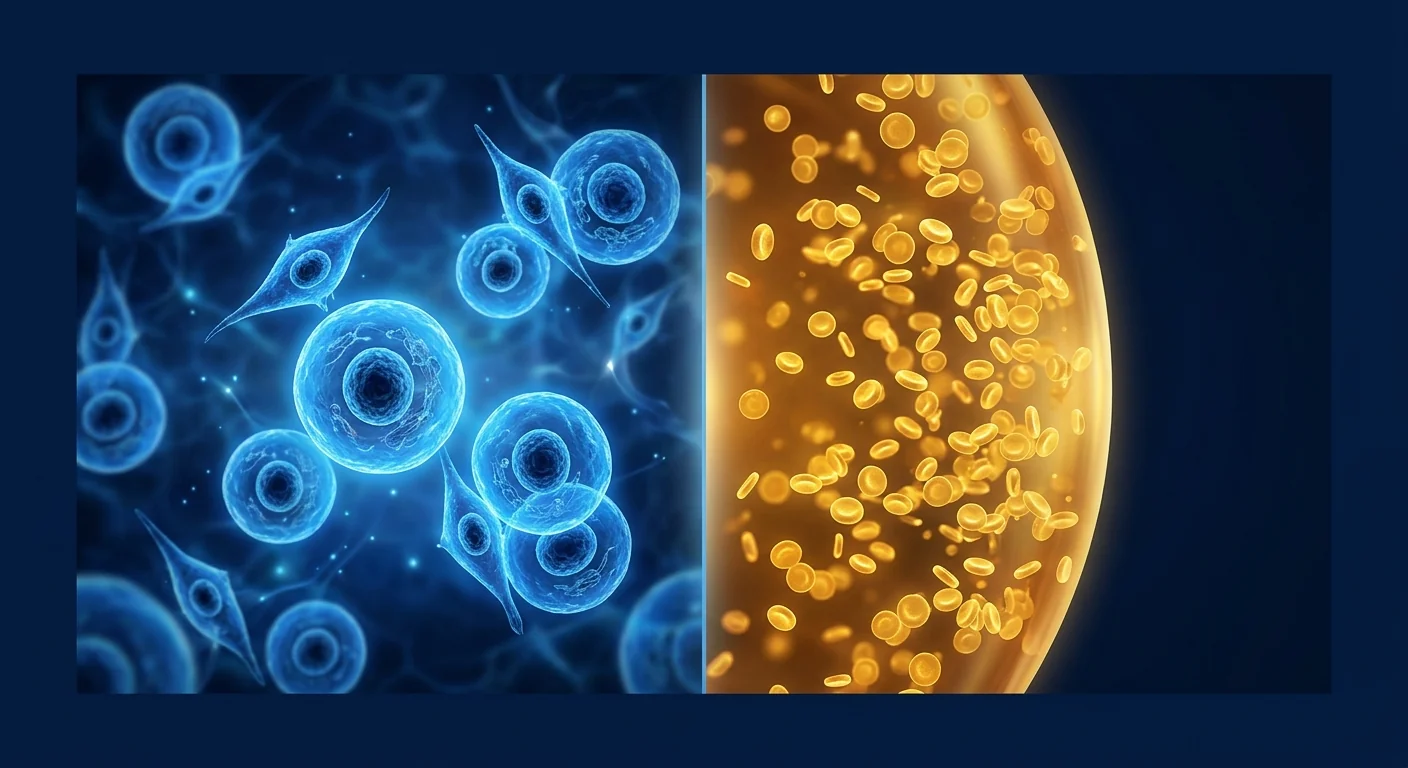
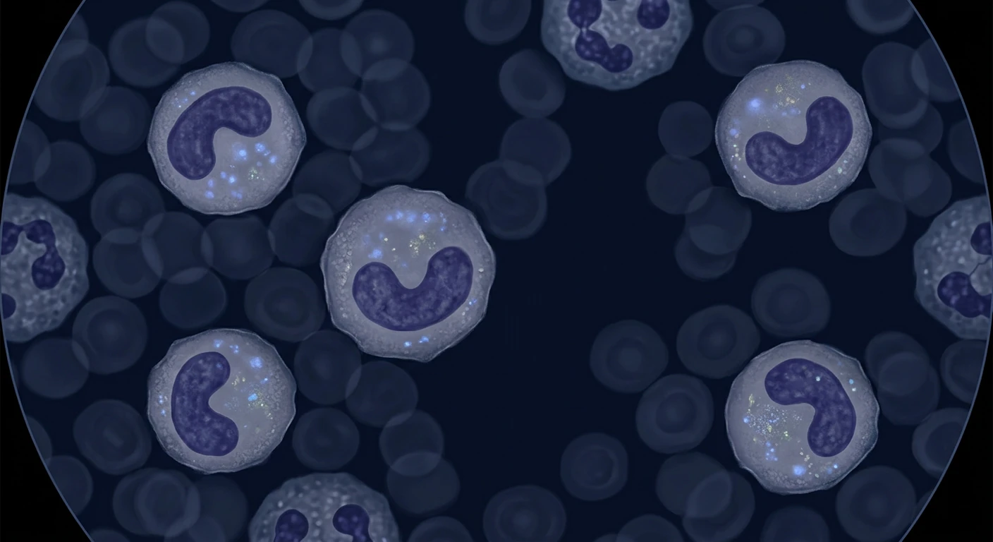
EDIM means epitope detection in monocytes. Monocytes are circulating white blood cells that ingest cellular material and later mature into macrophages in tissues. The EDIM hypothesis is that monocytes can engulf material shed by abnormal cells and carry recognizable protein fragments—epitopes—in their interior. A blood sample can then be stained with antibodies and analysed by flow cytometry to identify monocytes containing Apo10 or TKTL1-associated signals [1].
Apo10 is associated with DNaseX, an enzyme involved in programmed cell-death pathways. Research proposes that accumulation of Apo10-related material may reflect impaired apoptosis, a hallmark relevant to malignant transformation. TKTL1, transketolase-like 1, has been studied in relation to altered glucose metabolism, redox balance, and cell survival. Cancer cells often reprogramme metabolism, but neither biomarker is unique to one tumour type and neither independently constitutes a cancer diagnosis [2].
The laboratory usually identifies a monocyte gate with cell-surface markers, measures intracellular staining, and combines the Apo10 and TKTL1 findings into a score or risk category. Exact cut-offs and reporting formats depend on the validated laboratory protocol. A coloured category can make a report easier to read, but colour does not transform a probabilistic biomarker into a definitive yes-or-no test.
How is the blood sample collected and analysed?
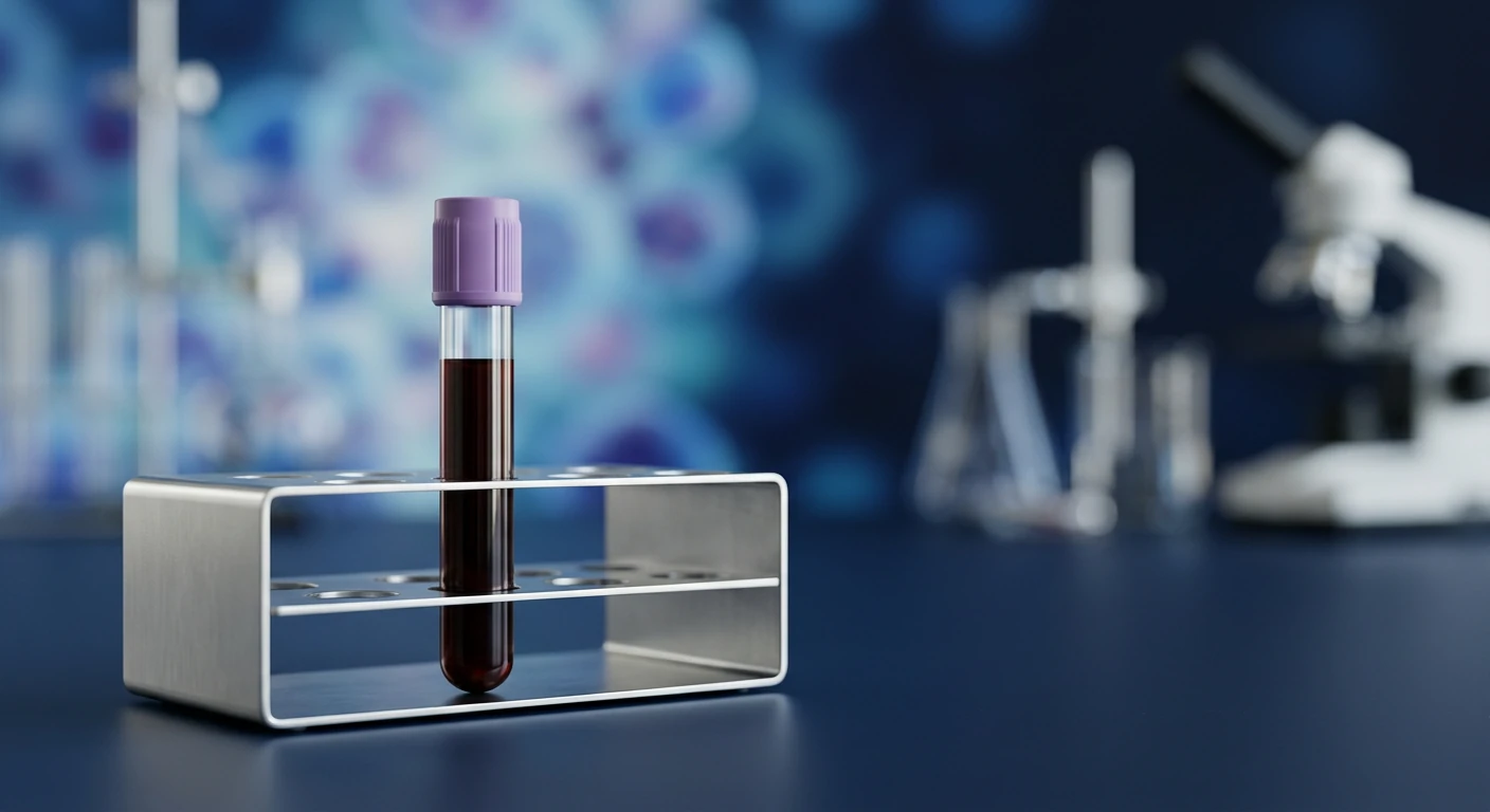
EDIM begins with an ordinary venous blood draw, followed by specialised laboratory handling and multiparameter flow cytometry. Patients should follow the collecting clinic’s preparation instructions and disclose current illnesses, medicines, recent procedures, and previous cancer treatment. The clinically important quality issues are not only the volume drawn but also correct tubes, identification, transport time, temperature, instrument controls, and a laboratory-specific validated workflow.
- Pre-test review: a clinician documents why the test is being considered, current symptoms, family history, smoking and occupational exposure, prior cancer, and the standard screenings already due.
- Blood collection: labelled blood is obtained by venipuncture. Fasting is generally not the central biological requirement, but patients should follow local instructions.
- Cell preparation: white cells are prepared while preserving the monocyte population. Antibodies identify monocytes and stain intracellular Apo10 and TKTL1 epitopes.
- Flow cytometry: thousands of cells pass individually through lasers. Fluorescence intensity and gating rules distinguish cell populations and biomarker-positive events.
- Quality review: controls, sample adequacy, instrument performance, gating, and calculation are checked before release.
- Clinical interpretation: the score is reviewed with medical history and conventional investigations, not in isolation.
Pre-analytical variation matters. A delayed or mishandled specimen can change cell quality. Laboratory thresholds may not transfer perfectly between platforms, antibody lots, or populations. Patients can reasonably ask whether the laboratory participates in external quality systems, how inconclusive samples are handled, and whether a repeat result is interpreted independently of the first.
What do Apo10 and TKTL1 mean?
Apo10 and TKTL1 are biologically plausible tumour-associated signals, not tumour-specific labels. Apo10 research focuses on dysregulated apoptosis. Normal tissues use programmed cell death to remove damaged cells; disrupted pathways can permit abnormal survival. TKTL1 research focuses on metabolic adaptation, especially pathways related to glucose use and management of oxidative stress. These mechanisms are relevant to cancer biology, but relevant is not the same as exclusive.
The EDIM approach differs from assays that directly measure circulating tumour DNA. It looks inside immune cells thought to have phagocytosed abnormal material. That may amplify a biological signal when tumour-derived material is scarce, but it also adds layers of interpretation: monocyte behaviour, inflammation, antibody specificity, gating, and scoring. Published studies report associations and diagnostic-performance estimates in selected cohorts [3], while later prospective work has continued to examine clinical validity [4].
No biomarker should be interpreted without prevalence. In a lower-risk screening population, even a test with apparently good sensitivity and specificity can generate more false positives than a case-control paper suggests. Conversely, an early or biologically quiet tumour may release too little signal, producing false reassurance. Real-world performance depends on who is tested, why they were tested, tumour type and stage, the laboratory method, and the definition of a positive result.
What can an EDIM result tell me?
An EDIM result can indicate that Apo10/TKTL1-positive monocyte activity is below, near, or above a laboratory threshold. It may help a physician decide whether to review overdue screening, repeat the assay, take a more detailed history, or choose a targeted conventional investigation. Research has evaluated EDIM patterns across several solid tumours, but a positive result generally does not localise the organ [5].
| Result pattern | Reasonable interpretation | What it does not mean |
|---|---|---|
| Lower / green-range signal | No elevated signal under that laboratory’s current threshold | Not proof that cancer is absent; continue routine screening and investigate symptoms |
| Borderline / yellow-range signal | Uncertain finding that may merit clinical review or repeat testing | Not “early cancer” and not a reason for indiscriminate whole-body procedures |
| Higher / red-range signal | Signal requiring physician-led assessment and proportionate follow-up | Not a cancer diagnosis, stage, tumour location, or treatment indication |
| Invalid or inadequate | Quality criteria were not met | Neither positive nor negative; the laboratory should explain repeat options |
A change over time may be interesting, but serial testing introduces regression to the mean and ordinary analytical variation. A falling score after lifestyle change or treatment cannot by itself prove that cancer was prevented or treated. Likewise, a rising score needs confirmation rather than immediate attribution to malignancy.
The safety rule
EDIM does not diagnose cancer and must not replace guideline-recommended screening or diagnostic evaluation. A lump, unexplained bleeding, persistent cough, changing bowel habit, unintentional weight loss, suspicious imaging, or abnormal standard screening needs the usual clinical pathway even when EDIM is low.
How accurate is EDIM?
Accuracy estimates in EDIM publications are promising in some settings but are not a universal guarantee for an asymptomatic person. Early studies often compared people with known cancers against controls. Such designs help establish a signal, but they can overestimate performance relative to population screening, where cancers are rarer, early stages predominate, and benign conditions are diverse [1].
Sensitivity asks how often a test is positive among people who truly have cancer. Specificity asks how often it is negative among people who do not. Positive predictive value asks the question patients usually care about: “If my result is positive, what is the chance I actually have cancer?” That value changes sharply with baseline risk. It cannot be inferred from sensitivity alone.
Studies also differ in cancer mix, stage, comparison groups, thresholds, and whether analysis was blinded. Prospective research and independent replication are more informative than a retrospective case-control series. Publications from 2022 and 2023 broaden the evidence base, but EDIM still lacks the mature mortality-reduction evidence available for certain established screening programmes [6]. A test can detect a biological signal without yet proving that using it in screening reduces deaths or avoids more harm than benefit.
How does EDIM compare with liquid biopsy?
EDIM is blood-based, but its target differs from most tests described as liquid biopsy. Circulating tumour DNA assays search plasma for tumour-associated mutations, methylation patterns, or fragmentation. Other multi-cancer tests combine proteins and genomic signals. EDIM instead examines monocytes for engulfed Apo10 and TKTL1-associated epitopes.
| Approach | Signal measured | Potential strength | Important limitation |
|---|---|---|---|
| EDIM | Apo10/TKTL1 epitopes inside monocytes | Immune-cell capture may concentrate cellular signals | Does not reliably localise a tumour; clinical utility remains under study |
| ctDNA mutation assay | Tumour-associated DNA variants | Can support molecular profiling in known cancer | Early tumours may shed little DNA; clonal haematopoiesis can confound |
| Methylation MCED | Abnormal DNA methylation patterns | May predict tissue of origin for several cancers | Positive findings require diagnostic work-up; mortality benefit unproven |
| Multi-analyte test | Proteins plus genomic markers | Combines complementary biology | Performance varies by cancer and stage |
Multi-analyte and methylation studies illustrate both potential and uncertainty [7] [8] [9]. None justify abandoning proven organ-specific screening. A new blood test should ideally complement an organised pathway with predefined confirmation, false-positive management, and follow-up.
Who might discuss EDIM with a physician?
EDIM may be discussed by an informed adult who wants an additional, investigational risk signal and accepts that follow-up may be uncertain. The conversation may be especially relevant when family history, smoking, occupational exposure, previous cancer, or other factors raise concern. It should begin with a formal risk review, not a package purchase.
- Confirm which standard screenings are due based on age, sex, organs, smoking history, family history, and national guidance.
- Document symptoms that require direct diagnostic investigation rather than screening.
- Discuss what a positive, borderline, negative, or invalid result would change.
- Consider anxiety, incidental findings, radiation exposure, invasive follow-up, and cost.
- Agree who will interpret the report and coordinate any next step.
EDIM is not an appropriate way to postpone evaluation of symptoms. It is also not a stand-alone surveillance plan after cancer treatment unless the treating oncology team explicitly integrates it into care. People who are pregnant, acutely unwell, recently operated on, receiving immunomodulatory treatment, or living with inflammatory or haematological conditions should disclose this context because immune-cell biology and interpretation may differ.
What happens after a high or borderline result?
The next step is a structured clinical review, not a declaration of cancer and not automatic whole-body scanning. The physician should verify sample quality, examine the numerical components and threshold, review prior results, take a symptom history, perform a focused examination, and identify the patient’s most plausible organ-specific risks.
- Check for urgent clues: red-flag symptoms or examination findings follow standard urgent referral pathways.
- Bring screening up to date: perform overdue evidence-based tests first. Low-dose CT, for example, has defined eligibility based largely on age and smoking history [11]; colorectal screening has established age and modality choices [12].
- Use targeted diagnostics: imaging, endoscopy, cytology, or tissue biopsy should be chosen for the clinical question. Histopathology remains the basis of most definitive cancer diagnoses.
- Consider repeat measurement: if no target is identified and the patient is clinically stable, a repeat under consistent conditions may help assess persistence, but the interval and stopping rule should be specified.
- Record a closure plan: every abnormal signal needs a named clinician, results review, and an endpoint that avoids endless testing.
Broad imaging can discover benign nodules that trigger more scans or biopsies. The potential benefit of earlier detection must therefore be balanced against radiation, procedural complications, overdiagnosis, and distress. In prospective blood-test research, confirmation pathways are planned in advance for exactly this reason.
What happens after a low result?
A low EDIM result does not create a cancer-free certificate. Continue mammography, cervical screening, colorectal screening, lung screening when eligible, and other guideline care on schedule. Report new or persistent symptoms. If a doctor has already recommended imaging or biopsy because of a clinical abnormality, do not cancel it on the basis of EDIM.
False negatives can arise because a tumour is very small, sheds little material, has different biology, sits outside the test’s strongest evidence, or because analytical and pre-analytical variation obscures the signal. Screening intervals should be based on accepted guidance and individual risk rather than on a marketing claim that one blood test covers dozens of cancers.
Benefits, burdens, and unanswered questions
The potential benefit is convenience: one blood draw may provide an additional broad biological signal. The burdens are uncertainty, false positives, false negatives, lack of localisation, repeat tests, costs, and downstream procedures. The balance is personal, but it should be discussed explicitly before collection.
Important unanswered questions include the best population, interval, threshold, and confirmation algorithm; performance by stage and tumour type; effects of infection or inflammation; reproducibility across laboratories; and whether EDIM-guided screening improves outcomes that matter to patients. Mortality, advanced-stage incidence, quality of life, complications, and cost are stronger endpoints than the number of signals detected.
Research into EDIM and other multi-cancer approaches is scientifically worthwhile. Responsible communication separates analytical validity (the laboratory reliably measures a signal), clinical validity (the signal correlates with disease), and clinical utility (using it improves outcomes). Evidence at one level does not automatically establish the next.
Questions to ask before booking the test
- Why is EDIM appropriate for my risk profile, and what standard screening am I due for now?
- Which exact Apo10/TKTL1 assay, cut-off, and laboratory validation are used?
- How often are results invalid, borderline, false positive, or false negative in people like me?
- Who will explain the result and arrange follow-up?
- What is the plan for each possible category before my blood is taken?
- Could my current illness, treatment, or recent procedure affect interpretation?
- What follow-up costs, imaging, or invasive procedures might occur?
- Will my sample or data be stored, and how is consent handled?
Frequently Asked Questions
Does a positive EDIM result mean I have cancer?
No. It means the assay found an Apo10/TKTL1-associated monocyte signal above a laboratory threshold. A physician must interpret it and, when justified, use standard diagnostic tests. Cancer diagnosis usually requires organ-directed assessment and often tissue pathology.
Can a negative EDIM result rule out cancer?
No. Early, low-shedding, or biologically different cancers may not generate a detectable signal. Symptoms and abnormal conventional tests must still be investigated.
Does EDIM replace mammography, Pap/HPV testing, colonoscopy, stool tests, or low-dose CT?
No. EDIM does not replace any guideline-recommended screening. Those programmes have defined eligibility, intervals, follow-up, and—in several settings—evidence of reduced cancer mortality.
Can EDIM tell where cancer is located?
Not reliably. The biomarkers are not organ-specific. Clinical history and targeted conventional tests are needed to localise any concern.
Is EDIM the same as a circulating tumour DNA test?
No. EDIM analyses biomarker epitopes inside monocytes. Most ctDNA tests analyse tumour-associated DNA changes in plasma.
Should EDIM be repeated every year?
There is no universal evidence-based interval for population screening. Any repeat plan should be individualised, document what trend would trigger action, and never displace standard screening.
Can EDIM monitor cancer treatment?
That use remains investigational. Treatment monitoring should follow the oncology team’s established imaging, laboratory, pathology, and clinical protocols.
Limitations and medical disclaimer
EDIM is an adjunctive, investigational risk-assessment tool. Evidence varies by cohort, tumour type, stage, cut-off, and study design. Published accuracy cannot be assumed to apply identically to every asymptomatic population or laboratory. The test does not provide a tumour location, stage, histology, or treatment recommendation.
This article is educational and is not medical advice. EDIM does not diagnose cancer, rule cancer out, or replace guideline screening. Seek prompt medical care for concerning symptoms. Decisions about testing and follow-up should be made with a qualified clinician who can consider personal risk, benefits, harms, alternatives, and local guidelines.
References
- Grimm M, et al. Biomarker-based detection and characterization of carcinomas using Apo10 and TKTL1 in monocytes. BMC Cancer. 2013;13:569. doi:10.1186/1471-2407-13-569
- Coy JF. EDIM-TKTL1/Apo10 Blood Test: An Innate Immune System Based Liquid Biopsy for the Early Detection, Characterization and Targeted Treatment of Cancer. International Journal of Molecular Sciences. 2017;18(4):878. doi:10.3390/ijms18040878
- Saman S, Stagno MJ, Warmann SW, et al. Biomarkers Apo10 and TKTL1: Epitope-detection in monocytes (EDIM) as a new diagnostic approach for cholangiocellular, pancreatic and colorectal carcinoma. Cancer Biomarkers. 2020;27(1):129-137. doi:10.3233/CBM-190414
- Stagno MJ, Schmidt A, Bochem J, et al. Epitope detection in monocytes (EDIM) for liquid biopsy including identification of GD2 in childhood neuroblastoma—a pilot study. British Journal of Cancer. 2022;127(7):1324-1331. doi:10.1038/s41416-022-01855-x
- Urla C, Stagno MJ, Schmidt A, et al. Epitope Detection in Monocytes (EDIM) As a New Method of Liquid Biopsy in Pediatric Rhabdomyosarcoma. Biomedicines. 2022;10(8):1812. doi:10.3390/biomedicines10081812
- Burg S, Smeets R, Gosau M, et al. Case Report: Early detection of lung carcinoid in an asymptomatic individual by blood-test initiated PET-CT imaging. Frontiers in Oncology. 2023;13:1177237. doi:10.3389/fonc.2023.1177237
- Cohen JD, et al. Detection and localization of surgically resectable cancers with a multi-analyte blood test. Science. 2018;359:926–930. doi:10.1126/science.aar3247
- Lennon AM, et al. Feasibility of blood testing combined with PET-CT to screen for cancer. Science. 2020;369:eabb9601. doi:10.1126/science.abb9601
- Klein EA, et al. Clinical validation of a targeted methylation-based multi-cancer early detection test. Annals of Oncology. 2021;32:1167–1177. doi:10.1016/j.annonc.2021.05.806
- de Koning HJ, van der Aalst CM, de Jong PA, et al. Reduced Lung-Cancer Mortality with Volume CT Screening in a Randomized Trial. N Engl J Med. 2020;382:503-513. doi:10.1056/NEJMoa1911793
- US Preventive Services Task Force. Screening for lung cancer: recommendation statement. JAMA. 2021;325:962–970. doi:10.1001/jama.2021.1117
- US Preventive Services Task Force. Screening for colorectal cancer: recommendation statement. JAMA. 2021;325:1965–1977. doi:10.1001/jama.2021.6238
EDIM 血液检测是一项研究中的癌症风险评估,检测单核细胞内的 Apo10 与 TKTL1 两种肿瘤相关信号。EDIM 即“单核细胞表位检测”。实验室以流式细胞术识别单核细胞,并测量其中的特定蛋白表位。结果可以提示需要医生进一步评估的异常信号,但不能证明患有癌症、不能确定肿瘤部位,也不能以低分排除癌症。
患者必须知道
EDIM 不能诊断癌症,也不能取代指南规定的常规筛查。乳腺摄影、宫颈 HPV/细胞学检查、结直肠粪便检测或肠镜、符合条件者的低剂量肺 CT,以及其他按年龄和风险安排的筛查仍须按时完成。出现肿块、不明出血、持续咳嗽、排便改变或体重下降等症状时,无论 EDIM 结果如何,都应走标准诊断流程。
EDIM 检测的是什么?
单核细胞是可吞噬细胞碎片的白细胞。EDIM 的研究假设是:单核细胞吞噬异常细胞材料后,可在其内部检测到 Apo10 与 TKTL1 相关表位 [1]。Apo10 与异常细胞凋亡途径相关;TKTL1 与葡萄糖代谢、氧化还原平衡和细胞存活研究相关 [2]。这些机制与癌症生物学有关,但并非某一癌种独有,因此单个标志物不是诊断。
实验室通常先用表面标志物圈定单核细胞,再染色细胞内 Apo10 和 TKTL1,以流式细胞仪分析数千个细胞。两个信号被组合为分数或风险级别;具体阈值取决于实验室验证方案。绿、黄、红颜色便于阅读,却不能把概率性结果变成确定答案。
采血与实验室流程
- 检测前评估:记录症状、家族史、吸烟史、既往肿瘤和应做的常规筛查。
- 静脉采血:按采集机构说明准备,并告知近期感染、手术和用药。
- 细胞处理:分离或处理白细胞,保留单核细胞群并进行抗体染色。
- 流式分析:用激光和荧光信号识别细胞群及阳性事件。
- 质量审核:检查样本时效、对照、仪器表现、门控和计算。
- 临床解释:医生结合风险与常规检查解读,而不是只看颜色。
运输时间、温度、抗体批次和门控规则均可能影响结果。患者可询问实验室如何处理不合格或临界样本、是否有外部质量管理,以及不同平台的阈值能否比较。
如何理解结果?
| 报告类别 | 合理含义 | 不代表什么 |
|---|---|---|
| 较低/绿色 | 未超过该实验室阈值 | 不代表没有癌症;仍需常规筛查 |
| 临界/黄色 | 不确定,可能需要复核或重复 | 不等于“早癌” |
| 较高/红色 | 需要医生主导的评估 | 不是癌症诊断、分期或部位 |
| 无效 | 样本或质量标准未通过 | 既非阳性也非阴性 |
早期病例对照研究显示了 Apo10/TKTL1 信号与部分实体肿瘤的关联 [3],后续前瞻性研究继续评估其临床有效性 [4]。但在无症状、低患病率人群中,阳性预测值通常低于已确诊病例与健康对照直接比较的结果。肿瘤很小、释放信号少或生物学不同,也可能出现假阴性。
EDIM 与液体活检有何不同?
EDIM 查看单核细胞内的蛋白表位;循环肿瘤 DNA 检测通常查看血浆中的突变、甲基化或 DNA 片段特征。多癌早检研究显示血液检测具有潜力,但阳性后仍需定位和确诊流程 [7] [8] [9]。目前没有一种新型广谱血液检测可以替代已有器官特异性筛查。
谁可以与医生讨论 EDIM?
愿意接受检测不确定性、并希望在常规筛查之外获得附加风险信号的知情成人,可以与医生讨论。家族史、吸烟、职业暴露或既往癌症会改变基础风险。检测前应先明确:现在应做哪些常规筛查、不同结果会采取什么行动、由谁解释,以及可能发生的影像、活检、费用和焦虑。
EDIM 不适合用来推迟症状检查,也不应自行替代癌症治疗后的标准随访。急性感染、炎症、近期手术、妊娠、免疫调节治疗或血液疾病可能改变免疫细胞背景,应完整告知医生。
较高或临界结果之后怎么办?
- 核对样本质量、分项数值和阈值。
- 复查症状、体检、个人风险和家族史;红旗症状按紧急路径处理。
- 先补做逾期的循证筛查,例如符合条件者的低剂量肺 CT [11] 或结直肠筛查 [12]。
- 根据线索选择靶向影像、内镜或活检,而不是自动进行无边界全身检查。
- 若暂未找到目标,可在明确间隔、触发标准和停止规则下考虑复测。
癌症通常需要器官定向检查,最终诊断往往依赖组织病理。广泛影像也会发现大量良性结节,可能带来重复扫描、辐射和不必要活检,因此随访必须权衡收益与伤害。
较低结果之后怎么办?
继续所有指南筛查。不要因低 EDIM 分数取消医生已建议的乳腺摄影、HPV 检查、肠镜、CT、超声或活检。新出现或持续存在的症状仍须就医。一次结果也不应被宣传为覆盖数十种癌症的“无癌证明”。
收益、负担与尚未解决的问题
潜在收益是一次采血提供广泛的附加生物学信号。负担包括假阳性、假阴性、无法定位、复测、费用、下游检查和心理压力。研究仍需确定最佳人群、间隔、阈值、确认算法、不同癌种与分期的表现、炎症影响、跨实验室可重复性,以及使用 EDIM 是否真正降低晚期癌症或死亡。
评价新检测应区分三个层次:分析有效性(能否可靠测量)、临床有效性(是否与疾病相关)和临床效用(使用后是否改善患者结局)。前一层的证据不能自动证明后一层。
检测前可询问的问题
- 为什么此检测适合我的风险?我现在有哪些常规筛查到期?
- 实验室使用何种 Apo10/TKTL1 方法和阈值?
- 类似人群中的临界、无效、假阳性和假阴性有多少?
- 每种结果的书面随访计划是什么?
- 谁负责解释并确保随访完成?
- 近期疾病或治疗会否影响解释?
常见问题
EDIM 阳性就是癌症吗?
不是。它只是超过实验室阈值的相关信号,需要结合标准检查评估。
EDIM 阴性能排除癌症吗?
不能。早期或低信号肿瘤可能漏检,症状和常规异常仍须检查。
能取代乳腺摄影、宫颈筛查、肠镜或低剂量 CT 吗?
不能。EDIM 不取代任何指南推荐筛查。
能确定肿瘤在哪里吗?
通常不能可靠定位;需要病史和靶向常规检查。
多久做一次?
目前没有适用于所有人的循证间隔,应由医生个体化决定,且不能挤占常规筛查。
局限性与医疗声明
EDIM 是附加性的研究中风险工具,不提供肿瘤部位、分期、组织类型或治疗建议。不同研究人群、癌种、分期、阈值和实验室可能产生不同表现。本文仅供教育,不构成诊断或医疗建议。出现警示症状应及时就医,并由合格医生权衡检测收益、风险和替代方案。
参考文献
- Grimm M, et al. Biomarker-based detection and characterization of carcinomas using Apo10 and TKTL1 in monocytes. BMC Cancer. 2013;13:569. doi:10.1186/1471-2407-13-569
- Coy JF. EDIM-TKTL1/Apo10 Blood Test: An Innate Immune System Based Liquid Biopsy for the Early Detection, Characterization and Targeted Treatment of Cancer. International Journal of Molecular Sciences. 2017;18(4):878. doi:10.3390/ijms18040878
- Saman S, Stagno MJ, Warmann SW, et al. Biomarkers Apo10 and TKTL1: Epitope-detection in monocytes (EDIM) as a new diagnostic approach for cholangiocellular, pancreatic and colorectal carcinoma. Cancer Biomarkers. 2020;27(1):129-137. doi:10.3233/CBM-190414
- Stagno MJ, Schmidt A, Bochem J, et al. Epitope detection in monocytes (EDIM) for liquid biopsy including identification of GD2 in childhood neuroblastoma—a pilot study. British Journal of Cancer. 2022;127(7):1324-1331. doi:10.1038/s41416-022-01855-x
- Urla C, Stagno MJ, Schmidt A, et al. Epitope Detection in Monocytes (EDIM) As a New Method of Liquid Biopsy in Pediatric Rhabdomyosarcoma. Biomedicines. 2022;10(8):1812. doi:10.3390/biomedicines10081812
- Burg S, Smeets R, Gosau M, et al. Case Report: Early detection of lung carcinoid in an asymptomatic individual by blood-test initiated PET-CT imaging. Frontiers in Oncology. 2023;13:1177237. doi:10.3389/fonc.2023.1177237
- Cohen JD, et al. Detection and localization of surgically resectable cancers with a multi-analyte blood test. Science. 2018;359:926–930. doi:10.1126/science.aar3247
- Lennon AM, et al. Feasibility of blood testing combined with PET-CT to screen for cancer. Science. 2020;369:eabb9601. doi:10.1126/science.abb9601
- Klein EA, et al. Clinical validation of a targeted methylation-based multi-cancer early detection test. Annals of Oncology. 2021;32:1167–1177. doi:10.1016/j.annonc.2021.05.806
- de Koning HJ, van der Aalst CM, de Jong PA, et al. Reduced Lung-Cancer Mortality with Volume CT Screening in a Randomized Trial. N Engl J Med. 2020;382:503-513. doi:10.1056/NEJMoa1911793
- US Preventive Services Task Force. Screening for lung cancer: recommendation statement. JAMA. 2021;325:962–970. doi:10.1001/jama.2021.1117
- US Preventive Services Task Force. Screening for colorectal cancer: recommendation statement. JAMA. 2021;325:1965–1977. doi:10.1001/jama.2021.6238
اختبار EDIM للدم هو تقييم بحثي لمخاطر السرطان يقيس إشارتين مرتبطتين بالأورام، Apo10 وTKTL1، داخل الخلايا الوحيدة. يشير الاسم إلى كشف الحاتمات في الخلايا الوحيدة. يستخدم المختبر قياس التدفق الخلوي، لكن النتيجة لا تثبت وجود السرطان، ولا تحدد موقعه، ولا تستبعده عندما تكون منخفضة.
قاعدة الأمان للمريض
لا يشخّص EDIM السرطان ولا يحل محل الفحوص المعيارية الموصى بها في الإرشادات. يجب الاستمرار في تصوير الثدي، وفحص عنق الرحم، وفحوص البراز أو تنظير القولون، والتصوير المقطعي منخفض الجرعة للمؤهلين، وسائر البرامج المناسبة للعمر والخطر. تستلزم الكتلة أو النزف غير المفسر أو السعال المستمر أو تغير الأمعاء أو نقص الوزن تقييماً تقليدياً مهما كانت نتيجة EDIM.
ماذا يقيس EDIM؟
الخلايا الوحيدة نوع من كريات الدم البيضاء يبتلع المواد الخلوية. تفترض تقنية EDIM أن هذه الخلايا قد تحمل حاتمات من مواد غير طبيعية ابتلعتها؛ ثم تكشف الأجسام المضادة Apo10 وTKTL1 داخلها [1]. يرتبط Apo10 بأبحاث اضطراب الموت الخلوي المبرمج، بينما يرتبط TKTL1 باستقلاب الغلوكوز والتوازن التأكسدي وبقاء الخلايا [2]. هذه الآليات مهمة في بيولوجيا السرطان لكنها ليست خاصة بورم واحد، ولذلك ليست تشخيصاً.
يحدد المختبر مجموعة الخلايا الوحيدة بعلامات سطحية، ثم يقيس التلوين داخل الخلية ويجمع الإشارتين في درجة أو فئة. تختلف الحدود حسب بروتوكول المختبر. الألوان الخضراء والصفراء والحمراء تسهّل القراءة، لكنها لا تجعل الاختبار الاحتمالي جواباً قطعياً.
كيف تُجمع العينة وتُحلل؟
- مراجعة قبل الاختبار: توثيق الأعراض والتاريخ العائلي والتدخين والسرطان السابق والفحوص الدورية المستحقة.
- سحب وريدي: اتباع تعليمات المركز وإبلاغه بالمرض الحديث والجراحة والأدوية.
- تحضير الخلايا: الحفاظ على مجموعة الخلايا الوحيدة وصبغ العلامات المطلوبة.
- قياس التدفق: تمر آلاف الخلايا عبر الليزر وتُحلل شدة الفلورة وقواعد البوابات.
- ضبط الجودة: فحص الوقت والحرارة والعينة والضوابط والجهاز والحساب.
- التفسير السريري: يراجع الطبيب الدرجة مع المخاطر والفحوص التقليدية.
قد يؤثر تأخر النقل أو سوء الحرارة أو اختلاف دفعة الأجسام المضادة أو قواعد البوابات في النتيجة. من حق المريض السؤال عن التحقق المخبري، وضبط الجودة الخارجي، وكيفية التعامل مع العينة غير الصالحة أو النتيجة الحدية.
كيف تُفهم النتيجة؟
| الفئة | التفسير المعقول | ما لا تعنيه |
|---|---|---|
| منخفضة/خضراء | لا توجد إشارة فوق حد المختبر | ليست دليلاً على غياب السرطان |
| حدية/صفراء | نتيجة غير مؤكدة قد تحتاج مراجعة أو إعادة | لا تعني «سرطاناً مبكراً» |
| مرتفعة/حمراء | إشارة تستدعي تقييماً يقوده الطبيب | ليست تشخيصاً أو مرحلة أو موقعاً |
| غير صالحة | لم تتحقق معايير الجودة | ليست إيجابية ولا سلبية |
أظهرت الدراسات الأولى ارتباطات في مجموعات مختارة [3]، واستمرت الدراسات اللاحقة في تقييم الصلاحية السريرية [4]. لكن الأداء في أشخاص بلا أعراض يختلف عن مقارنة مرضى معروفين بأشخاص أصحاء. عندما يكون السرطان نادراً، تنخفض القيمة التنبؤية الإيجابية حتى لو بدت الحساسية والنوعية جيدتين. وقد يفلت ورم صغير أو قليل الإطلاق من الكشف.
ما الفرق بين EDIM والخزعة السائلة؟
يفحص EDIM حاتمات بروتينية داخل الخلايا الوحيدة. أما اختبارات DNA الورمي الدوراني فتفحص غالباً طفرات أو مثيلة أو أنماط تجزؤ DNA في البلازما. توضح دراسات الاختبارات متعددة السرطان الإمكانات والحاجة إلى مسار تأكيد منظم [7] [8] [9]. لا يبرر أي منها ترك الفحص المعياري الخاص بكل عضو.
من يمكنه مناقشة الاختبار؟
يمكن لشخص بالغ مطلع يريد إشارة إضافية ويقبل عدم اليقين مناقشة EDIM مع الطبيب. يزيد التاريخ العائلي والتدخين والتعرض المهني والسرطان السابق من الخطر الأساسي. ينبغي أولاً تحديد الفحوص المستحقة، وما الذي سيتغير مع كل نتيجة، ومن سيتولى تفسيرها، وتكاليف التصوير أو الخزعة والقلق المحتمل.
لا ينبغي استخدام EDIM لتأجيل تقييم الأعراض أو لاستبدال متابعة الأورام المقررة. يجب الإفصاح عن العدوى الحادة والالتهاب والجراحة الحديثة والحمل والعلاج المناعي وأمراض الدم لأنها قد تغيّر سياق الخلايا المناعية.
ماذا يحدث بعد نتيجة مرتفعة أو حدية؟
- التحقق من جودة العينة والقيم التفصيلية والحد المستخدم.
- مراجعة الأعراض والفحص والمخاطر؛ تُحال علامات الإنذار عبر المسار العاجل المعتاد.
- استكمال الفحوص المعيارية المتأخرة، مثل التصوير المقطعي منخفض الجرعة للمؤهلين [11] وفحص القولون والمستقيم [12].
- اختيار تصوير أو تنظير أو خزعة موجهة للسؤال السريري، لا فحص شامل عشوائي.
- إذا لم يظهر هدف وكان الشخص مستقراً، يمكن التفكير في إعادة محددة الموعد مع قاعدة واضحة للتصرف والتوقف.
يبقى علم الأمراض النسيجي أساس معظم التشخيصات النهائية. قد يكشف التصوير الواسع عقداً حميدة تقود إلى أشعة وخزعات إضافية، لذا يجب موازنة الكشف المبكر المحتمل مع الإشعاع والمضاعفات وفرط التشخيص والضيق النفسي.
ماذا يحدث بعد نتيجة منخفضة؟
استمر في كل فحوص الإرشادات. لا تلغِ تصويراً أو تنظيراً أو خزعة أو اختبار HPV أو تصوير ثدي أو CT أوصى به الطبيب بسبب نتيجة EDIM منخفضة. قد تكون بعض الأورام صغيرة أو ضعيفة الإطلاق أو مختلفة بيولوجياً. ولا ينبغي تسويق نتيجة واحدة كشهادة خلو من عشرات السرطانات.
الفوائد والأعباء والأسئلة المفتوحة
الفائدة المحتملة هي الحصول على إشارة بيولوجية واسعة إضافية من سحب دم واحد. تشمل الأعباء النتائج الإيجابية والسلبية الكاذبة، وعدم تحديد الموقع، وإعادة الاختبار، والتكاليف، والإجراءات اللاحقة والقلق. ما زالت الأبحاث بحاجة إلى تحديد السكان والفاصل والحد وخوارزمية التأكيد المثلى، والأداء حسب النوع والمرحلة، وتأثير الالتهاب، وقابلية التكرار بين المختبرات، وهل يؤدي الاستخدام فعلاً إلى تقليل السرطان المتقدم أو الوفيات.
يجب فصل الصلاحية التحليلية—قياس الإشارة بثبات—عن الصلاحية السريرية—ارتباطها بالمرض—وعن المنفعة السريرية—تحسن نتائج المرضى عند استخدامها. إثبات مستوى لا يثبت المستوى التالي تلقائياً.
أسئلة قبل الحجز
- لماذا يناسب EDIM ملف مخاطرّي، وما الفحوص المعيارية المستحقة الآن؟
- ما طريقة Apo10/TKTL1 والحد والتحقق المخبري المستخدم؟
- ما معدلات النتائج الحدية وغير الصالحة والكاذبة في أشخاص مثلي؟
- ما الخطة المكتوبة لكل نتيجة، ومن ينسق المتابعة؟
- هل يؤثر مرضي أو علاجي الحديث في التفسير؟
- ما الفحوص والتكاليف والمخاطر اللاحقة المحتملة؟
الأسئلة الشائعة
هل تعني النتيجة الإيجابية أنني مصاب بالسرطان؟
لا. تعني وجود إشارة فوق حد المختبر، وتحتاج إلى تفسير وفحوص تشخيصية معيارية عند اللزوم.
هل تستبعد النتيجة السلبية السرطان؟
لا. قد لا يولد الورم المبكر أو قليل الإطلاق إشارة قابلة للكشف.
هل يحل EDIM محل تصوير الثدي أو عنق الرحم أو تنظير القولون أو CT للرئة؟
لا. لا يحل EDIM محل أي فحص موصى به في الإرشادات.
هل يحدد مكان الورم؟
ليس بصورة موثوقة؛ يلزم التاريخ السريري وفحوص موجهة.
هل هو اختبار DNA ورمي؟
لا. يقيس حاتمات داخل الخلايا الوحيدة، بينما تحلل اختبارات ctDNA تغيرات DNA في البلازما.
كم مرة ينبغي تكراره؟
لا يوجد فاصل موحد قائم على الدليل للسكان عامة. يجب تخصيص القرار من دون إزاحة الفحص المعياري.
القيود وإخلاء المسؤولية الطبية
EDIM أداة إضافية بحثية لتقييم الخطر، ولا يقدم موقع الورم أو مرحلته أو نسيجه أو توصية علاجية. يختلف الأداء باختلاف السكان والنوع والمرحلة والحد والمختبر. هذه المقالة للتثقيف وليست تشخيصاً أو نصيحة طبية. اطلب الرعاية سريعاً عند وجود أعراض مقلقة، وناقش المنافع والأضرار والبدائل مع طبيب مؤهل.
المراجع
- Grimm M, et al. Biomarker-based detection and characterization of carcinomas using Apo10 and TKTL1 in monocytes. BMC Cancer. 2013;13:569. doi:10.1186/1471-2407-13-569
- Coy JF. EDIM-TKTL1/Apo10 Blood Test: An Innate Immune System Based Liquid Biopsy for the Early Detection, Characterization and Targeted Treatment of Cancer. International Journal of Molecular Sciences. 2017;18(4):878. doi:10.3390/ijms18040878
- Saman S, Stagno MJ, Warmann SW, et al. Biomarkers Apo10 and TKTL1: Epitope-detection in monocytes (EDIM) as a new diagnostic approach for cholangiocellular, pancreatic and colorectal carcinoma. Cancer Biomarkers. 2020;27(1):129-137. doi:10.3233/CBM-190414
- Stagno MJ, Schmidt A, Bochem J, et al. Epitope detection in monocytes (EDIM) for liquid biopsy including identification of GD2 in childhood neuroblastoma—a pilot study. British Journal of Cancer. 2022;127(7):1324-1331. doi:10.1038/s41416-022-01855-x
- Urla C, Stagno MJ, Schmidt A, et al. Epitope Detection in Monocytes (EDIM) As a New Method of Liquid Biopsy in Pediatric Rhabdomyosarcoma. Biomedicines. 2022;10(8):1812. doi:10.3390/biomedicines10081812
- Burg S, Smeets R, Gosau M, et al. Case Report: Early detection of lung carcinoid in an asymptomatic individual by blood-test initiated PET-CT imaging. Frontiers in Oncology. 2023;13:1177237. doi:10.3389/fonc.2023.1177237
- Cohen JD, et al. Detection and localization of surgically resectable cancers with a multi-analyte blood test. Science. 2018;359:926–930. doi:10.1126/science.aar3247
- Lennon AM, et al. Feasibility of blood testing combined with PET-CT to screen for cancer. Science. 2020;369:eabb9601. doi:10.1126/science.abb9601
- Klein EA, et al. Clinical validation of a targeted methylation-based multi-cancer early detection test. Annals of Oncology. 2021;32:1167–1177. doi:10.1016/j.annonc.2021.05.806
- de Koning HJ, van der Aalst CM, de Jong PA, et al. Reduced Lung-Cancer Mortality with Volume CT Screening in a Randomized Trial. N Engl J Med. 2020;382:503-513. doi:10.1056/NEJMoa1911793
- US Preventive Services Task Force. Screening for lung cancer: recommendation statement. JAMA. 2021;325:962–970. doi:10.1001/jama.2021.1117
- US Preventive Services Task Force. Screening for colorectal cancer: recommendation statement. JAMA. 2021;325:1965–1977. doi:10.1001/jama.2021.6238