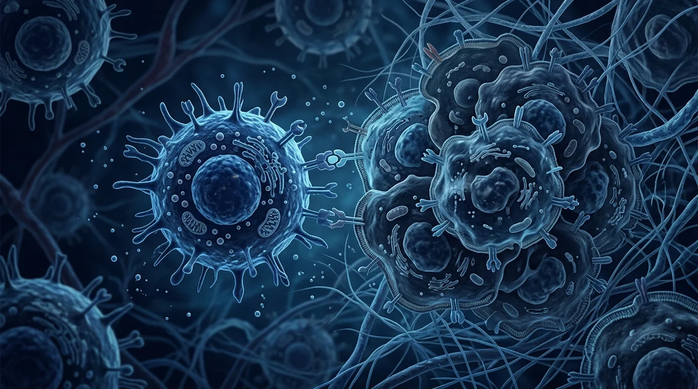
Natural killer (NK) cells are innate lymphocytes that detect and destroy stressed, infected or transformed cells without needing prior exposure to a specific antigen, using a balance of activating and inhibitory surface receptors rather than a single target-specific receptor. That receptor arithmetic is the whole of NK cell biology, and it explains both why NK cells matter in cancer and why NK-cell therapy has been harder to translate than its biology suggests [1][5].
NK cells work by subtraction, not recognition. A cytotoxic T cell asks a specific question: is this exact peptide present? An NK cell asks a broader one: does this cell still look normal? Loss of normal self-markers, or gain of stress ligands, is enough to trigger killing.
That makes NK cells fast but blunt. They act within hours rather than days, need no clonal expansion, and can attack tumour cells that have escaped T-cell surveillance by shedding antigen presentation machinery. They are also easily suppressed, short-lived after transfer, and heavily constrained inside solid tumours.
This article is about the biology, not a treatment offer. Understanding how NK cells recognise, engage and kill a target is the prerequisite for judging any NK-cell therapy claim honestly — including at clinics in Bangkok. Adoptive NK-cell therapy for cancer remains investigational outside defined transplant settings.
Why the mechanism matters to patients
Almost every exaggerated NK claim — "boosts your natural killer cells", "kills any cancer cell", "restores immune surveillance" — collapses when the receptor logic is spelled out. Knowing what NK cells actually detect lets you ask whether a specific proposal is biologically coherent for a specific tumour.
What are natural killer cells?
Natural killer cells are a distinct lineage of innate lymphoid cells that make up roughly 5–15% of circulating lymphocytes in healthy adults and can kill target cells directly without prior sensitisation. They were named for exactly that property: cytotoxicity that required no immunisation and no antigen-specific priming [1].
Human NK cells are conventionally identified as CD56-positive, CD3-negative lymphocytes, and they split into functionally different subsets. CD56dimCD16bright cells dominate the blood and are the more potent direct killers. CD56bright cells are enriched in tissue and lymph nodes and are stronger cytokine producers, releasing interferon-gamma and TNF rather than killing preferentially [11].
Modern single-cell work has complicated that neat division considerably. Human NK populations show substantial phenotypic and functional diversity shaped by development, tissue residence, cytomegalovirus exposure and prior activation, and the field now describes a spectrum rather than two boxes [19][21]. NK cells also sit within the broader innate lymphoid cell family, which matters because some tissue-resident populations that look NK-like behave quite differently in tumours [20].
Practical implication: "NK cells" is not one product. Two infusions with the same label can contain different subsets with different killing capacity and different persistence — which is why manufacturing detail matters, as covered in our article on NK cell manufacturing and quality control.
How do NK cells recognise cancer cells?
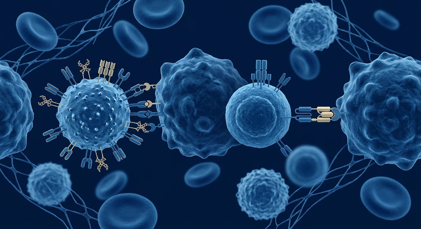
NK cells recognise abnormal cells by integrating signals from inhibitory receptors that read normal self-markers against activating receptors that read stress and damage — killing occurs when activation outweighs inhibition. There is no single "cancer receptor"; the decision is a summed threshold [6].
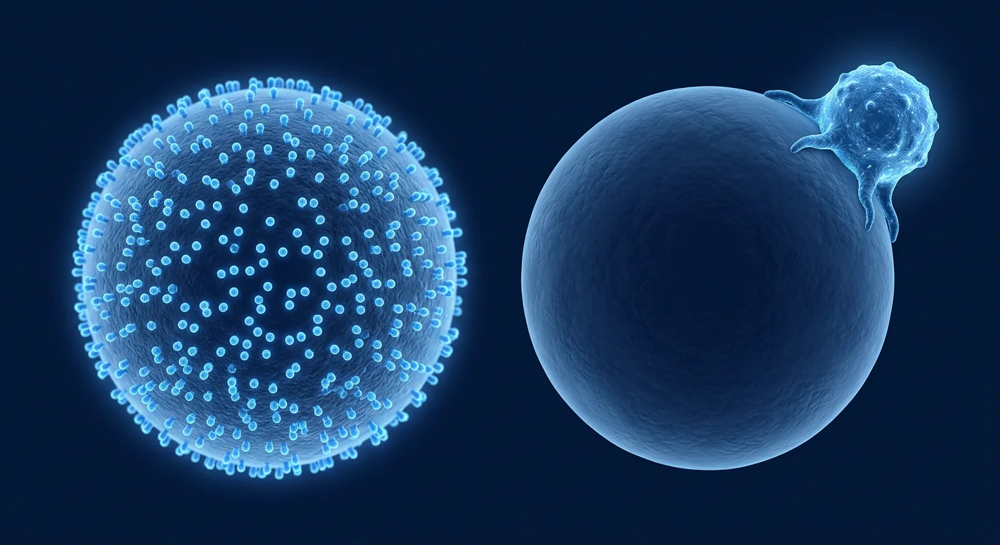
Missing self: the founding principle
The core insight came from a 1986 observation that lymphoma variants lacking MHC class I were selectively rejected, suggesting an immune strategy that targets the absence of self-markers rather than the presence of foreign ones [3]. That became the "missing self" hypothesis: MHC class I molecules act as a licence to live, read by NK inhibitory receptors [4].
In humans the main inhibitory receptors are the killer-cell immunoglobulin-like receptors (KIR), which bind HLA-A, -B and -C allotypes, and NKG2A, which binds HLA-E. When a virus-infected or transformed cell downregulates HLA class I — a common escape route from cytotoxic T cells — it simultaneously loses NK inhibition. Escaping one arm of immunity exposes it to the other.
Induced self: activating ligands
Loss of inhibition alone is usually insufficient. NK cells also require positive signals through activating receptors, most prominently NKG2D, which recognises stress-inducible ligands such as MICA, MICB and the ULBP family [7]. These ligands are largely absent from healthy tissue and induced by DNA damage, oncogene activation, proliferative stress and viral infection [8].
Other activating inputs include the natural cytotoxicity receptors NKp30, NKp44 and NKp46, DNAM-1 (CD226) binding CD155/CD112, and — importantly for therapy — CD16 (FcγRIIIa), which binds antibody Fc regions and triggers antibody-dependent cellular cytotoxicity [5].
Licensing and education
NK cells are not born fully functional. During development they undergo "education" or licensing: cells whose inhibitory receptors engage self-HLA become more responsive, while those without a matched self-ligand remain hyporesponsive. This calibration is what stops NK cells from attacking healthy HLA-low tissue, and it also explains why donor–recipient HLA/KIR combinations affect NK reactivity in transplantation [6].
How do NK cells kill a target cell?
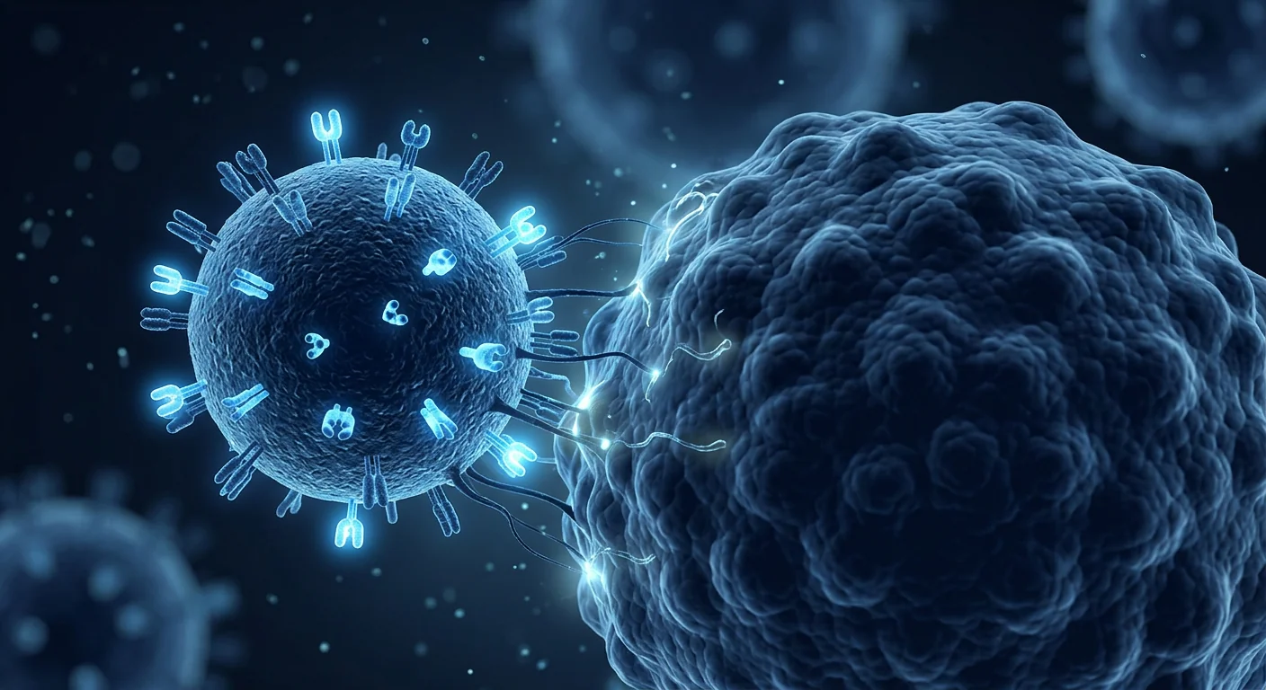
Once activation exceeds the inhibitory threshold, an NK cell forms a tight immunological synapse with the target, polarises pre-formed cytotoxic granules toward the contact site, and releases perforin and granzymes that trigger target-cell apoptosis within minutes. The machinery is largely pre-loaded, which is why NK killing is fast [9][10].
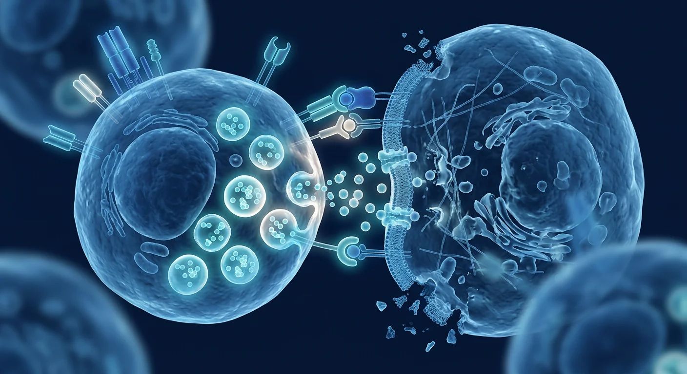
The sequence has four recognisable stages:
- Adhesion and synapse formation. Integrins including LFA-1 stabilise contact and commit the cell to a directed response.
- Granule polarisation. Lytic granules move along the microtubule network to the synapse and dock at the membrane.
- Degranulation. Perforin oligomerises in the target membrane, allowing granzymes to enter and activate caspase-dependent and caspase-independent apoptosis [9].
- Detachment and serial killing. The NK cell disengages and can kill again, though its capacity declines with each round.
Granule exocytosis is not the only route. NK cells also engage death receptors through FasL and TRAIL, and they secrete interferon-gamma and TNF that reshape the local immune response rather than killing directly [10]. Human genetic deficiencies in this pathway are informative: defects in perforin and its trafficking machinery cause severe immune dysregulation syndromes, confirming that the mechanism is functionally essential rather than redundant [9].
NK cells also act as immune organisers. In mouse models, NK-derived chemokines recruited conventional type 1 dendritic cells into tumours, linking innate killing to downstream T-cell priming [22]. This "conductor" role is a plausible reason NK activity correlates with outcomes beyond the number of cells it kills directly.
What is the evidence that NK cells matter in human cancer?
The strongest human evidence that NK function matters in cancer is epidemiological and immunogenetic rather than interventional: low natural cytotoxic activity predicted higher cancer incidence, and NK-deficient patients show characteristic disease patterns. These are association data, and they support relevance rather than proving that boosting NK cells prevents cancer.
An 11-year prospective follow-up of a Japanese general population found that low peripheral-blood natural cytotoxic activity was associated with increased cancer incidence, while medium and high activity were associated with reduced risk [12]. It remains one of the most cited pieces of human evidence for NK-mediated tumour surveillance — and it is observational, so reverse causation and confounding cannot be excluded.
Human NK cell deficiency states provide converging evidence. Patients with classical or functional NK deficiency present most strikingly with severe herpesvirus infections, and some have an increased burden of virus-associated malignancy [13]. Notably, they do not present with a universally elevated rate of all cancers, which argues against a simple model in which NK cells single-handedly police tumour development.
Within tumours, higher NK infiltration has been associated with better prognosis in several cancer types, but NK cells recovered from tumours are frequently dysfunctional — with downregulated activating receptors, impaired degranulation and altered metabolism [14][18]. The barrier is often not absence but suppression.
| Evidence type | What it shows | What it cannot show |
|---|---|---|
| Population cytotoxicity cohort [12] | Low NK activity tracks with higher cancer incidence | That raising NK activity lowers risk |
| NK deficiency syndromes [13] | NK cells are essential for herpesvirus control | Broad anti-cancer indispensability |
| Tumour-infiltration studies [14] | NK presence often correlates with prognosis | Direction of causation |
| Transplant alloreactivity | NK biology can be clinically consequential | Transferability to solid tumours |
| Early-phase NK trials [15][16] | Feasibility and acceptable early safety | Efficacy in most indications |
Why do tumours escape NK cells?
Tumours escape NK cells by hiding activating ligands, retaining or restoring inhibitory signals, and creating a microenvironment that starves and exhausts NK cells metabolically. Each mechanism is a direct counter to a step in the recognition-and-killing sequence described above.
- Ligand shedding. Tumours release soluble MICA/MICB that binds NKG2D without triggering it, downregulating the receptor on NK cells [8].
- Inhibitory retention. Selective preservation or induction of HLA-E sustains NKG2A-mediated inhibition even when classical HLA class I is reduced.
- Suppressive mediators. TGF-beta, adenosine, prostaglandin E2, IDO metabolites and regulatory T cells all impair NK cytotoxicity locally [14].
- Metabolic and physical barriers. Hypoxia, glucose competition and dense stroma limit NK infiltration and function in solid tumours [18].
- Exhaustion phenotypes. Chronic stimulation drives loss of effector capacity that resembles, but is not identical to, T-cell exhaustion.
These escape routes are why NK-based therapy strategies rarely rely on cells alone, and why receptor blockade, cytokine support and engager molecules feature so heavily in trial design — a topic we cover in NK cell therapy combinations.
Do NK cells have memory?
NK cells were long classified as purely innate and memory-free, but several forms of NK memory-like behaviour are now documented, blurring the innate–adaptive boundary. The evidence is real but narrower than popular summaries imply [2].
Two phenomena are best characterised. Adaptive NK cells — typically NKG2C-positive and expanded after human cytomegalovirus infection — persist for long periods with distinct epigenetic profiles. Cytokine-induced memory-like NK cells are generated by brief pre-activation with IL-12, IL-15 and IL-18, and show enhanced and more durable responses against myeloid leukaemia after transfer [17].
This matters therapeutically because it moves activation into the manufacturing step instead of exposing the patient to high-dose systemic cytokines. It does not mean an NK infusion confers lasting immunity comparable to vaccination, and durability data in solid tumours remain limited.
How does NK biology translate into therapy — and what is unproven?
NK biology supports several therapeutic strategies, but in most cancer indications adoptive NK-cell therapy remains investigational and should be accessed through registered clinical trials or regulated pathways. The mechanism is not the evidence.
Current translational approaches follow directly from the biology: supplying missing recognition through antibodies or engineered receptors; removing inhibition with KIR or NKG2A blockade; supplying survival signals with IL-15-family cytokines; pre-programming memory-like function during manufacturing; and choosing cell sources — peripheral blood, cord blood, or induced pluripotent stem cells — that yield different subset composition and scalability [15][16]. Our guides on NK cell sources, NK versus CAR-T and CAR-NK architecture examine those choices in detail.
What the biology does not support is any of the following claims: that an infusion "restores immune surveillance"; that NK therapy works across tumour types because the mechanism is antigen-independent; that a higher cell dose is necessarily better; or that a blood NK-activity number predicts an individual's treatment response. Reported clinical activity has been most encouraging in specific B-cell and myeloid haematological settings and markedly weaker in solid tumours [16][18].
Frequently asked questions
In short: NK cells kill by comparing inhibitory self-signals against activating stress signals, they act within hours using pre-formed cytotoxic granules, and their relevance to human cancer is well supported biologically while NK-cell therapy remains largely investigational.
What is the difference between NK cells and T cells?
T cells recognise specific antigen peptides presented on MHC molecules and require priming and clonal expansion. NK cells respond to the loss of self-markers and the presence of stress ligands without prior sensitisation, acting faster but with less target specificity [1].
Can a blood test measure my NK cell activity?
Yes, NK cell counts and functional cytotoxicity assays exist and were used in the epidemiological cohort described above [12]. However, results vary with assay method, timing, infection and stress, and no validated threshold predicts an individual's cancer risk or treatment benefit.
Do NK cells attack healthy cells?
Generally no, because healthy cells display normal HLA class I and few stress ligands, and because NK education calibrates responsiveness against self. Pathological NK activation occurs in specific disease states rather than as a routine consequence of activation.
Can lifestyle changes boost NK cells?
Exercise, sleep and stress produce measurable short-term changes in circulating NK numbers and cytotoxicity, and severe stress or sleep deprivation can reduce activity. It has not been demonstrated that such changes translate into altered cancer outcomes, so lifestyle claims should be kept modest.
Why is NK therapy less effective in solid tumours?
Because solid tumours add barriers that are absent in circulating malignancies: limited infiltration, hypoxia, suppressive metabolites, dense stroma and functional exhaustion of infiltrating NK cells [18].
Is NK cell therapy available as standard treatment in Bangkok?
Adoptive NK-cell therapy is not an approved standard treatment for most cancers. Outside defined transplant contexts, it should be pursued through registered clinical trials or regulated pathways, and it should never replace guideline-recommended diagnosis and treatment.
Limitations and medical disclaimer
Several limitations apply to everything above. Much of the mechanistic detail derives from in-vitro and murine work that does not always reproduce in humans; the epidemiological association between NK activity and cancer incidence is observational; NK activity assays are not standardised across laboratories; "NK cell" describes a heterogeneous population whose behaviour depends on subset and tissue context; and clinical results from one product or platform do not transfer to another.
This article is educational and does not describe a VELAR treatment protocol, recommend any NK-cell product, or constitute medical advice. NK-cell therapies for cancer are largely investigational. Patients should not delay, substitute or discontinue guideline-recommended cancer screening, diagnosis or treatment in order to pursue an unproven cell therapy, and should discuss any proposal — including its trial registration, evidence, monitoring plan and documented risks — with a qualified oncologist.
References
- Vivier E, Tomasello E, Baratin M, Walzer T, Ugolini S. Functions of natural killer cells. Nature Immunology. 2008;9:503–510. doi:10.1038/ni1582 ↩
- Vivier E, Raulet DH, Moretta A, et al. Innate or adaptive immunity? The example of natural killer cells. Science. 2011;331:44–49. doi:10.1126/science.1198687 ↩
- Kärre K, Ljunggren HG, Piontek G, Kiessling R. Selective rejection of H-2-deficient lymphoma variants suggests alternative immune defence strategy. Nature. 1986;319:675–678. doi:10.1038/319675a0 ↩
- Ljunggren HG, Kärre K. In search of the 'missing self': MHC molecules and NK cell recognition. Immunology Today. 1990;11:237–244. doi:10.1016/0167-5699(90)90097-S ↩
- Lanier LL. NK cell recognition. Annual Review of Immunology. 2005;23:225–274. doi:10.1146/annurev.immunol.23.021704.115526 ↩
- Long EO, Kim HS, Liu D, Peterson ME, Rajagopalan S. Controlling natural killer cell responses: integration of signals for activation and inhibition. Annual Review of Immunology. 2013;31:227–258. doi:10.1146/annurev-immunol-020711-075005 ↩
- Bauer S, Groh V, Wu J, et al. Activation of NK cells and T cells by NKG2D, a receptor for stress-inducible MICA. Science. 1999;285:727–729. doi:10.1126/science.285.5428.727 ↩
- Raulet DH, Guerra N. Oncogenic stress sensed by the immune system: role of natural killer cell receptors. Nature Reviews Immunology. 2009;9:568–580. doi:10.1038/nri2604 ↩
- Voskoboinik I, Whisstock JC, Trapani JA. Perforin and granzymes: function, dysfunction and human pathology. Nature Reviews Immunology. 2015;15:388–400. doi:10.1038/nri3839 ↩
- Prager I, Watzl C. Mechanisms of natural killer cell-mediated cellular cytotoxicity — a comprehensive review. Journal of Leukocyte Biology. 2019;105:1319–1329. doi:10.1002/JLB.MR0718-269R ↩
- Cooper MA, Fehniger TA, Caligiuri MA. The biology of human natural killer-cell subsets. Trends in Immunology. 2001;22:633–640. doi:10.1016/S1471-4906(01)02060-9 ↩
- Imai K, Matsuyama S, Miyake S, Suga K, Nakachi K. Natural cytotoxic activity of peripheral-blood lymphocytes and cancer incidence: an 11-year follow-up study of a general population. The Lancet. 2000;356:1795–1799. doi:10.1016/S0140-6736(00)03231-1 ↩
- Orange JS. Natural killer cell deficiency. Journal of Allergy and Clinical Immunology. 2013;132:515–525. doi:10.1016/j.jaci.2013.07.020 ↩
- Guillerey C, Huntington ND, Smyth MJ. Targeting natural killer cells in cancer immunotherapy. Nature Immunology. 2016;17:1025–1036. doi:10.1038/ni.3518 ↩
- Shimasaki N, Jain A, Campana D. NK cells for cancer immunotherapy. Nature Reviews Drug Discovery. 2020;19:200–218. doi:10.1038/s41573-019-0052-1 ↩
- Myers JA, Miller JS. Exploring the NK cell platform for cancer immunotherapy. Nature Reviews Clinical Oncology. 2021;18:85–100. doi:10.1038/s41571-020-0426-7 ↩
- Romee R, Rosario M, Berrien-Elliott MM, et al. Cytokine-induced memory-like natural killer cells exhibit enhanced responses against myeloid leukemia. Science Translational Medicine. 2016;8:357ra123. doi:10.1126/scitranslmed.aaf2341 ↩
- Bald T, Krummel MF, Smyth MJ, Barry KC. The NK cell–cancer cycle: advances and new challenges in NK cell-based immunotherapies. Nature Immunology. 2020;21:835–847. doi:10.1038/s41590-020-0728-z ↩
- Freud AG, Mundy-Bosse BL, Yu J, Caligiuri MA. The broad spectrum of human natural killer cell diversity. Immunity. 2017;47:820–833. doi:10.1016/j.immuni.2017.10.008 ↩
- Chiossone L, Dumas PY, Vienne M, Vivier E. Natural killer cells and other innate lymphoid cells in cancer. Nature Reviews Immunology. 2018;18:671–688. doi:10.1038/s41577-018-0061-z ↩
- Cichocki F, Grzywacz B, Miller JS. Human NK cell development: one road or many? Frontiers in Immunology. 2019;10:2078. doi:10.3389/fimmu.2019.02078 ↩
- Böttcher JP, Bonavita E, Chakravarty P, et al. NK cells stimulate recruitment of cDC1 into the tumor microenvironment promoting cancer immune control. Cell. 2018;172:1022–1037. doi:10.1016/j.cell.2018.01.004 ↩
自然杀伤(NK)细胞是先天免疫系统中的淋巴细胞,无需事先接触特定抗原即可识别并清除应激、感染或恶性转化的细胞;它依靠激活性与抑制性表面受体之间的平衡,而不是某一个特异性受体。这套“受体加减法”构成了 NK 细胞生物学的全部核心,也解释了为什么 NK 细胞在肿瘤中很重要,而 NK 细胞治疗的临床转化却比其生物学听起来困难得多 [1][5]。
NK 细胞靠“减法”工作,而不是靠特异识别。细胞毒性 T 细胞提出的是一个具体问题:这个特定的多肽在不在?NK 细胞提出的问题更宽泛:这个细胞看起来还正常吗?失去正常的自我标志,或出现应激配体,就足以触发杀伤。
这让 NK 细胞反应迅速但不够精准。它们在数小时内起效,不需要克隆扩增,能够攻击那些通过下调抗原提呈机制而逃脱 T 细胞监视的肿瘤细胞。但它们同样容易被抑制、输注后存续时间短,并在实体瘤内部受到严重限制。
本文讨论的是生物学机制,而不是治疗推荐。理解 NK 细胞如何识别、结合并杀伤靶细胞,是客观评估任何 NK 细胞治疗宣称(包括在曼谷)的前提。除特定移植情境外,过继性 NK 细胞治疗癌症目前仍属研究性质。
机制为什么对患者重要
几乎所有夸大的 NK 宣称——“提升您的自然杀伤细胞”“杀死任何癌细胞”“恢复免疫监视”——一旦把受体逻辑讲清楚就站不住脚。了解 NK 细胞究竟识别什么,才能判断某个具体方案对某个具体肿瘤是否在生物学上说得通。
什么是自然杀伤细胞?
自然杀伤细胞是一类独立的先天淋巴细胞谱系,约占健康成年人循环淋巴细胞的 5%–15%,可在未经致敏的情况下直接杀伤靶细胞。它们正因这一特性而得名:无需免疫接种、无需抗原特异性预激活的细胞毒性 [1]。
人类 NK 细胞通常定义为 CD56 阳性、CD3 阴性的淋巴细胞,并分为功能不同的亚群。CD56dimCD16bright 细胞在血液中占主导,是更强的直接杀伤细胞;CD56bright 细胞富集于组织与淋巴结,更擅长分泌干扰素-γ 和 TNF 等细胞因子,而非优先执行杀伤 [11]。
近年的单细胞研究让这一简洁的二分法复杂了许多。人类 NK 群体在发育、组织定居、巨细胞病毒暴露与既往激活的影响下呈现显著的表型与功能多样性,目前学界更倾向于用连续谱而非两个类别来描述 [19][21]。NK 细胞也属于更广义的先天淋巴细胞家族,这一点很重要,因为某些组织定居的“类 NK”群体在肿瘤中的行为并不相同 [20]。
实际含义:“NK 细胞”并不是单一产品。两份标签相同的输注制剂,其亚群构成、杀伤能力与存续时间可能完全不同——这正是制造细节至关重要的原因,详见我们关于 NK 细胞制造与质量控制 的文章。
NK 细胞如何识别癌细胞?
NK 细胞通过整合两类信号来识别异常细胞:读取正常自我标志的抑制性受体,与读取应激和损伤的激活性受体;当激活信号超过抑制信号时才发生杀伤。并不存在单一的“癌症受体”,这个决定是一个求和后的阈值判断 [6]。
缺失自我:奠基性原理
核心洞见来自 1986 年的一项观察:缺乏 MHC I 类分子的淋巴瘤变异株被选择性排斥,提示存在一种针对“自我标志缺失”而非“外来标志存在”的免疫策略 [3]。这就是“缺失自我”假说:MHC I 类分子相当于一张生存许可证,由 NK 抑制性受体读取 [4]。
在人类中,主要抑制性受体包括结合 HLA-A、-B、-C 等位基因的杀伤细胞免疫球蛋白样受体(KIR),以及结合 HLA-E 的 NKG2A。当病毒感染或转化的细胞下调 HLA I 类分子(这是逃避细胞毒性 T 细胞的常见途径)时,它同时也失去了对 NK 的抑制。逃过免疫系统的一条臂膀,就暴露在另一条臂膀之下。
诱导自我:激活性配体
仅仅失去抑制通常还不够。NK 细胞同样需要来自激活性受体的正向信号,其中最重要的是 NKG2D,它识别 MICA、MICB 与 ULBP 家族等应激诱导配体 [7]。这些配体在健康组织中基本不表达,由 DNA 损伤、癌基因激活、增殖应激与病毒感染诱导产生 [8]。
其他激活性输入还包括天然细胞毒性受体 NKp30、NKp44、NKp46,结合 CD155/CD112 的 DNAM-1(CD226),以及对治疗尤为关键的 CD16(FcγRIIIa)——它结合抗体 Fc 段,触发抗体依赖性细胞毒作用 [5]。
“执照”与教育
NK 细胞并非天生即具备完整功能。在发育过程中它们经历“教育”或获得执照:抑制性受体能与自身 HLA 结合的细胞反应性更强,而缺乏匹配自身配体的细胞则保持低反应状态。这一校准机制避免了 NK 细胞攻击 HLA 低表达的健康组织,也解释了为何供受者的 HLA/KIR 组合会影响移植中的 NK 反应性 [6]。
NK 细胞如何杀伤靶细胞?
一旦激活信号超过抑制阈值,NK 细胞便与靶细胞形成紧密的免疫突触,将预先形成的细胞毒性颗粒极化至接触部位,并释放穿孔素与颗粒酶,在数分钟内诱导靶细胞凋亡。由于杀伤机器是预装好的,NK 杀伤才如此迅速 [9][10]。
整个过程可分为四个阶段:
- 黏附与突触形成。包括 LFA-1 在内的整合素稳定接触,使细胞进入定向反应状态。
- 颗粒极化。溶解性颗粒沿微管网络移动至突触并在膜上停靠。
- 脱颗粒。穿孔素在靶细胞膜上寡聚成孔,使颗粒酶进入并激活依赖或不依赖 caspase 的凋亡通路 [9]。
- 脱离与连续杀伤。NK 细胞脱离后可再次杀伤,但每一轮之后能力都会下降。
颗粒胞吐并非唯一途径。NK 细胞还通过 FasL 与 TRAIL 激活死亡受体通路,并分泌干扰素-γ 与 TNF,从而重塑局部免疫反应而非直接杀伤 [10]。人类相关基因缺陷很有说明性:穿孔素及其转运机制的缺陷会导致严重的免疫失调综合征,证明该机制在功能上不可替代 [9]。
NK 细胞同时扮演免疫组织者的角色。在小鼠模型中,NK 来源的趋化因子将 cDC1 型树突状细胞招募进肿瘤,把先天杀伤与下游 T 细胞激活联系起来 [22]。这种“指挥者”作用,很可能是 NK 活性与预后的相关性超出其直接杀伤数量的原因之一。
NK 细胞在人类癌症中重要性的证据是什么?
支持 NK 功能与人类癌症相关性的最有力证据属于流行病学与免疫遗传学,而非干预性研究:较低的自然细胞毒活性与更高的癌症发生率相关,NK 缺陷患者也表现出特征性的疾病谱。这些是关联性数据,支持“相关”而非证明“提升 NK 可预防癌症”。
一项针对日本一般人群的 11 年前瞻性随访发现,外周血自然细胞毒活性较低者癌症发生率升高,而中、高活性者风险降低 [12]。它至今仍是支持 NK 介导肿瘤监视的最常被引用的人类证据之一——同时它是观察性研究,无法排除反向因果与混杂因素。
人类 NK 细胞缺陷状态提供了相互印证的证据。经典或功能性 NK 缺陷患者最突出的表现是严重疱疹病毒感染,部分患者的病毒相关恶性肿瘤负担增加 [13]。值得注意的是,他们并未普遍出现所有癌症发病率的升高,这否定了“NK 细胞独自守卫肿瘤发生”的简单模型。
在肿瘤内部,多种癌症中较高的 NK 浸润与较好预后相关,但从肿瘤中分离出的 NK 细胞常常功能失调——激活性受体下调、脱颗粒受损、代谢改变 [14][18]。障碍往往不是“缺席”,而是“被压制”。
| 证据类型 | 能说明什么 | 不能说明什么 |
|---|---|---|
| 人群细胞毒活性队列 [12] | 低 NK 活性与更高癌症发生率相关 | 提高 NK 活性可降低风险 |
| NK 缺陷综合征 [13] | NK 细胞对控制疱疹病毒不可或缺 | 对所有癌症具有普遍不可替代性 |
| 肿瘤浸润研究 [14] | NK 浸润常与预后相关 | 因果方向 |
| 移植中的同种反应性 | NK 生物学可具临床意义 | 可直接外推至实体瘤 |
| 早期 NK 临床试验 [15][16] | 可行性与早期安全性可接受 | 多数适应证中的疗效 |
肿瘤为何能逃脱 NK 细胞?
肿瘤通过隐藏激活性配体、保留或恢复抑制性信号,以及营造使 NK 细胞代谢耗竭的微环境来实现逃逸。每一种机制都精确对应上述识别与杀伤流程中的某一步。
- 配体脱落。肿瘤释放可溶性 MICA/MICB,与 NKG2D 结合却不激活它,导致 NK 表面受体下调 [8]。
- 抑制信号保留。选择性保留或诱导 HLA-E,使 NKG2A 介导的抑制在经典 HLA I 类下调时仍然存在。
- 抑制性介质。TGF-β、腺苷、前列腺素 E2、IDO 代谢产物与调节性 T 细胞均在局部削弱 NK 细胞毒性 [14]。
- 代谢与物理屏障。缺氧、葡萄糖竞争与致密间质限制 NK 在实体瘤中的浸润与功能 [18]。
- 耗竭表型。慢性刺激导致效应能力丧失,其形式类似但不完全等同于 T 细胞耗竭。
这些逃逸途径解释了为什么以 NK 为基础的治疗策略很少单独使用细胞,也解释了受体阻断、细胞因子支持与衔接分子为何在试验设计中占据如此重要的位置——相关内容见 NK 细胞联合治疗。
NK 细胞有记忆吗?
NK 细胞长期被归类为纯粹的先天、无记忆细胞,但目前已有多种“类记忆”行为被记录,模糊了先天与适应性免疫的界线。这些证据真实存在,但比大众化说法所暗示的要有限得多 [2]。
其中两种现象研究得最充分。适应性 NK 细胞通常为 NKG2C 阳性,在人巨细胞病毒感染后扩增,可长期存续并具有独特的表观遗传特征。细胞因子诱导的类记忆 NK 细胞由 IL-12、IL-15、IL-18 短暂预激活产生,输注后对髓系白血病表现出更强、更持久的反应 [17]。
这一点在治疗上有意义,因为它把激活步骤移到制造环节,避免患者暴露于大剂量全身细胞因子。但这并不意味着一次 NK 输注能带来类似疫苗的持久免疫,实体瘤中的持久性数据仍然有限。
NK 生物学如何转化为治疗——哪些尚未被证实?
NK 生物学支持若干治疗策略,但在大多数癌症适应证中,过继性 NK 细胞治疗仍属研究性质,应通过注册临床试验或受监管的途径获得。机制不等于证据。
目前的转化路径直接源自生物学:通过抗体或工程化受体提供缺失的识别信号;用 KIR 或 NKG2A 阻断解除抑制;用 IL-15 家族细胞因子提供存活信号;在制造过程中预先编程类记忆功能;以及选择外周血、脐带血或诱导多能干细胞等不同细胞来源,它们的亚群构成与可扩展性各不相同 [15][16]。我们关于 NK 细胞来源、NK 与 CAR-T 的比较 与 CAR-NK 结构 的文章对这些选择有更详细的讨论。
生物学不支持的宣称包括:输注可“恢复免疫监视”;因为机制不依赖抗原,所以 NK 治疗对各类肿瘤都有效;细胞剂量越高越好;或者一个血液 NK 活性数值可以预测某位患者的治疗反应。已报告的临床活性在特定 B 细胞与髓系血液系统疾病中最为鼓舞人心,而在实体瘤中明显更弱 [16][18]。
常见问题
简而言之:NK 细胞通过比较抑制性自我信号与激活性应激信号来决定杀伤,依靠预先形成的细胞毒性颗粒在数小时内起效;其与人类癌症的相关性在生物学上有充分支持,而 NK 细胞治疗在很大程度上仍属研究阶段。
NK 细胞与 T 细胞有什么区别?
T 细胞识别 MHC 分子提呈的特异性抗原多肽,需要预激活与克隆扩增。NK 细胞对自我标志缺失与应激配体出现作出反应,无需事先致敏,反应更快但靶向特异性较低 [1]。
血液检查能测出我的 NK 细胞活性吗?
可以,NK 细胞计数与功能性细胞毒试验确实存在,上述流行病学队列即使用了此类检测 [12]。但结果会随检测方法、时间、感染与压力而变化,目前没有任何经验证的阈值能预测个体的癌症风险或治疗获益。
NK 细胞会攻击健康细胞吗?
通常不会,因为健康细胞表达正常的 HLA I 类分子且应激配体很少,而且 NK 教育机制会针对自身进行反应性校准。病理性 NK 激活出现于特定疾病状态,而非激活的常规后果。
改变生活方式能提升 NK 细胞吗?
运动、睡眠与压力确实会在短期内改变循环 NK 数量与细胞毒性,严重压力或睡眠剥夺可降低活性。但尚未证明这些变化会转化为癌症结局的改变,因此有关生活方式的宣称应保持克制。
为什么 NK 治疗在实体瘤中效果较差?
因为实体瘤增加了循环系统恶性肿瘤所没有的屏障:浸润受限、缺氧、抑制性代谢产物、致密间质,以及浸润 NK 细胞的功能耗竭 [18]。
在曼谷可以把 NK 细胞治疗作为常规治疗吗?
过继性 NK 细胞治疗尚未成为多数癌症的获批标准治疗。除特定移植情境外,应通过注册临床试验或受监管途径进行,且绝不应替代指南推荐的诊断与治疗。
局限性与医疗声明
以上内容存在若干局限。许多机制细节来自体外与小鼠研究,未必能在人体重现;NK 活性与癌症发生率之间的流行病学关联属于观察性证据;NK 活性检测在各实验室之间尚未标准化;“NK 细胞”描述的是一个异质群体,其行为取决于亚群与组织环境;某一产品或平台的临床结果不能外推到另一个平台。
本文仅供教育参考,不描述 VELAR 的治疗方案,不推荐任何 NK 细胞产品,也不构成医疗建议。用于癌症的 NK 细胞治疗在很大程度上仍属研究性质。患者不应为追求未经证实的细胞治疗而延误、替代或中断指南推荐的癌症筛查、诊断与治疗,并应就任何方案(包括其试验注册、证据、监测计划与已记录的风险)咨询合格的肿瘤科医师。
参考文献
- Vivier E, Tomasello E, Baratin M, Walzer T, Ugolini S. Functions of natural killer cells. Nature Immunology. 2008;9:503–510. doi:10.1038/ni1582
- Vivier E, Raulet DH, Moretta A, et al. Innate or adaptive immunity? The example of natural killer cells. Science. 2011;331:44–49. doi:10.1126/science.1198687
- Kärre K, Ljunggren HG, Piontek G, Kiessling R. Selective rejection of H-2-deficient lymphoma variants suggests alternative immune defence strategy. Nature. 1986;319:675–678. doi:10.1038/319675a0
- Ljunggren HG, Kärre K. In search of the 'missing self': MHC molecules and NK cell recognition. Immunology Today. 1990;11:237–244. doi:10.1016/0167-5699(90)90097-S
- Lanier LL. NK cell recognition. Annual Review of Immunology. 2005;23:225–274. doi:10.1146/annurev.immunol.23.021704.115526
- Long EO, Kim HS, Liu D, Peterson ME, Rajagopalan S. Controlling natural killer cell responses: integration of signals for activation and inhibition. Annual Review of Immunology. 2013;31:227–258. doi:10.1146/annurev-immunol-020711-075005
- Bauer S, Groh V, Wu J, et al. Activation of NK cells and T cells by NKG2D, a receptor for stress-inducible MICA. Science. 1999;285:727–729. doi:10.1126/science.285.5428.727
- Raulet DH, Guerra N. Oncogenic stress sensed by the immune system: role of natural killer cell receptors. Nature Reviews Immunology. 2009;9:568–580. doi:10.1038/nri2604
- Voskoboinik I, Whisstock JC, Trapani JA. Perforin and granzymes: function, dysfunction and human pathology. Nature Reviews Immunology. 2015;15:388–400. doi:10.1038/nri3839
- Prager I, Watzl C. Mechanisms of natural killer cell-mediated cellular cytotoxicity — a comprehensive review. Journal of Leukocyte Biology. 2019;105:1319–1329. doi:10.1002/JLB.MR0718-269R
- Cooper MA, Fehniger TA, Caligiuri MA. The biology of human natural killer-cell subsets. Trends in Immunology. 2001;22:633–640. doi:10.1016/S1471-4906(01)02060-9
- Imai K, Matsuyama S, Miyake S, Suga K, Nakachi K. Natural cytotoxic activity of peripheral-blood lymphocytes and cancer incidence: an 11-year follow-up study of a general population. The Lancet. 2000;356:1795–1799. doi:10.1016/S0140-6736(00)03231-1
- Orange JS. Natural killer cell deficiency. Journal of Allergy and Clinical Immunology. 2013;132:515–525. doi:10.1016/j.jaci.2013.07.020
- Guillerey C, Huntington ND, Smyth MJ. Targeting natural killer cells in cancer immunotherapy. Nature Immunology. 2016;17:1025–1036. doi:10.1038/ni.3518
- Shimasaki N, Jain A, Campana D. NK cells for cancer immunotherapy. Nature Reviews Drug Discovery. 2020;19:200–218. doi:10.1038/s41573-019-0052-1
- Myers JA, Miller JS. Exploring the NK cell platform for cancer immunotherapy. Nature Reviews Clinical Oncology. 2021;18:85–100. doi:10.1038/s41571-020-0426-7
- Romee R, Rosario M, Berrien-Elliott MM, et al. Cytokine-induced memory-like natural killer cells exhibit enhanced responses against myeloid leukemia. Science Translational Medicine. 2016;8:357ra123. doi:10.1126/scitranslmed.aaf2341
- Bald T, Krummel MF, Smyth MJ, Barry KC. The NK cell–cancer cycle: advances and new challenges in NK cell-based immunotherapies. Nature Immunology. 2020;21:835–847. doi:10.1038/s41590-020-0728-z
- Freud AG, Mundy-Bosse BL, Yu J, Caligiuri MA. The broad spectrum of human natural killer cell diversity. Immunity. 2017;47:820–833. doi:10.1016/j.immuni.2017.10.008
- Chiossone L, Dumas PY, Vienne M, Vivier E. Natural killer cells and other innate lymphoid cells in cancer. Nature Reviews Immunology. 2018;18:671–688. doi:10.1038/s41577-018-0061-z
- Cichocki F, Grzywacz B, Miller JS. Human NK cell development: one road or many? Frontiers in Immunology. 2019;10:2078. doi:10.3389/fimmu.2019.02078
- Böttcher JP, Bonavita E, Chakravarty P, et al. NK cells stimulate recruitment of cDC1 into the tumor microenvironment promoting cancer immune control. Cell. 2018;172:1022–1037. doi:10.1016/j.cell.2018.01.004
الخلايا القاتلة الطبيعية (NK) هي خلايا لمفية من المناعة الفطرية تكتشف الخلايا المُجهدة أو المصابة أو المتحولة سرطانياً وتقضي عليها دون حاجة إلى تعرّض مسبق لمستضد محدد، وذلك بالاعتماد على توازن بين مستقبلات سطحية مُنشِّطة وأخرى كابتة بدلاً من مستقبل واحد نوعي للهدف. هذه «الحسابات المستقبِلية» هي جوهر بيولوجيا الخلايا القاتلة الطبيعية بالكامل، وهي تفسّر أهمية هذه الخلايا في السرطان، كما تفسّر لماذا كان تحويل العلاج بها إلى الممارسة أصعب بكثير مما تشير إليه بيولوجيتها [1][5].
الخلايا القاتلة الطبيعية تعمل بالطرح لا بالتعرّف النوعي. الخلية التائية السامة تطرح سؤالاً محدداً: هل هذا الببتيد بالتحديد موجود؟ أما الخلية القاتلة الطبيعية فتطرح سؤالاً أوسع: هل ما زالت هذه الخلية تبدو طبيعية؟ إن فقدان علامات الذات الطبيعية، أو ظهور روابط الإجهاد، يكفي لإطلاق القتل.
هذا يجعلها سريعة لكن غير دقيقة. فهي تعمل خلال ساعات لا أيام، ولا تحتاج إلى تكاثر نسيلي، وتستطيع مهاجمة خلايا سرطانية أفلتت من مراقبة الخلايا التائية بتخليها عن آلية عرض المستضدات. لكنها في المقابل سهلة الكبت، وقصيرة البقاء بعد النقل، ومُقيَّدة بشدة داخل الأورام الصلبة.
هذا المقال عن البيولوجيا، وليس عرضاً علاجياً. فهم كيفية تعرّف هذه الخلايا على هدفها والارتباط به وقتله هو الشرط المسبق لتقييم أي ادعاء بشأن العلاج بالخلايا القاتلة الطبيعية بصدق — بما في ذلك في بانكوك. ويبقى النقل التبنّي للخلايا القاتلة الطبيعية في السرطان بحثياً خارج سياقات زرع محددة.
لماذا تهم الآلية المرضى
معظم الادعاءات المبالَغ فيها — «تنشيط خلاياك القاتلة الطبيعية»، «تقتل أي خلية سرطانية»، «تستعيد المراقبة المناعية» — تنهار حين يُشرح منطق المستقبلات بوضوح. ومعرفة ما تكتشفه هذه الخلايا فعلاً تتيح لك السؤال عمّا إذا كان اقتراح محدد متسقاً بيولوجياً مع ورم محدد.
ما هي الخلايا القاتلة الطبيعية؟
الخلايا القاتلة الطبيعية سلالة متميزة من الخلايا اللمفية الفطرية تشكّل نحو 5–15% من الخلايا اللمفية الدائرة عند البالغين الأصحاء، وتستطيع قتل الخلايا المستهدفة مباشرة دون تحسيس مسبق. وقد سُمّيت بذلك تحديداً لهذه الخاصية: سُمّية خلوية لا تتطلب تمنيعاً ولا تحضيراً نوعياً للمستضد [1].
تُعرَّف الخلايا القاتلة الطبيعية البشرية عادةً بأنها خلايا لمفية موجبة لـ CD56 وسالبة لـ CD3، وتنقسم إلى مجموعات فرعية مختلفة وظيفياً. فخلايا CD56dimCD16bright تسيطر على الدم وهي الأقوى في القتل المباشر، أما خلايا CD56bright فتتركز في الأنسجة والعقد اللمفية وتنتج السيتوكينات مثل إنترفيرون غاما و TNF أكثر من ممارستها القتل [11].
وقد عقّدت دراسات الخلية الواحدة هذا التقسيم البسيط كثيراً. فالمجموعات البشرية تُظهر تنوعاً ظاهرياً ووظيفياً كبيراً يتشكّل بالنمو والإقامة النسيجية والتعرّض للفيروس المضخم للخلايا والتنشيط السابق، ويوصف المجال اليوم بطيف متصل لا بصندوقين [19][21]. كما تنتمي هذه الخلايا إلى عائلة أوسع من الخلايا اللمفية الفطرية، وهذا مهم لأن بعض المجموعات المقيمة في الأنسجة والتي تبدو مشابهة لها تتصرف بشكل مختلف داخل الأورام [20].
الدلالة العملية: «الخلايا القاتلة الطبيعية» ليست منتجاً واحداً. فحقنتان تحملان الوصف ذاته قد تحتويان مجموعات فرعية مختلفة بقدرات قتل ومدد بقاء مختلفة — ولهذا تفاصيل التصنيع مهمة، كما نوضح في مقالنا عن تصنيع الخلايا القاتلة الطبيعية ومراقبة الجودة.
كيف تتعرّف الخلايا القاتلة الطبيعية على الخلايا السرطانية؟
تتعرّف الخلايا القاتلة الطبيعية على الخلايا الشاذة بدمج إشارات المستقبلات الكابتة التي تقرأ علامات الذات الطبيعية مقابل المستقبلات المُنشِّطة التي تقرأ الإجهاد والتلف — ويحدث القتل عندما يتجاوز التنشيط الكبت. لا يوجد «مستقبل سرطان» واحد؛ القرار حاصل جمع يتخطى عتبة معينة [6].
الذات المفقودة: المبدأ التأسيسي
جاءت الفكرة الأساسية من ملاحظة عام 1986 أن متغيرات اللمفوما التي تفقد الصنف الأول من MHC تُرفض انتقائياً، ما يشير إلى استراتيجية مناعية تستهدف غياب علامات الذات بدلاً من وجود علامات غريبة [3]. وصار ذلك فرضية «الذات المفقودة»: جزيئات MHC من الصنف الأول بمثابة رخصة للبقاء تقرأها المستقبلات الكابتة [4].
وفي الإنسان، المستقبلات الكابتة الرئيسية هي مستقبلات KIR الشبيهة بالغلوبولين المناعي التي ترتبط بأنماط HLA-A و-B و-C، ومستقبل NKG2A الذي يرتبط بـ HLA-E. وعندما تُخفّض خلية مصابة بفيروس أو متحولة تعبير الصنف الأول من HLA — وهو مسار شائع للإفلات من الخلايا التائية — فإنها تفقد في الوقت نفسه كبت الخلايا القاتلة الطبيعية. الإفلات من أحد جناحي المناعة يعرّضها للجناح الآخر.
الذات المستحثّة: الروابط المُنشِّطة
فقدان الكبت وحده لا يكفي عادةً. فالخلايا القاتلة الطبيعية تحتاج أيضاً إشارات موجبة عبر مستقبلات مُنشِّطة، أبرزها NKG2D الذي يتعرّف على روابط مستحثّة بالإجهاد مثل MICA و MICB وعائلة ULBP [7]. وهذه الروابط غائبة إلى حد كبير عن الأنسجة السليمة وتُستحث بتلف الحمض النووي وتنشيط الجينات الورمية والإجهاد التكاثري والعدوى الفيروسية [8].
وتشمل المُدخلات المُنشِّطة الأخرى مستقبلات السُمّية الطبيعية NKp30 و NKp44 و NKp46، ومستقبل DNAM-1 (CD226) الذي يرتبط بـ CD155/CD112، والأهم علاجياً CD16 (FcγRIIIa) الذي يرتبط بمناطق Fc للأجسام المضادة فيُطلق السُمّية الخلوية المعتمدة على الأجسام المضادة [5].
الترخيص والتعليم
لا تُولد هذه الخلايا كاملة الوظيفة. فهي تخضع أثناء النمو لـ«تعليم» أو ترخيص: الخلايا التي ترتبط مستقبلاتها الكابتة بـ HLA الذاتي تصبح أكثر استجابة، أما التي تفتقر إلى رابط ذاتي مطابق فتبقى ضعيفة الاستجابة. هذه المعايرة هي ما يمنع مهاجمة الأنسجة السليمة منخفضة HLA، وتفسّر أيضاً تأثير توافق HLA/KIR بين المتبرع والمتلقي على التفاعلية في الزرع [6].
كيف تقتل الخلية القاتلة الطبيعية هدفها؟
عندما يتجاوز التنشيط عتبة الكبت، تُشكّل الخلية القاتلة الطبيعية تشابكاً مناعياً محكماً مع هدفها، وتوجّه حبيباتها السامة المُعدّة سلفاً نحو موضع التلامس، وتُحرّر البيرفورين والغرانزيمات التي تُحدث موتاً مبرمجاً في الخلية الهدف خلال دقائق. فالآلية محمّلة مسبقاً، ولهذا يكون القتل سريعاً [9][10].
وللعملية أربع مراحل واضحة:
- الالتصاق وتكوين التشابك. تعمل الإنتغرينات ومنها LFA-1 على تثبيت التلامس وإلزام الخلية باستجابة موجّهة.
- استقطاب الحبيبات. تنتقل الحبيبات الحالّة عبر شبكة الأنيبيبات الدقيقة إلى التشابك وترتكز على الغشاء.
- إطلاق الحبيبات. يتبلمر البيرفورين في غشاء الهدف فيسمح للغرانزيمات بالدخول وتنشيط الموت المبرمج المعتمد وغير المعتمد على الكاسباز [9].
- الانفصال والقتل المتسلسل. تنفصل الخلية ويمكنها القتل مرة أخرى، لكن قدرتها تتراجع مع كل دورة.
وإطلاق الحبيبات ليس المسار الوحيد. فهذه الخلايا تُفعّل أيضاً مستقبلات الموت عبر FasL و TRAIL، وتُفرز إنترفيرون غاما و TNF التي تعيد تشكيل الاستجابة المناعية المحلية بدل القتل المباشر [10]. والنقائص الجينية البشرية في هذا المسار دالة: فعيوب البيرفورين وآليات نقله تسبب اضطرابات مناعية شديدة، مما يؤكد أن الآلية أساسية لا زائدة [9].
كما تعمل هذه الخلايا كمنظّم مناعي. ففي نماذج الفئران، جذبت الكيموكينات المشتقة منها الخلايا التغصّنية من نوع cDC1 إلى داخل الورم، فربطت القتل الفطري بتحضير الخلايا التائية لاحقاً [22]. وهذا الدور «القيادي» تفسير محتمل لارتباط نشاطها بالنتائج بما يتجاوز عدد الخلايا التي تقتلها مباشرة.
ما الدليل على أهمية هذه الخلايا في السرطان البشري؟
أقوى الأدلة البشرية على أهمية وظيفة الخلايا القاتلة الطبيعية في السرطان هي وبائية ومناعية-وراثية لا تداخلية: فانخفاض السُمّية الخلوية الطبيعية تنبّأ بزيادة حدوث السرطان، ويُظهر المصابون بنقصها أنماط مرض مميزة. وهذه بيانات ارتباط تدعم الصلة لا تُثبت أن تعزيز هذه الخلايا يمنع السرطان.
وجدت متابعة مستقبلية على مدى 11 عاماً لعموم السكان في اليابان أن انخفاض السُمّية الخلوية الطبيعية في الدم المحيطي ارتبط بزيادة حدوث السرطان، بينما ارتبط النشاط المتوسط والمرتفع بانخفاض الخطر [12]. وتبقى من أكثر الأدلة البشرية استشهاداً على المراقبة الورمية — وهي دراسة رصدية، فلا يمكن استبعاد السببية العكسية والعوامل المُربِكة.
وتوفّر حالات نقص هذه الخلايا دليلاً متقارباً. فالمصابون بنقص كلاسيكي أو وظيفي يعانون أبرز ما يعانون من عدوى فيروس الحلأ الشديدة، وبعضهم يحمل عبئاً أكبر من الأورام المرتبطة بالفيروسات [13]. والملاحظ أنهم لا يظهرون ارتفاعاً شاملاً في معدلات كل السرطانات، وهو ما يعارض نموذجاً بسيطاً تكون فيه هذه الخلايا وحدها حارسة نشوء الورم.
وداخل الأورام، ارتبط ارتفاع تسلل هذه الخلايا بنتائج أفضل في عدة أنواع، لكن الخلايا المستخلَصة من الأورام كثيراً ما تكون معطّلة الوظيفة — بمستقبلات مُنشِّطة منخفضة وإطلاق حبيبات ضعيف واستقلاب متغيّر [14][18]. فالعقبة غالباً ليست الغياب بل الكبت.
| نوع الدليل | ما يُظهره | ما لا يُظهره |
|---|---|---|
| دراسة السُمّية السكانية [12] | انخفاض النشاط يواكب زيادة حدوث السرطان | أن رفع النشاط يخفّض الخطر |
| متلازمات النقص [13] | ضرورة هذه الخلايا لضبط فيروس الحلأ | ضرورة شاملة لكل السرطانات |
| دراسات التسلل الورمي [14] | ارتباط الوجود بالنتائج غالباً | اتجاه السببية |
| التفاعلية في الزرع | أن البيولوجيا قد تكون ذات أثر سريري | إمكان نقلها إلى الأورام الصلبة |
| تجارب مبكرة الطور [15][16] | الجدوى والسلامة المبكرة المقبولة | الفعالية في معظم الاستطبابات |
لماذا تُفلت الأورام من الخلايا القاتلة الطبيعية؟
تُفلت الأورام بإخفاء الروابط المُنشِّطة، والحفاظ على الإشارات الكابتة أو استعادتها، وخلق بيئة مجهرية تُجهد هذه الخلايا استقلابياً حتى الإنهاك. وكل آلية تقابل مباشرة خطوة من خطوات التعرّف والقتل السابقة.
- سلخ الروابط. تُحرّر الأورام MICA/MICB ذوّابة ترتبط بـ NKG2D دون تنشيطه فتُخفّض المستقبل على سطح الخلية [8].
- الإبقاء على الكبت. الحفاظ الانتقائي على HLA-E أو استحثاثه يُبقي الكبت عبر NKG2A قائماً حتى مع انخفاض الصنف الأول التقليدي.
- الوسائط الكابتة. يُضعف TGF-بيتا والأدينوزين والبروستاغلاندين E2 ونواتج IDO والخلايا التائية التنظيمية السُمّية الخلوية موضعياً [14].
- حواجز استقلابية وفيزيائية. نقص الأكسجين والتنافس على الغلوكوز والسدى الكثيف تحدّ من التسلل والوظيفة في الأورام الصلبة [18].
- أنماط الإنهاك. التنبيه المزمن يؤدي إلى فقدان القدرة المؤثِّرة بصورة تشبه إنهاك الخلايا التائية دون أن تطابقه.
ولهذا نادراً ما تعتمد الاستراتيجيات العلاجية على الخلايا وحدها، ولهذا يحتل حجب المستقبلات ودعم السيتوكينات وجزيئات الوصل مكانة كبيرة في تصميم التجارب — وهو موضوع نتناوله في العلاجات التوليفية بالخلايا القاتلة الطبيعية.
هل لهذه الخلايا ذاكرة؟
صُنّفت الخلايا القاتلة الطبيعية طويلاً كخلايا فطرية بلا ذاكرة، لكن عدة أشكال من السلوك الشبيه بالذاكرة موثّقة اليوم، وهي تُذيب الحد بين المناعة الفطرية والتكيّفية. والأدلة حقيقية لكنها أضيق مما توحي به الملخصات الشائعة [2].
وأفضل ظاهرتين توصيفاً هما: الخلايا التكيّفية الموجبة عادةً لـ NKG2C والتي تتوسّع بعد عدوى الفيروس المضخم للخلايا البشري وتبقى مدداً طويلة بسمات وراثية-تخلّقية مميزة؛ والخلايا الشبيهة بالذاكرة المستحثّة بالسيتوكينات التي تُولَّد بتنشيط مسبق قصير بـ IL-12 و IL-15 و IL-18 وتُظهر بعد النقل استجابات أقوى وأطول ضد الأورام النقوية [17].
ولهذا أهمية علاجية لأنه ينقل التنشيط إلى مرحلة التصنيع بدل تعريض المريض لجرعات سيتوكين جهازية عالية. لكنه لا يعني أن حقنة واحدة تمنح مناعة دائمة مماثلة للتطعيم، وتبقى بيانات الاستدامة في الأورام الصلبة محدودة.
كيف تُترجم البيولوجيا إلى علاج — وما غير المُثبت؟
تدعم البيولوجيا عدة استراتيجيات علاجية، لكن في معظم استطبابات السرطان يبقى النقل التبنّي للخلايا القاتلة الطبيعية بحثياً وينبغي الوصول إليه عبر تجارب سريرية مسجّلة أو مسارات منظّمة. فالآلية ليست دليلاً.
وتنبع المقاربات الحالية مباشرة من البيولوجيا: تأمين التعرّف المفقود عبر أجسام مضادة أو مستقبلات مهندسة؛ إزالة الكبت بحجب KIR أو NKG2A؛ تأمين إشارات البقاء بسيتوكينات عائلة IL-15؛ برمجة وظيفة شبيهة بالذاكرة أثناء التصنيع؛ واختيار مصادر خلوية — دم محيطي أو دم حبل سُرّي أو خلايا جذعية محفَّزة متعددة القدرات — تعطي تركيبات فرعية وقابلية توسّع مختلفة [15][16]. وتتناول أدلّتنا عن مصادر الخلايا القاتلة الطبيعية ومقارنتها بـ CAR-T وبنية CAR-NK تلك الخيارات بتفصيل.
وما لا تدعمه البيولوجيا يشمل: أن الحقنة «تستعيد المراقبة المناعية»؛ أن العلاج ينفع في كل أنواع الأورام لأن آليته لا تعتمد على المستضد؛ أن الجرعة الأعلى أفضل بالضرورة؛ أو أن رقم نشاط في الدم يتنبأ باستجابة فرد للعلاج. وقد كان النشاط السريري المُبلَّغ أكثر تشجيعاً في أورام دموية بائية ونقوية محددة، وأضعف بوضوح في الأورام الصلبة [16][18].
أسئلة شائعة
باختصار: تقتل الخلايا القاتلة الطبيعية بمقارنة إشارات الذات الكابتة بإشارات الإجهاد المُنشِّطة، وتعمل خلال ساعات عبر حبيبات سامة مُعدّة سلفاً، وصلتها بالسرطان البشري مدعومة بيولوجياً جيداً بينما يبقى العلاج بها بحثياً في معظمه.
ما الفرق بين الخلايا القاتلة الطبيعية والخلايا التائية؟
تتعرّف الخلايا التائية على ببتيدات مستضدية نوعية معروضة على جزيئات MHC وتحتاج تحضيراً وتكاثراً نسيلياً. أما الخلايا القاتلة الطبيعية فتستجيب لفقدان علامات الذات ووجود روابط الإجهاد دون تحسيس مسبق، فتكون أسرع وأقل نوعية [1].
هل يمكن لتحليل دم قياس نشاط خلاياي القاتلة الطبيعية؟
نعم، توجد اختبارات للعدد والسُمّية الوظيفية وقد استُخدمت في الدراسة الوبائية المذكورة [12]. لكن النتائج تتغير بطريقة الاختبار والتوقيت والعدوى والإجهاد، ولا توجد عتبة مُصادق عليها تتنبأ بخطر السرطان أو منفعة العلاج لفرد بعينه.
هل تهاجم هذه الخلايا الأنسجة السليمة؟
لا عادةً، لأن الخلايا السليمة تعرض HLA من الصنف الأول بشكل طبيعي وقليلاً من روابط الإجهاد، ولأن التعليم يعاير الاستجابة تجاه الذات. والتنشيط المرضي يحدث في حالات مرضية محددة لا كنتيجة روتينية للتنشيط.
هل يرفع تغيير نمط الحياة هذه الخلايا؟
تُحدث الرياضة والنوم والإجهاد تغيّرات قصيرة الأمد قابلة للقياس في العدد والسُمّية، وقد يُخفّض الإجهاد الشديد أو الحرمان من النوم النشاط. لكن لم يُثبت أن هذه التغيّرات تُترجم إلى تغيّر في نتائج السرطان، فينبغي أن تبقى الادعاءات متحفظة.
لماذا يكون العلاج أضعف في الأورام الصلبة؟
لأن الأورام الصلبة تضيف حواجز غائبة في الأورام الدائرة: تسلل محدود، ونقص أكسجين، ونواتج استقلابية كابتة، وسدى كثيف، وإنهاك وظيفي للخلايا المتسللة [18].
هل العلاج بالخلايا القاتلة الطبيعية متاح كعلاج معياري في بانكوك؟
النقل التبنّي لهذه الخلايا ليس علاجاً معيارياً معتمداً لمعظم السرطانات. وخارج سياقات الزرع المحددة ينبغي السعي إليه عبر تجارب مسجّلة أو مسارات منظّمة، ولا ينبغي أبداً أن يحل محل التشخيص والعلاج الموصى بهما في الأدلة الإرشادية.
القيود وإخلاء المسؤولية الطبية
تنطبق قيود عدة على كل ما سبق. فكثير من التفاصيل الآلية مستمد من أعمال مخبرية وفأرية لا تُستنسخ دائماً في الإنسان؛ والارتباط الوبائي بين النشاط وحدوث السرطان رصدي؛ واختبارات النشاط غير موحّدة بين المعامل؛ و«الخلية القاتلة الطبيعية» تصف مجموعة غير متجانسة يعتمد سلوكها على المجموعة الفرعية والسياق النسيجي؛ ونتائج منتج أو منصة لا تُنقل إلى أخرى.
هذا المقال تعليمي ولا يصف بروتوكول علاج في VELAR ولا يوصي بأي منتج ولا يُغني عن المشورة الطبية. والعلاجات المذكورة بحثية في معظمها. وينبغي ألا يؤخّر المرضى أو يستبدلوا أو يوقفوا الفحص والتشخيص والعلاج الموصى بهما في الأدلة الإرشادية سعياً إلى علاج خلوي غير مُثبت، وأن يناقشوا أي اقتراح — بما في ذلك تسجيله البحثي وأدلته وخطة مراقبته ومخاطره الموثّقة — مع طبيب أورام مؤهل.
المراجع
- Vivier E, Tomasello E, Baratin M, Walzer T, Ugolini S. Functions of natural killer cells. Nature Immunology. 2008;9:503–510. doi:10.1038/ni1582
- Vivier E, Raulet DH, Moretta A, et al. Innate or adaptive immunity? The example of natural killer cells. Science. 2011;331:44–49. doi:10.1126/science.1198687
- Kärre K, Ljunggren HG, Piontek G, Kiessling R. Selective rejection of H-2-deficient lymphoma variants suggests alternative immune defence strategy. Nature. 1986;319:675–678. doi:10.1038/319675a0
- Ljunggren HG, Kärre K. In search of the 'missing self': MHC molecules and NK cell recognition. Immunology Today. 1990;11:237–244. doi:10.1016/0167-5699(90)90097-S
- Lanier LL. NK cell recognition. Annual Review of Immunology. 2005;23:225–274. doi:10.1146/annurev.immunol.23.021704.115526
- Long EO, Kim HS, Liu D, Peterson ME, Rajagopalan S. Controlling natural killer cell responses: integration of signals for activation and inhibition. Annual Review of Immunology. 2013;31:227–258. doi:10.1146/annurev-immunol-020711-075005
- Bauer S, Groh V, Wu J, et al. Activation of NK cells and T cells by NKG2D, a receptor for stress-inducible MICA. Science. 1999;285:727–729. doi:10.1126/science.285.5428.727
- Raulet DH, Guerra N. Oncogenic stress sensed by the immune system: role of natural killer cell receptors. Nature Reviews Immunology. 2009;9:568–580. doi:10.1038/nri2604
- Voskoboinik I, Whisstock JC, Trapani JA. Perforin and granzymes: function, dysfunction and human pathology. Nature Reviews Immunology. 2015;15:388–400. doi:10.1038/nri3839
- Prager I, Watzl C. Mechanisms of natural killer cell-mediated cellular cytotoxicity — a comprehensive review. Journal of Leukocyte Biology. 2019;105:1319–1329. doi:10.1002/JLB.MR0718-269R
- Cooper MA, Fehniger TA, Caligiuri MA. The biology of human natural killer-cell subsets. Trends in Immunology. 2001;22:633–640. doi:10.1016/S1471-4906(01)02060-9
- Imai K, Matsuyama S, Miyake S, Suga K, Nakachi K. Natural cytotoxic activity of peripheral-blood lymphocytes and cancer incidence: an 11-year follow-up study of a general population. The Lancet. 2000;356:1795–1799. doi:10.1016/S0140-6736(00)03231-1
- Orange JS. Natural killer cell deficiency. Journal of Allergy and Clinical Immunology. 2013;132:515–525. doi:10.1016/j.jaci.2013.07.020
- Guillerey C, Huntington ND, Smyth MJ. Targeting natural killer cells in cancer immunotherapy. Nature Immunology. 2016;17:1025–1036. doi:10.1038/ni.3518
- Shimasaki N, Jain A, Campana D. NK cells for cancer immunotherapy. Nature Reviews Drug Discovery. 2020;19:200–218. doi:10.1038/s41573-019-0052-1
- Myers JA, Miller JS. Exploring the NK cell platform for cancer immunotherapy. Nature Reviews Clinical Oncology. 2021;18:85–100. doi:10.1038/s41571-020-0426-7
- Romee R, Rosario M, Berrien-Elliott MM, et al. Cytokine-induced memory-like natural killer cells exhibit enhanced responses against myeloid leukemia. Science Translational Medicine. 2016;8:357ra123. doi:10.1126/scitranslmed.aaf2341
- Bald T, Krummel MF, Smyth MJ, Barry KC. The NK cell–cancer cycle: advances and new challenges in NK cell-based immunotherapies. Nature Immunology. 2020;21:835–847. doi:10.1038/s41590-020-0728-z
- Freud AG, Mundy-Bosse BL, Yu J, Caligiuri MA. The broad spectrum of human natural killer cell diversity. Immunity. 2017;47:820–833. doi:10.1016/j.immuni.2017.10.008
- Chiossone L, Dumas PY, Vienne M, Vivier E. Natural killer cells and other innate lymphoid cells in cancer. Nature Reviews Immunology. 2018;18:671–688. doi:10.1038/s41577-018-0061-z
- Cichocki F, Grzywacz B, Miller JS. Human NK cell development: one road or many? Frontiers in Immunology. 2019;10:2078. doi:10.3389/fimmu.2019.02078
- Böttcher JP, Bonavita E, Chakravarty P, et al. NK cells stimulate recruitment of cDC1 into the tumor microenvironment promoting cancer immune control. Cell. 2018;172:1022–1037. doi:10.1016/j.cell.2018.01.004