The EDIM blood test does not look for cancer cells. It looks for a metabolic fingerprint — and that fingerprint comes from a century-old observation about how tumours burn sugar. Understanding the Warburg effect, and the enzyme TKTL1 that sits inside it, is the only honest way to judge what an EDIM score can and cannot mean.
The observation is old, the interpretation is not settled. In 1956 Otto Warburg reported that tumour slices consumed glucose at extraordinary rates and produced lactate even when oxygen was plentiful [1]. Warburg believed this reflected damaged respiration. Modern work has largely rejected his causal explanation while confirming the phenomenon itself [2], [4].
Reprogrammed metabolism is now treated as a core cancer trait. It appears in the widely cited hallmarks framework as "deregulating cellular energetics," retained and expanded in the 2022 revision [6], [7]. That does not make any single metabolic marker diagnostic.
This article explains the biology the EDIM test rests on. It is educational only. EDIM is investigational, is not a stand-alone cancer diagnosis, and does not replace guideline-recommended screening such as mammography, colonoscopy, low-dose CT or HPV testing.
Key message
The Warburg effect is a real, reproducible feature of many tumours, and TKTL1 is one enzyme associated with it. Neither fact establishes that a TKTL1-based blood score detects cancer reliably in an individual. Treat an EDIM result as a signal to discuss with a physician within a standard diagnostic pathway — never as a verdict.
What is the Warburg effect, in plain terms?
The Warburg effect is the tendency of many cancer cells to convert glucose to lactate even when oxygen is available — a process called aerobic glycolysis. Normal cells under good oxygen conditions mostly finish glucose breakdown in the mitochondria, which yields far more ATP per glucose molecule. Tumour cells frequently take the fast, wasteful route instead [2].
Why choose a less efficient pathway? The leading explanation is that proliferating cells do not primarily need ATP — they need building blocks. Rapid glycolysis supplies carbon skeletons for nucleotides, amino acids and lipids, and regenerates the reducing power needed for biosynthesis [3], [8]. Speed of flux, not yield per molecule, is the advantage.
Warburg's original interpretation was wrong in an important way. He proposed irreversible mitochondrial injury as the cause of cancer. Most tumours in fact retain functional mitochondria and use oxidative phosphorylation, glutamine oxidation and fatty-acid oxidation alongside glycolysis [4], [9], [21]. A 2021 physiological reappraisal argues the effect is better understood as a consequence of oncogenic signalling and microenvironmental pressure than as a primary defect [5].
The clinical relevance is already proven in one specific place: imaging. FDG-PET works because tumours take up a labelled glucose analogue avidly. Standardised acquisition and response criteria exist precisely because glucose avidity is measurable and reproducible enough for clinical use [18], [19]. That is the strongest real-world evidence that tumour glucose handling differs from normal tissue — and it is imaging evidence, not blood-test evidence.
Where does the pentose phosphate pathway fit in?
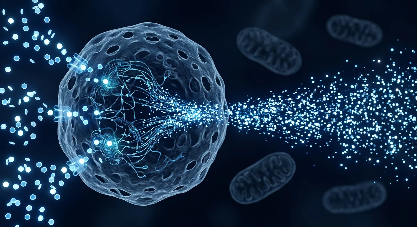
The pentose phosphate pathway (PPP) is the metabolic branch that turns glucose into ribose for DNA and RNA, and into NADPH for antioxidant defence and lipid synthesis. It is a side road off glycolysis, and in proliferating cells it carries heavy traffic [11].
The PPP has two arms. The oxidative arm generates NADPH. The non-oxidative arm, run by transketolase and transaldolase enzymes, interconverts sugars so that a cell can flexibly produce ribose without being locked into NADPH production. Cancer cells exploit this flexibility, and the PPP has therefore been proposed repeatedly as a therapeutic target [12].
NADPH matters for survival, not only growth. It fuels glutathione regeneration, which buffers the oxidative stress that rapid proliferation and many cancer treatments create. This is one reason PPP activity has been linked to treatment resistance in laboratory models [11], [12].
What is TKTL1 and why did it attract attention?
TKTL1 (transketolase-like protein 1) is a transketolase-family gene proposed to support the non-oxidative pentose phosphate pathway in tumours. Interest began with tissue studies reporting that strong TKTL1 staining in colon and urothelial carcinoma was associated with worse patient survival [13].
Functional work followed. Knockdown experiments reported that reducing TKTL1 slowed proliferation and reduced viability in certain cancer cell lines [14]. Expression analyses in colorectal cancer described modulation of TKTL1 across progression and metastasis [15], and a separate study reported that TKTL1 can be activated by promoter hypomethylation, offering an epigenetic route to overexpression [16].
The evidence is genuinely contested, and that matters here. An independent group reported that glucose metabolism in malignant cells was not regulated by TKTL1 in their experimental systems, and raised concerns about antibody specificity and transketolase activity attribution [17]. Any patient-facing description of TKTL1 that omits this disagreement is incomplete.
Honest summary of TKTL1
TKTL1 is a plausible, studied, and disputed marker of altered tumour sugar metabolism. It is not an established diagnostic biomarker with consensus performance characteristics, and it is not specific to any single cancer.
How does a metabolic trait end up measurable in blood?
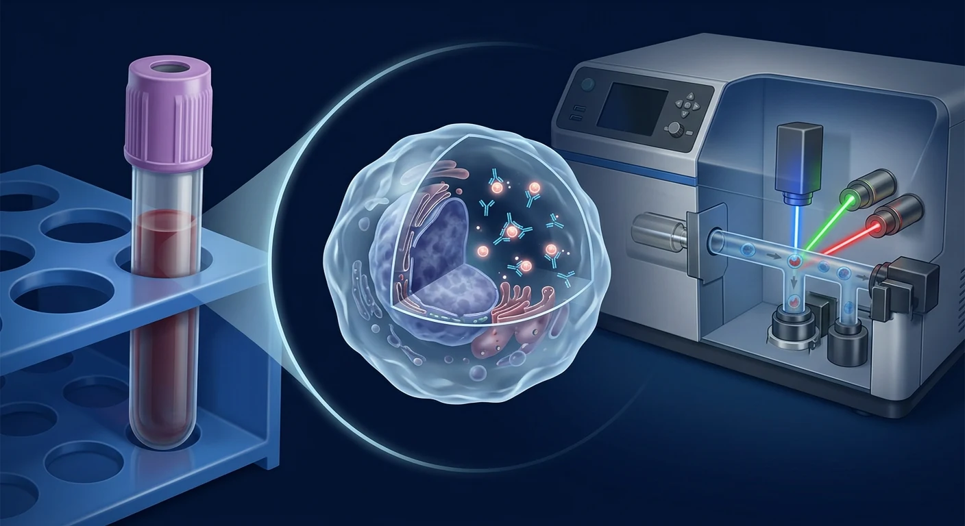
EDIM measures epitopes inside circulating monocytes rather than in tumour tissue. Monocytes and macrophages continuously survey tissue, ingest debris and present or retain fragments of what they have engulfed [22]. Their activation states are heterogeneous and context-dependent, which is well documented in immunology consensus work [23].
The proposed chain of reasoning runs: tumour metabolism → altered proteins → phagocytosed material → detectable epitope inside a monocyte. Each link is biologically plausible. The clinical question is whether the final signal is specific enough to separate cancer from inflammation, infection, tissue injury and benign proliferation in a real screening population.
Tumour metabolism also changes the local immune environment. Lactate accumulation from high glycolytic flux has been shown to blunt T-cell and NK-cell surveillance in experimental models [20]. That is relevant context for anyone reading about both metabolic testing and NK cell biology, but it is mechanistic evidence, not proof of diagnostic accuracy.
What the Warburg effect does not prove about EDIM
A real biological mechanism does not automatically produce a reliable test. Four gaps deserve explicit statement:
- Universality. Not all tumours are strongly glycolytic, and metabolic phenotypes vary between and within tumours [10], [9].
- Specificity. Activated immune cells, healing tissue and chronically inflamed tissue also shift toward glycolysis, so the metabolic signature is not exclusive to malignancy.
- Marker-level dispute. The link between TKTL1 protein detection and actual transketolase-driven metabolic flux remains contested [17].
- Population performance. Sensitivity and specificity reported in enriched cohorts do not transfer to low-prevalence screening; positive predictive value falls sharply when disease is rare.
These points are expanded in our review of EDIM evidence, accuracy and limitations and in the discussion of inflammation and false-positive results.
How this compares with other blood-based cancer testing
EDIM is a cell-based indirect metabolic assay; liquid biopsy is a direct molecular assay. Circulating tumour DNA testing detects tumour-derived genetic material and has entered clinical practice in defined settings, with its own well-documented implementation challenges around sensitivity at low tumour burden, clonal haematopoiesis and result interpretation [24].
The difference is what each test would miss. ctDNA requires shed tumour DNA above the detection threshold. A metabolic-epitope assay requires phagocytic sampling and stable epitope retention. Neither approach is currently a substitute for organ-specific guideline screening, and a full comparison is covered in EDIM versus liquid biopsy.
How a patient in Bangkok should use this information
Use metabolic-marker testing as a conversation starter with a physician, not as a decision-maker. At VELAR Center in Bangkok, EDIM testing is offered only with medical consultation, and results are interpreted alongside history, examination, risk factors and — where indicated — standard imaging or specialist referral.
Practical expectations for patients in Thailand:
- An elevated score triggers clinical assessment, not treatment.
- A normal score does not cancel the need for age-appropriate screening.
- Recent infection, surgery, vaccination or a flare of an inflammatory condition can affect immune-cell measurements and should be disclosed before testing.
- Serial testing without a defined clinical plan tends to generate anxiety rather than information.
Preparation details are described in EDIM test preparation and laboratory workflow.
How researchers actually measure tumour metabolism
Metabolic claims are only as good as the method used to make them, and the available methods measure different things. Confusing them is one reason public discussion of "cancer metabolism" drifts so far from the evidence.
| Method | What it measures | Clinical status |
|---|---|---|
| FDG-PET imaging | Glucose-analogue uptake in living tissue, per anatomic region | Established, with standardised protocols and response criteria [18] |
| Immunohistochemistry on tissue | Presence of a protein in a biopsy section | Research and pathology use; antibody specificity matters greatly [13] |
| Isotope-tracing flux studies | Actual carbon routing through metabolic pathways | Research only; the closest thing to ground truth [21] |
| Cell-line knockdown | Whether removing a gene changes growth in culture | Mechanistic evidence, not clinical accuracy [14] |
| Blood-based epitope assays (EDIM) | Protein epitopes inside circulating monocytes | Investigational; no consensus performance standard |
Notice what sits at the two ends of that table. The strongest metabolic evidence in oncology comes from imaging and tracing studies that observe metabolism where it happens. A blood epitope assay is several inferential steps removed from the tumour, which is precisely why its performance has to be demonstrated empirically rather than argued from mechanism.
Why "the mechanism is real" is a weak argument for any test
Plausible biology has repeatedly failed to translate into useful screening tests, and that history should inform expectations here. Markers can be genuinely associated with disease at the group level and still perform poorly for an individual, because association and discrimination are different statistical properties.
Three recurring failure modes are worth naming. First, spectrum bias: studies enriched with advanced, symptomatic disease inflate apparent sensitivity. Second, verification bias: when only test-positive patients receive confirmatory work-up, false negatives go uncounted. Third, prevalence effects: at a 1% disease prevalence, even 95% specificity produces far more false positives than true positives.
None of this means metabolic testing is worthless. It means the decisive evidence is prospective, blinded, adequately powered studies in the population the test is actually sold to — not mechanistic elegance. Until those exist, the responsible framing is investigational adjunct, and the responsible clinical use is inside a physician-led pathway rather than as a self-directed screen.
Frequently Asked Questions
Does the Warburg effect mean sugar feeds cancer?
No. Tumour cells consume glucose avidly, but this does not mean dietary sugar causes or accelerates cancer in humans. All cells use glucose, blood glucose is tightly regulated, and no clinical guideline recommends eliminating carbohydrates as a cancer treatment.
Is TKTL1 a proven cancer biomarker?
No. TKTL1 has supportive tissue and functional studies as well as published contradictory findings. It is best described as an investigational marker associated with altered tumour metabolism, not a validated diagnostic biomarker.
Can an EDIM test tell me where a cancer is?
No. EDIM is not organ-specific. Any abnormal result requires conventional localisation through imaging, endoscopy or tissue sampling ordered by a physician.
Why does FDG-PET work if the Warburg effect is disputed?
The phenomenon of high tumour glucose uptake is not disputed — Warburg's explanation of its cause is. FDG-PET exploits the uptake itself and is supported by standardised clinical procedure and response criteria.
Should I have an EDIM test instead of a colonoscopy or mammogram?
No. EDIM is investigational and does not replace any guideline-recommended screening test. Organ-specific screening programmes have documented mortality benefit that EDIM has not demonstrated.
Medical disclaimer
This article is provided for general educational purposes only and does not constitute medical advice, diagnosis or treatment. EDIM Apo10/TKTL1 testing is investigational and is not approved as a stand-alone cancer diagnostic. It does not replace guideline-recommended cancer screening or clinical evaluation. Always consult a qualified physician regarding symptoms, screening decisions and interpretation of any test result. Individual results vary and no outcome is guaranteed.
References
- Warburg O. On the origin of cancer cells. Science. 1956;123(3191):309-314. doi:10.1126/science.123.3191.309 ↩
- Vander Heiden MG, Cantley LC, Thompson CB. Understanding the Warburg effect: the metabolic requirements of cell proliferation. Science. 2009;324(5930):1029-1033. doi:10.1126/science.1160809 ↩
- Liberti MV, Locasale JW. The Warburg effect: how does it benefit cancer cells? Trends in Biochemical Sciences. 2016;41(3):211-218. doi:10.1016/j.tibs.2015.12.001 ↩
- Koppenol WH, Bounds PL, Dang CV. Otto Warburg's contributions to current concepts of cancer metabolism. Nature Reviews Cancer. 2011;11(5):325-337. doi:10.1038/nrc3038 ↩
- Vaupel P, Multhoff G. Revisiting the Warburg effect: historical dogma versus current understanding. The Journal of Physiology. 2021;599(6):1745-1757. doi:10.1113/JP278810 ↩
- Hanahan D, Weinberg RA. Hallmarks of cancer: the next generation. Cell. 2011;144(5):646-674. doi:10.1016/j.cell.2011.02.013 ↩
- Hanahan D. Hallmarks of cancer: new dimensions. Cancer Discovery. 2022;12(1):31-46. doi:10.1158/2159-8290.CD-21-1059 ↩
- Pavlova NN, Thompson CB. The emerging hallmarks of cancer metabolism. Cell Metabolism. 2016;23(1):27-47. doi:10.1016/j.cmet.2015.12.006 ↩
- Martínez-Reyes I, Chandel NS. Cancer metabolism: looking forward. Nature Reviews Cancer. 2021;21(10):669-680. doi:10.1038/s41568-021-00378-6 ↩
- DeBerardinis RJ, Chandel NS. Fundamentals of cancer metabolism. Science Advances. 2016;2(5):e1600200. doi:10.1126/sciadv.1600200 ↩
- Patra KC, Hay N. The pentose phosphate pathway and cancer. Trends in Biochemical Sciences. 2014;39(8):347-354. doi:10.1016/j.tibs.2014.06.005 ↩
- Cho ES, Cha YH, Kim HS, Kim NH, Yook JI. The pentose phosphate pathway as a potential target for cancer therapy. Biomolecules & Therapeutics. 2018;26(1):29-38. doi:10.4062/biomolther.2017.179 ↩
- Langbein S, Zerilli M, Zur Hausen A, et al. Expression of transketolase TKTL1 predicts colon and urothelial cancer patient survival: Warburg effect reinterpreted. British Journal of Cancer. 2006;94(4):578-585. doi:10.1038/sj.bjc.6602962 ↩
- Xu X, Zur Hausen A, Coy JF, Löchelt M. Transketolase-like protein 1 (TKTL1) is required for rapid cell growth and full viability of human tumor cells. International Journal of Cancer. 2009;124(6):1330-1337. doi:10.1002/ijc.24078 ↩
- Diaz-Moralli S, Tarrado-Castellarnau M, Alenda C, Castells A, Cascante M. Transketolase-like 1 expression is modulated during colorectal cancer progression and metastasis formation. PLoS ONE. 2011;6(9):e25323. doi:10.1371/journal.pone.0025323 ↩
- Sun W, Liu Y, Glazer CA, et al. TKTL1 is activated by promoter hypomethylation and contributes to head and neck squamous cell carcinoma carcinogenesis. Clinical Cancer Research. 2010;16(3):857-866. doi:10.1158/1078-0432.CCR-09-2604 ↩
- Mayer A, Von Wallbrunn A, Vaupel P. Glucose metabolism of malignant cells is not regulated by transketolase-like (TKTL)-1. International Journal of Oncology. 2010;37(2):265-271. doi:10.3892/ijo_00000674 ↩
- Boellaard R, Delgado-Bolton R, Oyen WJG, et al. FDG PET/CT: EANM procedure guidelines for tumour imaging, version 2.0. European Journal of Nuclear Medicine and Molecular Imaging. 2015;42(2):328-354. doi:10.1007/s00259-014-2961-x ↩
- Wahl RL, Jacene H, Kasamon Y, Lodge MA. From RECIST to PERCIST: evolving considerations for PET response criteria in solid tumors. Journal of Nuclear Medicine. 2009;50(Suppl 1):122S-150S. doi:10.2967/jnumed.108.057307 ↩
- Brand A, Singer K, Koehl GE, et al. LDHA-associated lactic acid production blunts tumor immunosurveillance by T and NK cells. Cell Metabolism. 2016;24(5):657-671. doi:10.1016/j.cmet.2016.08.011 ↩
- DeBerardinis RJ, Mancuso A, Daikhin E, et al. Beyond aerobic glycolysis: transformed cells can engage in glutamine metabolism that exceeds the requirement for protein and nucleotide synthesis. Proceedings of the National Academy of Sciences. 2007;104(49):19345-19350. doi:10.1073/pnas.0709747104 ↩
- Ginhoux F, Jung S. Monocytes and macrophages: developmental pathways and tissue homeostasis. Nature Reviews Immunology. 2014;14(6):392-404. doi:10.1038/nri3671 ↩
- Murray PJ, Allen JE, Biswas SK, et al. Macrophage activation and polarization: nomenclature and experimental guidelines. Immunity. 2014;41(1):14-20. doi:10.1016/j.immuni.2014.06.008 ↩
- Ignatiadis M, Sledge GW, Jeffrey SS. Liquid biopsy enters the clinic — implementation issues and future challenges. Nature Reviews Clinical Oncology. 2021;18(5):297-312. doi:10.1038/s41571-020-00457-x ↩
EDIM血液检测并不寻找癌细胞,它寻找的是一种代谢指纹——而这个指纹源自一个关于肿瘤如何燃烧糖分的百年观察。理解Warburg效应及其中的TKTL1酶,是判断一份EDIM评分能说明什么、不能说明什么的唯一诚实途径。
观察由来已久,解释却远未定论。1956年,Otto Warburg报告肿瘤组织切片以极高速率消耗葡萄糖,并且即使在氧气充足时仍产生乳酸[1]。Warburg认为这反映了呼吸功能受损。现代研究在确认该现象本身的同时,基本否定了他的因果解释[2],[4]。
代谢重编程如今被视为癌症的核心特征之一。它在广受引用的"癌症标志"框架中被称为"细胞能量代谢失调",并在2022年的修订版中得到保留与扩展[6],[7]。但这并不使任何单一代谢标志物具有诊断价值。
本文解释EDIM检测所依据的生物学原理。内容仅供教育参考。EDIM属于研究性方法,不是独立的癌症诊断,也不能替代指南推荐的筛查,例如乳腺X线摄影、结肠镜检查、低剂量CT或HPV检测。
核心信息
Warburg效应是许多肿瘤中真实且可重复的特征,TKTL1是与之相关的一种酶。这两点都不能证明基于TKTL1的血液评分能够可靠地在个体中检测出癌症。应将EDIM结果视为在标准诊断路径内与医生讨论的信号,而绝非结论。
什么是Warburg效应?
Warburg效应是指许多癌细胞即使在有氧条件下仍将葡萄糖转化为乳酸的倾向——这一过程称为有氧糖酵解。在氧气充足时,正常细胞大多在线粒体中完成葡萄糖分解,每分子葡萄糖产生的ATP要多得多。而肿瘤细胞常常选择快速但低效的途径[2]。
为何选择效率更低的途径?主流解释是增殖细胞首要需要的并非ATP,而是构建材料。快速糖酵解为核苷酸、氨基酸和脂质提供碳骨架,并再生生物合成所需的还原力[3],[8]。优势在于通量速度,而非单分子产率。
Warburg最初的解释在一个重要方面是错误的。他提出不可逆的线粒体损伤是癌症的病因。事实上,多数肿瘤保留有功能的线粒体,并与糖酵解并行使用氧化磷酸化、谷氨酰胺氧化和脂肪酸氧化[4],[9],[21]。2021年的一项生理学再评估认为,该效应更适合理解为致癌信号与微环境压力的结果,而非原发缺陷[5]。
其临床相关性已在一个具体领域得到证实:影像学。FDG-PET之所以有效,是因为肿瘤大量摄取标记的葡萄糖类似物。之所以存在标准化的采集与疗效评价标准,正是因为葡萄糖亲和性可测量且足够可重复以供临床使用[18],[19]。这是肿瘤葡萄糖处理不同于正常组织的最强真实世界证据——但这是影像学证据,而非血液检测证据。
戊糖磷酸途径的作用是什么?
戊糖磷酸途径(PPP)是将葡萄糖转化为用于DNA和RNA的核糖,以及用于抗氧化防御和脂质合成的NADPH的代谢分支。它是糖酵解的一条支路,在增殖细胞中承载着繁重的"流量"[11]。
PPP有两个分支。氧化分支产生NADPH。由转酮醇酶和转醛醇酶驱动的非氧化分支可相互转化糖类,使细胞能够灵活生成核糖而不被锁定于NADPH的产生。癌细胞利用这种灵活性,因此PPP被反复提议作为治疗靶点[12]。
NADPH关乎生存,而不仅是生长。它为谷胱甘肽再生提供燃料,从而缓冲快速增殖和多种癌症治疗所产生的氧化应激。这也是实验模型中PPP活性与治疗抵抗相关联的原因之一[11],[12]。
TKTL1是什么?为何受到关注?
TKTL1(转酮醇酶样蛋白1)是转酮醇酶家族基因,被认为在肿瘤中支持非氧化戊糖磷酸途径。相关关注始于组织学研究,这些研究报告结肠癌和尿路上皮癌中强TKTL1染色与较差的患者生存率相关[13]。
随后是功能学研究。敲低实验报告,降低TKTL1可减缓某些癌细胞系的增殖并降低其活力[14]。结直肠癌的表达分析描述了TKTL1在疾病进展与转移过程中的变化[15];另一项研究报告TKTL1可由启动子低甲基化激活,提供了一条过表达的表观遗传途径[16]。
证据确实存在争议,这一点很重要。一个独立研究组报告,在其实验体系中恶性细胞的葡萄糖代谢并非由TKTL1调控,并对抗体特异性与转酮醇酶活性归因提出了质疑[17]。任何面向患者的TKTL1描述若忽略这一分歧,都是不完整的。
关于TKTL1的诚实总结
TKTL1是一个具有合理性、经过研究但存在争议的肿瘤糖代谢改变标志物。它并非具有共识性能参数的成熟诊断生物标志物,也不针对任何单一癌种具有特异性。
代谢特征如何变成血液中可测量的信号?
EDIM测量的是循环单核细胞内部的表位,而非肿瘤组织。单核细胞和巨噬细胞持续巡视组织、吞噬碎片,并呈递或保留所吞噬物质的片段[22]。它们的活化状态具有异质性并依赖环境,这在免疫学共识文献中已有充分记载[23]。
推理链条为:肿瘤代谢 → 蛋白质改变 → 被吞噬的物质 → 单核细胞内可检测的表位。每一环节在生物学上都是合理的。临床问题在于,最终信号是否足够特异,能够在真实筛查人群中将癌症与炎症、感染、组织损伤和良性增殖区分开来。
肿瘤代谢也会改变局部免疫环境。实验模型显示,高糖酵解通量导致的乳酸堆积会削弱T细胞和NK细胞的免疫监视[20]。这对同时关注代谢检测与NK细胞生物学的读者是相关背景,但这属于机制证据,而非诊断准确性的证明。
Warburg效应无法证明EDIM的哪些方面
真实的生物学机制并不自动产生可靠的检测。有四项缺口需要明确指出:
- 普遍性。并非所有肿瘤都强烈依赖糖酵解,代谢表型在肿瘤之间及肿瘤内部都存在差异[10],[9]。
- 特异性。活化的免疫细胞、愈合组织和慢性炎症组织同样会转向糖酵解,因此该代谢特征并非恶性肿瘤所独有。
- 标志物层面的争议。TKTL1蛋白检测与实际转酮醇酶驱动的代谢通量之间的关联仍有争论[17]。
- 人群表现。在富集队列中报告的敏感性与特异性无法迁移到低患病率的筛查场景;当疾病罕见时,阳性预测值会急剧下降。
这些要点在我们关于EDIM证据、准确性与局限性的综述以及炎症与假阳性结果的讨论中有进一步展开。
与其他血液癌症检测的比较
EDIM是基于细胞的间接代谢检测;液体活检是直接的分子检测。循环肿瘤DNA检测直接检出肿瘤来源的遗传物质,并已在特定场景进入临床实践,同时在低肿瘤负荷下的敏感性、克隆性造血以及结果解读方面存在充分记录的落地挑战[24]。
区别在于各自会漏掉什么。ctDNA需要肿瘤释放的DNA超过检测阈值;代谢表位检测则依赖吞噬取样和表位的稳定保留。目前两者都不能替代针对特定器官的指南筛查,完整比较见EDIM与液体活检。
曼谷的患者应如何使用这些信息
把代谢标志物检测当作与医生讨论的起点,而不是决策者。在曼谷的VELAR Center,EDIM检测仅在医疗咨询下提供,结果会结合病史、体格检查、风险因素,并在有指征时结合标准影像学或专科转诊来解读。
泰国患者的实际预期:
- 评分升高触发的是临床评估,而非治疗。
- 评分正常并不取消按年龄进行常规筛查的必要性。
- 近期感染、手术、疫苗接种或炎症性疾病发作可能影响免疫细胞测量,检测前应予以告知。
- 缺乏明确临床计划的连续检测往往带来焦虑而非信息。
准备细节参见EDIM检测准备与实验室流程。
研究者实际如何测量肿瘤代谢
代谢结论的可靠性取决于所用方法,而现有各种方法测量的其实是不同的东西。混淆它们,是公众关于"癌症代谢"的讨论常常偏离证据的原因之一。
| 方法 | 测量内容 | 临床地位 |
|---|---|---|
| FDG-PET影像 | 活体组织按解剖区域的葡萄糖类似物摄取 | 成熟应用,具有标准化方案与疗效标准[18] |
| 组织免疫组化 | 活检切片中某蛋白的存在 | 研究与病理用途;抗体特异性至关重要[13] |
| 同位素示踪通量研究 | 碳在代谢途径中的实际走向 | 仅限研究;最接近"真实值"[21] |
| 细胞系敲低实验 | 去除某基因是否改变培养中的生长 | 机制证据,而非临床准确性[14] |
| 血液表位检测(EDIM) | 循环单核细胞内部的蛋白表位 | 研究性;无共识性能标准 |
请注意表格两端的差异。肿瘤学中最强的代谢证据来自在代谢实际发生之处进行观察的影像与示踪研究。血液表位检测与肿瘤之间隔着若干推理步骤,正因如此,其性能必须通过实证验证,而不能凭机制推演。
为什么"机制是真实的"并不足以支持一项检测
生物学上合理的设想屡次未能转化为有用的筛查工具,这段历史应当影响我们的预期。一个标志物可以在群体层面与疾病确有关联,却在个体层面表现不佳,因为"关联"与"区分能力"是不同的统计属性。
有三类反复出现的失败模式值得点名。第一,谱系偏倚:以晚期、有症状患者为主的研究会夸大表观敏感性。第二,验证偏倚:当只有检测阳性者接受确认检查时,假阴性被漏计。第三,患病率效应:在1%的患病率下,即使特异性达95%,假阳性也会远多于真阳性。
这并不意味着代谢检测毫无价值。它意味着决定性证据来自在该检测实际面向人群中开展的前瞻性、盲法、样本量充足的研究,而非机制上的优雅。在这些证据出现之前,负责任的定位是研究性辅助手段,负责任的临床用法是纳入医生主导的路径,而非由个人自行筛查。
常见问题
Warburg效应是否意味着糖分"喂养"癌症?
不是。肿瘤细胞大量消耗葡萄糖,但这并不意味着膳食糖分会在人体中导致或加速癌症。所有细胞都使用葡萄糖,血糖受到严格调控,且没有任何临床指南推荐以完全去除碳水化合物作为癌症治疗。
TKTL1是已证实的癌症生物标志物吗?
不是。TKTL1既有支持性的组织学与功能学研究,也有已发表的相反结果。更恰当的描述是:它是一个与肿瘤代谢改变相关的研究性标志物,而非经过验证的诊断生物标志物。
EDIM检测能告诉我癌症的位置吗?
不能。EDIM不具有器官特异性。任何异常结果都需要通过医生开具的影像学、内镜或组织取样进行常规定位。
如果Warburg效应存在争议,为何FDG-PET有效?
存在争议的是Warburg对成因的解释,而非肿瘤高葡萄糖摄取这一现象本身。FDG-PET利用的正是摄取现象,并有标准化的临床操作与疗效评价标准支持。
我可以用EDIM检测代替结肠镜或乳腺X线摄影吗?
不可以。EDIM属于研究性方法,不能替代任何指南推荐的筛查检查。针对特定器官的筛查项目具有已记录的死亡率获益,而EDIM尚未证明这一点。
医疗免责声明
本文仅供一般教育参考,不构成医疗建议、诊断或治疗。EDIM Apo10/TKTL1检测属于研究性方法,未被批准为独立的癌症诊断工具,也不能替代指南推荐的癌症筛查或临床评估。关于症状、筛查决策以及任何检测结果的解读,请务必咨询合格医师。个体结果各异,不保证任何疗效。
参考文献
- Warburg O. Science. 1956;123(3191):309-314. doi:10.1126/science.123.3191.309
- Vander Heiden MG, et al. Science. 2009;324(5930):1029-1033. doi:10.1126/science.1160809
- Liberti MV, Locasale JW. Trends in Biochemical Sciences. 2016;41(3):211-218. doi:10.1016/j.tibs.2015.12.001
- Koppenol WH, et al. Nature Reviews Cancer. 2011;11(5):325-337. doi:10.1038/nrc3038
- Vaupel P, Multhoff G. The Journal of Physiology. 2021;599(6):1745-1757. doi:10.1113/JP278810
- Hanahan D, Weinberg RA. Cell. 2011;144(5):646-674. doi:10.1016/j.cell.2011.02.013
- Hanahan D. Cancer Discovery. 2022;12(1):31-46. doi:10.1158/2159-8290.CD-21-1059
- Pavlova NN, Thompson CB. Cell Metabolism. 2016;23(1):27-47. doi:10.1016/j.cmet.2015.12.006
- Martínez-Reyes I, Chandel NS. Nature Reviews Cancer. 2021;21(10):669-680. doi:10.1038/s41568-021-00378-6
- DeBerardinis RJ, Chandel NS. Science Advances. 2016;2(5):e1600200. doi:10.1126/sciadv.1600200
- Patra KC, Hay N. Trends in Biochemical Sciences. 2014;39(8):347-354. doi:10.1016/j.tibs.2014.06.005
- Cho ES, et al. Biomolecules & Therapeutics. 2018;26(1):29-38. doi:10.4062/biomolther.2017.179
- Langbein S, et al. British Journal of Cancer. 2006;94(4):578-585. doi:10.1038/sj.bjc.6602962
- Xu X, et al. International Journal of Cancer. 2009;124(6):1330-1337. doi:10.1002/ijc.24078
- Diaz-Moralli S, et al. PLoS ONE. 2011;6(9):e25323. doi:10.1371/journal.pone.0025323
- Sun W, et al. Clinical Cancer Research. 2010;16(3):857-866. doi:10.1158/1078-0432.CCR-09-2604
- Mayer A, et al. International Journal of Oncology. 2010;37(2):265-271. doi:10.3892/ijo_00000674
- Boellaard R, et al. European Journal of Nuclear Medicine and Molecular Imaging. 2015;42(2):328-354. doi:10.1007/s00259-014-2961-x
- Wahl RL, et al. Journal of Nuclear Medicine. 2009;50(Suppl 1):122S-150S. doi:10.2967/jnumed.108.057307
- Brand A, et al. Cell Metabolism. 2016;24(5):657-671. doi:10.1016/j.cmet.2016.08.011
- DeBerardinis RJ, et al. PNAS. 2007;104(49):19345-19350. doi:10.1073/pnas.0709747104
- Ginhoux F, Jung S. Nature Reviews Immunology. 2014;14(6):392-404. doi:10.1038/nri3671
- Murray PJ, et al. Immunity. 2014;41(1):14-20. doi:10.1016/j.immuni.2014.06.008
- Ignatiadis M, et al. Nature Reviews Clinical Oncology. 2021;18(5):297-312. doi:10.1038/s41571-020-00457-x
اختبار EDIM في الدم لا يبحث عن الخلايا السرطانية، بل يبحث عن بصمة أيضية — وهذه البصمة تعود إلى ملاحظة عمرها قرن كامل حول طريقة حرق الأورام للسكر. إن فهم تأثير واربورغ، وإنزيم TKTL1 الذي يقع ضمنه، هو الطريقة الأمينة الوحيدة للحكم على ما يمكن وما لا يمكن أن تعنيه نتيجة EDIM.
الملاحظة قديمة، أما تفسيرها فلم يُحسم بعد. في عام 1956 أفاد أوتو واربورغ بأن شرائح الأورام تستهلك الغلوكوز بمعدلات استثنائية وتنتج اللاكتات حتى مع وفرة الأكسجين [1]. اعتقد واربورغ أن ذلك يعكس تلفاً في التنفس الخلوي. وقد رفضت الأبحاث الحديثة تفسيره السببي إلى حد كبير، مع تأكيد الظاهرة نفسها [2]، [4].
تُعد إعادة برمجة الأيض اليوم سمة أساسية من سمات السرطان. وهي مذكورة في إطار "سمات السرطان" الواسع الانتشار بوصفها "اختلال تنظيم طاقة الخلية"، وقد جرى الإبقاء عليها وتوسيعها في مراجعة عام 2022 [6]، [7]. لكن ذلك لا يجعل أي واسم أيضي منفرد صالحاً للتشخيص.
يشرح هذا المقال البيولوجيا التي يستند إليها اختبار EDIM. والمحتوى تثقيفي فقط. فاختبار EDIM بحثي، وليس تشخيصاً مستقلاً للسرطان، ولا يحل محل برامج الفحص الموصى بها في الإرشادات مثل تصوير الثدي الشعاعي، أو تنظير القولون، أو الأشعة المقطعية منخفضة الجرعة، أو فحص فيروس الورم الحليمي البشري.
رسالة رئيسية
تأثير واربورغ سمة حقيقية وقابلة للتكرار في كثير من الأورام، وTKTL1 أحد الإنزيمات المرتبطة به. ولا تثبت أي من هاتين الحقيقتين أن نتيجة دم مبنية على TKTL1 تكشف السرطان بموثوقية لدى فرد بعينه. تعامل مع نتيجة EDIM كإشارة تستدعي مناقشة الطبيب ضمن مسار تشخيصي قياسي — لا كحكم نهائي.
ما هو تأثير واربورغ ببساطة؟
تأثير واربورغ هو ميل كثير من الخلايا السرطانية إلى تحويل الغلوكوز إلى لاكتات حتى مع توافر الأكسجين — وهي عملية تُسمى تحلل السكر الهوائي. فالخلايا الطبيعية في ظروف أكسجين جيدة تُكمل تفكيك الغلوكوز في الميتوكوندريا، وهو ما ينتج طاقة أكبر بكثير لكل جزيء غلوكوز. أما الخلايا الورمية فكثيراً ما تسلك المسار السريع المهدر بدلاً من ذلك [2].
لماذا تختار مساراً أقل كفاءة؟ التفسير السائد هو أن الخلايا المتكاثرة لا تحتاج أساساً إلى الطاقة بل إلى مواد البناء. فتحلل السكر السريع يوفر الهياكل الكربونية للنيوكليوتيدات والأحماض الأمينية والدهون، ويجدد القدرة الاختزالية اللازمة للتخليق الحيوي [3]، [8]. الميزة هنا في سرعة التدفق لا في العائد لكل جزيء.
كان تفسير واربورغ الأصلي خاطئاً في نقطة مهمة. فقد اقترح أن تلفاً لا رجعة فيه في الميتوكوندريا هو سبب السرطان. والواقع أن معظم الأورام تحتفظ بميتوكوندريا وظيفية وتستخدم الفسفرة التأكسدية وأكسدة الغلوتامين والأحماض الدهنية إلى جانب تحلل السكر [4]، [9]، [21]. وتذهب مراجعة فسيولوجية عام 2021 إلى أن الظاهرة تُفهم بشكل أفضل كنتيجة للإشارات الورمية وضغوط البيئة المجهرية لا كخلل أولي [5].
الأهمية السريرية مثبتة بالفعل في مجال واحد محدد: التصوير. يعمل تصوير FDG-PET لأن الأورام تلتقط نظيراً موسوماً للغلوكوز بنهم. وتوجد معايير موحدة للتصوير وتقييم الاستجابة تحديداً لأن نهم الغلوكوز قابل للقياس وقابل للتكرار بما يكفي للاستخدام السريري [18]، [19]. هذا أقوى دليل واقعي على اختلاف تعامل الأورام مع الغلوكوز عن الأنسجة الطبيعية — وهو دليل تصويري لا دليل من اختبار دم.
أين يقع مسار فوسفات البنتوز؟
مسار فوسفات البنتوز هو الفرع الأيضي الذي يحوّل الغلوكوز إلى ريبوز للحمض النووي والحمض النووي الريبي، وإلى NADPH للدفاع المضاد للأكسدة وتخليق الدهون. وهو طريق جانبي متفرع عن تحلل السكر، ويحمل حركة كثيفة في الخلايا المتكاثرة [11].
للمسار ذراعان. الذراع التأكسدي ينتج NADPH. أما الذراع غير التأكسدي، الذي تديره إنزيمات الترانسكيتولاز والترانسألدولاز، فيحوّل السكريات فيما بينها بحيث تستطيع الخلية إنتاج الريبوز بمرونة دون أن ترتبط بإنتاج NADPH. وتستغل الخلايا السرطانية هذه المرونة، ولذلك اقتُرح المسار مراراً كهدف علاجي [12].
أهمية NADPH تتعلق بالبقاء لا بالنمو فقط. فهو يغذي تجديد الغلوتاثيون الذي يخفف الإجهاد التأكسدي الناتج عن التكاثر السريع وعن كثير من علاجات السرطان. وهذا أحد أسباب ربط نشاط المسار بمقاومة العلاج في النماذج المخبرية [11]، [12].
ما هو TKTL1 ولماذا جذب الاهتمام؟
TKTL1 هو جين من عائلة الترانسكيتولاز يُقترح أنه يدعم المسار غير التأكسدي لفوسفات البنتوز في الأورام. وقد بدأ الاهتمام بدراسات نسيجية أفادت بأن التلوّن القوي لـTKTL1 في سرطان القولون والمسالك البولية يرتبط ببقاء أسوأ للمرضى [13].
ثم تلت ذلك أعمال وظيفية. أفادت تجارب تثبيط التعبير بأن خفض TKTL1 أبطأ التكاثر وقلل الحيوية في خطوط خلوية سرطانية معينة [14]. ووصفت تحليلات التعبير في سرطان القولون والمستقيم تغير TKTL1 عبر مراحل التقدم والانتقال [15]، وأفادت دراسة منفصلة بأن TKTL1 يمكن تنشيطه بنقص مثيلة المُحفّز [16].
الأدلة متنازع عليها فعلاً، وهذا أمر مهم هنا. فقد أفادت مجموعة بحثية مستقلة بأن أيض الغلوكوز في الخلايا الخبيثة لم يكن منظَّماً بواسطة TKTL1 في أنظمتها التجريبية، وأثارت تحفظات حول نوعية الأجسام المضادة ونسبة نشاط الترانسكيتولاز [17]. وأي وصف موجه للمرضى يغفل هذا الخلاف يكون ناقصاً.
خلاصة أمينة حول TKTL1
TKTL1 واسم معقول ومدروس ومتنازع عليه لتغير أيض السكر في الأورام. وهو ليس واسماً تشخيصياً راسخاً بخصائص أداء متوافق عليها، وليس نوعياً لأي سرطان بعينه.
كيف تصبح سمة أيضية قابلة للقياس في الدم؟
يقيس EDIM نواتج فوق سطحية داخل الخلايا الوحيدة المنتشرة في الدم بدلاً من نسيج الورم. فالخلايا الوحيدة والبلاعم تمسح الأنسجة باستمرار وتبتلع الحطام وتحتفظ بشظايا مما التهمته [22]. وحالات تنشيطها متغايرة وتعتمد على السياق، وهو أمر موثق جيداً في أدبيات المناعة [23].
سلسلة الاستدلال المقترحة هي: أيض الورم ← بروتينات متغيرة ← مادة مبتلعة ← نواتج قابلة للكشف داخل خلية وحيدة. كل حلقة معقولة بيولوجياً. والسؤال السريري هو ما إذا كانت الإشارة النهائية نوعية بما يكفي لفصل السرطان عن الالتهاب والعدوى وإصابة الأنسجة والتكاثر الحميد في مجتمع فحص حقيقي.
يغيّر أيض الورم أيضاً البيئة المناعية المحلية. فقد تبيّن أن تراكم اللاكتات الناتج عن التدفق السكري العالي يضعف مراقبة الخلايا التائية والخلايا القاتلة الطبيعية في نماذج تجريبية [20]. وهذا سياق مهم لمن يقرأ عن الفحص الأيضي وبيولوجيا الخلايا القاتلة الطبيعية معاً، لكنه دليل آلي لا إثبات لدقة تشخيصية.
ما لا يثبته تأثير واربورغ عن EDIM
وجود آلية بيولوجية حقيقية لا ينتج تلقائياً اختباراً موثوقاً. وهناك أربع فجوات تستحق التصريح بها:
- الشمولية. ليست كل الأورام شديدة الاعتماد على تحلل السكر، وتتباين الأنماط الأيضية بين الأورام وداخلها [10]، [9].
- النوعية. الخلايا المناعية المنشطة والأنسجة الملتئمة والملتهبة مزمناً تتحول أيضاً نحو تحلل السكر، فالبصمة الأيضية ليست حكراً على الخباثة.
- الخلاف على مستوى الواسم. لا تزال العلاقة بين كشف بروتين TKTL1 والتدفق الأيضي الفعلي محل نزاع [17].
- الأداء السكاني. الحساسية والنوعية المُبلغ عنها في مجموعات مُثراة لا تنتقل إلى الفحص منخفض الانتشار؛ إذ تنخفض القيمة التنبؤية الإيجابية بحدة عندما يكون المرض نادراً.
وتُوسَّع هذه النقاط في مراجعتنا حول أدلة EDIM ودقته وقيوده وفي مناقشة الالتهاب والنتائج الإيجابية الكاذبة.
المقارنة مع فحوص السرطان الدموية الأخرى
EDIM فحص أيضي غير مباشر قائم على الخلايا، أما الخزعة السائلة ففحص جزيئي مباشر. فاختبار الحمض النووي الورمي المنتشر يكشف مادة وراثية مصدرها الورم وقد دخل الممارسة السريرية في سياقات محددة، مع تحديات تطبيق موثقة تتعلق بالحساسية عند العبء الورمي المنخفض وتكوّن الدم النسيلي وتفسير النتائج [24].
الفرق يكمن فيما قد يفوت كل اختبار. فالحمض النووي الورمي المنتشر يتطلب تجاوز عتبة الكشف، بينما يتطلب فحص النواتج الأيضية أخذ عينات بالبلعمة واحتفاظاً مستقراً بالنواتج. ولا يشكل أي منهما حالياً بديلاً عن الفحص الموصى به لكل عضو، والمقارنة الكاملة في EDIM مقابل الخزعة السائلة.
كيف ينبغي للمريض في بانكوك استخدام هذه المعلومات
استخدم فحص الواسمات الأيضية كنقطة انطلاق لحوار مع الطبيب لا كصانع قرار. في مركز VELAR في بانكوك، يُقدَّم فحص EDIM فقط مع استشارة طبية، وتُفسَّر النتائج مع التاريخ المرضي والفحص السريري وعوامل الخطر، ومع التصوير القياسي أو الإحالة التخصصية عند الحاجة.
توقعات عملية للمرضى في تايلاند:
- النتيجة المرتفعة تستدعي تقييماً سريرياً لا علاجاً.
- النتيجة الطبيعية لا تلغي الحاجة إلى الفحص المناسب للعمر.
- العدوى أو الجراحة أو التطعيم أو نوبة مرض التهابي حديثة قد تؤثر على قياسات الخلايا المناعية ويجب ذكرها قبل الفحص.
- الفحص المتكرر دون خطة سريرية واضحة يولّد القلق أكثر مما يولّد المعلومة.
تفاصيل التحضير موضحة في تحضير اختبار EDIM وسير العمل المخبري.
كيف يقيس الباحثون أيض الورم فعلياً
قيمة أي ادعاء أيضي لا تتجاوز قيمة الطريقة المستخدمة لإثباته، والطرق المتاحة تقيس أشياء مختلفة. والخلط بينها أحد أسباب ابتعاد النقاش العام حول "أيض السرطان" عن الأدلة.
| الطريقة | ما تقيسه | الوضع السريري |
|---|---|---|
| تصوير FDG-PET | التقاط نظير الغلوكوز في النسيج الحي حسب المنطقة التشريحية | راسخ، ببروتوكولات ومعايير استجابة موحدة [18] |
| الكيمياء المناعية النسيجية | وجود بروتين في شريحة خزعة | استخدام بحثي ومرضي؛ نوعية الأجسام المضادة حاسمة [13] |
| دراسات التتبع بالنظائر | المسار الفعلي للكربون عبر المسارات الأيضية | بحثي فقط؛ الأقرب إلى الحقيقة المرجعية [21] |
| تثبيط الجين في خطوط خلوية | هل يغيّر حذف الجين النمو في المزرعة | دليل آلي لا دقة سريرية [14] |
| فحوص النواتج الدموية (EDIM) | نواتج بروتينية داخل الخلايا الوحيدة المنتشرة | بحثي؛ لا معيار أداء متوافق عليه |
لاحظ طرفي الجدول. أقوى الأدلة الأيضية في علم الأورام تأتي من التصوير ودراسات التتبع التي تراقب الأيض حيث يحدث. أما فحص النواتج الدموية فيبعد عن الورم عدة خطوات استدلالية، ولهذا تحديداً يجب إثبات أدائه تجريبياً لا استنتاجه من الآلية.
لماذا لا تكفي "الآلية حقيقية" كحجة لأي اختبار
لطالما أخفقت أفكار بيولوجية معقولة في التحول إلى اختبارات فحص مفيدة، وهذا التاريخ ينبغي أن يضبط التوقعات هنا. فقد يرتبط واسم بالمرض فعلاً على مستوى المجموعة ويظل أداؤه ضعيفاً لدى الفرد، لأن الارتباط والقدرة على التمييز خاصيتان إحصائيتان مختلفتان.
ثلاثة أنماط إخفاق متكررة تستحق التسمية. الأول، تحيّز الطيف: الدراسات المُثراة بالمرض المتقدم والأعراض تضخم الحساسية الظاهرية. الثاني، تحيّز التحقق: حين يخضع الإيجابيون فقط للفحص التأكيدي، تبقى السلبيات الكاذبة غير محسوبة. الثالث، أثر الانتشار: عند انتشار 1%، تنتج نوعية 95% إيجابيات كاذبة أكثر بكثير من الإيجابيات الحقيقية.
لا يعني ذلك أن الفحص الأيضي بلا قيمة. بل يعني أن الدليل الحاسم هو دراسات مستقبلية معماة وبحجم عينة كافٍ في المجتمع الذي يُسوَّق له الاختبار فعلاً، لا الأناقة الآلية. وإلى أن توجد تلك الدراسات، فالتأطير المسؤول هو "أداة بحثية مساعدة"، والاستخدام السريري المسؤول يكون ضمن مسار يقوده طبيب لا كفحص ذاتي.
الأسئلة الشائعة
هل يعني تأثير واربورغ أن السكر يغذي السرطان؟
لا. تستهلك الخلايا الورمية الغلوكوز بنهم، لكن ذلك لا يعني أن سكر الطعام يسبب السرطان أو يسرّعه لدى البشر. فكل الخلايا تستخدم الغلوكوز، وسكر الدم منظَّم بدقة، ولا توصي أي إرشادات سريرية بإلغاء الكربوهيدرات كعلاج للسرطان.
هل TKTL1 واسم سرطاني مثبت؟
لا. لدى TKTL1 دراسات نسيجية ووظيفية داعمة ونتائج منشورة متناقضة أيضاً. ويُوصف بدقة أكبر بأنه واسم بحثي مرتبط بتغير أيض الورم، لا واسم تشخيصي مُتحقق منه.
هل يستطيع اختبار EDIM تحديد موضع السرطان؟
لا. فاختبار EDIM ليس نوعياً لعضو معين. وأي نتيجة غير طبيعية تتطلب تحديد الموضع بالطرق التقليدية عبر التصوير أو التنظير أو أخذ عينة نسيجية بأمر من الطبيب.
لماذا ينجح FDG-PET إذا كان تأثير واربورغ محل خلاف؟
الخلاف يدور حول تفسير واربورغ للسبب لا حول ظاهرة الالتقاط المرتفع للغلوكوز نفسها. ويستفيد FDG-PET من الالتقاط ذاته، وتدعمه إجراءات سريرية ومعايير استجابة موحدة.
هل أستبدل تنظير القولون أو تصوير الثدي باختبار EDIM؟
لا. اختبار EDIM بحثي ولا يحل محل أي فحص موصى به في الإرشادات. فبرامج الفحص الخاصة بكل عضو لها فائدة موثقة في خفض الوفيات لم يثبتها EDIM.
إخلاء المسؤولية الطبية
هذا المقال لأغراض تثقيفية عامة فقط ولا يشكل نصيحة طبية أو تشخيصاً أو علاجاً. اختبار EDIM Apo10/TKTL1 بحثي وغير معتمد كتشخيص مستقل للسرطان، ولا يحل محل الفحص الموصى به في الإرشادات أو التقييم السريري. استشر دائماً طبيباً مؤهلاً بشأن الأعراض وقرارات الفحص وتفسير أي نتيجة. تختلف النتائج بين الأفراد ولا تُضمن أي نتيجة.
المراجع
- Warburg O. Science. 1956;123(3191):309-314. doi:10.1126/science.123.3191.309
- Vander Heiden MG, et al. Science. 2009;324(5930):1029-1033. doi:10.1126/science.1160809
- Liberti MV, Locasale JW. Trends in Biochemical Sciences. 2016;41(3):211-218. doi:10.1016/j.tibs.2015.12.001
- Koppenol WH, et al. Nature Reviews Cancer. 2011;11(5):325-337. doi:10.1038/nrc3038
- Vaupel P, Multhoff G. The Journal of Physiology. 2021;599(6):1745-1757. doi:10.1113/JP278810
- Hanahan D, Weinberg RA. Cell. 2011;144(5):646-674. doi:10.1016/j.cell.2011.02.013
- Hanahan D. Cancer Discovery. 2022;12(1):31-46. doi:10.1158/2159-8290.CD-21-1059
- Pavlova NN, Thompson CB. Cell Metabolism. 2016;23(1):27-47. doi:10.1016/j.cmet.2015.12.006
- Martínez-Reyes I, Chandel NS. Nature Reviews Cancer. 2021;21(10):669-680. doi:10.1038/s41568-021-00378-6
- DeBerardinis RJ, Chandel NS. Science Advances. 2016;2(5):e1600200. doi:10.1126/sciadv.1600200
- Patra KC, Hay N. Trends in Biochemical Sciences. 2014;39(8):347-354. doi:10.1016/j.tibs.2014.06.005
- Cho ES, et al. Biomolecules & Therapeutics. 2018;26(1):29-38. doi:10.4062/biomolther.2017.179
- Langbein S, et al. British Journal of Cancer. 2006;94(4):578-585. doi:10.1038/sj.bjc.6602962
- Xu X, et al. International Journal of Cancer. 2009;124(6):1330-1337. doi:10.1002/ijc.24078
- Diaz-Moralli S, et al. PLoS ONE. 2011;6(9):e25323. doi:10.1371/journal.pone.0025323
- Sun W, et al. Clinical Cancer Research. 2010;16(3):857-866. doi:10.1158/1078-0432.CCR-09-2604
- Mayer A, et al. International Journal of Oncology. 2010;37(2):265-271. doi:10.3892/ijo_00000674
- Boellaard R, et al. European Journal of Nuclear Medicine and Molecular Imaging. 2015;42(2):328-354. doi:10.1007/s00259-014-2961-x
- Wahl RL, et al. Journal of Nuclear Medicine. 2009;50(Suppl 1):122S-150S. doi:10.2967/jnumed.108.057307
- Brand A, et al. Cell Metabolism. 2016;24(5):657-671. doi:10.1016/j.cmet.2016.08.011
- DeBerardinis RJ, et al. PNAS. 2007;104(49):19345-19350. doi:10.1073/pnas.0709747104
- Ginhoux F, Jung S. Nature Reviews Immunology. 2014;14(6):392-404. doi:10.1038/nri3671
- Murray PJ, et al. Immunity. 2014;41(1):14-20. doi:10.1016/j.immuni.2014.06.008
- Ignatiadis M, et al. Nature Reviews Clinical Oncology. 2021;18(5):297-312. doi:10.1038/s41571-020-00457-x