Inflammation can complicate EDIM interpretation because the test measures biomarker material inside activated phagocytic monocytes, cells that also participate in infection, tissue injury, surgery, and repair. An elevated score may deserve investigation, but it is not proof that cancer is present.
The practical problem is biological overlap. Apo10 and TKTL1 are intended to capture abnormal apoptosis and metabolic reprogramming, yet the immune-cell carrier is responsive to many non-cancer processes. A result must therefore be read alongside recent illness, symptoms, medical history, standard screening eligibility, imaging, and—when a lesion is found—pathology.
The safest rule is simple. Do not use EDIM as a stand-alone cancer diagnosis, and do not use a low score to dismiss warning symptoms or delay guideline-recommended screening.
Direct answer
Current studies suggest that inflammation and tissue-remodeling states can influence EDIM signals, especially TKTL1-related scoring, but the size and duration of that effect are not reliably quantified for every condition. Recent infection, surgery, dental extraction, or active inflammatory disease should be disclosed before testing, and an abnormal result should be confirmed through standard clinical pathways.
Why can inflammation affect an EDIM blood test?
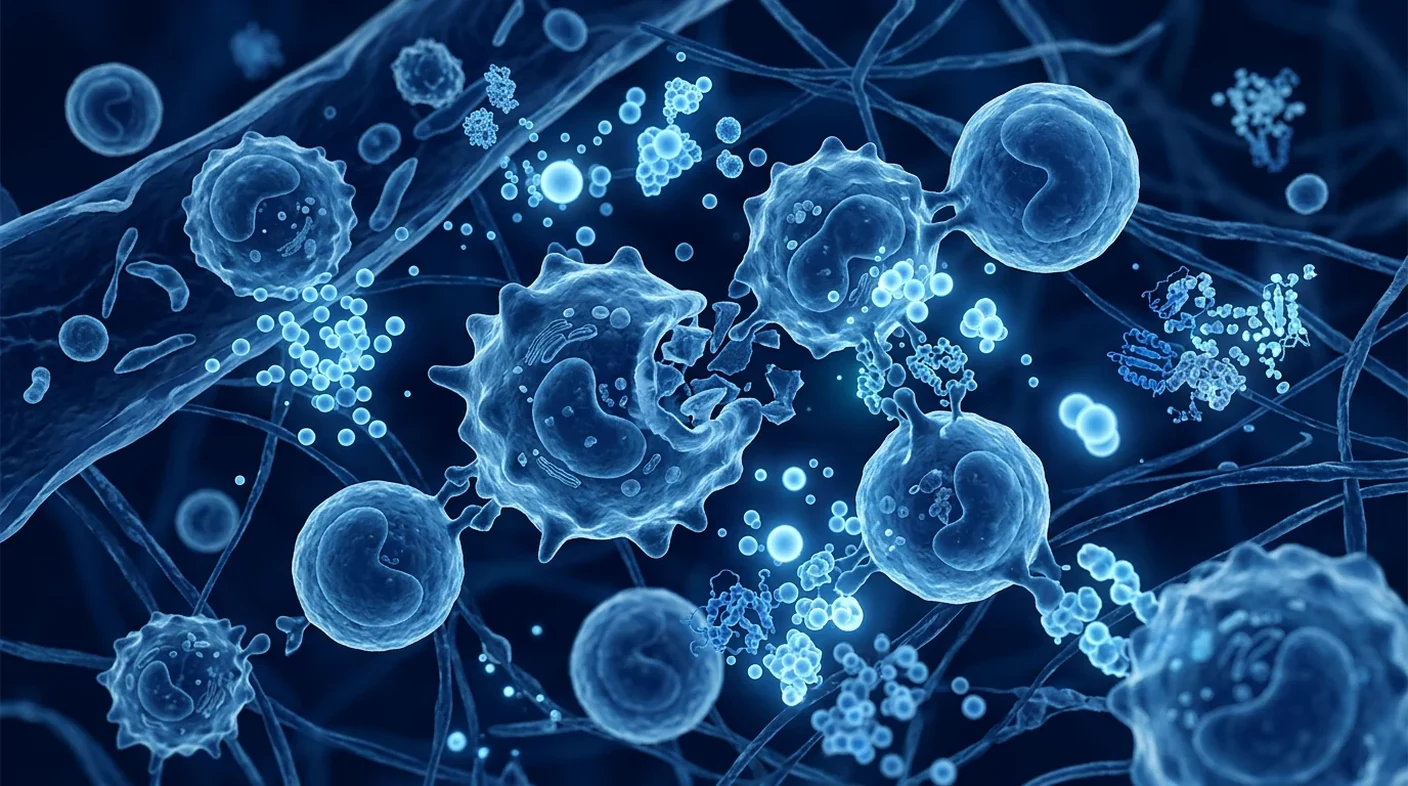
Inflammation may affect EDIM because activated CD14+/CD16+ monocytes and macrophage-like cells are both the assay’s carrier cells and central participants in immune cleanup. They engulf cellular debris, migrate between tissue and blood, and change their abundance and activation state during infection, trauma, wound healing, and chronic inflammatory disease.
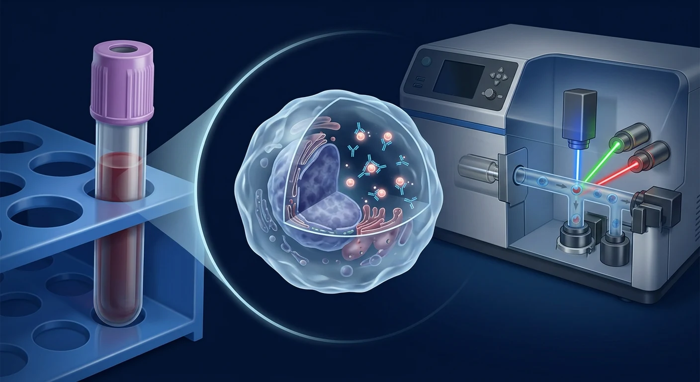
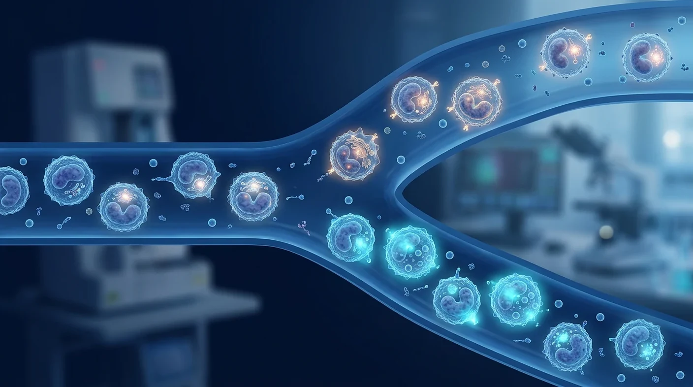
EDIM—epitope detection in monocytes—uses flow cytometry to identify a gated immune-cell population and then detect intracellular Apo10 and TKTL1 signals. The model is that phagocytes ingest material from abnormal tissue and concentrate otherwise diluted epitopes. Published cancer cohorts support the biological plausibility of finding these signals in circulating macrophage-like cells [1]–[4].
But a carrier cell does not know why tissue is abnormal. Innate immune cells respond whenever the body removes damaged cells. That means the assay can be influenced by two layers at once: the biology of the measured epitope and the biology of the monocyte carrying it. This is why context is not an optional footnote; it is part of interpreting the test.
What do Apo10 and TKTL1 represent?
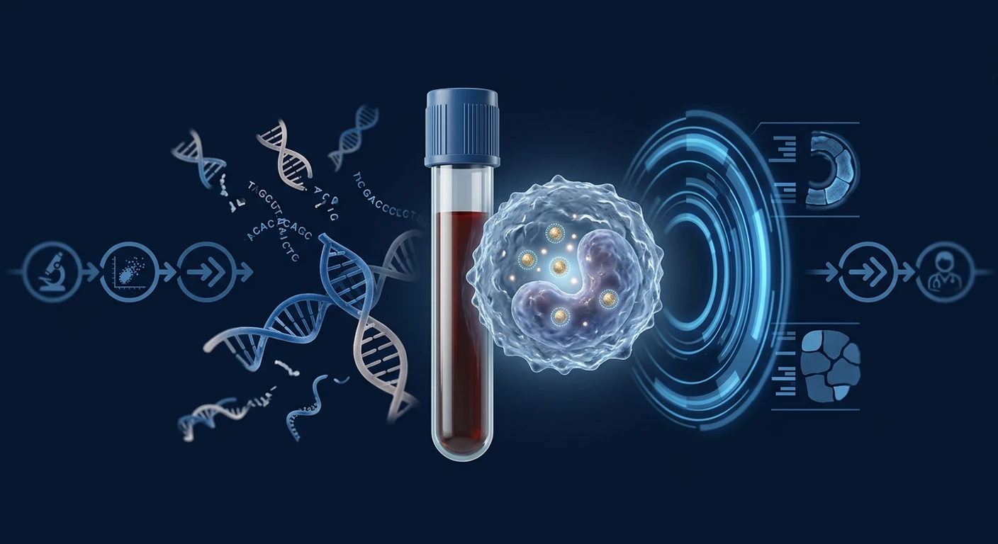
Apo10 is described as an epitope associated with DNaseX and disrupted apoptosis, while TKTL1 is associated in the EDIM literature with altered glucose metabolism and invasive cellular behavior. The combined score is intended to capture complementary biological features rather than one organ-specific tumor marker.
Apo10-related staining has been proposed as a signal of cells that have escaped normal programmed death. TKTL1-related staining is linked to metabolic adaptation, including fermentation-like glucose use under conditions in which malignant cells may gain survival advantages. Studies in oral, prostate, breast, gastrointestinal, and pediatric tumor cohorts have reported higher scores in patients with known cancers than in selected controls [2]–[8].
Neither marker is a postal code for a tumor. A combined score does not reliably tell a clinician where an abnormal process is located, what histologic cancer type it might be, or whether treatment is needed. Localization and diagnosis still require organ-directed assessment.
Which inflammatory situations may matter before testing?
Recent infection, surgery, tissue injury, dental procedures, and active inflammatory disease are the main contexts worth reporting because they can activate monocytes or increase tissue turnover. The exact influence is not established well enough to prescribe one universal waiting period.
- Acute infection: fever, respiratory infection, urinary infection, gastrointestinal infection, or another active illness can alter innate immune-cell activity.
- Recent surgery or invasive procedures: healing produces cell debris, metabolic demand, and macrophage recruitment.
- Dental extraction or substantial dental inflammation: published EDIM discussions specifically note tooth extraction and tissue remodeling as potential contexts for elevated TKTL1-related signals.
- Wounds, fractures, or burns: repair involves phagocytosis, angiogenesis, and metabolic reprogramming.
- Autoimmune or chronic inflammatory disease: rheumatoid arthritis, inflammatory bowel disease, vasculitis, and other active disorders may change monocyte phenotype or abundance.
- Immunosuppressive medication: corticosteroids, biologics, and other agents may alter the measured immune-cell compartment; some studies excluded such patients, limiting generalizability.
- Recent cancer treatment: surgery, radiation, chemotherapy, immunotherapy, and tumor breakdown can change both marker release and immune-cell behavior.
The gastrointestinal EDIM study included 13 participants with inflammatory conditions. One crossed the combined cut-off, while the group’s combined scores were higher than those of healthy individuals; the authors explicitly noted that inflammation might influence the result [1]. That observation is important but small. It cannot estimate false-positive rates across the broad spectrum of infections and inflammatory disorders seen in practice.
Does inflammation cause a false-positive EDIM result?
Inflammation can be a plausible contributor to an elevated EDIM score, but a result should not be labelled “false positive” until an appropriate reference-standard evaluation and follow-up find no target cancer. A positive blood score and a negative first scan are not always enough to settle the question.
Several explanations remain possible: transient inflammatory interference; analytical variation near the threshold; a benign process expressing related biology; a lesion too small for current localization; or a cancer unrelated to the initial imaging field. Conversely, an extensive search can discover incidental benign abnormalities and create a cascade of unnecessary procedures.
The term false positive also depends on intended use. A threshold developed in known cancer cases versus healthy controls may behave differently in asymptomatic screening, where cancer prevalence is low and benign inflammatory conditions are common. Even high sensitivity and specificity can yield more false than true positives when a disease is rare. This is why predictive value—not just sensitivity—is critical.
How should predictive value be explained?
The chance that an abnormal result represents cancer depends strongly on the patient’s baseline risk, not only on the assay’s reported sensitivity and specificity. Age, smoking exposure, family history, inherited syndromes, previous cancer, symptoms, and prevalence in the tested population all change interpretation.
Consider a hypothetical test with 95% sensitivity and 95% specificity used in 10,000 people where 1% actually have the target cancer. About 95 of 100 cancer cases might test positive, but roughly 495 of 9,900 people without cancer could also test positive. In that simplified example, only about one in six positive results would represent cancer. This is an illustration, not a performance estimate for EDIM.
Case-control accuracy can look impressive when researchers compare established cancer with healthy volunteers. Real screening populations include small tumors, benign nodules, inflammatory disease, multiple medications, and incomplete follow-up. STARD and QUADAS-2 emphasize representative enrollment, prespecified thresholds, blinded reference standards, and transparent handling of indeterminate results [9], [10]. Spectrum bias can otherwise exaggerate apparent accuracy [11].
When should testing be postponed?
Testing may reasonably be postponed when a person has an acute febrile illness, is immediately recovering from major surgery or tissue injury, or has an uncontrolled inflammatory flare—unless the ordering clinician has a specific reason not to wait. No peer-reviewed evidence supports one universal delay for every situation.
A clinic should not invent a rigid “washout” interval and present it as validated. The appropriate timing depends on the event, recovery, urgency, symptoms, and purpose of testing. A stable patient exploring optional risk assessment is different from a patient with a suspicious mass or unexplained bleeding who needs immediate standard diagnostic work-up.
Before collection, document the date and severity of recent infection, vaccination, surgery, dental work, wound, inflammatory flare, and medication change. If testing proceeds, this information should travel with the result. If a borderline result is repeated, the same laboratory and method may reduce avoidable analytical variation, but repetition must not substitute for investigating red-flag symptoms.
What should happen after an abnormal EDIM result?
An abnormal EDIM result should trigger a clinician-led risk assessment and a proportionate confirmatory plan, not a declaration of cancer. The first step is to review symptoms, baseline risk, recent inflammatory events, the exact assay, the laboratory threshold, and whether the sample met quality criteria.
- Check urgency: investigate alarm symptoms—such as a suspicious mass, bleeding, persistent weight loss, progressive jaundice, haematuria, or focal neurological signs—through standard pathways without waiting for a repeat EDIM test.
- Review confounders: note infection, inflammation, surgery, dental work, tissue injury, medication, and recent treatment.
- Continue established screening: mammography, cervical screening, colorectal screening, and low-dose CT for eligible high-risk smokers remain governed by guideline criteria.
- Choose focused confirmation: examination, organ-specific laboratory tests, ultrasound, mammography, CT, MRI, endoscopy, or another appropriate test should be selected from the clinical picture rather than automatically ordering every scan.
- Confirm suspicious tissue: imaging can localize; pathology usually establishes cancer type and guides treatment.
- Plan follow-up: if initial evaluation is reassuring, define whether observation, a time-limited repeat assay, or additional targeted investigation is appropriate.
Whole-body imaging has trade-offs. CT and PET/CT can expose patients to radiation; contrast can cause adverse reactions; and sensitive imaging finds incidental abnormalities that may lead to biopsies or repeated scans. The diagnostic pathway must balance the harm of missing cancer against the harm of indiscriminate investigation.
Can repeating EDIM distinguish inflammation from cancer?
A repeat result may show whether a signal is persistent, but it cannot by itself distinguish inflammation from cancer. Falling after recovery may support a transient explanation; persistence may justify further review; neither pattern provides histological proof.
Published surgical monitoring studies reported reductions in Apo10/TKTL1-related scores after removal of oral or prostate tumors [5], [6]. These studies support investigation of serial measurement, but postoperative change is not the same as validating repeat testing after infection in asymptomatic people. Regression to the mean and technical variation can also make a second value look improved.
A repeat strategy should specify timing, what magnitude of change matters, what happens if the score stays elevated, and when standard diagnostics take priority. Without such rules, serial testing can become an open-ended cycle of anxiety and cost.
How does EDIM fit with established cancer screening?
EDIM should be treated as an adjunctive investigational risk signal and not as a replacement for screening methods proven in defined populations. Established programs have organ targets, eligibility criteria, intervals, quality standards, and evidence connecting the pathway to meaningful outcomes.
| Method | Primary signal | Role of inflammation | What confirms cancer? |
|---|---|---|---|
| EDIM Apo10/TKTL1 | Intracellular epitopes in activated monocytes | May alter carrier-cell activation or measured signal; effect incompletely quantified | Targeted imaging/endoscopy and pathology |
| Conventional tumor markers | Proteins or antigens in blood | Many markers can rise in benign inflammatory or organ conditions | Clinical work-up and pathology |
| ctDNA/MCED assays | Tumor-associated DNA features in plasma | Inflammation is not the same signal, but clonal hematopoiesis and low tumor shedding can confound | Localization and pathology |
| Organ-specific screening | Imaging, cytology, endoscopy, or validated molecular tests | Varies by method | Defined diagnostic pathway, often pathology |
Biomarker development proceeds through phases from discovery and clinical assay development to prospective screening, cancer-control benefit, and implementation [12]. A biomarker may be biologically meaningful without yet being clinically useful [13]. Low-dose CT illustrates the higher bar: its use in selected high-risk people rests on a randomized trial demonstrating reduced lung-cancer mortality, while documenting false positives and downstream procedures [14].
Questions to ask before an EDIM test in Bangkok
Patients should ask how the laboratory controls pre-analytic variables and exactly what pathway follows an abnormal result. A responsible service should be able to answer without turning preliminary evidence into a guarantee.
- What is the intended use—optional risk assessment, evaluation of symptoms, or monitoring after known cancer?
- Which Apo10 and TKTL1 method, antibodies, instrument, gating strategy, and cut-off are used?
- How quickly must the blood be processed, and what makes a sample invalid?
- Which infections, inflammatory diseases, procedures, or medications should be reported?
- Is there a documented policy for postponement or repeat collection, and what evidence supports it?
- What is the expected positive predictive value for someone with my baseline risk?
- Who reviews an abnormal result, and how are imaging and specialist referrals selected?
- Will standard cancer screening continue regardless of the EDIM score?
Frequently Asked Questions
Can a cold or flu make EDIM positive?
An acute infection can activate monocytes and is therefore a plausible confounder, but the probability and magnitude of an EDIM score change are not well quantified. Report current or recent illness and let the ordering clinician decide whether postponement is appropriate.
How long should I wait after surgery or dental extraction?
There is no universally validated waiting period for EDIM after every procedure. Timing should reflect the scale of tissue injury, recovery, symptoms, and urgency; a clinic should distinguish local policy from peer-reviewed evidence.
Does a high TKTL1 score prove aggressive cancer?
No. TKTL1 is a biological signal, not a pathology report. A high score does not establish cancer type, location, stage, or aggressiveness and requires standard confirmation.
Can inflammation explain every abnormal result?
No. Inflammation is one possible explanation, not a dismissal. An abnormal score needs proportionate evaluation so that a potentially important lesion is not missed.
Can a normal EDIM result replace mammography or colon screening?
No. A normal score does not rule out cancer and should not change eligibility for guideline-recommended breast, cervical, colorectal, or lung screening. EDIM is not a stand-alone cancer diagnosis and is not a replacement for established screening.
Should EDIM be repeated before imaging?
Not automatically. Alarm symptoms or a suspicious examination should move directly through standard diagnostics; repeat testing may be considered only when it will not delay necessary care and a clinician has defined how the result will change management.
Limitations and medical disclaimer
The central limitation is that EDIM evidence has not yet quantified inflammatory interference across representative real-world screening populations. Published studies are generally small, disease-specific, and often compare known cancer with healthy controls. Some exclude immunosuppressed patients or active infection, leaving uncertainty precisely where interpretation is hardest.
Conflicts of interest and links to assay development are disclosed in parts of the literature. Such links do not automatically invalidate findings, but they make independent, preregistered, multicentre validation especially important. Future studies should enroll consecutive participants, record inflammatory states prospectively, lock thresholds before analysis, verify outcomes consistently, and report cancer-stage sensitivity, false-positive procedures, indeterminate samples, and long-term clinical utility.
This article is educational and not personal medical advice. EDIM is not presented here as a stand-alone diagnosis or a substitute for established screening. Anyone with warning symptoms should seek prompt medical evaluation regardless of any biomarker result.
References
- Saman S, Stagno MJ, Warmann SW, Malek NP, Plentz RR, Schmid E. Biomarkers Apo10 and TKTL1: epitope-detection in monocytes (EDIM) as a new diagnostic approach for cholangiocellular, pancreatic and colorectal carcinoma. Cancer Biomarkers. 2020;27(1):129–137. doi:10.3233/CBM-190414 ↩
- Coy JF. EDIM-TKTL1/Apo10 Blood Test: An Innate Immune System Based Liquid Biopsy for the Early Detection, Characterization and Targeted Treatment of Cancer. International Journal of Molecular Sciences. 2017;18(4):878. doi:10.3390/ijms18040878 ↩
- Grimm M, Schmitt S, Teriete P, et al. A biomarker based detection and characterization of carcinomas exploiting two fundamental biophysical mechanisms in mammalian cells. BMC Cancer. 2013;13:569. doi:10.1186/1471-2407-13-569 ↩
- Feyen O, Coy JF, Prasad V, Schierl R, Saenger J, Baum RP. EDIM-TKTL1 blood test: a noninvasive method to detect upregulated glucose metabolism in patients with malignancies. Future Oncology. 2012;8(10):1349–1359. doi:10.2217/fon.12.98 ↩
- Grimm M, Kraut W, Hoefert S, et al. Evaluation of a biomarker based blood test for monitoring surgical resection of oral squamous cell carcinomas. Clinical Oral Investigations. 2016;20(2):329–338. doi:10.1007/s00784-015-1518-0 ↩
- Todenhöfer T, Hennenlotter J, Keller G, Neumann T, Stenzl A, Bedke J. Effect of radical prostatectomy on levels of cancer related epitopes in circulating macrophages of patients with clinically localized prostate cancer. Prostate. 2017;77(12):1251–1258. doi:10.1002/pros.23384 ↩
- Stagno MJ, Schmidt A, Schmid E, et al. Epitope detection in monocytes (EDIM) for liquid biopsy including identification of GD2 in childhood neuroblastoma—a pilot study. British Journal of Cancer. 2022;127:1324–1331. doi:10.1038/s41416-022-01855-x ↩
- Urla C, Warmann SW, Stagno MJ, et al. Epitope Detection in Monocytes (EDIM) as a New Method of Liquid Biopsy in Pediatric Rhabdomyosarcoma. Biomedicines. 2022;10(8):1812. doi:10.3390/biomedicines10081812 ↩
- Bossuyt PM, Reitsma JB, Bruns DE, et al. STARD 2015: an updated list of essential items for reporting diagnostic accuracy studies. BMJ. 2015;351:h5527. doi:10.1136/bmj.h5527 ↩
- Whiting PF, Rutjes AWS, Westwood ME, et al. QUADAS-2: a revised tool for the quality assessment of diagnostic accuracy studies. Annals of Internal Medicine. 2011;155(8):529–536. doi:10.7326/0003-4819-155-8-201110180-00009 ↩
- Lachs MS, Nachamkin I, Edelstein PH, Goldman J, Feinstein AR, Schwartz JS. Spectrum bias in the evaluation of diagnostic tests: lessons from the rapid dipstick test for urinary tract infection. Annals of Internal Medicine. 1992;117(2):135–140. doi:10.7326/0003-4819-117-2-135 ↩
- Pepe MS, Etzioni R, Feng Z, et al. Phases of biomarker development for early detection of cancer. Journal of the National Cancer Institute. 2001;93(14):1054–1061. doi:10.1093/jnci/93.14.1054 ↩
- Califf RM. Biomarker definitions and their applications. Experimental Biology and Medicine. 2018;243(3):213–221. doi:10.1177/1535370217750088 ↩
- National Lung Screening Trial Research Team. Reduced lung-cancer mortality with low-dose computed tomographic screening. New England Journal of Medicine. 2011;365(5):395–409. doi:10.1056/NEJMoa1102873 ↩
炎症会使 EDIM 的解释更复杂,因为该检测测量的是活化吞噬性单核细胞内的生物标志物,而这些细胞也参与感染、组织损伤、手术后反应和修复。分数升高值得评估,但不能证明患有癌症。
核心问题是生物学重叠。Apo10 与 TKTL1 旨在反映异常凋亡和代谢重编程,但携带信号的免疫细胞会对许多非癌症过程作出反应。因此,结果必须结合近期疾病、症状、病史、标准筛查资格、影像以及发现病灶后的病理来判断。
直接结论
现有研究提示,炎症和组织重塑状态可能影响 EDIM 信号,尤其是 TKTL1 相关评分,但不同疾病影响的幅度与持续时间尚未被可靠量化。近期感染、手术、拔牙或活动性炎症疾病都应在检测前报告;异常结果必须通过标准临床路径确认。
为什么炎症会影响 EDIM 血液检测?
炎症可能影响 EDIM,是因为 CD14+/CD16+ 单核细胞和巨噬细胞样细胞既是检测载体,也是免疫清除的核心参与者。它们吞噬细胞碎片,在组织与血液间迁移,并在感染、创伤、伤口愈合及慢性炎症期间改变数量和活化状态。
EDIM 使用流式细胞术筛选免疫细胞群,再检测细胞内 Apo10 与 TKTL1。其理论是吞噬细胞摄取异常组织材料,把原本稀释的表位浓缩在细胞内。癌症队列支持这种生物学合理性 [1]–[4],但载体细胞并不知道组织为何异常;任何需要清除受损细胞的过程都可能激活它。
Apo10 和 TKTL1 分别代表什么?
Apo10 被描述为与 DNaseX 和异常凋亡有关的表位,TKTL1 则在 EDIM 文献中与改变的葡萄糖代谢及侵袭性细胞行为相关。综合分数试图捕捉互补的生物学特征,而不是某个器官特异性肿瘤标志物。
口腔、前列腺、乳腺、胃肠道及儿童肿瘤队列报告患者评分高于选定对照 [2]–[8]。但两种标志物都不能定位肿瘤,也不能给出组织学类型、分期或治疗指征;这些问题仍需器官定向检查和病理。
检测前哪些炎症情况值得注意?
近期感染、手术、组织损伤、牙科操作和活动性炎症疾病最值得报告,因为它们会激活单核细胞或增加组织周转。现有证据不足以规定一个适用于所有情况的等待期。
- 急性感染:发热、呼吸道、尿路或胃肠道感染会改变先天免疫活动。
- 近期手术或侵入性操作:愈合会产生碎片、代谢需求和巨噬细胞募集。
- 拔牙或严重牙科炎症:EDIM 文献把拔牙和组织重塑列为 TKTL1 信号可能升高的情境。
- 伤口、骨折或烧伤:修复涉及吞噬、血管生成和代谢重编程。
- 自身免疫或慢性炎症病:类风湿关节炎、炎症性肠病和血管炎可改变单核细胞表型。
- 免疫抑制药物:激素和生物制剂可改变受测细胞群;部分研究排除了这类患者。
- 近期癌症治疗:手术、放疗、化疗和免疫治疗会改变标志物释放与免疫行为。
一项胃肠道癌研究纳入 13 名炎症患者,其中 1 人综合分数越过阈值,且该组综合分数高于健康人;作者指出炎症可能影响结果 [1]。这一观察很重要,但样本太小,不能估计真实世界各种炎症状态下的假阳性率。
炎症会造成“假阳性”吗?
炎症可能导致分数升高,但只有在适当参考标准和随访未发现目标癌症后,才能称为假阳性。阳性血液分数加一次阴性影像并不总能结束评估。
可能解释包括短暂炎症干扰、阈值附近分析波动、良性过程表达相似生物学、病灶太小无法定位,或初始影像范围之外的疾病。另一方面,无选择地全身搜索也会发现偶然良性异常,导致不必要检查。
阈值若来自已知癌症患者与健康人对比,在无症状低患病率人群中表现可能完全不同。即使敏感度和特异度较高,当疾病罕见时,假阳性仍可能多于真阳性。
如何理解阳性预测值?
异常结果代表癌症的概率高度依赖个人基础风险,而不只取决于敏感度和特异度。年龄、吸烟、家族史、遗传综合征、既往癌症和症状都会改变解释。
假设某检测敏感度、特异度均为 95%,用于 10,000 名患病率 1% 的人群:约 95 名癌症患者阳性,但约 495 名无癌者也可能阳性。此简化示例中,阳性者约六分之一真正患癌;这只是统计说明,不是 EDIM 性能估计。
STARD 和 QUADAS-2 要求代表性入组、预设阈值、盲法参考标准和透明报告不确定结果 [9]、[10]。否则疾病谱偏倚会夸大准确性 [11]。
什么时候应考虑推迟检测?
急性发热、重大手术或组织损伤后的早期恢复期,以及未控制的炎症急性发作时,可合理考虑推迟可选检测,除非医生有明确理由立即进行。没有同行评审证据支持统一等待时间。
时机应依据事件规模、恢复情况、症状、紧迫性和检测目的。正在评估可选风险的稳定人士,与有可疑肿块或不明出血、需要立即标准诊断的人完全不同。
采血前应记录感染、疫苗接种、手术、牙科治疗、伤口、炎症发作及用药变化的日期和严重度。重复检测不能代替对警示症状的检查。
EDIM 异常后应该做什么?
异常结果应启动医生主导的风险评估和相称的确认计划,而不是宣布癌症。首先复核症状、基础风险、近期炎症事件、具体检测方法、实验室阈值和样本质量。
- 判断紧迫性:肿块、出血、持续体重下降、黄疸、血尿或局灶神经体征应立即按标准路径检查。
- 审查混杂因素:感染、炎症、手术、牙科操作、损伤、药物与近期治疗。
- 继续标准筛查:乳腺摄影、宫颈和结直肠筛查以及符合条件者的低剂量 CT 不应停止。
- 选择有针对性的确认:根据临床情况使用查体、器官特异性化验、超声、CT、MRI 或内镜,而非自动进行所有扫描。
- 确认可疑组织:影像负责定位,病理通常确立癌种并指导治疗。
- 制定随访:若首轮评估无异常,明确观察、限时复测或进一步定向检查。
全身影像也有代价:CT/PET-CT 的辐射、造影剂风险,以及偶然发现导致的活检和重复扫描。路径必须在漏诊风险与过度检查伤害之间平衡。
复测能区分炎症和癌症吗?
复测可以显示信号是否持续,但不能单独区分炎症与癌症。康复后下降可能支持短暂因素,持续升高可能需要进一步评估;两者都不是组织学证明。
口腔和前列腺肿瘤切除后评分下降的研究支持连续监测的研究价值 [5]、[6],但不能等同于验证无症状感染后复测。回归均值和技术波动也会造成第二次结果改善。
EDIM 与标准癌症筛查如何配合?
EDIM 应被视为辅助性的研究风险信号,而不能替代在特定人群中已证实有效的筛查。标准项目有明确器官、适用人群、间隔、质量体系及结局证据。
| 方法 | 主要信号 | 炎症影响 | 如何确诊 |
|---|---|---|---|
| EDIM Apo10/TKTL1 | 活化单核细胞内表位 | 可能改变载体细胞与信号,幅度未充分量化 | 定向影像/内镜和病理 |
| 传统肿瘤标志物 | 血中蛋白或抗原 | 许多标志物在良性炎症或器官病中也升高 | 临床检查和病理 |
| ctDNA/MCED | 血浆肿瘤相关 DNA 特征 | 信号不同,但克隆性造血与低脱落会混杂 | 定位和病理 |
| 器官特异性筛查 | 影像、细胞学、内镜或验证分子检测 | 依方法而异 | 既定诊断路径,常需病理 |
生物标志物应从发现、临床测定、前瞻筛查逐步走向癌症控制和实施 [12]。有生物学意义不等于已有临床效用 [13]。低剂量 CT 的门槛更高:随机试验证实高危人群肺癌死亡下降,同时记录假阳性和后续操作 [14]。
在曼谷检测 EDIM 前应问什么?
患者应询问实验室如何控制采血前变量,以及异常结果后具体执行什么路径。
- 用途是可选风险评估、症状检查还是已知癌症监测?
- 使用何种抗体、仪器、门控方法和阈值?
- 血液必须多快处理,什么情况判定样本无效?
- 哪些感染、炎症病、操作和药物需要报告?
- 推迟或复测政策有什么证据?
- 在我的基础风险下,预期阳性预测值是多少?
- 谁解释异常结果,如何选择影像和专科转诊?
- 无论 EDIM 分数如何,是否继续标准筛查?
常见问题
感冒或流感会使 EDIM 阳性吗?
急性感染会激活单核细胞,因此是合理的混杂因素,但影响概率和幅度尚未充分量化。应报告近期疾病,由医生判断是否推迟。
手术或拔牙后要等多久?
没有适用于所有操作的统一验证等待期。应根据损伤范围、恢复、症状和紧迫性决定,并区分机构政策与同行评审证据。
TKTL1 高就证明侵袭性癌症吗?
不能。TKTL1 是生物信号,不是病理报告,不能确立癌种、位置、分期或侵袭性。
所有异常都能用炎症解释吗?
不能。炎症只是可能解释之一,不能用来轻率否定结果;仍需相称评估。
正常 EDIM 能代替乳腺或肠癌筛查吗?
不能。正常分数不能排除癌症,也不应改变指南推荐筛查资格。
影像前是否应先复测?
不应自动这样做。警示症状或可疑查体应直接进入标准诊断;只有不延误必要照护且结果会改变管理时才考虑复测。
局限性与医疗声明
关键局限是 EDIM 证据尚未在代表性真实筛查人群中量化各种炎症干扰。研究通常规模小、癌种特异,并把已知癌症与健康对照比较;部分排除免疫抑制或活动性感染患者。
部分文献披露与检测开发的利益关联,这不自动否定结果,但更需要独立、预注册、多中心验证。未来研究应前瞻记录炎症状态、预先锁定阈值、一致验证结局,并报告分期敏感度、假阳性操作、无效样本和长期临床效用。
本文仅供教育,不构成个人医疗建议。EDIM 不是独立诊断,也不能替代标准筛查。出现警示症状者,无论任何生物标志物结果,都应及时就医。
参考文献
- Saman S, Stagno MJ, Warmann SW, Malek NP, Plentz RR, Schmid E. Biomarkers Apo10 and TKTL1: epitope-detection in monocytes (EDIM) as a new diagnostic approach for cholangiocellular, pancreatic and colorectal carcinoma. Cancer Biomarkers. 2020;27(1):129–137. doi:10.3233/CBM-190414 ↩
- Coy JF. EDIM-TKTL1/Apo10 Blood Test: An Innate Immune System Based Liquid Biopsy for the Early Detection, Characterization and Targeted Treatment of Cancer. International Journal of Molecular Sciences. 2017;18(4):878. doi:10.3390/ijms18040878 ↩
- Grimm M, Schmitt S, Teriete P, et al. A biomarker based detection and characterization of carcinomas exploiting two fundamental biophysical mechanisms in mammalian cells. BMC Cancer. 2013;13:569. doi:10.1186/1471-2407-13-569 ↩
- Feyen O, Coy JF, Prasad V, Schierl R, Saenger J, Baum RP. EDIM-TKTL1 blood test: a noninvasive method to detect upregulated glucose metabolism in patients with malignancies. Future Oncology. 2012;8(10):1349–1359. doi:10.2217/fon.12.98 ↩
- Grimm M, Kraut W, Hoefert S, et al. Evaluation of a biomarker based blood test for monitoring surgical resection of oral squamous cell carcinomas. Clinical Oral Investigations. 2016;20(2):329–338. doi:10.1007/s00784-015-1518-0 ↩
- Todenhöfer T, Hennenlotter J, Keller G, Neumann T, Stenzl A, Bedke J. Effect of radical prostatectomy on levels of cancer related epitopes in circulating macrophages of patients with clinically localized prostate cancer. Prostate. 2017;77(12):1251–1258. doi:10.1002/pros.23384 ↩
- Stagno MJ, Schmidt A, Schmid E, et al. Epitope detection in monocytes (EDIM) for liquid biopsy including identification of GD2 in childhood neuroblastoma—a pilot study. British Journal of Cancer. 2022;127:1324–1331. doi:10.1038/s41416-022-01855-x ↩
- Urla C, Warmann SW, Stagno MJ, et al. Epitope Detection in Monocytes (EDIM) as a New Method of Liquid Biopsy in Pediatric Rhabdomyosarcoma. Biomedicines. 2022;10(8):1812. doi:10.3390/biomedicines10081812 ↩
- Bossuyt PM, Reitsma JB, Bruns DE, et al. STARD 2015: an updated list of essential items for reporting diagnostic accuracy studies. BMJ. 2015;351:h5527. doi:10.1136/bmj.h5527 ↩
- Whiting PF, Rutjes AWS, Westwood ME, et al. QUADAS-2: a revised tool for the quality assessment of diagnostic accuracy studies. Annals of Internal Medicine. 2011;155(8):529–536. doi:10.7326/0003-4819-155-8-201110180-00009 ↩
- Lachs MS, Nachamkin I, Edelstein PH, Goldman J, Feinstein AR, Schwartz JS. Spectrum bias in the evaluation of diagnostic tests: lessons from the rapid dipstick test for urinary tract infection. Annals of Internal Medicine. 1992;117(2):135–140. doi:10.7326/0003-4819-117-2-135 ↩
- Pepe MS, Etzioni R, Feng Z, et al. Phases of biomarker development for early detection of cancer. Journal of the National Cancer Institute. 2001;93(14):1054–1061. doi:10.1093/jnci/93.14.1054 ↩
- Califf RM. Biomarker definitions and their applications. Experimental Biology and Medicine. 2018;243(3):213–221. doi:10.1177/1535370217750088 ↩
- National Lung Screening Trial Research Team. Reduced lung-cancer mortality with low-dose computed tomographic screening. New England Journal of Medicine. 2011;365(5):395–409. doi:10.1056/NEJMoa1102873 ↩
قد يجعل الالتهاب تفسير EDIM أكثر تعقيداً لأن الاختبار يقيس مادة واسمات حيوية داخل الوحيدات البلعمية المنشطة، وهي خلايا تشارك أيضاً في العدوى وإصابة الأنسجة والجراحة والإصلاح. قد تستحق الدرجة المرتفعة التقييم، لكنها لا تثبت وجود السرطان.
المشكلة العملية هي التداخل البيولوجي. صُمم Apo10 وTKTL1 لالتقاط اضطراب الاستماتة وإعادة البرمجة الأيضية، لكن الخلية المناعية الحاملة تستجيب لعمليات كثيرة غير سرطانية. لذلك تُقرأ النتيجة مع المرض الحديث والأعراض والتاريخ الطبي والفحص القياسي والتصوير، ومع علم الأمراض عند العثور على آفة.
الإجابة المباشرة
تشير الدراسات الحالية إلى أن الالتهاب وحالات إعادة تشكيل الأنسجة قد تؤثر في إشارات EDIM، ولا سيما درجة TKTL1، لكن حجم التأثير ومدته غير محددين لكل حالة. ينبغي الإفصاح عن عدوى أو جراحة أو خلع سن أو مرض التهابي نشط، وتأكيد النتيجة غير الطبيعية بمسار تشخيص السرطان القياسي.
لماذا قد يؤثر الالتهاب في فحص EDIM؟
قد يؤثر الالتهاب لأن الوحيدات CD14+/CD16+ والخلايا الشبيهة بالبلعميات هي ناقل القياس وفي الوقت نفسه عناصر أساسية في تنظيف الأنسجة. تبتلع الحطام وتنتقل بين الدم والأنسجة وتتغير أثناء العدوى والرضوض والتئام الجروح والالتهاب المزمن.
يستخدم EDIM قياس التدفق الخلوي لتحديد مجموعة مناعية ثم كشف Apo10 وTKTL1 داخلها. تفترض الفكرة أن الخلايا البلعمية تجمع مادة من نسيج غير طبيعي وتُركز الحواتم داخلها. تدعم مجموعات السرطان المعقولية البيولوجية [1]–[4]، لكن الخلية الناقلة لا تعرف سبب تلف النسيج؛ فهي تنشط في عمليات غير خبيثة أيضاً.
ماذا يمثل Apo10 وTKTL1؟
يوصف Apo10 بأنه حاتمة مرتبطة بـDNaseX واضطراب الاستماتة، بينما يرتبط TKTL1 في أدبيات EDIM بتغير استقلاب الغلوكوز والسلوك الخلوي الاجتياحي. تجمع الدرجة بين ميزتين بيولوجيتين، ولا تعمل كواسم لعضو واحد.
أبلغت دراسات أورام الفم والبروستاتا والثدي والجهاز الهضمي والأطفال عن درجات أعلى لدى مرضى السرطان المعروف مقارنة بضوابط مختارة [2]–[8]. لكن النتيجة لا تحدد الموقع أو النوع النسيجي أو المرحلة ولا تقرر العلاج؛ يلزم تقييم موجّه للعضو وعلم الأمراض.
ما الحالات الالتهابية المهمة قبل الفحص؟
تستحق العدوى الحديثة والجراحة وإصابة الأنسجة وإجراءات الأسنان والمرض الالتهابي النشط الإفصاح لأنها قد تنشط الوحيدات أو تزيد دوران الأنسجة. لا تكفي الأدلة لتحديد مدة انتظار موحدة.
- عدوى حادة: الحمى وعدوى التنفس أو البول أو الجهاز الهضمي تغير نشاط المناعة الفطرية.
- جراحة أو إجراء غازٍ حديث: يولّد الشفاء حطاماً خلوياً ويستدعي البلعميات.
- خلع سن أو التهاب سني مهم: تذكر مناقشات EDIM خلع الأسنان وإعادة تشكيل الأنسجة كظروف محتملة لارتفاع TKTL1.
- جروح أو كسور أو حروق: يتضمن الإصلاح البلعمة وتكوين الأوعية وإعادة البرمجة الأيضية.
- مرض مناعي ذاتي أو التهابي مزمن: قد تغير الروماتويد وأمراض الأمعاء الالتهابية والتهاب الأوعية نمط الوحيدات.
- أدوية مثبطة للمناعة: تغير الستيرويدات والعلاجات البيولوجية مجموعة الخلايا المقاسة، وقد استبعدت بعض الدراسات هؤلاء المرضى.
- علاج سرطان حديث: تغير الجراحة والإشعاع والعلاج الكيميائي والمناعي إطلاق الواسمات.
شملت دراسة هضمية 13 مشاركاً لديهم التهاب؛ تجاوز واحد الحد المركب وكانت درجات المجموعة أعلى من الأصحاء، وذكر الباحثون أن الالتهاب قد يؤثر في النتيجة [1]. الملاحظة مهمة لكنها صغيرة ولا تقدر معدل الإيجابيات الكاذبة في الواقع.
هل يسبب الالتهاب نتيجة EDIM إيجابية كاذبة؟
قد يساهم الالتهاب في ارتفاع الدرجة، لكن لا تُسمى النتيجة إيجابية كاذبة إلا بعد تقييم مرجعي مناسب ومتابعة لا تجد السرطان المستهدف. نتيجة دم إيجابية وصورة أولى سلبية لا تحسم دائماً.
قد يكون السبب تداخلاً التهابياً عابراً، أو تفاوتاً تحليلياً قرب الحد، أو عملية حميدة ذات بيولوجيا متشابهة، أو آفة أصغر من أن تُحدد، أو مرضاً خارج مجال التصوير الأول. وبالمقابل، البحث الشامل قد يكشف مصادفات حميدة تقود إلى إجراءات لا لزوم لها.
يتغير معنى الإيجابية حسب الاستخدام. فالحد المطور من مقارنة سرطان معروف بأصحاء قد يؤدي أداء مختلفاً في فحص أشخاص بلا أعراض، حيث الانتشار منخفض والالتهاب الحميد شائع.
كيف تُشرح القيمة التنبؤية؟
يعتمد احتمال أن تمثل النتيجة غير الطبيعية سرطاناً على الخطر الأساسي للشخص، وليس فقط على الحساسية والنوعية. العمر والتدخين والتاريخ العائلي والمتلازمات الوراثية والسرطان السابق والأعراض عوامل تغير التفسير.
في مثال افتراضي، إذا بلغت الحساسية والنوعية 95% واستخدم الفحص لدى 10,000 شخص مع انتشار 1%، فقد تظهر إيجابية لدى 95 من 100 حالة سرطان ونحو 495 من 9,900 شخص بلا سرطان. أي إن واحداً تقريباً من كل ست إيجابيات يمثل السرطان. هذا شرح إحصائي وليس تقديراً لأداء EDIM.
تؤكد STARD وQUADAS-2 ضرورة التسجيل التمثيلي والحدود المحددة مسبقاً والمرجع المعمى والإبلاغ عن النتائج غير الحاسمة [9]، [10]. وإلا فقد يبالغ تحيز الطيف في الدقة [11].
متى يُؤجل الاختبار؟
قد يكون التأجيل معقولاً أثناء مرض حموي حاد أو التعافي المبكر من جراحة كبيرة أو إصابة نسيجية أو نوبة التهابية غير مضبوطة، ما لم توجد ضرورة سريرية محددة. لا يوجد تأخير عالمي مثبت لكل ظرف.
يعتمد التوقيت على حجم الحدث والتعافي والأعراض والاستعجال والغرض. يختلف تقييم اختياري لشخص مستقر عن شخص لديه كتلة أو نزف غير مفسر ويحتاج إلى تشخيص قياسي فوري.
ينبغي تسجيل تاريخ وشدة العدوى والتطعيم والجراحة وعمل الأسنان والجروح ونوبات الالتهاب وتغير الأدوية. ولا يجوز أن يحل التكرار محل فحص أعراض الإنذار.
ماذا بعد نتيجة EDIM غير طبيعية؟
ينبغي أن تطلق النتيجة تقييماً يقوده طبيب وخطة تأكيد متناسبة، لا إعلان السرطان. تبدأ المراجعة بالأعراض والخطر الأساسي والأحداث الالتهابية والطريقة المخبرية والحد وجودة العينة.
- تحديد الاستعجال: الكتلة أو النزف أو نقص الوزن أو اليرقان أو الدم في البول أو العلامات العصبية تحتاج إلى المسار القياسي فوراً.
- مراجعة المربكات: العدوى والالتهاب والجراحة والأسنان والإصابة والأدوية والعلاج الحديث.
- متابعة الفحص المثبت: لا تتوقف صورة الثدي وفحص عنق الرحم والقولون وCT منخفض الجرعة للمؤهلين.
- اختيار تأكيد مركّز: الفحص والاختبارات الخاصة بالعضو والموجات فوق الصوتية أو CT أو MRI أو التنظير حسب الصورة السريرية.
- تأكيد النسيج المشتبه: يحدد التصوير الموقع ويثبت علم الأمراض النوع ويوجه العلاج.
- خطة متابعة: بعد تقييم مطمئن، يُحدد الرصد أو إعادة محدودة زمنياً أو فحص موجّه إضافي.
للتصوير الشامل أضرار محتملة: إشعاع CT وPET/CT، ومخاطر الصبغة، والموجودات العرضية التي تقود إلى خزعات أو صور متكررة. يجب موازنة تفويت السرطان مع الإفراط في التحقيق.
هل يميز تكرار EDIM الالتهاب من السرطان؟
قد يوضح التكرار استمرار الإشارة لكنه لا يميز وحده الالتهاب من السرطان. الانخفاض بعد التعافي قد يدعم عاملاً عابراً، والاستمرار قد يستدعي مراجعة، لكن أياً منهما ليس إثباتاً نسيجياً.
أظهرت دراسات بعد إزالة أورام الفم والبروستاتا انخفاض الدرجات [5]، [6]. تدعم هذه النتائج بحث القياس المتسلسل لكنها لا تثبت إعادة الفحص بعد العدوى لدى أشخاص بلا أعراض. كما قد يسبب الارتداد نحو المتوسط والتفاوت التقني تحسناً ظاهرياً.
كيف يتكامل EDIM مع فحص السرطان المثبت؟
ينبغي اعتبار EDIM إشارة خطر بحثية مساعدة، لا بديلاً لطرق أثبتت فائدتها في مجموعات محددة. للبرامج القائمة أعضاء مستهدفة ومعايير أهلية وفواصل وضبط جودة وأدلة نتائج.
| الطريقة | الإشارة الأساسية | دور الالتهاب | ما الذي يؤكد السرطان؟ |
|---|---|---|---|
| EDIM Apo10/TKTL1 | حواتم داخل الوحيدات المنشطة | قد يغير الخلايا الحاملة أو الإشارة؛ التأثير غير محدد بالكامل | تصوير/تنظير موجّه وعلم الأمراض |
| واسمات الورم التقليدية | بروتينات أو مستضدات دموية | قد ترتفع في حالات حميدة والتهابية | التقييم السريري وعلم الأمراض |
| ctDNA/MCED | سمات DNA ورمية في البلازما | إشارة مختلفة، لكن تكون الدم النسيلي وقلة الطرح مربكان | تحديد الموقع وعلم الأمراض |
| الفحص الخاص بعضو | تصوير أو خلايا أو تنظير أو اختبار جزيئي مثبت | يختلف حسب الطريقة | مسار محدد وغالباً علم الأمراض |
يمر تطوير الواسم من الاكتشاف إلى الفحص المستقبلي ثم إثبات التحكم في السرطان والتطبيق [12]. المعنى البيولوجي لا يساوي المنفعة السريرية [13]. أما CT منخفض الجرعة فاستند إلى تجربة عشوائية أظهرت خفض وفيات سرطان الرئة لدى مرتفعي الخطر مع توثيق الإيجابيات الكاذبة [14].
أسئلة قبل فحص EDIM في بانكوك
ينبغي السؤال عن ضبط المتغيرات قبل التحليل وعن الخطة الدقيقة بعد النتيجة غير الطبيعية.
- هل الغرض تقييم اختياري أم أعراض أم مراقبة سرطان معروف؟
- ما الأجسام المضادة والجهاز والبوابات والحد المستخدم؟
- متى تعالج العينة وما الذي يجعلها غير صالحة؟
- أي عدوى أو التهاب أو إجراء أو دواء يجب الإفصاح عنه؟
- ما دليل سياسة التأجيل أو التكرار؟
- ما القيمة التنبؤية المتوقعة عند خطري الأساسي؟
- من يراجع النتيجة وكيف يُختار التصوير والاختصاصي؟
- هل يستمر الفحص القياسي مهما كانت الدرجة؟
الأسئلة الشائعة
هل يمكن للزكام أو الإنفلونزا جعل EDIM إيجابياً؟
العدوى الحادة تنشط الوحيدات ولذلك هي مربك معقول، لكن احتمال وحجم تغير الدرجة غير محددين جيداً. أخبر الطبيب بالمرض الحديث ليقرر التوقيت.
كم أنتظر بعد الجراحة أو خلع السن؟
لا توجد مدة موحدة مثبتة لكل إجراء. يعتمد القرار على إصابة النسيج والتعافي والأعراض والاستعجال، ويجب تمييز سياسة المركز من الدليل المنشور.
هل TKTL1 المرتفع يثبت سرطاناً عدوانياً؟
لا. إنه إشارة بيولوجية وليس تقرير علم أمراض، ولا يثبت النوع أو الموقع أو المرحلة أو العدوانية.
هل يفسر الالتهاب كل نتيجة غير طبيعية؟
لا. الالتهاب تفسير محتمل وليس سبباً لتجاهل النتيجة؛ يلزم تقييم متناسب حتى لا تضيع آفة مهمة.
هل تحل نتيجة طبيعية محل صورة الثدي أو فحص القولون؟
لا. لا تنفي الدرجة الطبيعية السرطان ولا تغير الأهلية للفحص الموصى به.
هل أكرر EDIM قبل التصوير؟
ليس تلقائياً. أعراض الإنذار أو الفحص المشتبه يتطلبان تشخيصاً قياسياً؛ يُنظر في التكرار فقط إذا لم يؤخر الرعاية وكان سيغير القرار.
القيود وإخلاء المسؤولية الطبية
القيد المركزي أن أدلة EDIM لم تحدد بعد أثر الالتهاب في مجموعات فحص واقعية ممثلة. الدراسات صغيرة وخاصة بأمراض، وغالباً تقارن سرطاناً معروفاً بأصحاء؛ واستبعد بعضها العدوى النشطة أو كبت المناعة.
تكشف بعض الأوراق عن صلات بتطوير الاختبار. لا يبطل ذلك النتائج تلقائياً لكنه يزيد الحاجة إلى تحقق مستقل مسجل مسبقاً ومتعدد المراكز، مع توثيق الالتهاب وتثبيت الحدود والتحقق المتسق والإبلاغ عن الحساسية حسب المرحلة والإجراءات الكاذبة والعينات غير الحاسمة والمنفعة الطويلة.
هذا المقال للتثقيف وليس نصيحة شخصية. لا يُعرض EDIM كتشخيص مستقل أو بديل للفحص المثبت. يجب تقييم أعراض الإنذار بسرعة بغض النظر عن أي واسم حيوي.
المراجع
- Saman S, Stagno MJ, Warmann SW, Malek NP, Plentz RR, Schmid E. Biomarkers Apo10 and TKTL1: epitope-detection in monocytes (EDIM) as a new diagnostic approach for cholangiocellular, pancreatic and colorectal carcinoma. Cancer Biomarkers. 2020;27(1):129–137. doi:10.3233/CBM-190414 ↩
- Coy JF. EDIM-TKTL1/Apo10 Blood Test: An Innate Immune System Based Liquid Biopsy for the Early Detection, Characterization and Targeted Treatment of Cancer. International Journal of Molecular Sciences. 2017;18(4):878. doi:10.3390/ijms18040878 ↩
- Grimm M, Schmitt S, Teriete P, et al. A biomarker based detection and characterization of carcinomas exploiting two fundamental biophysical mechanisms in mammalian cells. BMC Cancer. 2013;13:569. doi:10.1186/1471-2407-13-569 ↩
- Feyen O, Coy JF, Prasad V, Schierl R, Saenger J, Baum RP. EDIM-TKTL1 blood test: a noninvasive method to detect upregulated glucose metabolism in patients with malignancies. Future Oncology. 2012;8(10):1349–1359. doi:10.2217/fon.12.98 ↩
- Grimm M, Kraut W, Hoefert S, et al. Evaluation of a biomarker based blood test for monitoring surgical resection of oral squamous cell carcinomas. Clinical Oral Investigations. 2016;20(2):329–338. doi:10.1007/s00784-015-1518-0 ↩
- Todenhöfer T, Hennenlotter J, Keller G, Neumann T, Stenzl A, Bedke J. Effect of radical prostatectomy on levels of cancer related epitopes in circulating macrophages of patients with clinically localized prostate cancer. Prostate. 2017;77(12):1251–1258. doi:10.1002/pros.23384 ↩
- Stagno MJ, Schmidt A, Schmid E, et al. Epitope detection in monocytes (EDIM) for liquid biopsy including identification of GD2 in childhood neuroblastoma—a pilot study. British Journal of Cancer. 2022;127:1324–1331. doi:10.1038/s41416-022-01855-x ↩
- Urla C, Warmann SW, Stagno MJ, et al. Epitope Detection in Monocytes (EDIM) as a New Method of Liquid Biopsy in Pediatric Rhabdomyosarcoma. Biomedicines. 2022;10(8):1812. doi:10.3390/biomedicines10081812 ↩
- Bossuyt PM, Reitsma JB, Bruns DE, et al. STARD 2015: an updated list of essential items for reporting diagnostic accuracy studies. BMJ. 2015;351:h5527. doi:10.1136/bmj.h5527 ↩
- Whiting PF, Rutjes AWS, Westwood ME, et al. QUADAS-2: a revised tool for the quality assessment of diagnostic accuracy studies. Annals of Internal Medicine. 2011;155(8):529–536. doi:10.7326/0003-4819-155-8-201110180-00009 ↩
- Lachs MS, Nachamkin I, Edelstein PH, Goldman J, Feinstein AR, Schwartz JS. Spectrum bias in the evaluation of diagnostic tests: lessons from the rapid dipstick test for urinary tract infection. Annals of Internal Medicine. 1992;117(2):135–140. doi:10.7326/0003-4819-117-2-135 ↩
- Pepe MS, Etzioni R, Feng Z, et al. Phases of biomarker development for early detection of cancer. Journal of the National Cancer Institute. 2001;93(14):1054–1061. doi:10.1093/jnci/93.14.1054 ↩
- Califf RM. Biomarker definitions and their applications. Experimental Biology and Medicine. 2018;243(3):213–221. doi:10.1177/1535370217750088 ↩
- National Lung Screening Trial Research Team. Reduced lung-cancer mortality with low-dose computed tomographic screening. New England Journal of Medicine. 2011;365(5):395–409. doi:10.1056/NEJMoa1102873 ↩