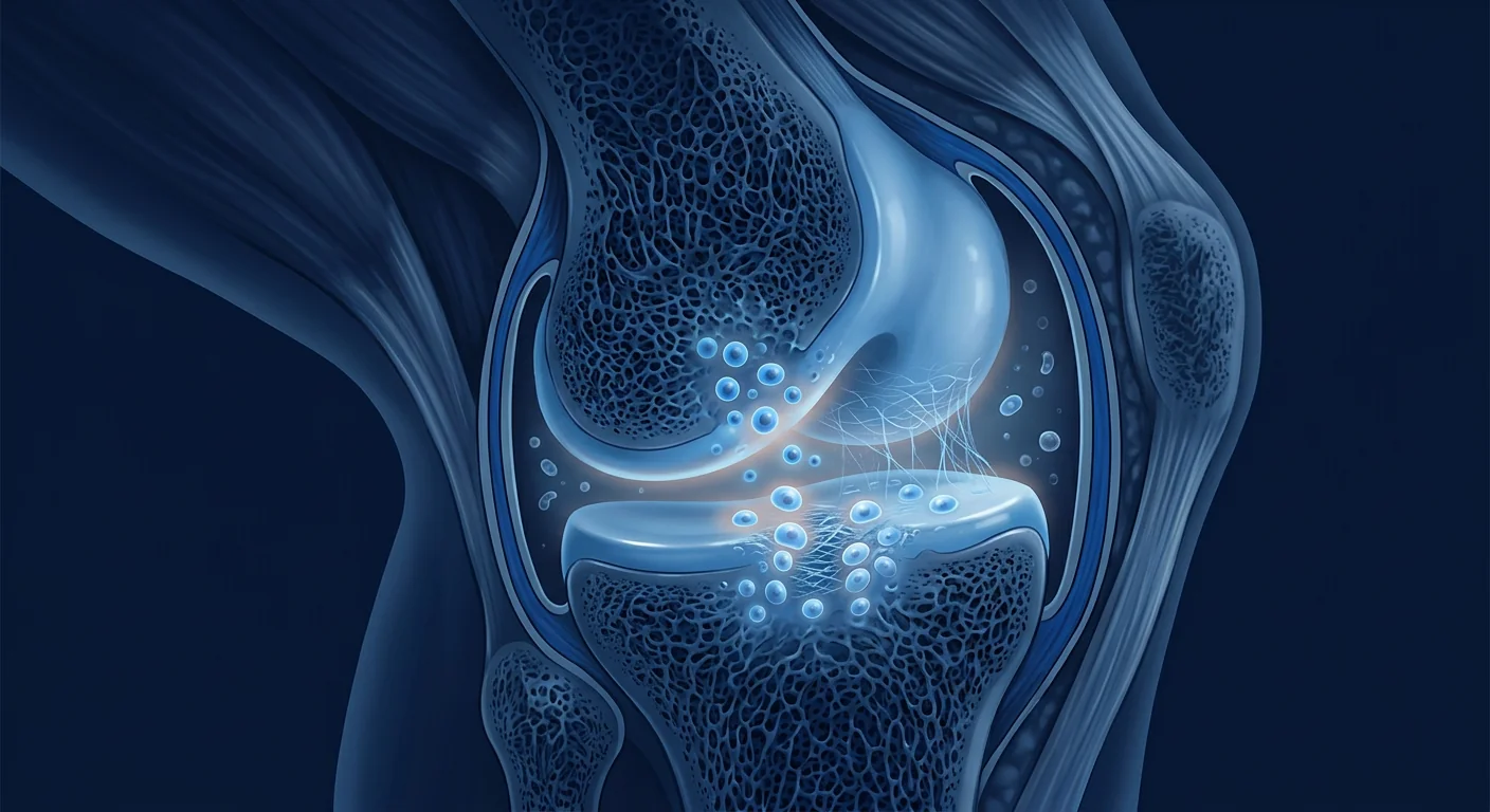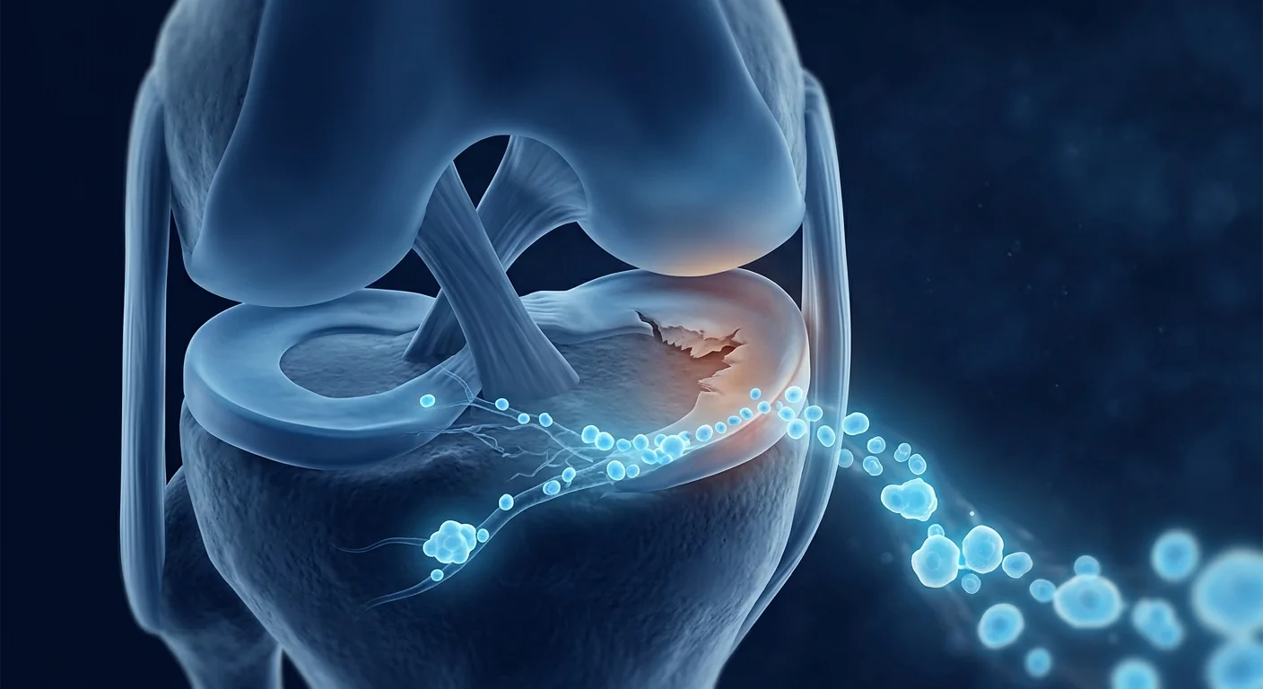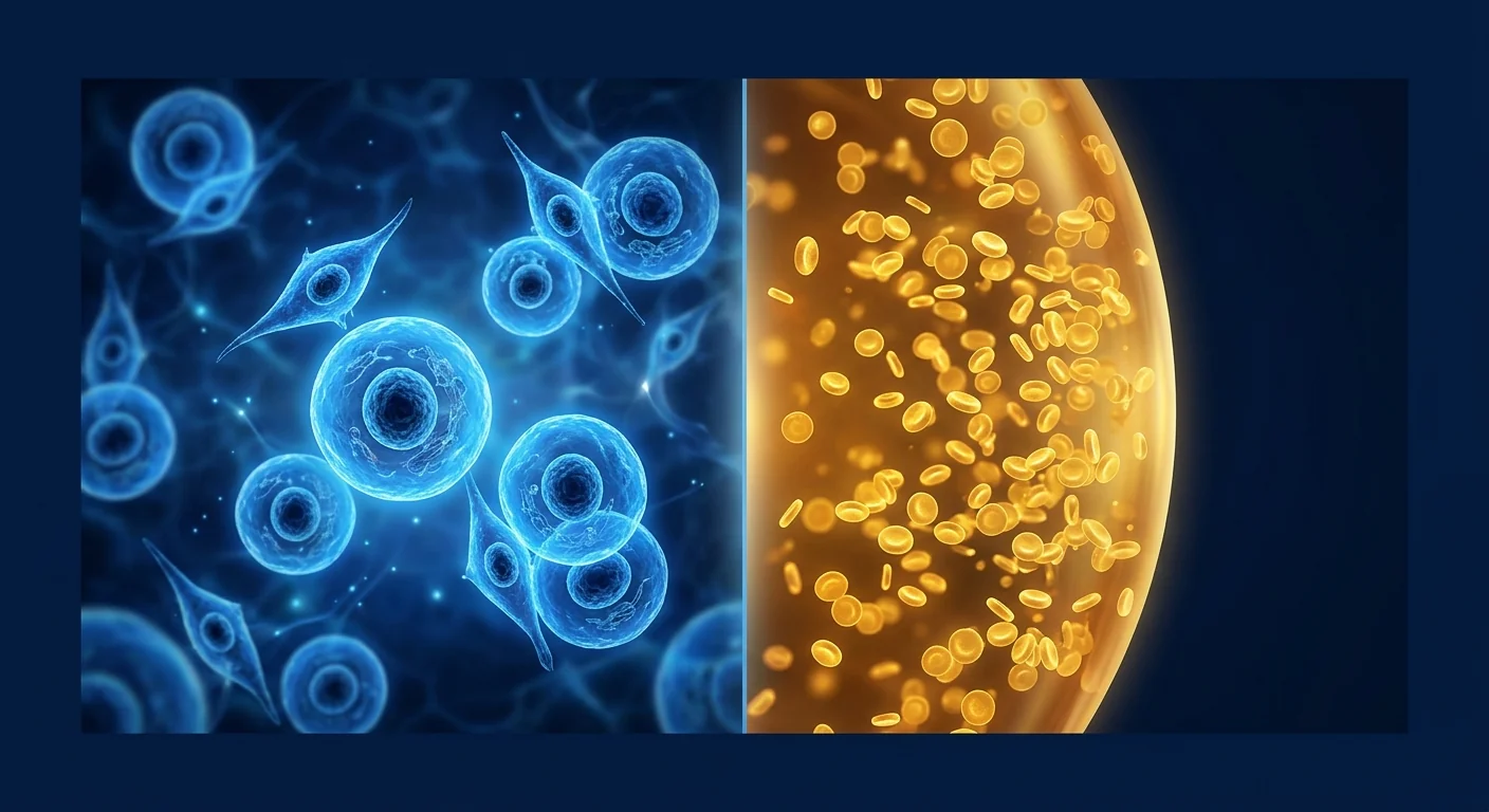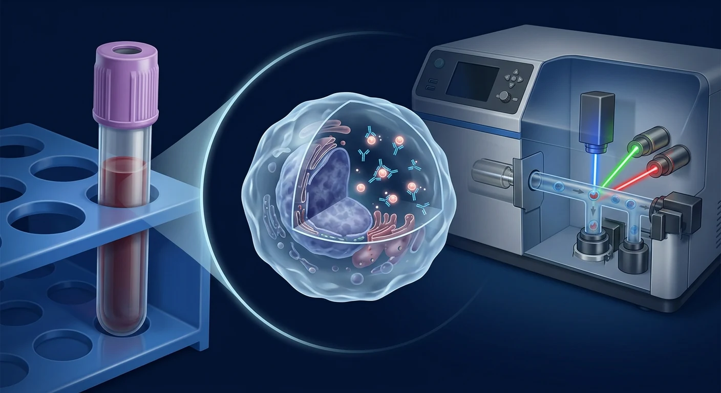
EDIM—epitope detection in monocytes—is an investigational blood-test approach that looks for selected intracellular epitopes inside activated CD14+/CD16+ monocytes or macrophage-like cells. In the best-known implementation, flow cytometry is used to quantify Apo10, an epitope associated with DNaseX and abnormal apoptosis, and TKTL1, a protein linked in the supporting literature to altered glucose metabolism. The proposed biological idea is indirect: phagocytic immune cells ingest material from abnormal tissue, return to the circulation, and carry detectable molecular traces.
That mechanism is scientifically interesting. It does not, by itself, establish a validated population-screening test. Evidence must answer several different questions: can the laboratory measure the signal reliably; does the result distinguish cancer from non-cancer in the intended population; and does acting on the result improve outcomes enough to outweigh downstream harm? Those are analytic validity, diagnostic accuracy, and clinical utility. They are related, but they are not interchangeable.
Bottom line
Small, mostly case-control or disease-specific studies report promising separation between known cancer cases and healthy controls. The evidence base is not yet sufficient to treat a positive EDIM result as a cancer diagnosis, a negative result as an all-clear, or the test as a replacement for recommended screening, diagnostic imaging, endoscopy, or tissue pathology. Any abnormal result requires confirmation through standard clinical pathways.
What exactly does an EDIM test measure?
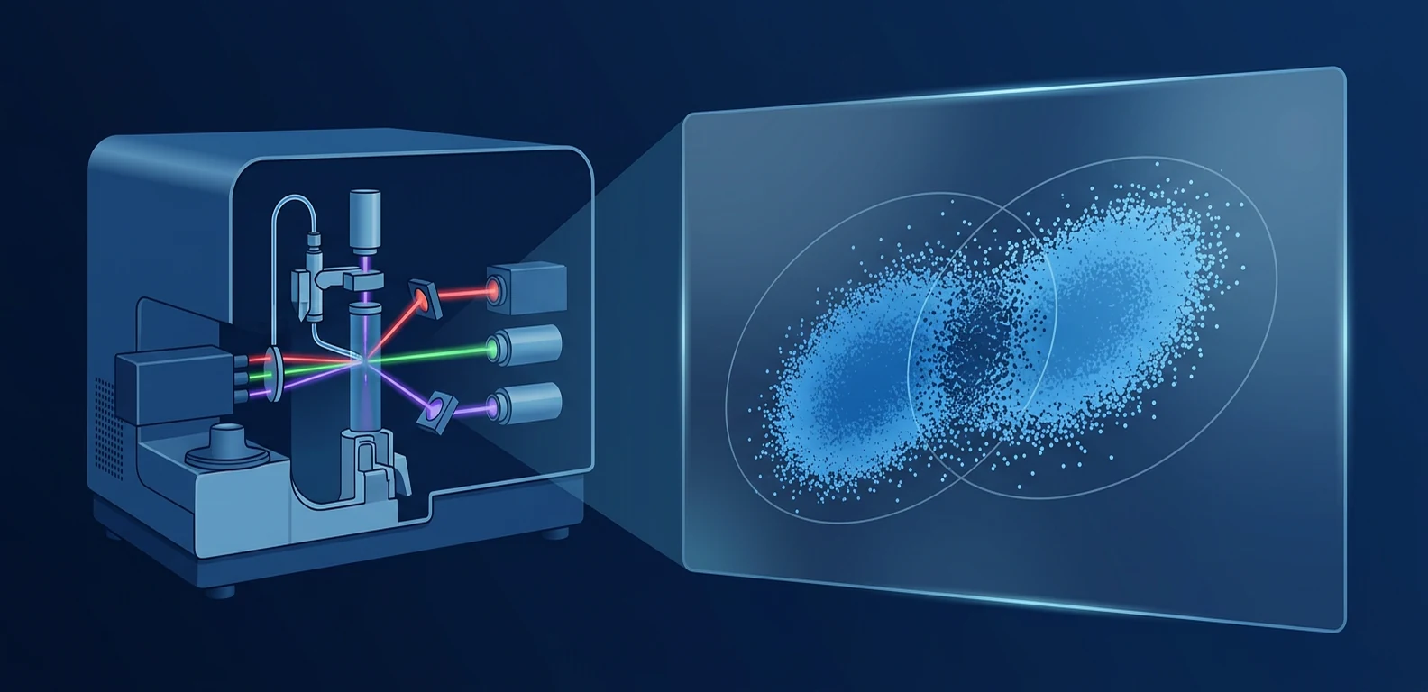
The assay gates a subset of circulating cells using surface markers, commonly CD14 and CD16, then stains intracellular targets after permeabilization. A laboratory derives marker scores and may combine Apo10 and TKTL1 into a composite score. This differs from circulating-tumor-DNA assays, which seek tumor-derived nucleic acids directly in plasma, and from conventional serum tumor markers. EDIM instead uses immune cells as biological collectors.
The concept has supporting observations. In oral squamous cell carcinoma, EDIM scores correlated with tissue expression and often declined after surgical removal; related studies described higher marker-bearing monocytes in patients than controls [1]–[4]. Pilot work has extended the approach to gastrointestinal cancers, neuroblastoma, and rhabdomyosarcoma [5]–[8]. These findings support biological plausibility and hypothesis generation. They do not demonstrate that one threshold performs consistently across all cancers, stages, laboratories, or asymptomatic populations.
1. Analytic validity: can the laboratory result be trusted?
Analytic validity concerns the measurement process, not whether the patient has cancer. A robust assay should show repeatability within a run, reproducibility across days and operators, stability during collection and transport, and comparable performance across instruments and reagent lots. Pre-analytic variables may include anticoagulant, time to processing, storage temperature, cell viability, recent infection, inflammatory activity, medication, and the proportion of CD14+/CD16+ cells available for analysis.
Flow cytometry adds technical choices: compensation, gating boundaries, antibody specificity, permeabilization, fluorescence intensity, instrument calibration, and the formula that converts cell-level signals into a score. A published cut-off is not automatically portable to another laboratory unless the method is standardized and externally validated. Laboratories should document quality controls, invalid-result rules, precision around the decision threshold, and what happens when the sample contains too few suitable cells.
The available clinical papers generally describe the assay method and biological correlations, but publicly available peer-reviewed evidence is limited regarding broad inter-laboratory reproducibility, lot-to-lot performance, interference studies, and prospective proficiency testing. Therefore, a claim that the test is analytically measurable is narrower than a claim that every commercial implementation is analytically equivalent.
2. Diagnostic accuracy: how well does EDIM classify patients?
Diagnostic accuracy compares the index test with a reference standard in a defined population. Sensitivity is the proportion of people with the target disease who test positive. Specificity is the proportion without it who test negative. Positive and negative predictive values answer a different question—what a result means for this patient—and depend strongly on disease prevalence.
Several EDIM studies report high sensitivities. A mixed study of cholangiocarcinoma, pancreatic cancer, and colorectal cancer found a positive combined score in 62 of 62 cancer patients, no positives among 29 healthy controls, and one positive among 13 participants with inflammatory conditions [5]. An earlier synthesis reported 95.8% sensitivity and 97.3% specificity across selected prostate, breast, and oral cancer cohorts [6]. Neuroblastoma and rhabdomyosarcoma pilot studies also found high detection rates against healthy controls [7], [8].
These numbers should be read with their denominators, designs, and confidence intervals. Many cohorts are small and contain patients with already established, sometimes clinically apparent disease. A test can separate advanced cases from unusually healthy controls and still perform less well among asymptomatic people with benign nodules, chronic inflammation, autoimmune disease, metabolic illness, recent surgery, or very small tumors. Accuracy estimates are properties of a test in a specific context, not permanent attributes of a brand.
Spectrum bias: why case-control results may look better than screening
Spectrum bias occurs when the people studied do not represent the patients in whom the test will actually be used. Classic case-control designs often compare clear disease with healthy volunteers. Real clinical practice contains a difficult middle: early lesions, pre-cancer, benign disease, inflammatory conditions, multiple comorbidities, and ambiguous imaging. Sensitivity may fall when tumors are smaller or shed less material; specificity may fall when non-malignant processes activate monocytes or affect the measured epitopes.
This is not a criticism unique to EDIM. It is a central principle of diagnostic-test evaluation, formalized in STARD and QUADAS-2 guidance [10]–[12]. The strongest next study would prospectively enroll consecutive participants from the intended-use population, define the threshold before seeing outcomes, apply an appropriate reference standard regardless of EDIM result, blind interpreters, account for missing and indeterminate results, and report accuracy by cancer type and stage.
False positives: what can a positive result mean?
A false positive is a positive EDIM result in a person who does not have the target cancer under the reference standard. Possible explanations include biological overlap, inflammatory activity, technical variation near a threshold, an occult abnormality not established during follow-up, or a marker signal that is not specific to malignant tissue. The small gastrointestinal study's positive result in an inflammatory participant is a reminder that non-cancer controls must be clinically representative [5].
The practical consequences are not abstract. A positive result can lead to anxiety, repeated blood tests, radiation exposure from CT or PET/CT, contrast reactions, endoscopy, biopsy, incidental findings, cost, and occasionally complications. Broad whole-body localization after a non-localizing blood test also raises the chance of discovering unrelated abnormalities. A responsible pathway specifies who reviews the result, which organ-specific investigation is justified, and when surveillance is safer than an invasive procedure.
A positive EDIM result is not histological proof of cancer and does not identify a tumor's location with certainty. It should trigger a clinician-led risk assessment, not an assertion that cancer is present.
False negatives: why a reassuring result cannot rule out cancer
A false negative occurs when cancer is present but the test is below threshold or invalid. Plausible causes include a small or biologically quiet lesion, tumor heterogeneity, variable epitope expression, insufficient phagocytosis, low numbers of gated monocytes, treatment effects, specimen delay, or analytical error. A proposed saturation effect in advanced disease further shows that marker score may not increase linearly with burden.
A negative result must never override symptoms, examination, imaging, pathology, family history, or eligibility for established screening. Someone with rectal bleeding still needs appropriate colorectal evaluation; a suspicious breast lesion still needs breast imaging and possible biopsy; haematuria, unexplained weight loss, persistent lymphadenopathy, or an abnormal cervical test still require standard work-up. The greatest harm from a false negative is delayed diagnosis caused by misplaced reassurance.
3. Clinical utility: does using EDIM improve health outcomes?
Clinical utility asks whether a test changes management in a way that produces more benefit than harm. High sensitivity and specificity are not enough. A useful screening strategy should detect disease at a stage when treatment is more effective, reduce cancer-specific or overall mortality where feasible to demonstrate, avoid excessive overdiagnosis, and have an acceptable balance of procedures, complications, anxiety, and cost.
The EDIM literature includes monitoring studies in which scores declined after surgery and a colon-cancer case report in which a signal preceded radiological detection of metastasis [2], [4], [9]. Such evidence is useful for designing trials but cannot establish clinical utility. A before-and-after biomarker change does not prove that acting on the change improves survival. A single early-detection case cannot quantify how many false alarms, missed cancers, or unnecessary procedures occur in a screened population.
Screening history shows why outcome trials matter. Low-dose CT lung screening demonstrated a mortality benefit in a defined high-risk population, while also documenting false positives and downstream procedures [13]. EDIM would require analogous prospective evidence for its intended population and pathway. At present, there is insufficient peer-reviewed evidence that EDIM-guided screening reduces mortality or improves quality-adjusted survival compared with established care.
| Evidence domain | Question | Current interpretation |
|---|---|---|
| Biological plausibility | Can monocytes contain measured epitopes? | Supported by laboratory and disease-cohort observations. |
| Analytic validity | Is measurement reproducible across settings? | Some methods are described; broad independent standardization evidence remains limited. |
| Diagnostic accuracy | Does it distinguish disease from non-disease? | Promising in small selected cohorts; generalizability to screening is uncertain. |
| Clinical utility | Does EDIM-guided care improve outcomes? | Not established by randomized or adequately controlled outcome studies. |
Confirmatory standard testing is essential
EDIM should be considered an investigational risk signal, not a standalone diagnosis. Confirmation depends on the clinical scenario and may include focused history and examination, guideline-based imaging, ultrasound, mammography, CT, MRI, endoscopy, organ-specific laboratory testing, and ultimately pathology when a suspicious lesion is sampled. Tissue diagnosis also provides tumor type, grade, molecular features, and information needed for treatment selection—information a pan-cancer score cannot supply.
Likewise, EDIM should not replace established screening for cervical, breast, colorectal, or lung cancer in eligible people. Each standard program has defined populations, intervals, quality controls, and management pathways. Adding a new test without knowing the incremental benefit can increase procedures without improving outcomes.
Monitoring after cancer treatment: a separate intended use
Monitoring is not the same as screening. In a patient with a known tumor that expressed the relevant epitopes, a serial score might be studied as an adjunct to clinical review and standard surveillance. Surgical studies showing score reductions provide a rationale for this question [2], [4]. But meaningful monitoring evidence needs predefined sampling times, analytical change thresholds, comparison with imaging and pathology, and proof that earlier action helps.
Scores can vary because of biology or measurement. Testing too often can produce noise and cascade into low-value investigations. A rise should not be equated automatically with recurrence, and a fall should not prove complete response. The result must be interpreted against the known cancer, treatment, symptoms, and accepted follow-up protocol.
How patients can appraise an EDIM offer
- Ask the intended use: asymptomatic screening, investigation of symptoms, treatment monitoring, or recurrence surveillance?
- Ask about the exact assay: markers, threshold, laboratory accreditation, sample stability, invalid rate, and repeatability near the cut-off.
- Ask for the matching evidence: was performance measured prospectively in people like you, or in known cancers versus healthy controls?
- Request absolute numbers: sensitivity, specificity, confidence intervals, and expected positive predictive value at your baseline risk.
- Agree on next steps before testing: who interprets a positive result, what confirmatory test follows, and who pays for downstream investigation?
- Do not stop standard care: continue recommended screening and investigate warning symptoms regardless of an EDIM score.
What evidence would change confidence?
Confidence would increase with independent, preregistered, multicentre studies using a locked assay and threshold; consecutive asymptomatic or diagnostically relevant participants; adequate representation of benign and inflammatory conditions; stage-specific sensitivity; complete reference-standard verification; and transparent reporting of indeterminate samples. External validation should occur in laboratories and populations separate from the developers.
For clinical utility, a trial or carefully controlled implementation study should compare an EDIM-guided pathway with usual care and measure late-stage cancer incidence, mortality where appropriate, time to diagnosis, false-positive procedures, overdiagnosis, complications, anxiety, and cost. Publication of favorable and unfavorable results is necessary to reduce reporting bias.
Frequently asked questions
Is EDIM a proven early-cancer screening test?
No. Published studies are promising but predominantly small and selected. Broad population-screening performance and outcome benefit have not been established.
Does a positive EDIM score mean I have cancer?
No. It indicates that the assay crossed its decision threshold. Cancer must be evaluated and, when a lesion is found, generally confirmed through standard diagnostic testing and pathology.
Can a negative score rule cancer out?
No. False negatives are possible, particularly where stage-specific sensitivity is uncertain. Symptoms and recommended screening should never be ignored.
Do major guidelines endorse EDIM?
This review does not identify EDIM as an endorsed replacement for established population cancer-screening or diagnostic pathways. Availability, a commercial certificate, or biological plausibility is not the same as a guideline recommendation.
Can EDIM identify where a cancer is?
The Apo10/TKTL1 composite is not organ-specific. Localization requires clinical assessment and appropriately targeted imaging or endoscopy, followed by tissue confirmation when indicated.
Limitations and medical disclaimer
The evidence is evolving, terminology and commercial implementations may vary, and results from one assay version should not be transferred automatically to another. Several publications involve investigators or entities connected with assay intellectual property or development; conflict-of-interest disclosure does not invalidate findings, but it strengthens the case for independent replication. This article is educational and is not personal medical advice.
The safest interpretation is deliberately narrow: EDIM is a biologically plausible investigational liquid-biopsy approach with encouraging signals in selected cohorts, substantial uncertainty in real-world screening, and no standalone diagnostic role. Decisions must remain anchored to symptoms, baseline risk, established screening, confirmatory imaging or endoscopy, and pathology.
References
- Grimm M, et al. A biomarker based detection and characterization of carcinomas exploiting two fundamental biophysical mechanisms in mammalian cells. BMC Cancer. 2013;13:569. doi:10.1186/1471-2407-13-569
- Grimm M, et al. Evaluation of a biomarker based blood test for monitoring surgical resection of oral squamous cell carcinomas. Clinical Oral Investigations. 2016;20:329–338. doi:10.1007/s00784-015-1518-0
- Grimm M, et al. Analysis of circulating CD14+/CD16+ monocyte-derived macrophages in oral squamous cell carcinoma. Oral Surgery, Oral Medicine, Oral Pathology and Oral Radiology. 2016;121:301–306. doi:10.1016/j.oooo.2015.10.024
- Todenhöfer T, et al. Effect of radical prostatectomy on levels of cancer related epitopes in circulating macrophages. Prostate. 2017;77:1251–1258. doi:10.1002/pros.23384
- Saman S, et al. Biomarkers Apo10 and TKTL1: EDIM as a new diagnostic approach for cholangiocellular, pancreatic and colorectal carcinoma. Cancer Biomarkers. 2020;27:129–137. doi:10.3233/CBM-190414
- Coy JF. EDIM-TKTL1/Apo10 Blood Test: An Innate Immune System Based Liquid Biopsy. International Journal of Molecular Sciences. 2017;18:878. doi:10.3390/ijms18040878
- Stagno MJ, et al. EDIM for liquid biopsy including identification of GD2 in childhood neuroblastoma—a pilot study. British Journal of Cancer. 2022;127:1324–1331. doi:10.1038/s41416-022-01855-x
- Urla C, et al. EDIM as a new method of liquid biopsy in pediatric rhabdomyosarcoma. Biomedicines. 2022;10:1812. doi:10.3390/biomedicines10081812
- Jansen N, Coy JF. Diagnostic use of EDIM blood test for early detection of colon cancer metastasis. Future Oncology. 2013;9:605–609. doi:10.2217/fon.13.8
- Bossuyt PM, et al. STARD 2015: an updated list of essential items for reporting diagnostic accuracy studies. BMJ. 2015;351:h5527. doi:10.1136/bmj.h5527
- Whiting PF, et al. QUADAS-2: a revised tool for quality assessment of diagnostic accuracy studies. Annals of Internal Medicine. 2011;155:529–536. doi:10.7326/0003-4819-155-8-201110180-00009
- Lachs MS, et al. Spectrum bias in the evaluation of diagnostic tests. Annals of Internal Medicine. 1992;117:135–140. doi:10.7326/0003-4819-117-2-135
- National Lung Screening Trial Research Team. Reduced lung-cancer mortality with low-dose CT screening. New England Journal of Medicine. 2011;365:395–409. doi:10.1056/NEJMoa1102873
EDIM(单核细胞表位检测)是一种研究中的血液检测方法。它通过流式细胞术,在活化的 CD14+/CD16+ 单核细胞或巨噬细胞样细胞内检测 Apo10、TKTL1 等表位。理论上,这些吞噬细胞摄取异常组织材料后,把可检测的分子信号带回外周血。
这一机制具有科学价值,但不能单独证明其适合普通人群癌症筛查。评价必须分开回答三个问题:实验室能否稳定测量(分析有效性);结果能否在目标人群中区分癌症与非癌症(诊断准确性);依据结果采取行动能否改善结局且收益大于伤害(临床效用)。
核心结论
小型、主要为病例对照或特定癌种的研究显示已知癌症患者与健康对照之间存在良好区分。但现有证据不足以把阳性结果当作癌症诊断、把阴性结果当作“排除癌症”,也不能替代标准筛查、影像、内镜或组织病理。异常结果必须通过标准诊疗路径确认。
EDIM 测量什么?
检测先用 CD14/CD16 标记筛选细胞,通透后染色细胞内靶点,再计算单项与综合分数。它不同于直接寻找血浆肿瘤 DNA 的检测,也不同于传统血清肿瘤标志物;EDIM 把免疫细胞视为“生物采集器”。口腔鳞癌手术前后研究及胃肠道癌、神经母细胞瘤和横纹肌肉瘤试点研究支持其生物学合理性 [1]–[8],但不能证明同一阈值适用于所有癌种、分期、实验室和无症状人群。
1. 分析有效性
分析有效性关注测量本身,而不是患者是否患癌。应验证同批次重复性、不同日期和操作者间再现性、运输稳定性、仪器和抗体批次一致性。抗凝剂、处理延迟、温度、细胞活力、炎症、药物以及可供分析的细胞数量都可能影响结果。流式细胞术的门控、补偿、通透、校准和评分公式也必须标准化。
已发表论文描述了方法和部分生物学关联,但关于跨实验室再现性、干扰试验、批间差异和外部能力验证的同行评审证据仍有限。能测到信号,不等于所有商业实验室都能给出等效结果。
2. 诊断准确性
诊断准确性必须把指数检测与参考标准在明确人群中比较。敏感度是患者中阳性的比例,特异度是无病者中阴性的比例;阳性和阴性预测值则强烈依赖患病率。
一项胆管、胰腺及结直肠癌研究中,62/62 癌症患者综合分数阳性,29 名健康对照均阴性,13 名炎症患者中 1 名阳性 [5]。其他选定队列也报告较高准确性 [6]–[8]。但样本通常较小,患者常已有明确疾病;这不能直接代表无症状筛查。
疾病谱偏倚
疾病谱偏倚是指研究对象与实际使用人群不同。明显癌症与健康志愿者容易区分,而临床中存在早期病变、癌前病变、良性结节、慢性炎症、自身免疫病和多种共病。肿瘤较小时敏感度可能下降,非恶性炎症激活单核细胞时特异度也可能下降。STARD、QUADAS-2 和疾病谱偏倚研究都强调应在连续、真实目标人群中前瞻验证 [10]–[12]。
假阳性
假阳性会带来焦虑、重复抽血、CT 或 PET/CT 辐射、造影剂风险、内镜、活检、偶然发现和费用。原因可能包括炎症、阈值附近技术波动、生物学重叠,或随访尚未证实的异常。EDIM 阳性不是癌症组织学证据,也不能确定肿瘤位置。它只能触发医生主导的风险评估。
假阴性
肿瘤较小、表位表达异质、吞噬不足、目标细胞数量少、治疗影响、样本延迟或分析误差均可造成假阴性。阴性结果不得推翻症状、体检、影像、病理、家族史或标准筛查资格。便血、可疑乳腺病灶、血尿、体重下降或持续淋巴结肿大仍需常规检查。
3. 临床效用
临床效用问的是:使用检测是否真正改善健康结局。高敏感度和特异度并不够;筛查应在治疗更有效的阶段发现疾病,减少晚期癌症或死亡,同时控制过度诊断、侵入性检查、并发症、焦虑和成本。
EDIM 文献包括术后分数下降以及结肠癌转移影像发现前出现信号的个案 [2]、[4]、[9]。这可用于设计试验,但不能证明依据该信号行动会改善生存。低剂量 CT 肺癌筛查之所以成为标准,是因为在特定高危人群中证实死亡率获益,并同时记录假阳性伤害 [13]。EDIM 尚缺少类似结局证据。
| 证据层面 | 问题 | 当前判断 |
|---|---|---|
| 生物学合理性 | 单核细胞内能否检测相关表位? | 实验室及疾病队列观察支持。 |
| 分析有效性 | 跨环境测量是否可重复? | 已有方法描述,但广泛独立标准化证据有限。 |
| 诊断准确性 | 能否区分患病与无病? | 选定小队列有希望;筛查普适性不确定。 |
| 临床效用 | EDIM 指导的照护能否改善结局? | 尚未由随机或充分对照的结局研究证实。 |
必须进行标准确认检测
EDIM 应被视为研究性的风险信号,而非独立诊断。根据情境,确认可包括病史和体检、指南推荐影像、超声、乳腺摄影、CT、MRI、内镜、器官特异性检查,以及发现可疑病灶后的组织病理。病理还能提供癌种、分级和治疗所需分子信息。EDIM 不应替代符合条件人群的宫颈、乳腺、结直肠或肺癌标准筛查。
治疗后监测是不同用途
已知癌症患者的监测与普通筛查不同。若原肿瘤表达相应表位,连续分数可作为研究中的辅助指标,但需要预设时间、变化阈值,并与影像和病理比较。升高不能自动等同复发,下降也不能证明完全缓解。
患者应提出的问题
- 用途是无症状筛查、症状诊断还是治疗后监测?
- 具体标志物、阈值、无效率和实验室质量体系是什么?
- 证据是否来自与自己相似的前瞻性人群?
- 敏感度、特异度、置信区间及自身基线风险下的预测值是多少?
- 阳性后由谁解释、做何种确认、下游费用由谁承担?
- 无论结果如何,都继续标准筛查并检查警示症状。
什么证据能提高可信度?
需要独立、预注册、多中心研究,锁定检测与阈值,连续纳入真实目标人群,充分包含良性与炎症疾病,报告各癌种和分期敏感度,并对所有参与者采用适当参考标准。临床效用研究还应比较 EDIM 路径与常规照护,测量晚期癌症、死亡、假阳性操作、过度诊断、并发症、焦虑及成本。
常见问题
EDIM 是已证实的早癌筛查吗?
不是。现有小型选定队列有希望,但普通人群筛查表现及结局获益尚未确立。
阳性就是癌症吗?
不是。它只表示超过检测阈值,必须进行标准诊断,发现病灶时通常需病理确认。
阴性能排除癌症吗?
不能。假阴性存在,症状和推荐筛查不能被忽略。
指南是否推荐 EDIM?
本文未把 EDIM 认定为替代现行人群癌症筛查或诊断路径的指南推荐项目。可购买或具商业认证不等于获得临床指南推荐。
局限性与医疗声明
证据仍在发展,不同商业版本可能不同。部分研究者或机构与检测知识产权或开发有关,这不自动否定研究,但更需要独立重复验证。本文仅供教育,不构成个人医疗建议。最安全的结论是:EDIM 是具有生物学合理性、在选定队列显示积极信号的研究性液体活检方法,但真实筛查环境仍有重大不确定性,不具备独立诊断作用。
参考文献
- Grimm M, et al. A biomarker based detection and characterization of carcinomas exploiting two fundamental biophysical mechanisms in mammalian cells. BMC Cancer. 2013;13:569. doi:10.1186/1471-2407-13-569
- Grimm M, et al. Evaluation of a biomarker based blood test for monitoring surgical resection of oral squamous cell carcinomas. Clinical Oral Investigations. 2016;20:329–338. doi:10.1007/s00784-015-1518-0
- Grimm M, et al. Analysis of circulating CD14+/CD16+ monocyte-derived macrophages in oral squamous cell carcinoma. Oral Surgery, Oral Medicine, Oral Pathology and Oral Radiology. 2016;121:301–306. doi:10.1016/j.oooo.2015.10.024
- Todenhöfer T, et al. Effect of radical prostatectomy on levels of cancer related epitopes in circulating macrophages. Prostate. 2017;77:1251–1258. doi:10.1002/pros.23384
- Saman S, et al. Biomarkers Apo10 and TKTL1: EDIM as a new diagnostic approach for cholangiocellular, pancreatic and colorectal carcinoma. Cancer Biomarkers. 2020;27:129–137. doi:10.3233/CBM-190414
- Coy JF. EDIM-TKTL1/Apo10 Blood Test: An Innate Immune System Based Liquid Biopsy. International Journal of Molecular Sciences. 2017;18:878. doi:10.3390/ijms18040878
- Stagno MJ, et al. EDIM for liquid biopsy including identification of GD2 in childhood neuroblastoma—a pilot study. British Journal of Cancer. 2022;127:1324–1331. doi:10.1038/s41416-022-01855-x
- Urla C, et al. EDIM as a new method of liquid biopsy in pediatric rhabdomyosarcoma. Biomedicines. 2022;10:1812. doi:10.3390/biomedicines10081812
- Jansen N, Coy JF. Diagnostic use of EDIM blood test for early detection of colon cancer metastasis. Future Oncology. 2013;9:605–609. doi:10.2217/fon.13.8
- Bossuyt PM, et al. STARD 2015: an updated list of essential items for reporting diagnostic accuracy studies. BMJ. 2015;351:h5527. doi:10.1136/bmj.h5527
- Whiting PF, et al. QUADAS-2: a revised tool for quality assessment of diagnostic accuracy studies. Annals of Internal Medicine. 2011;155:529–536. doi:10.7326/0003-4819-155-8-201110180-00009
- Lachs MS, et al. Spectrum bias in the evaluation of diagnostic tests. Annals of Internal Medicine. 1992;117:135–140. doi:10.7326/0003-4819-117-2-135
- National Lung Screening Trial Research Team. Reduced lung-cancer mortality with low-dose CT screening. New England Journal of Medicine. 2011;365:395–409. doi:10.1056/NEJMoa1102873
EDIM، أي كشف الحواتم داخل الوحيدات، هو نهج بحثي لفحص الدم. يستخدم قياس التدفق الخلوي للكشف عن حواتم مثل Apo10 وTKTL1 داخل خلايا CD14+/CD16+ المنشطة. تفترض الفكرة أن الخلايا البلعمية تبتلع مواد من نسيج غير طبيعي ثم تحمل إشارات جزيئية إلى الدم المحيطي.
الآلية مثيرة للاهتمام، لكنها لا تثبت وحدها صلاحية الفحص السكاني. يجب فصل ثلاثة أسئلة: هل القياس المخبري ثابت؟ وهذه الصلاحية التحليلية. هل يميز السرطان من عدمه في السكان المقصودين؟ وهذه الدقة التشخيصية. وهل يؤدي استخدامه إلى نتائج صحية أفضل مع ضرر أقل؟ وهذه المنفعة السريرية.
الخلاصة
أظهرت دراسات صغيرة، غالبها حالات وشواهد أو سرطانات محددة، فصلاً واعداً بين حالات سرطان معروفة وأصحاء. لكنها لا تكفي لاعتبار النتيجة الإيجابية تشخيصاً، أو السلبية نفياً للسرطان، أو لاستبدال الفحص الموصى به والتصوير والتنظير وعلم الأمراض. كل نتيجة غير طبيعية تحتاج إلى تأكيد بالمسار الطبي القياسي.
ماذا يقيس EDIM؟
تُحدَّد الخلايا بعلامات CD14 وCD16، ثم تُصبغ الأهداف داخلها بعد جعل الغشاء نافذاً، وتُحسب درجات منفردة أو مركبة. يختلف ذلك عن اختبارات DNA الورم الحر التي تبحث مباشرة عن أحماض نووية ورمية. تدعم دراسات سرطان الفم قبل الجراحة وبعدها، وتجارب سرطانات الجهاز الهضمي والورم الأرومي العصبي والساركومة العضلية المخططة، المعقولية البيولوجية [1]–[8]؛ لكنها لا تثبت صلاحية عتبة واحدة لكل السرطانات والمراحل والمختبرات والأصحاء بلا أعراض.
1. الصلاحية التحليلية
الصلاحية التحليلية تخص موثوقية القياس، لا وجود السرطان. ينبغي إثبات التكرار داخل التشغيل، وإعادة الإنتاج بين الأيام والمشغلين، وثبات العينة أثناء النقل، واتساق الأجهزة ودفعات الكواشف. قد تؤثر مادة منع التخثر، ووقت المعالجة، والحرارة، وحيوية الخلايا، والعدوى أو الالتهاب، والأدوية، وعدد الخلايا المؤهلة. كما يجب توحيد التعويض والبوابات والأجسام المضادة والنفاذية والمعايرة ومعادلة النتيجة.
تصف الدراسات المنشورة الطريقة وبعض الارتباطات، لكن الأدلة المحكمة الواسعة على إعادة الإنتاج بين المختبرات، ودراسات التداخل، والاختبار الخارجي للكفاءة ما تزال محدودة. قابلية قياس إشارة لا تعني تكافؤ كل تطبيق تجاري.
2. الدقة التشخيصية
تُقارن نتيجة الاختبار بمرجع تشخيصي ضمن سكان محددين. الحساسية هي نسبة المصابين الذين تظهر لديهم نتيجة إيجابية، والنوعية نسبة غير المصابين ذوي النتيجة السلبية. أما القيم التنبؤية فتعتمد كثيراً على انتشار المرض.
وجدت دراسة لسرطانات القنوات الصفراوية والبنكرياس والقولون نتيجة مركبة إيجابية لدى 62 من 62 مريضاً، وسلبية لدى 29 سليماً، مع إيجابية واحدة بين 13 مصاباً بالالتهاب [5]. وأبلغت مجموعات مختارة أخرى عن أرقام مرتفعة [6]–[8]. إلا أن العينات صغيرة غالباً والمرض معروف مسبقاً؛ فلا يمكن نقل الأرقام مباشرة إلى فحص أشخاص بلا أعراض.
تحيز طيف المرض
ينشأ تحيز الطيف عندما لا يشبه المشاركون الأشخاص الذين سيُستخدم الفحص لديهم فعلياً. فصل سرطان واضح عن متطوع سليم أسهل من الواقع الذي يضم أوراماً مبكرة، وآفات حميدة، والتهاباً مزمناً، وأمراض مناعة ذاتية ومراضات متعددة. قد تنخفض الحساسية مع الورم الصغير، وتنخفض النوعية عندما تنشط حالات غير خبيثة الوحيدات. تؤكد STARD وQUADAS-2 وأبحاث تحيز الطيف ضرورة التحقق المستقبلي في سكان الاستخدام الحقيقي [10]–[12].
النتائج الإيجابية الكاذبة
قد تنتج عن الالتهاب، أو التداخل البيولوجي، أو التفاوت التقني قرب العتبة. ويمكن أن تقود إلى القلق، وتكرار التحاليل، والتعرض للإشعاع في CT أو PET/CT، ومخاطر الصبغة، والتنظير والخزعة والموجودات العرضية والتكاليف. النتيجة الإيجابية ليست دليلاً نسيجياً ولا تحدد موضع الورم يقيناً. إنها تستدعي تقييماً طبياً منظماً لا إعلان وجود سرطان.
النتائج السلبية الكاذبة
قد تحدث بسبب ورم صغير، أو اختلاف التعبير عن الحواتم، أو بلعمة غير كافية، أو قلة الخلايا، أو تأثير العلاج، أو تأخر العينة أو خطأ تحليلي. لا يجوز أن تتغلب نتيجة سلبية على الأعراض أو الفحص أو التصوير أو علم الأمراض أو التاريخ العائلي أو أهلية الفحص القياسي. النزف الشرجي، أو كتلة الثدي المشبوهة، أو الدم في البول، أو نقص الوزن، تظل بحاجة إلى تقييم معتاد.
3. المنفعة السريرية
المنفعة السريرية تسأل إن كان استخدام الاختبار يغير الرعاية بما يحسن الصحة أكثر مما يسبب الضرر. الحساسية والنوعية العاليتان لا تكفيان. ينبغي للفحص اكتشاف المرض في مرحلة يصبح العلاج فيها أنفع، وتقليل السرطان المتأخر أو الوفيات، مع ضبط الإفراط في التشخيص والإجراءات والقلق والكلفة.
تشمل أدبيات EDIM انخفاض الدرجات بعد الجراحة وتقرير حالة سبق فيه المؤشر اكتشاف نقيلة سرطان القولون بالتصوير [2]، [4]، [9]. هذه ملاحظات لتصميم الدراسات وليست برهاناً على تحسن البقاء. أثبت فحص الرئة بـCT منخفض الجرعة انخفاض الوفيات في فئة عالية الخطورة، ووثق أيضاً الإيجابيات الكاذبة [13]. لا تتوفر لـEDIM حتى الآن أدلة نتائج مماثلة.
| مجال الدليل | السؤال | التفسير الحالي |
|---|---|---|
| المعقولية البيولوجية | هل توجد الحواتم داخل الوحيدات؟ | تدعمها ملاحظات مخبرية ومرضية. |
| الصلاحية التحليلية | هل القياس قابل لإعادة الإنتاج؟ | الطريقة موصوفة؛ التوحيد المستقل الواسع محدود. |
| الدقة التشخيصية | هل يميز المرض؟ | واعد في مجموعات صغيرة مختارة؛ التعميم غير مؤكد. |
| المنفعة السريرية | هل يحسن المسار الموجه بـEDIM النتائج؟ | غير مثبتة بدراسات نتائج عشوائية أو مضبوطة كافية. |
التأكيد بالفحوص القياسية ضروري
ينبغي اعتبار EDIM إشارة خطر بحثية لا تشخيصاً مستقلاً. قد يشمل التأكيد التاريخ والفحص، والتصوير الموافق للإرشادات، والموجات فوق الصوتية أو تصوير الثدي أو CT أو MRI، والتنظير، ثم علم الأمراض عند أخذ عينة. ويحدد النسيج نوع الورم ودرجته وخصائصه اللازمة للعلاج، وهي معلومات لا توفرها درجة شاملة. ولا ينبغي أن يحل EDIM محل فحوص عنق الرحم والثدي والقولون والرئة المعتمدة للمؤهلين.
المراقبة بعد العلاج استعمال مختلف
المراقبة لمريض معروف السرطان ليست فحصاً سكانياً. قد تُدرس الدرجة المتسلسلة كعامل مساعد إذا كان الورم الأصلي يعبر الحواتم، لكن يلزم توقيت وعتبة تغير محددان ومقارنة بالتصوير وعلم الأمراض. الارتفاع لا يثبت النكس والانخفاض لا يثبت الاستجابة الكاملة.
أسئلة ينبغي للمريض طرحها
- هل الغرض فحص بلا أعراض، أم تشخيص أعراض، أم مراقبة بعد العلاج؟
- ما العلامات والعتبة ومعدل النتائج غير الصالحة ونظام جودة المختبر؟
- هل دُرس الأداء مستقبلياً في أشخاص يشبهونني؟
- ما الحساسية والنوعية وفواصل الثقة والقيمة التنبؤية عند خطري الأساسي؟
- من يفسر الإيجابية وما فحص التأكيد ومن يتحمل التكاليف؟
- استمر في الفحص القياسي وتقييم علامات الإنذار مهما كانت النتيجة.
ما الدليل المطلوب؟
ترتفع الثقة بدراسات مستقلة، مسجلة مسبقاً، متعددة المراكز، تستخدم اختباراً وعتبة مقفلين، وتضم بالتتابع سكان الاستخدام الحقيقي وحالات حميدة والتهابية، وتبلغ الحساسية حسب النوع والمرحلة، وتطبق المرجع على الجميع. وتحتاج المنفعة السريرية مقارنة مسار EDIM بالرعاية المعتادة وقياس السرطان المتأخر والوفيات والإجراءات الكاذبة والإفراط في التشخيص والمضاعفات والقلق والكلفة.
أسئلة شائعة
هل EDIM فحص مثبت للكشف المبكر؟
لا. النتائج واعدة في مجموعات صغيرة مختارة، لكن أداء الفحص السكاني وفائدته النهائية غير مثبتين.
هل الإيجابية تعني السرطان؟
لا. تعني تجاوز عتبة الاختبار، ويلزم تشخيص قياسي وعادة تأكيد نسيجي عند العثور على آفة.
هل السلبية تنفي السرطان؟
لا. قد تحدث سلبية كاذبة، ولا يجوز تجاهل الأعراض أو الفحص الموصى به.
هل توصي الإرشادات بـEDIM؟
لا تقدم هذه المراجعة EDIM كبديل موصى به في الإرشادات لمسارات فحص السرطان أو تشخيصه. التوفر التجاري أو الشهادة لا يساوي توصية إرشادية.
القيود وإخلاء المسؤولية
الأدلة تتطور وقد تختلف التطبيقات التجارية. يرتبط بعض الباحثين أو الجهات بملكية الاختبار أو تطويره؛ لا يبطل ذلك النتائج تلقائياً لكنه يجعل التكرار المستقل ضرورياً. المقال للتثقيف وليس نصيحة طبية شخصية. التفسير الأكثر أماناً: EDIM نهج خزعة سائلة بحثي ذو معقولية بيولوجية وإشارات واعدة في مجموعات مختارة، مع عدم يقين كبير في الفحص الواقعي ودون دور تشخيصي مستقل.
المراجع
- Grimm M, et al. A biomarker based detection and characterization of carcinomas exploiting two fundamental biophysical mechanisms in mammalian cells. BMC Cancer. 2013;13:569. doi:10.1186/1471-2407-13-569
- Grimm M, et al. Evaluation of a biomarker based blood test for monitoring surgical resection of oral squamous cell carcinomas. Clinical Oral Investigations. 2016;20:329–338. doi:10.1007/s00784-015-1518-0
- Grimm M, et al. Analysis of circulating CD14+/CD16+ monocyte-derived macrophages in oral squamous cell carcinoma. Oral Surgery, Oral Medicine, Oral Pathology and Oral Radiology. 2016;121:301–306. doi:10.1016/j.oooo.2015.10.024
- Todenhöfer T, et al. Effect of radical prostatectomy on levels of cancer related epitopes in circulating macrophages. Prostate. 2017;77:1251–1258. doi:10.1002/pros.23384
- Saman S, et al. Biomarkers Apo10 and TKTL1: EDIM as a new diagnostic approach for cholangiocellular, pancreatic and colorectal carcinoma. Cancer Biomarkers. 2020;27:129–137. doi:10.3233/CBM-190414
- Coy JF. EDIM-TKTL1/Apo10 Blood Test: An Innate Immune System Based Liquid Biopsy. International Journal of Molecular Sciences. 2017;18:878. doi:10.3390/ijms18040878
- Stagno MJ, et al. EDIM for liquid biopsy including identification of GD2 in childhood neuroblastoma—a pilot study. British Journal of Cancer. 2022;127:1324–1331. doi:10.1038/s41416-022-01855-x
- Urla C, et al. EDIM as a new method of liquid biopsy in pediatric rhabdomyosarcoma. Biomedicines. 2022;10:1812. doi:10.3390/biomedicines10081812
- Jansen N, Coy JF. Diagnostic use of EDIM blood test for early detection of colon cancer metastasis. Future Oncology. 2013;9:605–609. doi:10.2217/fon.13.8
- Bossuyt PM, et al. STARD 2015: an updated list of essential items for reporting diagnostic accuracy studies. BMJ. 2015;351:h5527. doi:10.1136/bmj.h5527
- Whiting PF, et al. QUADAS-2: a revised tool for quality assessment of diagnostic accuracy studies. Annals of Internal Medicine. 2011;155:529–536. doi:10.7326/0003-4819-155-8-201110180-00009
- Lachs MS, et al. Spectrum bias in the evaluation of diagnostic tests. Annals of Internal Medicine. 1992;117:135–140. doi:10.7326/0003-4819-117-2-135
- National Lung Screening Trial Research Team. Reduced lung-cancer mortality with low-dose CT screening. New England Journal of Medicine. 2011;365:395–409. doi:10.1056/NEJMoa1102873