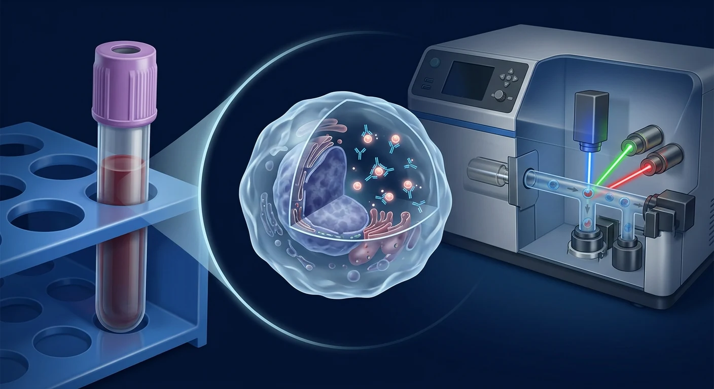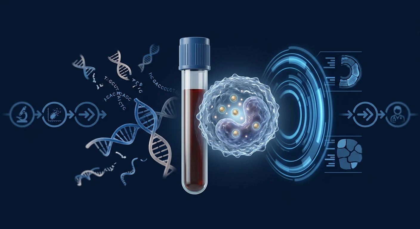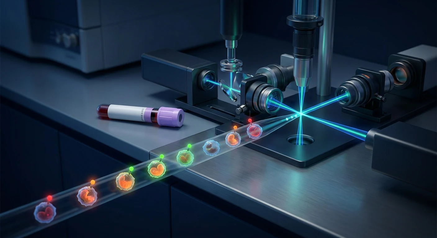
An EDIM Apo10/TKTL1 result is only as trustworthy as the blood sample and the laboratory workflow that produced it. The test counts fluorescence signals inside living, activated monocytes, so how the tube is drawn, stored, transported, and gated can change the number a patient eventually reads.
Why the pre-analytical phase deserves attention. In laboratory medicine generally, most reported errors happen before an instrument is ever switched on — in collection, identification, handling, and transport rather than in the analyser itself [13], [14]. A cell-based assay is more exposed to that phase than a simple serum chemistry, because the analyte is a fragile intact cell rather than a stable molecule.
What this article covers. It walks through the practical journey of an EDIM sample in Bangkok — preparation, draw, transport, staining, flow-cytometric gating, quality control, and reporting — and marks clearly where published standardisation exists and where it does not.
What it is not. This is not a claim that EDIM is a validated stand-alone cancer diagnosis. EDIM remains an investigational adjunct; guideline-recommended screening such as mammography, colonoscopy, cervical screening, or low-dose CT where indicated is not replaced by any blood biomarker score [14].
Direct answer
Preparation for an EDIM blood test is simple for the patient — a routine venous draw, no fasting requirement described in the published method papers — but the laboratory side is demanding: the sample must reach the flow-cytometry laboratory quickly, stay within a controlled temperature range, and be processed with calibrated instrument settings and defined monocyte gating. Because a published, internationally harmonised EDIM protocol is not available in the same way it is for accredited routine assays, the operating laboratory's own documented procedure and quality control matter a great deal.
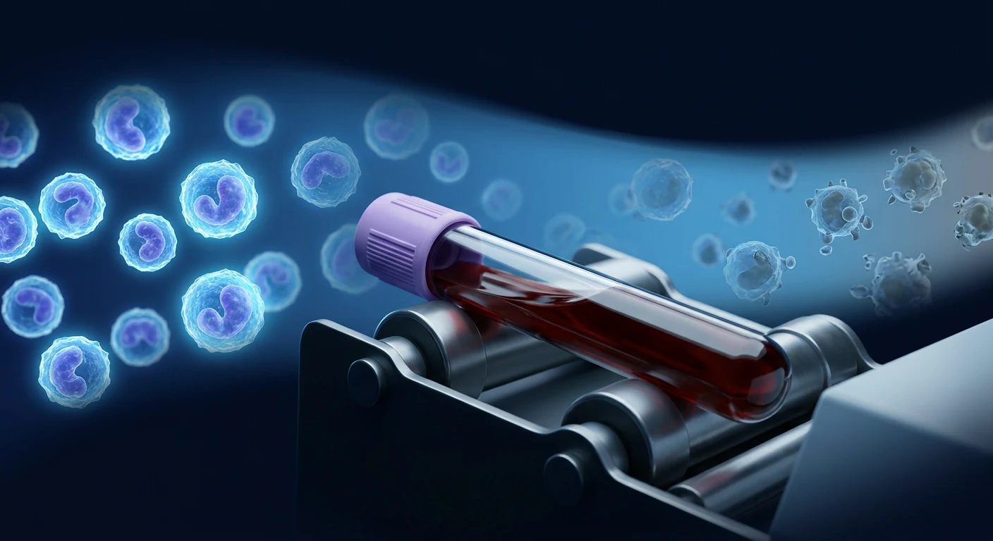
What actually happens between the blood draw and an EDIM score
EDIM measures tumour-associated epitope material inside circulating phagocytic monocytes rather than free molecules in plasma. Macrophages and monocytes ingest cellular debris; the method's premise is that some of that ingested material can be detected inside the cell by fluorescence-labelled antibodies and quantified by flow cytometry [1], [2].
In outline, the workflow has six stages: patient preparation and consent; venous collection into an anticoagulated tube; controlled transport to the laboratory; monocyte identification and permeabilisation for intracellular staining of Apo10 and TKTL1 targets; flow-cytometric acquisition with calibration and controls; and score generation with clinical reporting [3], [4].
Every stage adds variability. The published EDIM literature reports scores from research and pilot cohorts rather than from large multi-laboratory reproducibility programmes, so cross-laboratory comparability of an individual number should not be assumed [9], [10].
How to prepare for an EDIM blood test
Patient preparation for EDIM is straightforward, but the clinical history taken beforehand is the part that changes interpretation. The published method descriptions do not specify a fasting requirement, and the sample is a standard peripheral venous draw [1].
What should be disclosed before the draw:
- Recent infection or fever — monocytes are activated during infection, and activation states are part of the biology the assay observes.
- Surgery, biopsy, or dental extraction in recent weeks — tissue repair and remodelling change the monocyte population's activity.
- Active inflammatory or autoimmune disease, and current flare status.
- Immunosuppressive, corticosteroid, or chemotherapeutic treatment, which alters circulating monocyte counts and phenotype.
- Known cancer history and treatment dates, because post-treatment measurement is interpreted differently from a first screening measurement [5], [6].
- Vaccination in the preceding days, which produces a transient immune activation.
None of these automatically disqualify a person from testing. They determine whether the physician should postpone the draw, or read the result with wider uncertainty. See our companion article on EDIM, inflammation, and false-positive results for how those states are weighed.
Sample collection: tube type, volume, and labelling
EDIM requires whole blood with intact, viable leukocytes, so the sample is collected into an anticoagulant tube rather than a clot tube. Serum is unusable for this assay because the cellular fraction is discarded during clotting.
Three collection details matter more than patients usually expect:
- Anticoagulant choice. Different anticoagulants preserve leukocyte surface markers and viability differently, and the laboratory's validated tube type should be used without substitution.
- Fill volume. Under-filled tubes change the blood-to-anticoagulant ratio, which can affect cell morphology and staining behaviour.
- Gentle mixing, no vigorous shaking. Mechanical stress damages cells and can generate debris that complicates gating.
Difficult venepuncture — repeated attempts, prolonged tourniquet time, or drawing through a narrow line — is a recognised general source of pre-analytical error in laboratory medicine and should be documented when it occurs [14].
Why time and temperature matter
Cell-based assays deteriorate with delay. Studies of high-dimensional flow cytometry in multicentre clinical research have shown that blood storage time and handling conditions measurably change data quality and marker expression before acquisition [15].
The practical consequences are:
- Longer intervals between draw and staining allow cell activation, apoptosis, and debris accumulation.
- Temperature excursions during transport — a hot vehicle in Bangkok traffic, or a sample left against a cold pack — affect membrane integrity and antigen detectability.
- Delays disproportionately affect fragile subpopulations, and monocytes are among the more responsive cell types.
For patients this translates into one operational question worth asking: is the sample analysed locally in Bangkok, or shipped abroad, and what interval and temperature range does the laboratory guarantee? A laboratory that cannot state its accepted stability window is not demonstrating control of the most controllable part of the process.
Inside the laboratory: monocyte gating and intracellular staining
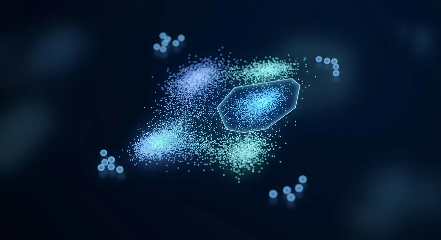
Once received, the laboratory must first identify the correct cells. Monocytes are a heterogeneous population — classical, intermediate, and non-classical subsets are defined by CD14 and CD16 expression, and each behaves differently in inflammation [16]. Which subsets are included in the analysis gate directly influences the measured signal.
Detecting Apo10 and TKTL1 material requires access to the inside of the cell, so the workflow involves fixation and permeabilisation before antibody staining, then washing to remove unbound antibody. Each of these steps has a published potential to alter epitope accessibility and background fluorescence, which is why immunology consensus guidelines devote extensive attention to panel design, compensation, and controls [17].
Quality control: calibration, controls, and reproducibility
A flow-cytometry number is meaningful only against a controlled instrument baseline. Standardisation practice in cytometry relies on calibration beads, defined photomultiplier settings, compensation matrices, and longitudinal instrument performance tracking so that a fluorescence intensity measured today is comparable to one measured last month [18].
Harmonisation efforts in large immunophenotyping programmes have shown that without shared reagents, protocols, and analysis rules, inter-site variability can exceed the biological differences being studied [19]. That is the central caution for EDIM: the published clinical papers come from a limited set of investigators and settings [3], [7], [8], and broad multi-laboratory reproducibility data are not established in the public literature.
Reasonable quality questions for any laboratory offering the test include whether it runs daily calibration, uses isotype or fluorescence-minus-one controls, documents a fixed gating strategy, participates in external quality assessment where available, and records operator and instrument identity with each result.
Turnaround time and what the report contains
Reports typically express Apo10 and TKTL1 as scored values with a combined interpretation band rather than a simple positive or negative. Our guide to understanding EDIM test results covers how those bands are read clinically.
Three reporting elements are worth checking on any report:
- Collection and analysis timestamps, which let a physician judge whether the stability window was respected.
- The reference or cut-off values used, since scoring bands are method-specific and not interchangeable between providers.
- An explicit statement of intended use — an adjunct risk-assessment result, not a diagnosis.
Where the workflow can fail, and how it shows up
Failures rarely announce themselves as obvious errors; they appear as plausible numbers. A delayed sample may lower detectable signal; an inflammatory state may raise it; a shifted gate may move a borderline case across a threshold.
This is why a single borderline value should trigger a considered clinical conversation rather than an immediate cascade of imaging. Predictive value depends heavily on the pre-test probability of disease in the person being tested, and the same test performs very differently in a high-risk clinic population and a low-risk asymptomatic one [20], [11]. Compare the approach in our article on EDIM versus liquid biopsy and standard screening.
What standardisation is still missing
Biomarker development is conventionally described in phases, moving from exploratory assay work through retrospective validation to prospective screening and, finally, evidence of reduced mortality or morbidity [12]. EDIM's published record sits in earlier phases: pilot and cohort studies in defined tumour groups, with reported associations rather than demonstrated screening benefit [1], [7], [8].
The specific gaps most relevant to this article are pre-analytical stability data published for the EDIM method itself, formal inter-laboratory reproducibility studies, and transparent reporting of analytical performance in line with diagnostic accuracy reporting standards [9], [10]. Analytical validity — does the assay measure what it claims, repeatably — logically precedes any clinical promise [21].
Questions to ask before booking an EDIM test in Bangkok
- Where is the sample analysed, and how long after the draw?
- What temperature and time window does the laboratory accept, and what happens if it is exceeded?
- What quality controls and calibration are run with each batch?
- Which monocyte populations are included in the gate?
- How will an abnormal result be followed up through standard diagnostic pathways?
- What will not be answered by this test?
Frequently Asked Questions
Do I need to fast before an EDIM blood test?
The published EDIM method descriptions do not describe a fasting requirement, and the sample is a routine venous draw. Follow the specific instructions given by the ordering clinic, since some providers combine EDIM with other tests that do require fasting.
How long does an EDIM test take to process?
Processing depends on where the flow cytometry is performed and how quickly the sample reaches that laboratory. Because the assay uses intact cells, laboratories define a limited acceptance window after collection rather than allowing indefinite storage.
Can inflammation change my EDIM result?
Inflammatory and tissue-repair states affect the monocyte population the test examines, so recent infection, surgery, or an autoimmune flare should be disclosed before testing. An abnormal score in that context is a reason for careful review, not a diagnosis.
Are EDIM scores comparable between different laboratories?
They should not be assumed comparable. Scoring bands are method-specific and depend on instrument calibration, gating, and reagents; published multi-laboratory reproducibility data for EDIM are limited.
Does a normal EDIM result mean I do not have cancer?
No. A normal score does not exclude cancer, and it does not replace guideline-recommended screening for breast, cervical, colorectal, lung, or other cancers where those programmes apply to you.
Limitations and medical disclaimer
EDIM is an investigational adjunct test. The evidence base consists largely of pilot and cohort studies with selected populations, and it does not currently demonstrate that EDIM-guided testing improves survival or reduces cancer mortality. Reported accuracy figures from selected cohorts may not transfer to general screening populations, a well-described phenomenon in diagnostic-test evaluation [11], [20].
This article is educational and does not constitute medical advice, diagnosis, or a treatment recommendation. Decisions about cancer screening, further imaging, or biopsy should be made with a qualified physician who knows your history. VELAR Center does not present EDIM as a stand-alone cancer diagnosis or as a substitute for guideline-recommended screening.
References
- Saman S, Stagno MJ, Warmann SW, Malek NP, Plentz RR, Schmid E. Biomarkers Apo10 and TKTL1: epitope-detection in monocytes (EDIM) as a new diagnostic approach for cholangiocellular, pancreatic and colorectal carcinoma. Cancer Biomarkers. 2020;27(1):129–137. doi:10.3233/CBM-190414 ↩
- Coy JF. EDIM-TKTL1/Apo10 Blood Test: An Innate Immune System Based Liquid Biopsy for the Early Detection, Characterization and Targeted Treatment of Cancer. International Journal of Molecular Sciences. 2017;18(4):878. doi:10.3390/ijms18040878 ↩
- Grimm M, Schmitt S, Teriete P, et al. A biomarker based detection and characterization of carcinomas exploiting two fundamental biophysical mechanisms in mammalian cells. BMC Cancer. 2013;13:569. doi:10.1186/1471-2407-13-569 ↩
- Feyen O, Coy JF, Prasad V, Schierl R, Saenger J, Baum RP. EDIM-TKTL1 blood test: a noninvasive method to detect upregulated glucose metabolism in patients with malignancies. Future Oncology. 2012;8(10):1349–1359. doi:10.2217/fon.12.98 ↩
- Grimm M, Kraut W, Hoefert S, et al. Evaluation of a biomarker based blood test for monitoring surgical resection of oral squamous cell carcinomas. Clinical Oral Investigations. 2016;20(2):329–338. doi:10.1007/s00784-015-1518-0 ↩
- Todenhöfer T, Hennenlotter J, Keller G, Neumann T, Stenzl A, Bedke J. Effect of radical prostatectomy on levels of cancer related epitopes in circulating macrophages of patients with clinically localized prostate cancer. Prostate. 2017;77(12):1251–1258. doi:10.1002/pros.23384 ↩
- Stagno MJ, Schmidt A, Schmid E, et al. Epitope detection in monocytes (EDIM) for liquid biopsy including identification of GD2 in childhood neuroblastoma—a pilot study. British Journal of Cancer. 2022;127:1324–1331. doi:10.1038/s41416-022-01855-x ↩
- Urla C, Warmann SW, Stagno MJ, et al. Epitope Detection in Monocytes (EDIM) as a New Method of Liquid Biopsy in Pediatric Rhabdomyosarcoma. Biomedicines. 2022;10(8):1812. doi:10.3390/biomedicines10081812 ↩
- Bossuyt PM, Reitsma JB, Bruns DE, et al. STARD 2015: an updated list of essential items for reporting diagnostic accuracy studies. BMJ. 2015;351:h5527. doi:10.1136/bmj.h5527 ↩
- Whiting PF, Rutjes AWS, Westwood ME, et al. QUADAS-2: a revised tool for the quality assessment of diagnostic accuracy studies. Annals of Internal Medicine. 2011;155(8):529–536. doi:10.7326/0003-4819-155-8-201110180-00009 ↩
- Lachs MS, Nachamkin I, Edelstein PH, Goldman J, Feinstein AR, Schwartz JS. Spectrum bias in the evaluation of diagnostic tests: lessons from the rapid dipstick test for urinary tract infection. Annals of Internal Medicine. 1992;117(2):135–140. doi:10.7326/0003-4819-117-2-135 ↩
- Pepe MS, Etzioni R, Feng Z, et al. Phases of biomarker development for early detection of cancer. Journal of the National Cancer Institute. 2001;93(14):1054–1061. doi:10.1093/jnci/93.14.1054 ↩
- Plebani M. Errors in clinical laboratories or errors in laboratory medicine? Clinical Chemistry and Laboratory Medicine. 2006;44(6):750–759. doi:10.1515/CCLM.2006.123 ↩
- Lippi G, Chance JJ, Church S, et al. Preanalytical quality improvement: from dream to reality. Clinical Chemistry and Laboratory Medicine. 2011;49(7):1113–1126. doi:10.1515/CCLM.2011.600 ↩
- Diks AM, Bonroy C, Teodosio C, et al. Impact of blood storage and sample handling on quality of high dimensional flow cytometric data in multicenter clinical research. Journal of Immunological Methods. 2019;475:112616. doi:10.1016/j.jim.2019.112616 ↩
- Ziegler-Heitbrock L, Ancuta P, Crowe S, et al. Nomenclature of monocytes and dendritic cells in blood. Blood. 2010;116(16):e74–e80. doi:10.1182/blood-2010-02-258558 ↩
- Cossarizza A, Chang HD, Radbruch A, et al. Guidelines for the use of flow cytometry and cell sorting in immunological studies (second edition). European Journal of Immunology. 2019;49(10):1457–1973. doi:10.1002/eji.201970107 ↩
- Wang L, Hoffman RA. Standardization, Calibration, and Control in Flow Cytometry. Current Protocols in Cytometry. 2017;79:1.3.1–1.3.27. doi:10.1002/cpcy.14 ↩
- Maecker HT, McCoy JP, Nussenblatt R. Standardizing immunophenotyping for the Human Immunology Project. Nature Reviews Immunology. 2012;12(3):191–200. doi:10.1038/nri3158 ↩
- Leeflang MMG, Rutjes AWS, Reitsma JB, Hooft L, Bossuyt PMM. Variation of a test's sensitivity and specificity with disease prevalence. CMAJ. 2013;185(11):E537–E544. doi:10.1503/cmaj.121286 ↩
- Califf RM. Biomarker definitions and their applications. Experimental Biology and Medicine. 2018;243(3):213–221. doi:10.1177/1535370217750088 ↩
EDIM Apo10/TKTL1 结果的可信度,取决于采集到的血液样本以及产生该结果的实验室流程。该检测计数的是活化单核细胞内部的荧光信号,因此采血、储存、运输与设门方式,都可能改变患者最终看到的数值。
为何分析前阶段值得重视。在检验医学中,多数已报告的误差发生在仪器启动之前——出现在采集、身份识别、样本处理与运输环节,而非分析仪本身 [13]、[14]。基于细胞的检测比普通血清生化更容易受此影响,因为被测对象是脆弱的完整细胞,而不是稳定的分子。
本文涵盖内容。本文按顺序说明一份 EDIM 样本在曼谷的实际旅程——准备、采血、运输、染色、流式细胞术设门、质量控制与报告,并明确指出哪些环节已有公开的标准化依据,哪些尚无。
本文不主张的内容。本文并不声称 EDIM 是经过验证的独立癌症诊断方法。EDIM 仍属研究性辅助手段;任何血液生物标志物评分都不能取代指南推荐的筛查,如乳腺X线摄影、结肠镜、宫颈筛查或在适应证下的低剂量CT [14]。
直接回答
对患者而言,EDIM 检测的准备很简单——常规静脉采血,已发表方法学文献未描述空腹要求;但实验室端要求较高:样本必须迅速送达流式细胞术实验室,保持在受控温度范围内,并以校准的仪器设置和明确的单核细胞设门策略进行处理。由于目前尚缺乏像认可常规检验那样公开且国际统一的 EDIM 方案,执行实验室自身的书面标准操作程序与质量控制就格外重要。
从采血到 EDIM 评分之间究竟发生了什么
EDIM 测量的是循环吞噬性单核细胞内部的肿瘤相关表位物质,而非血浆中的游离分子。巨噬细胞与单核细胞会吞噬细胞碎片;该方法的前提是其中部分被吞噬物质可由荧光标记抗体在细胞内被检出,并通过流式细胞术定量 [1]、[2]。
整体流程包含六个阶段:患者准备与知情同意;采集至抗凝管;受控运输至实验室;单核细胞识别与透膜处理以进行 Apo10 与 TKTL1 胞内染色;带校准与对照的流式采集;以及评分生成与临床报告 [3]、[4]。
每一阶段都会引入变异。已发表的 EDIM 文献报告的多为研究性与先导队列结果,而非大规模多实验室重复性研究,因此不应假定个体数值可跨实验室直接比较 [9]、[10]。
如何为 EDIM 血液检测做准备
患者准备并不复杂,但事前采集的临床病史才是影响解读的关键。已发表的方法描述未规定空腹要求,样本为标准外周静脉血 [1]。
采血前应主动告知:
- 近期感染或发热——感染期间单核细胞被激活,而活化状态正是该检测所观察的生物学的一部分。
- 近数周内的手术、活检或拔牙——组织修复与重塑会改变单核细胞群体的活动状态。
- 活动性炎症或自身免疫疾病及当前是否处于发作期。
- 免疫抑制剂、糖皮质激素或化疗,这些会改变循环单核细胞数量与表型。
- 已知癌症病史及治疗时间,因为治疗后测量的解读与首次筛查测量不同 [5]、[6]。
- 近日接种疫苗,会引起一过性免疫激活。
上述情况并不会自动排除检测资格,但会决定医师是否应推迟采血,或在更大的不确定性下解读结果。相关内容可参见《EDIM 与炎症:假阳性结果》。
样本采集:管型、采血量与标识
EDIM 需要含有完整、有活性白细胞的全血,因此采集使用抗凝管而非促凝管。血清不适用于该检测,因为凝血过程会丢弃细胞成分。
三个采集细节的重要性常被低估:
- 抗凝剂选择。不同抗凝剂对白细胞表面标志与活性的保存效果不同,应使用实验室已验证的管型,不可随意替换。
- 采血量。采血量不足会改变血液与抗凝剂比例,可能影响细胞形态与染色表现。
- 轻柔颠倒混匀,切勿剧烈震荡。机械应力会损伤细胞并产生碎片,干扰设门。
穿刺困难——反复穿刺、止血带时间过长或经细管路采血——在检验医学中是公认的分析前误差来源,发生时应予记录 [14]。
时间与温度为何关键
细胞类检测会随延迟而劣化。多中心临床研究中的高维流式细胞术研究显示,采集后的储存时间与处理条件会明显改变数据质量与标志物表达 [15]。
实际影响包括:
- 采血至染色间隔越长,细胞活化、凋亡与碎片累积越明显。
- 运输过程中的温度波动——曼谷交通中的高温车厢,或紧贴冰袋放置——会影响膜完整性与抗原可检出性。
- 延迟对脆弱亚群影响更大,而单核细胞属于反应较敏感的细胞类型之一。
对患者而言,可归结为一个务实问题:样本是在曼谷本地分析,还是送往境外?实验室承诺的时间与温度范围是什么?无法说明其可接受稳定期的实验室,等于未对流程中最可控的部分展现控制力。
实验室内部:单核细胞设门与胞内染色
样本接收后,实验室首先必须识别正确的细胞。单核细胞是异质群体——经典型、中间型与非经典型亚群由 CD14 与 CD16 表达界定,在炎症状态下表现各异 [16]。分析门中纳入哪些亚群,会直接影响测得的信号。
检测 Apo10 与 TKTL1 物质需进入细胞内部,因此流程包含固定与透膜,然后进行抗体染色并洗涤去除未结合抗体。上述每一步都可能改变表位可及性与背景荧光,这也是免疫学共识指南对抗体组合设计、补偿与对照高度重视的原因 [17]。
质量控制:校准、对照与重复性
流式细胞术数值只有在受控的仪器基线下才具意义。细胞术标准化实践依赖校准微球、固定的光电倍增管设置、补偿矩阵与仪器性能的纵向追踪,使今天测得的荧光强度可与上月的结果比较 [18]。
大型免疫表型项目的协调工作显示,若缺乏共享试剂、方案与分析规则,中心间变异可能超过所研究的生物学差异 [19]。这正是 EDIM 的核心警示:已发表临床论文来自有限的研究者与机构 [3]、[7]、[8],公开文献中缺乏广泛的多实验室重复性数据。
对任何提供该检测的实验室,合理的质量提问包括:是否执行每日校准、是否使用同型对照或荧光减一对照、是否有固定成文的设门策略、是否参加可获得的室间质量评价,以及是否记录每份结果的操作者与仪器信息。
周转时间与报告内容
报告通常将 Apo10 与 TKTL1 表示为评分值并给出综合解读区间,而非简单的阳性或阴性。关于如何在临床上解读这些区间,可参见《读懂 EDIM 检测结果》。
任何报告都值得核对三项内容:
- 采集与分析时间戳,便于医师判断是否符合稳定期要求。
- 所用参考值或临界值,因为评分区间与方法绑定,不同提供者之间不可互换。
- 明确的预期用途声明——辅助性风险评估结果,而非诊断。
流程可能在哪里出错,以及如何显现
失误很少以明显错误的形式出现,而是表现为看似合理的数值。样本延迟可能降低可检出信号;炎症状态可能升高信号;设门偏移可能使临界个案跨越阈值。
因此,单一临界值应引发审慎的临床讨论,而非立即启动一连串影像检查。预测值高度依赖受检者的验前概率,同一检测在高风险门诊人群与低风险无症状人群中的表现差异很大 [20]、[11]。可对照阅读《EDIM 与液体活检及标准筛查的比较》。
标准化仍缺少什么
生物标志物开发通常按阶段推进,从探索性检测研究,经回顾性验证,到前瞻性筛查,最终需证明降低死亡率或发病率 [12]。EDIM 已发表的证据处于较早阶段:特定肿瘤组的先导与队列研究,报告的是相关性而非已证实的筛查获益 [1]、[7]、[8]。
与本文最相关的具体缺口是:EDIM 方法本身已发表的分析前稳定性数据、正式的实验室间重复性研究,以及依照诊断准确性报告规范进行的分析性能透明报告 [9]、[10]。分析有效性——该检测是否可重复地测量其所声称的对象——在逻辑上先于任何临床承诺 [21]。
在曼谷预约 EDIM 前应询问的问题
- 样本在何处分析?采血后多久完成?
- 实验室接受的温度与时间窗是什么?超出后如何处理?
- 每批次运行哪些质控与校准?
- 分析门包含哪些单核细胞群体?
- 结果异常时将如何通过标准诊断路径随访?
- 这项检测无法回答哪些问题?
常见问题
做 EDIM 血液检测前需要空腹吗?
已发表的 EDIM 方法描述未提出空腹要求,样本为常规静脉采血。请遵循开单诊所的具体说明,因为部分机构会将 EDIM 与确需空腹的其他检查同时进行。
EDIM 检测需要多久出结果?
取决于流式检测在何处进行以及样本多快送达。由于该检测使用完整细胞,实验室会设定采集后的有限接收窗口,而非允许无限期储存。
炎症会改变我的 EDIM 结果吗?
炎症与组织修复状态会影响该检测所观察的单核细胞群体,因此近期感染、手术或自身免疫发作应在检测前告知。此背景下的异常评分是审慎评估的理由,而非诊断。
不同实验室的 EDIM 评分可以互相比较吗?
不应假定可比。评分区间与方法绑定,取决于仪器校准、设门与试剂;EDIM 的多实验室重复性公开数据有限。
EDIM 结果正常是否意味着我没有癌症?
不是。正常评分不能排除癌症,也不能取代适用于您的乳腺癌、宫颈癌、结直肠癌、肺癌等指南推荐筛查。
局限性与医疗免责声明
EDIM 属研究性辅助检测。其证据基础主要为选定人群的先导与队列研究,目前并未证明以 EDIM 指导的检测能改善生存或降低癌症死亡率。来自选定队列的准确性数据未必适用于一般筛查人群,这是诊断检测评估中已被充分描述的现象 [11]、[20]。
本文仅供教育参考,不构成医疗建议、诊断或治疗推荐。关于癌症筛查、进一步影像检查或活检的决定,应与了解您病史的合格医师共同作出。VELAR Center 不将 EDIM 作为独立的癌症诊断,也不作为指南推荐筛查的替代。
参考文献
- Saman S, et al. Biomarkers Apo10 and TKTL1: epitope-detection in monocytes (EDIM) as a new diagnostic approach for cholangiocellular, pancreatic and colorectal carcinoma. Cancer Biomarkers. 2020;27(1):129–137. doi:10.3233/CBM-190414
- Coy JF. EDIM-TKTL1/Apo10 Blood Test. Int J Mol Sci. 2017;18(4):878. doi:10.3390/ijms18040878
- Grimm M, et al. A biomarker based detection and characterization of carcinomas. BMC Cancer. 2013;13:569. doi:10.1186/1471-2407-13-569
- Feyen O, et al. EDIM-TKTL1 blood test. Future Oncology. 2012;8(10):1349–1359. doi:10.2217/fon.12.98
- Grimm M, et al. Evaluation of a biomarker based blood test for monitoring surgical resection of oral squamous cell carcinomas. Clin Oral Investig. 2016;20(2):329–338. doi:10.1007/s00784-015-1518-0
- Todenhöfer T, et al. Effect of radical prostatectomy on levels of cancer related epitopes in circulating macrophages. Prostate. 2017;77(12):1251–1258. doi:10.1002/pros.23384
- Stagno MJ, et al. EDIM for liquid biopsy including identification of GD2 in childhood neuroblastoma. Br J Cancer. 2022;127:1324–1331. doi:10.1038/s41416-022-01855-x
- Urla C, et al. EDIM as a New Method of Liquid Biopsy in Pediatric Rhabdomyosarcoma. Biomedicines. 2022;10(8):1812. doi:10.3390/biomedicines10081812
- Bossuyt PM, et al. STARD 2015. BMJ. 2015;351:h5527. doi:10.1136/bmj.h5527
- Whiting PF, et al. QUADAS-2. Ann Intern Med. 2011;155(8):529–536. doi:10.7326/0003-4819-155-8-201110180-00009
- Lachs MS, et al. Spectrum bias in the evaluation of diagnostic tests. Ann Intern Med. 1992;117(2):135–140. doi:10.7326/0003-4819-117-2-135
- Pepe MS, et al. Phases of biomarker development for early detection of cancer. JNCI. 2001;93(14):1054–1061. doi:10.1093/jnci/93.14.1054
- Plebani M. Errors in clinical laboratories or errors in laboratory medicine? Clin Chem Lab Med. 2006;44(6):750–759. doi:10.1515/CCLM.2006.123
- Lippi G, et al. Preanalytical quality improvement: from dream to reality. Clin Chem Lab Med. 2011;49(7):1113–1126. doi:10.1515/CCLM.2011.600
- Diks AM, et al. Impact of blood storage and sample handling on quality of high dimensional flow cytometric data. J Immunol Methods. 2019;475:112616. doi:10.1016/j.jim.2019.112616
- Ziegler-Heitbrock L, et al. Nomenclature of monocytes and dendritic cells in blood. Blood. 2010;116(16):e74–e80. doi:10.1182/blood-2010-02-258558
- Cossarizza A, et al. Guidelines for the use of flow cytometry and cell sorting in immunological studies (second edition). Eur J Immunol. 2019;49(10):1457–1973. doi:10.1002/eji.201970107
- Wang L, Hoffman RA. Standardization, Calibration, and Control in Flow Cytometry. Curr Protoc Cytom. 2017;79:1.3.1–1.3.27. doi:10.1002/cpcy.14
- Maecker HT, et al. Standardizing immunophenotyping for the Human Immunology Project. Nat Rev Immunol. 2012;12(3):191–200. doi:10.1038/nri3158
- Leeflang MMG, et al. Variation of a test's sensitivity and specificity with disease prevalence. CMAJ. 2013;185(11):E537–E544. doi:10.1503/cmaj.121286
- Califf RM. Biomarker definitions and their applications. Exp Biol Med. 2018;243(3):213–221. doi:10.1177/1535370217750088
نتيجة فحص EDIM لواسمَي Apo10 وTKTL1 لا تكون موثوقة أكثر من العينة الدموية وسير العمل المختبري الذي أنتجها. يقيس الاختبار إشارات فلورية داخل وحيدات نواة حية ومنشّطة، لذا فإن طريقة السحب والتخزين والنقل وتحديد البوابات التحليلية قد تغيّر الرقم الذي يقرأه المريض في النهاية.
لماذا تستحق المرحلة ما قبل التحليلية الاهتمام. في طب المختبرات عموماً، تحدث معظم الأخطاء المُبلغ عنها قبل تشغيل الجهاز أصلاً — في السحب وتحديد الهوية والمناولة والنقل، لا في المحلل نفسه [13]، [14]. والفحوص القائمة على الخلايا أكثر تعرضاً لهذه المرحلة من الكيمياء المصلية البسيطة، لأن المادة المقيسة خلية سليمة هشّة لا جزيء ثابت.
ما الذي يغطيه هذا المقال. يعرض الرحلة العملية لعينة EDIM في بانكوك — التحضير والسحب والنقل والتلوين وتحديد البوابات بقياس التدفق الخلوي وضبط الجودة وإصدار التقرير — مع توضيح أين توجد معايير منشورة وأين لا توجد.
ما لا يدّعيه هذا المقال. لا يُقدَّم EDIM هنا كتشخيص سرطاني مستقل ومُثبَت. يبقى EDIM أداة مساعدة قيد البحث؛ ولا يحل أي واسم دموي محل برامج الفحص الموصى بها في الإرشادات مثل تصوير الثدي الشعاعي أو تنظير القولون أو فحص عنق الرحم أو التصوير المقطعي منخفض الجرعة عند وجود دواعيه [14].
إجابة مباشرة
التحضير لفحص EDIM بسيط بالنسبة للمريض — سحب وريدي روتيني، ولا تصف الأوراق المنهجية المنشورة شرط صيام — لكن الجانب المختبري صعب: يجب أن تصل العينة سريعاً إلى مختبر قياس التدفق الخلوي، وأن تبقى ضمن نطاق حراري مضبوط، وأن تُعالَج بإعدادات جهاز معايَرة واستراتيجية بوابات محددة للوحيدات. ولأن بروتوكولاً منشوراً ومنسقاً دولياً لـ EDIM غير متاح كما هو الحال في الفحوص الروتينية المعتمدة، فإن الإجراء الموثق للمختبر المنفّذ وضبط جودته يكتسبان أهمية كبيرة.
ماذا يحدث فعلياً بين سحب الدم ودرجة EDIM
يقيس EDIM مادة حواتم مرتبطة بالورم داخل وحيدات النواة البلعمية الدائرة، لا جزيئات حرة في البلازما. تبتلع البلاعم والوحيدات الحطام الخلوي؛ وفرضية الطريقة أن جزءاً من هذه المادة المبتلعة يمكن كشفه داخل الخلية بأجسام مضادة موسومة بالفلورة وقياسه بقياس التدفق الخلوي [1]، [2].
يتألف سير العمل إجمالاً من ست مراحل: تحضير المريض والموافقة المستنيرة؛ السحب في أنبوب مانع للتخثر؛ النقل المضبوط إلى المختبر؛ تحديد الوحيدات وإنفاذ الأغشية للتلوين داخل الخلوي لهدفَي Apo10 وTKTL1؛ الاكتساب بقياس التدفق مع المعايرة والشواهد؛ ثم توليد الدرجة وإصدار التقرير السريري [3]، [4].
كل مرحلة تضيف تبايناً. تعرض المنشورات المتاحة درجات من مجموعات بحثية واستطلاعية لا من برامج استنساخية متعددة المختبرات، لذا لا ينبغي افتراض قابلية مقارنة رقم فردي بين المختبرات [9]، [10].
كيف تستعد لفحص دم EDIM
تحضير المريض مباشر، لكن السيرة السريرية المأخوذة مسبقاً هي ما يغيّر التفسير. لا تحدد الأوراق المنهجية المنشورة شرط صيام، والعينة سحب وريدي محيطي قياسي [1].
ما ينبغي الإفصاح عنه قبل السحب:
- عدوى أو حمى حديثة — تُنشَّط الوحيدات أثناء العدوى، وحالة التنشيط جزء من البيولوجيا التي يلاحظها الفحص.
- جراحة أو خزعة أو قلع سن خلال الأسابيع الأخيرة — يغيّر الترميم النسيجي نشاط تعداد الوحيدات.
- مرض التهابي أو مناعي ذاتي نشط وحالة الهجمة الراهنة.
- علاج مثبط للمناعة أو كورتيزوني أو كيميائي، إذ يغيّر عدد الوحيدات الدائرة ونمطها الظاهري.
- تاريخ سرطان معروف وتواريخ العلاج، لأن القياس بعد العلاج يُفسَّر بطريقة تختلف عن قياس فحص أولي [5]، [6].
- تطعيم في الأيام السابقة، لما يُحدثه من تنشيط مناعي عابر.
لا يستبعد أي من ذلك الشخص تلقائياً من الفحص، لكنه يحدد ما إذا كان على الطبيب تأجيل السحب أو قراءة النتيجة بهامش شك أوسع. راجع مقالنا المرافق عن EDIM والالتهاب والنتائج الإيجابية الكاذبة.
جمع العينة: نوع الأنبوب والحجم والتوسيم
يتطلب EDIM دماً كاملاً بكريات بيضاء سليمة وحيّة، لذا تُجمع العينة في أنبوب مانع للتخثر لا في أنبوب تخثر. المصل غير صالح لهذا الفحص لأن الجزء الخلوي يُستبعد أثناء التخثر.
ثلاث تفاصيل في الجمع أهم مما يتوقعه المرضى عادة:
- اختيار مانع التخثر. تحافظ مضادات التخثر المختلفة على واسمات سطح الكريات البيضاء وحيويتها بدرجات متفاوتة، وينبغي استخدام نوع الأنبوب المُعتمد في المختبر دون استبدال.
- حجم التعبئة. الأنابيب ناقصة التعبئة تغيّر نسبة الدم إلى مانع التخثر، ما قد يؤثر في شكل الخلايا وسلوك التلوين.
- مزج لطيف دون رجّ عنيف. يضر الإجهاد الميكانيكي بالخلايا وينتج حطاماً يعقّد تحديد البوابات.
صعوبة الوخز الوريدي — تكرار المحاولات أو إطالة زمن العاصبة أو السحب عبر خط ضيق — مصدر معروف للخطأ ما قبل التحليلي وينبغي توثيقه عند حدوثه [14].
لماذا يهم الزمن والحرارة
تتدهور الفحوص الخلوية مع التأخير. أظهرت دراسات قياس التدفق عالي الأبعاد في أبحاث سريرية متعددة المراكز أن زمن تخزين الدم وظروف المناولة يغيّران جودة البيانات والتعبير عن الواسمات بشكل قابل للقياس قبل الاكتساب [15].
النتائج العملية:
- الفترات الأطول بين السحب والتلوين تتيح تنشيط الخلايا والاستماتة وتراكم الحطام.
- تقلبات الحرارة أثناء النقل — مركبة حارّة في ازدحام بانكوك أو عينة ملاصقة لقالب تبريد — تؤثر في سلامة الغشاء وقابلية كشف المستضد.
- يؤثر التأخير في التجمعات الهشّة أكثر، والوحيدات من أكثر أنواع الخلايا استجابة.
بالنسبة للمريض يُترجَم ذلك إلى سؤال تشغيلي واحد يستحق الطرح: هل تُحلَّل العينة محلياً في بانكوك أم تُشحن للخارج، وما المدة والنطاق الحراري اللذان يضمنهما المختبر؟ المختبر الذي لا يستطيع ذكر نافذة الثبات المقبولة لديه لا يُظهر ضبطاً لأكثر أجزاء العملية قابلية للضبط.
داخل المختبر: بوابات الوحيدات والتلوين داخل الخلوي
بعد الاستلام، على المختبر أولاً تحديد الخلايا الصحيحة. الوحيدات تعداد غير متجانس — تُعرَّف التجمعات الكلاسيكية والوسيطة وغير الكلاسيكية بتعبير CD14 وCD16، ويسلك كل منها سلوكاً مختلفاً في الالتهاب [16]. وتحديد التجمعات المُدرجة في البوابة التحليلية يؤثر مباشرة في الإشارة المقيسة.
يتطلب كشف مادة Apo10 وTKTL1 الوصول إلى داخل الخلية، لذا يتضمن سير العمل التثبيت وإنفاذ الأغشية قبل التلوين بالأجسام المضادة ثم الغسل لإزالة غير المرتبط. ولكل خطوة قدرة منشورة على تغيير إتاحة الحاتمة والفلورة الخلفية، ولهذا تولي الإرشادات المناعية التوافقية اهتماماً واسعاً لتصميم اللوحات والتعويض والشواهد [17].
ضبط الجودة: المعايرة والشواهد وقابلية التكرار
لا يكون رقم قياس التدفق ذا معنى إلا مقابل خط أساس مضبوط للجهاز. تعتمد ممارسات التقييس على خرزات معايرة وإعدادات محددة لمضاعفات الضوء ومصفوفات تعويض وتتبع طولي لأداء الجهاز، بحيث تكون شدة الفلورة المقيسة اليوم قابلة للمقارنة بتلك المقيسة الشهر الماضي [18].
أظهرت جهود التنسيق في برامج التنميط المناعي الكبيرة أنه بدون كواشف وبروتوكولات وقواعد تحليل مشتركة قد يتجاوز التباين بين المواقع الفروق البيولوجية المدروسة [19]. وهذا هو التحذير المركزي بشأن EDIM: تأتي الأوراق السريرية المنشورة من مجموعة محدودة من الباحثين والمواقع [3]، [7]، [8]، وبيانات الاستنساخية واسعة النطاق بين المختبرات غير راسخة في الأدبيات العامة.
من الأسئلة المعقولة لأي مختبر يقدّم الفحص: هل يجري معايرة يومية، وهل يستخدم شواهد نمطية أو شواهد ناقص الفلورة، وهل يوثق استراتيجية بوابات ثابتة، وهل يشارك في تقييم جودة خارجي حيثما توفر، وهل يسجل هوية المشغّل والجهاز مع كل نتيجة.
زمن الإنجاز ومحتوى التقرير
تعرض التقارير عادة Apo10 وTKTL1 كقيم مُدرَّجة ضمن نطاق تفسير مجمّع لا كنتيجة إيجابية أو سلبية بسيطة. ويشرح دليلنا حول فهم نتائج فحص EDIM كيفية قراءة تلك النطاقات سريرياً.
ثلاثة عناصر تستحق التحقق في أي تقرير:
- توقيتا الجمع والتحليل، ليقدّر الطبيب ما إذا احتُرمت نافذة الثبات.
- القيم المرجعية أو الحدود الفاصلة المستخدمة، فنطاقات التدريج مرتبطة بالطريقة وغير قابلة للتبادل بين المزودين.
- بيان صريح للاستخدام المقصود — نتيجة مساعدة لتقييم الخطورة لا تشخيصاً.
أين قد يفشل سير العمل وكيف يظهر ذلك
نادراً ما تُعلن الأخطاء عن نفسها بوضوح؛ بل تظهر كأرقام معقولة. قد تخفض العينة المتأخرة الإشارة القابلة للكشف؛ وقد ترفعها حالة التهابية؛ وقد تنقل بوابة منزاحة حالة حدّية عبر العتبة.
لذلك ينبغي أن تستدعي القيمة الحدّية المفردة حواراً سريرياً متأنياً لا سلسلة فورية من التصوير. تعتمد القيمة التنبؤية بشدة على الاحتمال قبل الاختبار لدى الشخص المفحوص، ويؤدي الفحص نفسه أداءً مختلفاً جداً في عيادة عالية الخطورة مقارنة بمجموعة لا عرضية منخفضة الخطورة [20]، [11]. قارن ذلك بمقالنا عن EDIM مقابل الخزعة السائلة والفحص القياسي.
ما الذي ما زال ناقصاً في التقييس
يُوصف تطوير الواسمات الحيوية تقليدياً على مراحل، من العمل الاستكشافي إلى التحقق بأثر رجعي فالفحص المستقبلي، وأخيراً إثبات خفض الوفيات أو المراضة [12]. ويقع السجل المنشور لـ EDIM في مراحل أبكر: دراسات استطلاعية وأترابية في مجموعات ورمية محددة، بارتباطات مُبلَّغ عنها لا بفائدة فحص مُثبتة [1]، [7]، [8].
أكثر الفجوات صلة بهذا المقال هي بيانات الثبات ما قبل التحليلي المنشورة لطريقة EDIM ذاتها، ودراسات الاستنساخية الرسمية بين المختبرات، والإبلاغ الشفاف عن الأداء التحليلي وفق معايير الإبلاغ عن دقة التشخيص [9]، [10]. فالصلاحية التحليلية — هل يقيس الفحص ما يدّعيه بشكل متكرر — تسبق منطقياً أي وعد سريري [21].
أسئلة تُطرح قبل حجز فحص EDIM في بانكوك
- أين تُحلَّل العينة، وبعد كم من الوقت من السحب؟
- ما نافذة الزمن والحرارة التي يقبلها المختبر، وماذا يحدث عند تجاوزها؟
- ما الشواهد والمعايرات التي تُجرى مع كل دفعة؟
- أي تجمعات من الوحيدات تُدرَج في البوابة؟
- كيف ستُتابَع نتيجة غير طبيعية عبر المسارات التشخيصية القياسية؟
- ما الذي لن يجيب عنه هذا الفحص؟
الأسئلة الشائعة
هل أحتاج إلى الصيام قبل فحص دم EDIM؟
لا تصف الأوراق المنهجية المنشورة شرط صيام، والعينة سحب وريدي روتيني. اتبع تعليمات العيادة الطالبة تحديداً، إذ يجمع بعض المزودين EDIM مع فحوص أخرى تتطلب الصيام فعلاً.
كم يستغرق إنجاز فحص EDIM؟
يعتمد ذلك على مكان إجراء قياس التدفق وسرعة وصول العينة. ولأن الفحص يستخدم خلايا سليمة، تحدد المختبرات نافذة قبول محدودة بعد الجمع بدل السماح بتخزين غير محدد.
هل يمكن أن يغيّر الالتهاب نتيجتي؟
تؤثر حالات الالتهاب وترميم الأنسجة في تعداد الوحيدات الذي يفحصه الاختبار، لذا ينبغي الإفصاح عن عدوى أو جراحة أو هجمة مناعية ذاتية حديثة قبل الفحص. والدرجة غير الطبيعية في هذا السياق سبب لمراجعة متأنية لا تشخيص.
هل درجات EDIM قابلة للمقارنة بين المختبرات؟
لا ينبغي افتراض ذلك. فنطاقات التدريج مرتبطة بالطريقة وتعتمد على معايرة الجهاز والبوابات والكواشف، وبيانات الاستنساخية المنشورة بين المختبرات محدودة.
هل تعني نتيجة EDIM الطبيعية أنني لا أعاني سرطاناً؟
لا. لا تستبعد الدرجة الطبيعية السرطان، ولا تحل محل الفحوص الموصى بها في الإرشادات لسرطانات الثدي أو عنق الرحم أو القولون والمستقيم أو الرئة أو غيرها حيثما تنطبق عليك.
القيود وإخلاء المسؤولية الطبية
EDIM فحص مساعد قيد البحث. تتكون قاعدته الأدلّية أساساً من دراسات استطلاعية وأترابية بمجموعات منتقاة، ولا تثبت حالياً أن الفحص الموجَّه بـ EDIM يحسّن البقيا أو يخفض وفيات السرطان. وقد لا تنتقل أرقام الدقة من مجموعات منتقاة إلى مجموعات الفحص العامة، وهي ظاهرة موصوفة جيداً في تقييم الاختبارات التشخيصية [11]، [20].
هذا المقال تثقيفي ولا يشكل نصيحة طبية أو تشخيصاً أو توصية علاجية. وينبغي اتخاذ القرارات بشأن فحص السرطان أو التصوير الإضافي أو الخزعة مع طبيب مؤهل يعرف تاريخك. ولا يقدّم مركز VELAR فحص EDIM كتشخيص سرطاني مستقل ولا كبديل عن الفحوص الموصى بها في الإرشادات.
المراجع
- Saman S, et al. Biomarkers Apo10 and TKTL1: epitope-detection in monocytes (EDIM). Cancer Biomarkers. 2020;27(1):129–137. doi:10.3233/CBM-190414
- Coy JF. EDIM-TKTL1/Apo10 Blood Test. Int J Mol Sci. 2017;18(4):878. doi:10.3390/ijms18040878
- Grimm M, et al. A biomarker based detection and characterization of carcinomas. BMC Cancer. 2013;13:569. doi:10.1186/1471-2407-13-569
- Feyen O, et al. EDIM-TKTL1 blood test. Future Oncology. 2012;8(10):1349–1359. doi:10.2217/fon.12.98
- Grimm M, et al. Monitoring surgical resection of oral squamous cell carcinomas. Clin Oral Investig. 2016;20(2):329–338. doi:10.1007/s00784-015-1518-0
- Todenhöfer T, et al. Effect of radical prostatectomy on cancer related epitopes in circulating macrophages. Prostate. 2017;77(12):1251–1258. doi:10.1002/pros.23384
- Stagno MJ, et al. EDIM for liquid biopsy in childhood neuroblastoma. Br J Cancer. 2022;127:1324–1331. doi:10.1038/s41416-022-01855-x
- Urla C, et al. EDIM as a New Method of Liquid Biopsy in Pediatric Rhabdomyosarcoma. Biomedicines. 2022;10(8):1812. doi:10.3390/biomedicines10081812
- Bossuyt PM, et al. STARD 2015. BMJ. 2015;351:h5527. doi:10.1136/bmj.h5527
- Whiting PF, et al. QUADAS-2. Ann Intern Med. 2011;155(8):529–536. doi:10.7326/0003-4819-155-8-201110180-00009
- Lachs MS, et al. Spectrum bias in the evaluation of diagnostic tests. Ann Intern Med. 1992;117(2):135–140. doi:10.7326/0003-4819-117-2-135
- Pepe MS, et al. Phases of biomarker development for early detection of cancer. JNCI. 2001;93(14):1054–1061. doi:10.1093/jnci/93.14.1054
- Plebani M. Errors in clinical laboratories or errors in laboratory medicine? Clin Chem Lab Med. 2006;44(6):750–759. doi:10.1515/CCLM.2006.123
- Lippi G, et al. Preanalytical quality improvement: from dream to reality. Clin Chem Lab Med. 2011;49(7):1113–1126. doi:10.1515/CCLM.2011.600
- Diks AM, et al. Impact of blood storage and sample handling on high dimensional flow cytometric data. J Immunol Methods. 2019;475:112616. doi:10.1016/j.jim.2019.112616
- Ziegler-Heitbrock L, et al. Nomenclature of monocytes and dendritic cells in blood. Blood. 2010;116(16):e74–e80. doi:10.1182/blood-2010-02-258558
- Cossarizza A, et al. Guidelines for the use of flow cytometry and cell sorting in immunological studies. Eur J Immunol. 2019;49(10):1457–1973. doi:10.1002/eji.201970107
- Wang L, Hoffman RA. Standardization, Calibration, and Control in Flow Cytometry. Curr Protoc Cytom. 2017;79:1.3.1–1.3.27. doi:10.1002/cpcy.14
- Maecker HT, et al. Standardizing immunophenotyping for the Human Immunology Project. Nat Rev Immunol. 2012;12(3):191–200. doi:10.1038/nri3158
- Leeflang MMG, et al. Variation of a test's sensitivity and specificity with disease prevalence. CMAJ. 2013;185(11):E537–E544. doi:10.1503/cmaj.121286
- Califf RM. Biomarker definitions and their applications. Exp Biol Med. 2018;243(3):213–221. doi:10.1177/1535370217750088