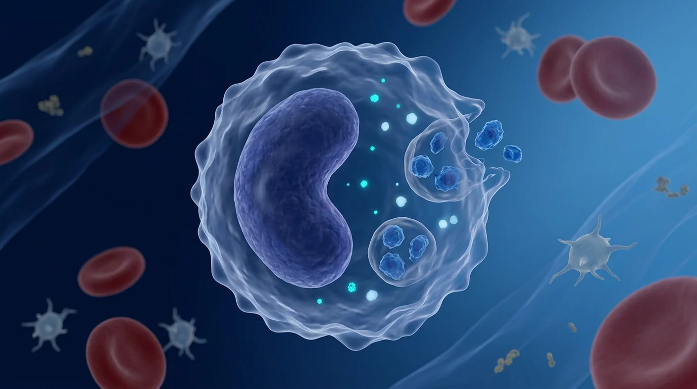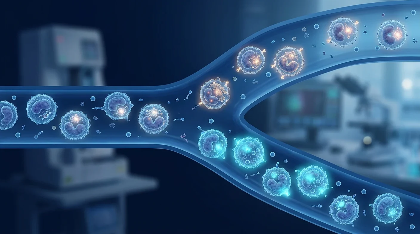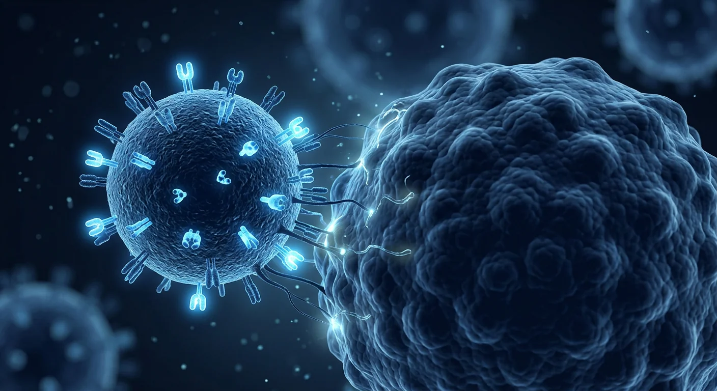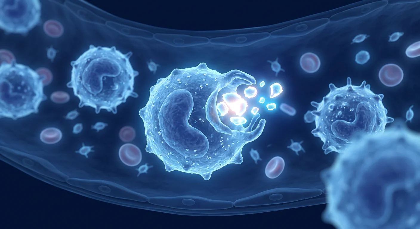
Monocytes and macrophages are the innate immune cells that make the EDIM blood test conceptually possible. EDIM — epitope detection in monocytes — does not look for tumour molecules floating free in plasma. It looks inside circulating monocytes for tumour-associated material those cells are presumed to have ingested in tissue.[1]
Why the cell biology matters to a patient. If the carrier cell is the measuring instrument, then anything that changes monocyte numbers, subsets, activation state or phagocytic appetite can change the number on the report — independently of whether cancer is present.
What this article covers. Monocyte subsets and lifespan, how macrophages take up tumour material, what tumour-associated macrophages actually do, and where the honest evidence limits of the EDIM concept sit. This is educational content about immune biology at VELAR Center in Bangkok, not a diagnostic claim.
Important framing
EDIM is investigational. It is not a stand-alone cancer diagnosis and does not replace guideline-recommended cancer screening, imaging, endoscopy or tissue pathology. A positive result does not establish cancer; a negative result does not exclude it.
What monocytes are and why the EDIM test uses them
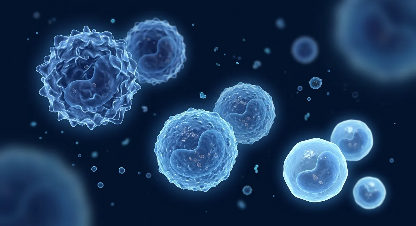
Monocytes are short-lived circulating white blood cells that patrol tissue, ingest cellular debris, and differentiate into macrophages and dendritic cells. They are part of the innate immune system — the arm that responds without needing prior antigen-specific training.[8]
Human blood monocytes are conventionally divided into three populations by surface CD14 and CD16 expression: classical (CD14++CD16−), intermediate (CD14++CD16+) and non-classical (CD14+CD16++).[5] This nomenclature was standardised because different laboratories were using incompatible definitions — a detail that directly affects how any monocyte-based assay is gated and compared between centres.
The subsets are not interchangeable. Classical monocytes dominate numerically and are strongly phagocytic and inflammatory. Non-classical monocytes patrol the vascular endothelium. Intermediate monocytes sit between the two and are often expanded in inflammation.[6][7] EDIM protocols specifically target activated CD14/CD16-positive fractions, which is why monocyte subset shifts are a plausible source of assay variation.
Lifespan is short. Circulating monocytes persist for a limited period before entering tissue or dying, which means the blood compartment reflects a recent, rolling sample of immune activity rather than a long-term record. A monocyte-carried signal is therefore a snapshot, not a cumulative history.
How macrophages take up tumour material
Macrophages ingest dying cells and cellular fragments through phagocytosis, and that uptake is the presumed mechanism by which tumour-associated epitopes enter a monocyte. Tumours are metabolically stressed, proliferative and frequently necrotic, so they release substantial cellular debris into surrounding tissue.
Two candidate tumour-associated markers are measured in EDIM protocols: Apo10, an epitope of the DNaseX/apoptosis-related protein linked to disturbed apoptosis, and TKTL1, transketolase-like 1, associated with altered glucose metabolism.[1] TKTL1 is biologically interesting because aerobic glycolysis — the Warburg effect — is a recognised feature of proliferating cells, not a cancer-exclusive event.[14] TKTL1 expression has also been reported to vary with progression in colorectal cancer tissue.[15]
Deregulated metabolism and evasion of apoptosis are recognised hallmarks of cancer.[13] That makes both markers biologically rational targets. It does not make them specific: normal proliferating tissue, regenerating tissue and inflamed tissue share elements of the same biology.
Tumour-associated macrophages: allies and accomplices
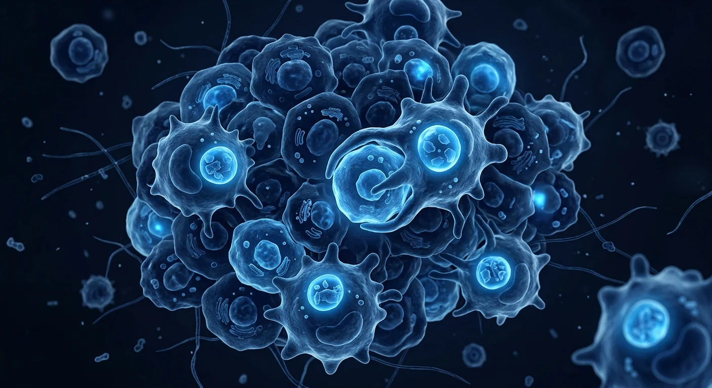
Tumour-associated macrophages (TAMs) are among the most abundant immune cells inside many solid tumours, and they frequently support rather than suppress tumour growth.[9] They can promote angiogenesis, matrix remodelling, metastatic seeding and local immune suppression.[10]
Because of this, TAMs are an active drug-development target in oncology — through CSF1R blockade, recruitment inhibition, and reprogramming strategies.[11] Those programmes remain largely investigational, and results have been mixed.
Monocyte heterogeneity in cancer patients is well documented. Circulating monocyte phenotype and function are altered in malignancy, including changes in subset distribution and suppressive capacity.[12] This cuts two ways for a monocyte-based assay: it supports the premise that circulating monocytes carry tumour-related information, and it warns that the carrier population itself is disturbed in the very patients being tested.
Why inflammation and infection complicate the signal
Monocytes are recruited aggressively during infection and inflammation, which changes both their number and their state.[18] Any assay expressed as a proportion of activated monocytes has a denominator that moves.
Practical consequences worth discussing with a clinician before testing:
- Recent infection, vaccination or surgery can alter monocyte activation.
- Chronic inflammatory or autoimmune disease shifts subset ratios.
- Corticosteroids, immunosuppressants and chemotherapy affect monocyte counts and function.
- Tissue repair after injury involves the same phagocytic machinery a tumour would engage.
None of this makes the measurement meaningless. It means an isolated number, read without clinical context, is easy to over-interpret in either direction. Our related discussion of EDIM and inflammation-driven false positives covers this in clinical detail.
What the published EDIM evidence actually shows
The EDIM literature consists mainly of small, selected-population studies, not large prospective screening trials. Reported work includes a cohort in cholangiocellular, pancreatic and colorectal carcinoma,[2] a paediatric rhabdomyosarcoma liquid-biopsy application,[3] and a pilot study in childhood neuroblastoma.[4]
Case-control design inflates apparent accuracy. When a study compares known cancer patients with healthy controls, sensitivity and specificity usually look better than they will in an unselected population where disease prevalence is low. Reporting and quality frameworks exist precisely because of this — STARD for reporting diagnostic accuracy studies,[16] and QUADAS-2 for appraising risk of bias.[17]
Compare this with the trajectory of plasma-based liquid biopsy. ctDNA methods took years of analytical validation, standardisation and prospective work before clinical implementation was seriously discussed.[19] EDIM has not accumulated a comparable independent evidence base. Our EDIM versus liquid biopsy comparison sets the two side by side.
Evidence checkpoint
Biological plausibility is necessary but not sufficient. A mechanism can be coherent and a test can still fail to change outcomes. The questions that matter are: which population was studied, what was the reference standard, was the assay independently reproduced, and did testing improve a patient-relevant result?
How a monocyte-based result should be used in practice
Treat an EDIM result as one contextual data point inside a clinician-led assessment, never as a verdict. A responsible pathway looks like this:
- Establish the clinical question first. Symptoms, family history, risk factors and screening status determine what testing is appropriate.
- Complete guideline screening. Organ-specific programmes have outcome evidence behind them; a blood biomarker does not substitute for them.
- Record confounders. Recent infection, inflammation, medication and surgery should be documented before the sample is drawn.
- Interpret with the full laboratory report. Gating strategy, subset definitions and quality control affect the number — see our note on EDIM preparation and laboratory workflow.
- Escalate through conventional pathways only. Abnormal findings are investigated with imaging, endoscopy or pathology, directed by the appropriate specialist.
At VELAR Center in Bangkok, cellular and immune-biology testing is discussed as adjunctive information within a physician assessment. No biomarker offered here is presented as a cancer diagnosis.
From blood draw to number: where monocyte biology meets laboratory reality
A monocyte-based assay is only as stable as the cells it measures, and monocytes change quickly outside the body. Time from venepuncture to analysis, storage temperature, anticoagulant choice and handling all influence monocyte activation state. This is a general property of flow-cytometric immune phenotyping, not a criticism unique to EDIM.
Gating is an interpretive act. Deciding which events count as activated CD14/CD16-positive monocytes requires operator-defined boundaries. Standardised subset nomenclature exists precisely because inconsistent gating made results incomparable across laboratories.[5] When comparing two results from different providers, the gating strategy matters as much as the headline score.
Serial results are more informative than single ones — with caveats. A trend measured in the same laboratory, with the same protocol, under comparable clinical conditions, carries more interpretive weight than one isolated number. But a rising trend still requires conventional diagnostic work-up to mean anything clinically, and a falling trend does not confirm treatment success.
Questions worth asking before any monocyte-based test
Ask about population, protocol and consequence before you ask about the price. The following questions separate a considered decision from a purchase:
- Who was studied? Case-control cohorts of known cancer patients versus healthy volunteers do not predict performance in people without symptoms.
- What is the reference standard? Accuracy claims are meaningless without knowing what the test was compared against — tissue pathology, imaging, or clinical follow-up.
- Has an independent laboratory reproduced it? Reproduction by groups without a commercial interest is the strongest single signal of robustness.
- What happens if the result is abnormal? If the answer is broad, unfocused imaging, the downstream harms — incidental findings, anxiety, invasive follow-up — belong in the decision.
- What happens if the result is normal? If a normal result would tempt anyone to defer guideline screening, that alone is a reason for caution.
These questions apply to every emerging biomarker, not only EDIM. They are the same principles that shaped the slow, disciplined path ctDNA testing has taken toward clinical use.[19]
Does a routine monocyte count on a blood panel tell you anything?
A monocyte count on a standard complete blood count measures quantity, not cargo — it says nothing about tumour-associated epitopes. Monocytosis, an elevated monocyte count, has a long differential that includes chronic infection, recovery from acute infection, inflammatory disease, certain haematological disorders and some malignancies. It is a prompt for clinical thought, not a cancer signal.
The two measurements answer different questions. A CBC tells a clinician how many monocytes are circulating. A monocyte-based epitope assay claims to describe what a subset of those cells contains. Because monocyte biology in cancer patients is demonstrably altered in both number and phenotype,[12] reading either measurement in isolation risks the same error: attributing a general immune response to one specific cause.
Practical takeaway. If a routine panel shows an abnormal monocyte count, the correct next step is a clinical review of the whole picture — symptoms, history, other blood indices, examination — not a reflexive purchase of an investigational biomarker test.
Monocyte biology and NK cells: two different immune stories
Monocytes are sampling and presenting cells; natural killer cells are cytotoxic effectors. Monocyte-derived information underlies a diagnostic concept, whereas NK-cell biology underlies a therapeutic one. Both are innate immunity, but the clinical questions are unrelated — and the evidence bases are separate. Readers interested in the effector side can start with how NK cells recognise and kill cancer cells.
Frequently Asked Questions
What do monocytes have to do with a cancer blood test?
EDIM measures tumour-associated epitopes inside monocytes rather than in plasma, on the premise that these cells ingest tumour material in tissue and carry it into circulation.[1] The monocyte is the carrier, not the tumour.
Can inflammation cause an abnormal monocyte-based cancer test?
It plausibly can. Monocytes are recruited and activated during infection, injury and chronic inflammation, which changes both counts and subset distribution.[18] Clinical context is essential before acting on any single result.
Are Apo10 and TKTL1 cancer-specific?
No. Apo10 relates to disturbed apoptosis and TKTL1 to altered glucose metabolism — both are hallmarks of proliferating cells generally, not features unique to malignancy.[13][14]
Does a normal result mean I can skip screening?
No. Guideline-recommended screening — mammography, cervical screening, colorectal testing, low-dose lung CT where indicated — should continue regardless of any investigational blood biomarker result.
Why are tumour-associated macrophages described as harmful?
Because in many solid tumours they promote angiogenesis, tissue remodelling and local immune suppression rather than tumour clearance.[9][10] They are being actively studied as drug targets.[11]
How strong is the EDIM evidence base in 2026?
Limited. Published studies are small, mostly case-control, and concentrated in a few tumour types.[2][3][4] Large independent prospective validation in unselected populations has not been established.
Bottom line
The immune biology behind EDIM is real and reasonably well characterised: monocytes circulate, enter tissue, phagocytose debris, and become macrophages that interact intimately with tumours. That biology makes a monocyte-carried tumour signal conceptually plausible.
Plausible is not the same as validated. The same biology that could carry a tumour signal also responds to infection, injury, inflammation, medication and normal tissue turnover. Until large, independent, prospective studies in unselected populations demonstrate that monocyte-based testing changes patient outcomes, the conservative position stands: use it, if at all, as adjunctive information alongside — never instead of — guideline-recommended screening and clinician-directed diagnosis.
Medical disclaimer
This article is educational and does not constitute medical advice, diagnosis or treatment. Discuss any cancer-screening decision, symptom or test result with a qualified physician. EDIM and other biomarker assays discussed here are investigational and are not a substitute for guideline-recommended cancer screening or diagnostic evaluation.
References
- Coy J. EDIM-TKTL1/Apo10 Blood Test: An Innate Immune System Based Liquid Biopsy for the Early Detection, Characterization and Targeted Treatment of Cancer. International Journal of Molecular Sciences. 2017;18:878. doi:10.3390/ijms18040878 ↩
- Saman S, Stagno M, Warmann S, et al.. Biomarkers Apo10 and TKTL1: Epitope-detection in monocytes (EDIM) as a new diagnostic approach for cholangiocellular, pancreatic and colorectal carcinoma. Cancer Biomarkers. 2019;27:129-137. doi:10.3233/CBM-190414 ↩
- Urla C, Stagno MJ, Schmidt A, et al.. Epitope Detection in Monocytes (EDIM) As a New Method of Liquid Biopsy in Pediatric Rhabdomyosarcoma. Biomedicines. 2022;10:1812. doi:10.3390/biomedicines10081812 ↩
- Stagno MJ, Schmidt A, Bochem J, et al.. Epitope detection in monocytes (EDIM) for liquid biopsy including identification of GD2 in childhood neuroblastoma—a pilot study. British Journal of Cancer. 2022;127:1324-1331. doi:10.1038/s41416-022-01855-x ↩
- Ziegler-Heitbrock L, Ancuta P, Crowe S, et al.. Nomenclature of monocytes and dendritic cells in blood. Blood. 2010;116:e74-e80. doi:10.1182/blood-2010-02-258558 ↩
- Guilliams M, Mildner A, Yona S. Developmental and Functional Heterogeneity of Monocytes. Immunity. 2018;49:595-613. doi:10.1016/j.immuni.2018.10.005 ↩
- Jakubzick CV, Randolph GJ, Henson PM. Monocyte differentiation and antigen-presenting functions. Nature Reviews Immunology. 2017;17:349-362. doi:10.1038/nri.2017.28 ↩
- Ginhoux F, Jung S. Monocytes and macrophages: developmental pathways and tissue homeostasis. Nature Reviews Immunology. 2014;14:392-404. doi:10.1038/nri3671 ↩
- Mantovani A, Marchesi F, Malesci A, et al.. Tumour-associated macrophages as treatment targets in oncology. Nature Reviews Clinical Oncology. 2017;14:399-416. doi:10.1038/nrclinonc.2016.217 ↩
- Noy R, Pollard JW. Tumor-Associated Macrophages: From Mechanisms to Therapy. Immunity. 2014;41:49-61. doi:10.1016/j.immuni.2014.06.010 ↩
- Cassetta L, Pollard JW. Targeting macrophages: therapeutic approaches in cancer. Nature Reviews Drug Discovery. 2018;17:887-904. doi:10.1038/nrd.2018.169 ↩
- Olingy CE, Dinh HQ, Hedrick CC. Monocyte heterogeneity and functions in cancer. Journal of Leukocyte Biology. 2019;106:309-322. doi:10.1002/JLB.4RI0818-311R ↩
- Hanahan D. Hallmarks of Cancer: New Dimensions. Cancer Discovery. 2022;12:31-46. doi:10.1158/2159-8290.CD-21-1059 ↩
- Vander Heiden MG, Cantley LC, Thompson CB. Understanding the Warburg Effect: The Metabolic Requirements of Cell Proliferation. Science. 2009;324:1029-1033. doi:10.1126/science.1160809 ↩
- Diaz-Moralli S, Tarrado-Castellarnau M, Alenda C, et al.. Transketolase-Like 1 Expression Is Modulated during Colorectal Cancer Progression and Metastasis Formation. PLoS ONE. 2011;6:e25323. doi:10.1371/journal.pone.0025323 ↩
- Bossuyt PM, Reitsma JB, Bruns DE, et al.. STARD 2015: an updated list of essential items for reporting diagnostic accuracy studies. BMJ. 2015:h5527. doi:10.1136/bmj.h5527 ↩
- Whiting PF, Rutjes AW, Westwood ME, et al.. QUADAS-2: A Revised Tool for the Quality Assessment of Diagnostic Accuracy Studies. Annals of Internal Medicine. 2011;155:529-536. doi:10.7326/0003-4819-155-8-201110180-00009 ↩
- Shi C, Pamer EG. Monocyte recruitment during infection and inflammation. Nature Reviews Immunology. 2011;11:762-774. doi:10.1038/nri3070 ↩
- Wan JCM, Massie C, Garcia-Corbacho J, et al.. Liquid biopsies come of age: towards implementation of circulating tumour DNA. Nature Reviews Cancer. 2017;17:223-238. doi:10.1038/nrc.2017.7 ↩
单核细胞与巨噬细胞是使 EDIM 血液检测在概念上成为可能的先天免疫细胞。EDIM(单核细胞内表位检测)并不检测血浆中游离的肿瘤分子,而是检测循环单核细胞内部被认为是在组织中吞噬所得的肿瘤相关物质。[1]
为什么细胞生物学对患者很重要。如果载体细胞本身就是测量仪器,那么任何改变单核细胞数量、亚群、活化状态或吞噬活性的因素,都可能改变报告上的数值——无论是否存在癌症。
本文内容。单核细胞亚群与寿命、巨噬细胞如何摄取肿瘤物质、肿瘤相关巨噬细胞的实际作用,以及 EDIM 概念在证据上的诚实界限。这是曼谷 VELAR Center 关于免疫生物学的科普内容,并非诊断声明。
重要说明
EDIM 属于研究性检测,不是独立的癌症诊断,也不能取代指南推荐的癌症筛查、影像学、内镜或组织病理学。阳性结果不能确立癌症;阴性结果也不能排除癌症。
单核细胞是什么,EDIM 为何使用它们
单核细胞是寿命较短的循环白细胞,负责巡视组织、吞噬细胞碎片,并可分化为巨噬细胞和树突状细胞。它们属于先天免疫系统,无需事先的抗原特异性训练即可反应。[8]
人血单核细胞按表面 CD14 与 CD16 表达通常分为三群:经典型(CD14++CD16−)、中间型(CD14++CD16+)与非经典型(CD14+CD16++)。[5]之所以统一命名,是因为各实验室曾使用互不兼容的定义——这一点直接影响任何基于单核细胞的检测如何设门与跨中心比较。
各亚群并不可互换。经典型单核细胞数量占优,吞噬与炎症活性强;非经典型沿血管内皮巡逻;中间型介于两者之间,在炎症时常扩增。[6][7]EDIM 方案专门针对活化的 CD14/CD16 阳性部分,因此单核细胞亚群的变化是检测波动的合理来源。
寿命很短。循环单核细胞存续时间有限,随后进入组织或死亡,这意味着血液中反映的是近期、滚动的免疫活动样本,而非长期记录。单核细胞携带的信号因此是快照,而不是累积病史。
巨噬细胞如何摄取肿瘤物质
巨噬细胞通过吞噬作用摄入凋亡细胞与细胞碎片,这被认为是肿瘤相关表位进入单核细胞的机制。肿瘤代谢应激、增殖活跃且常有坏死,会向周围组织释放大量细胞碎片。
EDIM 方案测量两种候选肿瘤相关标志物:Apo10(与凋亡紊乱相关的 DNaseX/凋亡相关蛋白表位)与 TKTL1(转酮醇酶样蛋白1,与糖代谢改变相关)。[1]TKTL1 在生物学上值得关注,因为有氧糖酵解(Warburg 效应)是增殖细胞的公认特征,并非癌症独有。[14]有研究报告 TKTL1 表达在结直肠癌组织中随进展而变化。[15]
代谢失调与逃避凋亡是公认的癌症标志。[13]这使两种标志物成为合理靶点,但并不意味着它们具有特异性:正常增殖组织、再生组织与炎症组织都具备部分相同的生物学特征。
肿瘤相关巨噬细胞:盟友还是帮凶
肿瘤相关巨噬细胞(TAM)是许多实体瘤中最丰富的免疫细胞之一,且常常促进而非抑制肿瘤生长。[9]它们可促进血管生成、基质重塑、转移播散与局部免疫抑制。[10]
因此,TAM 是肿瘤药物研发的活跃靶点——包括 CSF1R 阻断、招募抑制与重编程策略。[11]这些方案多数仍处于研究阶段,结果参差不齐。
癌症患者的单核细胞异质性有充分记录。恶性肿瘤中循环单核细胞的表型与功能会发生改变,包括亚群分布与抑制能力的变化。[12]这对基于单核细胞的检测有双重含义:既支持循环单核细胞携带肿瘤相关信息的前提,也提醒载体群体本身在受检者中已被扰动。
炎症与感染为何使信号复杂化
感染与炎症期间单核细胞被大量募集,其数量与状态均随之改变。[18]任何以活化单核细胞比例表达的检测,其分母本身就是变动的。
检测前值得与临床医师讨论的实际影响:
- 近期感染、疫苗接种或手术可改变单核细胞活化状态。
- 慢性炎症或自身免疫疾病会改变亚群比例。
- 糖皮质激素、免疫抑制剂与化疗会影响单核细胞计数与功能。
- 损伤后的组织修复动用与肿瘤相同的吞噬机制。
这并不意味着测量毫无意义,而是说:脱离临床背景的孤立数值,很容易在任一方向上被过度解读。相关讨论见EDIM 与炎症导致的假阳性。
已发表的 EDIM 证据究竟显示了什么
EDIM 文献主要由小样本、选择性人群研究构成,而非大型前瞻性筛查试验。已报道的工作包括胆管细胞癌、胰腺癌与结直肠癌队列,[2]儿童横纹肌肉瘤的液体活检应用,[3]以及儿童神经母细胞瘤的初步研究。[4]
病例对照设计会夸大表观准确性。当研究将已知癌症患者与健康对照比较时,敏感性与特异性通常优于在患病率较低的非选择人群中的真实表现。正因如此才有报告与质量框架——诊断准确性研究报告规范 STARD,[16]以及偏倚风险评估工具 QUADAS-2。[17]
可与血浆液体活检的发展轨迹对比。ctDNA 方法历经多年分析验证、标准化与前瞻性研究后,才被认真讨论临床应用。[19]EDIM 尚未积累可比的独立证据基础。参见EDIM 与液体活检对比。
证据要点
生物学上的合理性必要但不充分。机制可以自洽,检测仍可能无法改善结局。真正重要的问题是:研究了哪类人群、参考标准是什么、检测是否被独立重复、以及检测是否改善了与患者相关的结果。
基于单核细胞的结果在实践中应如何使用
应将 EDIM 结果视为医师主导评估中的一项背景数据,而绝非结论。负责任的路径如下:
- 先确立临床问题。症状、家族史、危险因素与筛查状态决定何种检测是适当的。
- 完成指南筛查。器官特异性筛查项目有结局证据支持;血液标志物无法替代。
- 记录混杂因素。采样前应记录近期感染、炎症、用药与手术情况。
- 结合完整实验室报告解读。设门策略、亚群定义与质控都会影响数值——参见EDIM 检测准备与实验室流程。
- 仅通过常规路径升级检查。异常结果应由相应专科医师主导,通过影像、内镜或病理进一步评估。
在曼谷 VELAR Center,细胞与免疫生物学检测均作为医师评估中的辅助信息进行讨论。本中心提供的任何标志物均不作为癌症诊断。
从抽血到数值:单核细胞生物学与实验室现实的交汇
基于单核细胞的检测,其稳定性取决于所测细胞,而单核细胞离体后变化很快。从静脉穿刺到分析的时间、储存温度、抗凝剂选择与操作方式都会影响单核细胞的活化状态。这是流式细胞免疫表型分析的普遍特性,并非 EDIM 独有的缺陷。
设门本身是一种解释行为。判定哪些事件属于活化的 CD14/CD16 阳性单核细胞需要操作者设定边界。统一亚群命名之所以出现,正是因为设门不一致会使结果无法跨实验室比较。[5]比较来自不同机构的两份结果时,设门策略与数值本身同样重要。
连续结果比单次结果更有参考价值——但仍有前提。在同一实验室、同一方案、可比临床条件下测得的趋势,解释权重高于孤立数值。但上升趋势仍需常规诊断检查才具临床意义,下降趋势也不能确认治疗成功。
在接受任何单核细胞检测前值得提出的问题
在询问价格之前,先询问人群、方案与后果。以下问题能将审慎决策与单纯消费区分开来:
- 研究了谁?已知癌症患者与健康志愿者的病例对照队列,无法预测无症状人群中的表现。
- 参考标准是什么?不知道与何种标准比较,准确性数据便没有意义。
- 是否有独立实验室重复?无商业利益关联的独立重复,是稳健性最强的单一信号。
- 结果异常会怎样?若答案是广泛而无重点的影像检查,则偶发发现、焦虑与侵入性随访等下游危害应纳入决策。
- 结果正常会怎样?若正常结果可能诱使任何人推迟指南筛查,这本身就是审慎的理由。
这些问题适用于所有新兴标志物,不限于 EDIM。它们正是 ctDNA 检测走向临床所遵循的同一原则。[19]
常规血常规中的单核细胞计数能说明什么?
血常规中的单核细胞计数测的是数量而非内容物,它无法说明肿瘤相关表位。单核细胞增多的鉴别诊断很广,包括慢性感染、急性感染恢复期、炎症性疾病、某些血液系统疾病与部分恶性肿瘤。它提示需要临床思考,而非癌症信号。
两种测量回答不同问题。血常规告诉医师循环中有多少单核细胞;基于单核细胞的表位检测则声称描述其中一部分细胞携带了什么。由于癌症患者的单核细胞在数量与表型上确有改变,[12]孤立解读任一指标都可能犯同一错误:把一般性免疫反应归因于某一特定原因。
实用结论。若常规检查显示单核细胞计数异常,正确的下一步是结合症状、病史、其他血液指标与体格检查进行整体临床复核,而不是反射性地购买研究性标志物检测。
单核细胞生物学与 NK 细胞:两个不同的免疫故事
单核细胞是取样与呈递细胞;自然杀伤细胞是细胞毒性效应细胞。单核细胞衍生的信息支撑的是诊断概念,而 NK 细胞生物学支撑的是治疗概念。两者同属先天免疫,但临床问题互不相关,证据基础也彼此独立。对效应端感兴趣的读者可从NK 细胞如何识别并杀伤癌细胞开始。
常见问题
单核细胞与癌症血液检测有什么关系?
EDIM 检测单核细胞内部而非血浆中的肿瘤相关表位,其前提是这些细胞在组织中吞噬肿瘤物质并带入循环。[1]单核细胞是载体,而非肿瘤本身。
炎症会导致基于单核细胞的癌症检测异常吗?
有可能。感染、损伤与慢性炎症会募集并活化单核细胞,改变计数与亚群分布。[18]在依据任何单一结果采取行动前,临床背景至关重要。
Apo10 与 TKTL1 对癌症具有特异性吗?
没有。Apo10 与凋亡紊乱相关,TKTL1 与糖代谢改变相关——两者都是增殖细胞的普遍特征,而非恶性肿瘤独有。[13][14]
结果正常就可以跳过筛查吗?
不可以。无论任何研究性血液标志物结果如何,指南推荐的筛查——乳腺X线、宫颈筛查、结直肠检测、必要时的低剂量肺部CT——都应继续。
为何说肿瘤相关巨噬细胞是有害的?
因为在许多实体瘤中,它们促进血管生成、组织重塑与局部免疫抑制,而非清除肿瘤。[9][10]它们正被作为药物靶点积极研究。[11]
2026 年 EDIM 的证据基础有多强?
有限。已发表研究样本小、多为病例对照,且集中于少数肿瘤类型。[2][3][4]尚未建立在非选择人群中的大规模独立前瞻性验证。
结论
EDIM 背后的免疫生物学是真实且研究相对充分的:单核细胞在循环中运行、进入组织、吞噬碎片,并成为与肿瘤密切互动的巨噬细胞。这一生物学使"单核细胞携带肿瘤信号"在概念上具有合理性。
但合理并不等于已验证。同样的生物学也会对感染、损伤、炎症、用药与正常组织更新作出反应。在大规模、独立、前瞻性的非选择人群研究证明单核细胞检测能改善患者结局之前,保守立场依然成立:若使用,也只能作为辅助信息,与指南推荐的筛查和医师主导的诊断并行,而绝不可取而代之。
医疗免责声明
本文仅供教育参考,不构成医疗建议、诊断或治疗。任何癌症筛查决定、症状或检测结果,请与合格医师讨论。本文讨论的 EDIM 及其他标志物检测均属研究性质,不能替代指南推荐的癌症筛查或诊断评估。
参考文献
- Coy J. EDIM-TKTL1/Apo10 Blood Test: An Innate Immune System Based Liquid Biopsy for the Early Detection, Characterization and Targeted Treatment of Cancer. International Journal of Molecular Sciences. 2017;18:878. doi:10.3390/ijms18040878 ↩
- Saman S, Stagno M, Warmann S, et al.. Biomarkers Apo10 and TKTL1: Epitope-detection in monocytes (EDIM) as a new diagnostic approach for cholangiocellular, pancreatic and colorectal carcinoma. Cancer Biomarkers. 2019;27:129-137. doi:10.3233/CBM-190414 ↩
- Urla C, Stagno MJ, Schmidt A, et al.. Epitope Detection in Monocytes (EDIM) As a New Method of Liquid Biopsy in Pediatric Rhabdomyosarcoma. Biomedicines. 2022;10:1812. doi:10.3390/biomedicines10081812 ↩
- Stagno MJ, Schmidt A, Bochem J, et al.. Epitope detection in monocytes (EDIM) for liquid biopsy including identification of GD2 in childhood neuroblastoma—a pilot study. British Journal of Cancer. 2022;127:1324-1331. doi:10.1038/s41416-022-01855-x ↩
- Ziegler-Heitbrock L, Ancuta P, Crowe S, et al.. Nomenclature of monocytes and dendritic cells in blood. Blood. 2010;116:e74-e80. doi:10.1182/blood-2010-02-258558 ↩
- Guilliams M, Mildner A, Yona S. Developmental and Functional Heterogeneity of Monocytes. Immunity. 2018;49:595-613. doi:10.1016/j.immuni.2018.10.005 ↩
- Jakubzick CV, Randolph GJ, Henson PM. Monocyte differentiation and antigen-presenting functions. Nature Reviews Immunology. 2017;17:349-362. doi:10.1038/nri.2017.28 ↩
- Ginhoux F, Jung S. Monocytes and macrophages: developmental pathways and tissue homeostasis. Nature Reviews Immunology. 2014;14:392-404. doi:10.1038/nri3671 ↩
- Mantovani A, Marchesi F, Malesci A, et al.. Tumour-associated macrophages as treatment targets in oncology. Nature Reviews Clinical Oncology. 2017;14:399-416. doi:10.1038/nrclinonc.2016.217 ↩
- Noy R, Pollard JW. Tumor-Associated Macrophages: From Mechanisms to Therapy. Immunity. 2014;41:49-61. doi:10.1016/j.immuni.2014.06.010 ↩
- Cassetta L, Pollard JW. Targeting macrophages: therapeutic approaches in cancer. Nature Reviews Drug Discovery. 2018;17:887-904. doi:10.1038/nrd.2018.169 ↩
- Olingy CE, Dinh HQ, Hedrick CC. Monocyte heterogeneity and functions in cancer. Journal of Leukocyte Biology. 2019;106:309-322. doi:10.1002/JLB.4RI0818-311R ↩
- Hanahan D. Hallmarks of Cancer: New Dimensions. Cancer Discovery. 2022;12:31-46. doi:10.1158/2159-8290.CD-21-1059 ↩
- Vander Heiden MG, Cantley LC, Thompson CB. Understanding the Warburg Effect: The Metabolic Requirements of Cell Proliferation. Science. 2009;324:1029-1033. doi:10.1126/science.1160809 ↩
- Diaz-Moralli S, Tarrado-Castellarnau M, Alenda C, et al.. Transketolase-Like 1 Expression Is Modulated during Colorectal Cancer Progression and Metastasis Formation. PLoS ONE. 2011;6:e25323. doi:10.1371/journal.pone.0025323 ↩
- Bossuyt PM, Reitsma JB, Bruns DE, et al.. STARD 2015: an updated list of essential items for reporting diagnostic accuracy studies. BMJ. 2015:h5527. doi:10.1136/bmj.h5527 ↩
- Whiting PF, Rutjes AW, Westwood ME, et al.. QUADAS-2: A Revised Tool for the Quality Assessment of Diagnostic Accuracy Studies. Annals of Internal Medicine. 2011;155:529-536. doi:10.7326/0003-4819-155-8-201110180-00009 ↩
- Shi C, Pamer EG. Monocyte recruitment during infection and inflammation. Nature Reviews Immunology. 2011;11:762-774. doi:10.1038/nri3070 ↩
- Wan JCM, Massie C, Garcia-Corbacho J, et al.. Liquid biopsies come of age: towards implementation of circulating tumour DNA. Nature Reviews Cancer. 2017;17:223-238. doi:10.1038/nrc.2017.7 ↩
الوحيدات (المونوسايت) والبلاعم هي خلايا المناعة الفطرية التي تجعل فحص الدم EDIM ممكنًا من الناحية المفاهيمية. لا يبحث EDIM — كشف الحواتم داخل الوحيدات — عن جزيئات ورمية سابحة في البلازما، بل يبحث داخل الوحيدات الدائرة عن مادة مرتبطة بالورم يُفترض أن هذه الخلايا التهمتها في الأنسجة.[1]
لماذا تهم بيولوجيا الخلية المريض. إذا كانت الخلية الحاملة هي أداة القياس نفسها، فإن أي عامل يغيّر عدد الوحيدات أو مجموعاتها الفرعية أو حالة تنشيطها أو نشاطها البلعمي قد يغيّر الرقم في التقرير — بصرف النظر عن وجود السرطان من عدمه.
ما يغطيه هذا المقال. المجموعات الفرعية للوحيدات وعمرها، كيفية التقاط البلاعم للمادة الورمية، ما تفعله البلاعم المرتبطة بالورم فعليًا، وأين تقف الحدود الصادقة لأدلة مفهوم EDIM. هذا محتوى تثقيفي عن بيولوجيا المناعة في مركز فيلار ببانكوك، وليس ادعاءً تشخيصيًا.
إطار مهم
يُعد EDIM فحصًا بحثيًا. ليس تشخيصًا مستقلاً للسرطان ولا يحل محل فحوص الكشف المبكر الموصى بها في الإرشادات، ولا التصوير، ولا التنظير، ولا علم الأمراض النسيجي. النتيجة الإيجابية لا تثبت السرطان، والنتيجة السلبية لا تستبعده.
ما هي الوحيدات ولماذا يستخدمها فحص EDIM
الوحيدات خلايا دم بيضاء قصيرة العمر تدور في الدم، تجوب الأنسجة وتبتلع الحطام الخلوي وتتمايز إلى بلاعم وخلايا متغصنة. وهي جزء من جهاز المناعة الفطري الذي يستجيب دون تدريب مسبق خاص بمستضد معين.[8]
تُقسَّم وحيدات الدم البشري تقليديًا إلى ثلاث مجموعات حسب تعبير CD14 وCD16 السطحي: الكلاسيكية (CD14++CD16−)، والوسيطة (CD14++CD16+)، وغير الكلاسيكية (CD14+CD16++).[5] وقد جرى توحيد هذه التسميات لأن المختبرات كانت تستخدم تعريفات غير متوافقة — وهي تفصيلة تؤثر مباشرة في كيفية بوابة أي فحص قائم على الوحيدات ومقارنته بين المراكز.
المجموعات الفرعية ليست متبادلة. تهيمن الوحيدات الكلاسيكية عدديًا وهي شديدة البلعمة والالتهاب، بينما تجوب غير الكلاسيكية بطانة الأوعية، وتقع الوسيطة بينهما وكثيرًا ما تتوسع أثناء الالتهاب.[6][7] تستهدف بروتوكولات EDIM تحديدًا الأجزاء المنشّطة الإيجابية لـ CD14/CD16، ولهذا تُعد تحولات المجموعات الفرعية مصدرًا معقولاً لتباين النتائج.
العمر قصير. تبقى الوحيدات الدائرة فترة محدودة قبل دخول الأنسجة أو الموت، ما يعني أن حجرة الدم تعكس عيّنة حديثة ومتجددة من النشاط المناعي وليس سجلاً طويل الأمد. لذلك فإن الإشارة المحمولة على الوحيدات لقطة لحظية وليست تاريخًا تراكميًا.
كيف تلتقط البلاعم المادة الورمية
تبتلع البلاعم الخلايا المحتضرة والشظايا الخلوية عبر البلعمة، وهذا هو الآلية المفترضة لدخول الحواتم المرتبطة بالورم إلى الوحيدة. الأورام مجهدة أيضيًا وسريعة التكاثر وكثيرًا ما تكون نخرية، فتطلق حطامًا خلويًا كبيرًا في الأنسجة المحيطة.
يقيس بروتوكول EDIM واسمين مرشحين: Apo10، وهو حاتمة مرتبطة ببروتين DNaseX المرتبط بالاستماتة المضطربة، وTKTL1، الشبيه بالترانسكيتولاز 1 المرتبط باضطراب أيض الغلوكوز.[1] وTKTL1 مثير للاهتمام بيولوجيًا لأن التحلل السكري الهوائي — تأثير واربورغ — سمة معروفة للخلايا المتكاثرة عمومًا وليست حكرًا على السرطان.[14] كما أُبلغ عن تغيّر تعبير TKTL1 مع تقدّم سرطان القولون والمستقيم.[15]
اضطراب الأيض والإفلات من الاستماتة سمتان معترف بهما للسرطان.[13] هذا يجعل كلا الواسمين هدفًا منطقيًا، لكنه لا يجعلهما نوعيين: فالأنسجة المتكاثرة الطبيعية والمتجددة والملتهبة تشترك في عناصر البيولوجيا نفسها.
البلاعم المرتبطة بالورم: حليف أم شريك
البلاعم المرتبطة بالورم من أكثر الخلايا المناعية وفرة داخل كثير من الأورام الصلبة، وهي غالبًا تدعم نمو الورم بدل كبحه.[9] إذ يمكنها تعزيز تكوّن الأوعية وإعادة تشكيل المطرس والانتشار النقيلي وكبت المناعة موضعيًا.[10]
لذلك تمثل هذه البلاعم هدفًا نشطًا لتطوير الأدوية في الأورام — عبر حصار CSF1R وتثبيط التجنيد واستراتيجيات إعادة البرمجة.[11] ولا تزال هذه البرامج بحثية إلى حد بعيد ونتائجها متفاوتة.
تباين الوحيدات لدى مرضى السرطان موثّق جيدًا. يتغيّر النمط الظاهري والوظيفة للوحيدات الدائرة في الأورام الخبيثة، بما يشمل توزيع المجموعات الفرعية والقدرة الكابتة.[12] وهذا سيف ذو حدين لأي فحص قائم على الوحيدات: فهو يدعم الفرضية، لكنه ينبّه إلى أن مجموعة الخلايا الحاملة نفسها مضطربة لدى من يُجرى لهم الفحص.
لماذا يعقّد الالتهاب والعدوى الإشارة
تُجنَّد الوحيدات بقوة أثناء العدوى والالتهاب، فيتغيّر عددها وحالتها معًا.[18] وأي فحص يُعبَّر عنه كنسبة من الوحيدات المنشّطة يكون مقامه نفسه متحركًا.
نتائج عملية تستحق النقاش مع الطبيب قبل الفحص:
- العدوى أو التطعيم أو الجراحة الحديثة قد تغيّر تنشيط الوحيدات.
- الأمراض الالتهابية أو المناعية الذاتية المزمنة تغيّر نسب المجموعات الفرعية.
- الكورتيكوستيرويدات ومثبطات المناعة والعلاج الكيميائي تؤثر في عدد الوحيدات ووظيفتها.
- إصلاح الأنسجة بعد الإصابة يستخدم الآلية البلعمية نفسها التي يستدعيها الورم.
هذا لا يجعل القياس بلا معنى، لكنه يعني أن رقمًا منعزلاً يُقرأ خارج السياق السريري يسهل المبالغة في تفسيره في أي اتجاه. راجع مناقشتنا حول EDIM والنتائج الإيجابية الكاذبة الناتجة عن الالتهاب.
ماذا تُظهر أدلة EDIM المنشورة فعليًا
تتألف أدبيات EDIM أساسًا من دراسات صغيرة على مجموعات منتقاة، لا من تجارب فحص مسبقة كبيرة. تشمل الأعمال المنشورة مجموعة في سرطان القنوات الصفراوية والبنكرياس والقولون والمستقيم،[2] وتطبيقًا في الساركوما العضلية المخططة لدى الأطفال،[3] ودراسة تمهيدية في ورم الأرومة العصبية عند الأطفال.[4]
تصميم الحالات والشواهد يضخّم الدقة الظاهرية. عند مقارنة مرضى سرطان معروفين بأصحاء، تبدو الحساسية والنوعية أفضل مما ستكون عليه في مجموعة غير منتقاة ذات انتشار منخفض للمرض. ولهذا وُضعت أطر التقرير والجودة — STARD لتقرير دراسات الدقة التشخيصية،[16] وQUADAS-2 لتقييم خطر التحيّز.[17]
قارن ذلك بمسار الخزعة السائلة البلازمية. استغرقت طرق ctDNA سنوات من التحقق التحليلي والتوحيد والعمل المسبق قبل أن تُناقش جديًا للاستخدام السريري.[19] ولم يراكم EDIM قاعدة أدلة مستقلة مماثلة. راجع مقارنة EDIM بالخزعة السائلة.
نقطة تحقق من الأدلة
المعقولية البيولوجية ضرورية لكنها غير كافية. قد تكون الآلية متماسكة ويظل الفحص عاجزًا عن تغيير النتائج. الأسئلة المهمة: أي مجموعة دُرست، وما المعيار المرجعي، وهل كُرِّر الفحص بشكل مستقل، وهل حسّن الفحص نتيجة تهم المريض.
كيف ينبغي استخدام نتيجة قائمة على الوحيدات عمليًا
تعامل مع نتيجة EDIM كنقطة بيانات سياقية ضمن تقييم يقوده الطبيب، لا كحكم نهائي. المسار المسؤول:
- حدّد السؤال السريري أولاً. الأعراض والتاريخ العائلي وعوامل الخطر وحالة الفحص تحدد ما هو مناسب.
- أكمل فحوص الإرشادات. برامج الكشف الخاصة بالأعضاء مدعومة بأدلة على النتائج؛ ولا يحل واسم دموي محلها.
- سجّل العوامل المربكة. ينبغي توثيق العدوى والالتهاب والأدوية والجراحة الحديثة قبل سحب العينة.
- فسّر مع التقرير المخبري الكامل. استراتيجية البوابة وتعريفات المجموعات الفرعية وضبط الجودة تؤثر في الرقم — راجع تحضير فحص EDIM وسير العمل المخبري.
- صعّد عبر المسارات التقليدية فقط. تُستقصى النتائج غير الطبيعية بالتصوير أو التنظير أو علم الأمراض بتوجيه من الاختصاصي المناسب.
في مركز فيلار ببانكوك، تُناقش فحوص البيولوجيا الخلوية والمناعية كمعلومات مساعدة ضمن تقييم طبي. ولا يُقدَّم أي واسم حيوي هنا بوصفه تشخيصًا للسرطان.
من سحب الدم إلى الرقم: حيث تلتقي بيولوجيا الوحيدات بواقع المختبر
الفحص القائم على الوحيدات مستقر بقدر استقرار الخلايا التي يقيسها، والوحيدات تتغير بسرعة خارج الجسم. الزمن بين سحب العينة والتحليل، ودرجة حرارة التخزين، ونوع مانع التخثر، وطريقة المناولة، كلها تؤثر في حالة تنشيط الوحيدات. هذه سمة عامة لتنميط المناعة بقياس التدفق الخلوي وليست انتقادًا خاصًا بـ EDIM.
البوابة فعل تفسيري. تحديد الأحداث التي تُحتسب وحيدات منشّطة إيجابية لـ CD14/CD16 يتطلب حدودًا يضعها المشغّل. وقد نشأ توحيد تسميات المجموعات الفرعية تحديدًا لأن اختلاف البوابات جعل النتائج غير قابلة للمقارنة بين المختبرات.[5] وعند مقارنة نتيجتين من جهتين مختلفتين، تكون استراتيجية البوابة بأهمية الرقم المعلن.
النتائج المتسلسلة أكثر إفادة من المنفردة — مع تحفظات. الاتجاه المقاس في المختبر نفسه وبالبروتوكول نفسه وتحت ظروف سريرية مماثلة يحمل وزنًا تفسيريًا أكبر من رقم منعزل. لكن الاتجاه المرتفع يظل بحاجة إلى استقصاء تشخيصي تقليدي ليعني شيئًا، والاتجاه المنخفض لا يؤكد نجاح العلاج.
أسئلة تستحق الطرح قبل أي فحص قائم على الوحيدات
اسأل عن المجموعة والبروتوكول والعاقبة قبل أن تسأل عن السعر. هذه الأسئلة تفصل القرار المدروس عن عملية الشراء:
- من دُرس؟ مجموعات الحالات والشواهد من مرضى معروفين مقابل متطوعين أصحاء لا تتنبأ بالأداء لدى من لا تظهر عليهم أعراض.
- ما المعيار المرجعي؟ ادعاءات الدقة بلا معنى دون معرفة ما قورن به الفحص.
- هل كرّره مختبر مستقل؟ التكرار من مجموعات بلا مصلحة تجارية أقوى إشارة منفردة على المتانة.
- ماذا يحدث إذا كانت النتيجة غير طبيعية؟ إذا كان الجواب تصويرًا واسعًا غير موجّه، فإن الأضرار اللاحقة — الاكتشافات العرضية والقلق والمتابعة الاجتياحية — جزء من القرار.
- ماذا يحدث إذا كانت النتيجة طبيعية؟ إذا كانت نتيجة طبيعية قد تغري أحدًا بتأجيل فحوص الإرشادات، فهذا وحده سبب للحذر.
تنطبق هذه الأسئلة على كل واسم ناشئ وليس على EDIM وحده. وهي المبادئ نفسها التي شكّلت المسار البطيء المنضبط لفحص ctDNA نحو الاستخدام السريري.[19]
هل يخبرك عدد الوحيدات في تحليل الدم الروتيني بشيء؟
عدد الوحيدات في تعداد الدم الكامل يقيس الكمية لا المحتوى — ولا يقول شيئًا عن الحواتم المرتبطة بالورم. لكثرة الوحيدات تشخيص تفريقي واسع يشمل العدوى المزمنة والتعافي من عدوى حادة والأمراض الالتهابية وبعض اضطرابات الدم وبعض الأورام الخبيثة. إنها دعوة للتفكير السريري لا إشارة سرطان.
القياسان يجيبان عن سؤالين مختلفين. تعداد الدم يخبر الطبيب بعدد الوحيدات الدائرة، بينما يدّعي فحص الحواتم وصف ما تحتويه شريحة منها. ولأن بيولوجيا الوحيدات لدى مرضى السرطان متغيرة عددًا ونمطًا ظاهريًا،[12] فإن قراءة أي منهما بمعزل عن الآخر تخاطر بالخطأ نفسه: نسب استجابة مناعية عامة إلى سبب واحد بعينه.
الخلاصة العملية. إذا أظهر تحليل روتيني عدد وحيدات غير طبيعي، فالخطوة الصحيحة مراجعة سريرية للصورة كاملة — الأعراض والتاريخ ومؤشرات الدم الأخرى والفحص السريري — لا شراء انعكاسي لفحص واسم بحثي.
بيولوجيا الوحيدات والخلايا القاتلة الطبيعية: قصتان مناعيتان مختلفتان
الوحيدات خلايا أخذ عينات وعرض؛ أما الخلايا القاتلة الطبيعية فهي خلايا مؤثِّرة سامة للخلايا. المعلومات المستمدة من الوحيدات تدعم مفهومًا تشخيصيًا، بينما تدعم بيولوجيا الخلايا القاتلة الطبيعية مفهومًا علاجيًا. كلاهما مناعة فطرية، لكن الأسئلة السريرية غير مرتبطة وقواعد الأدلة منفصلة. يمكن للمهتمين بالجانب المؤثِّر البدء من كيف تتعرف الخلايا القاتلة الطبيعية على الخلايا السرطانية وتقتلها.
الأسئلة الشائعة
ما علاقة الوحيدات بفحص دم للسرطان؟
يقيس EDIM حواتم مرتبطة بالورم داخل الوحيدات لا في البلازما، بافتراض أن هذه الخلايا تبتلع مادة ورمية في الأنسجة وتنقلها إلى الدورة الدموية.[1] الوحيدة هي الحامل وليست الورم.
هل يمكن أن يسبب الالتهاب نتيجة غير طبيعية؟
نعم على الأرجح. تُجنَّد الوحيدات وتُنشَّط أثناء العدوى والإصابة والالتهاب المزمن، فيتغير العدد وتوزيع المجموعات الفرعية.[18] السياق السريري ضروري قبل التصرف بناءً على أي نتيجة منفردة.
هل Apo10 وTKTL1 نوعيان للسرطان؟
لا. يرتبط Apo10 باضطراب الاستماتة وTKTL1 باضطراب أيض الغلوكوز — وكلاهما سمة عامة للخلايا المتكاثرة لا خاصية حصرية للورم الخبيث.[13][14]
هل تعني النتيجة الطبيعية إمكانية تخطي الفحوص؟
لا. ينبغي الاستمرار في الفحوص الموصى بها في الإرشادات — تصوير الثدي، وفحص عنق الرحم، وفحوص القولون والمستقيم، والتصوير المقطعي منخفض الجرعة للرئة عند الاستطباب — بغض النظر عن أي واسم بحثي.
لماذا تُوصف البلاعم المرتبطة بالورم بالضارة؟
لأنها في كثير من الأورام الصلبة تعزز تكوّن الأوعية وإعادة التشكيل النسيجي وكبت المناعة موضعيًا بدل إزالة الورم.[9][10] وهي قيد الدراسة النشطة كأهداف دوائية.[11]
ما مدى قوة قاعدة أدلة EDIM في 2026؟
محدودة. الدراسات المنشورة صغيرة وأغلبها حالات وشواهد ومركزة في أنواع أورام قليلة.[2][3][4] ولم يثبت تحقق مستقل مسبق واسع في مجموعات غير منتقاة.
الخلاصة
البيولوجيا المناعية وراء EDIM حقيقية وموصوفة بشكل معقول: الوحيدات تدور وتدخل الأنسجة وتبتلع الحطام وتصبح بلاعم تتفاعل عن قرب مع الأورام. هذه البيولوجيا تجعل فكرة إشارة ورمية محمولة على الوحيدات معقولة مفاهيميًا.
لكن المعقول ليس المُتحقق منه. البيولوجيا نفسها التي قد تحمل إشارة ورمية تستجيب أيضًا للعدوى والإصابة والالتهاب والأدوية وتجدد الأنسجة الطبيعي. وإلى أن تُظهر دراسات كبيرة ومستقلة ومسبقة في مجموعات غير منتقاة أن الفحص القائم على الوحيدات يغيّر نتائج المرضى، يبقى الموقف المتحفظ قائمًا: يُستخدم — إن استُخدم — كمعلومة مساعدة إلى جانب الفحوص الموصى بها والتشخيص الذي يقوده الطبيب، لا بديلاً عنها.
إخلاء مسؤولية طبية
هذا المقال تثقيفي ولا يشكل نصيحة طبية أو تشخيصًا أو علاجًا. ناقش أي قرار يتعلق بالكشف عن السرطان أو عرض أو نتيجة فحص مع طبيب مؤهل. فحص EDIM وغيره من فحوص الواسمات المذكورة هنا بحثية ولا تحل محل الفحوص الموصى بها في الإرشادات أو التقييم التشخيصي.
المراجع
- Coy J. EDIM-TKTL1/Apo10 Blood Test: An Innate Immune System Based Liquid Biopsy for the Early Detection, Characterization and Targeted Treatment of Cancer. International Journal of Molecular Sciences. 2017;18:878. doi:10.3390/ijms18040878 ↩
- Saman S, Stagno M, Warmann S, et al.. Biomarkers Apo10 and TKTL1: Epitope-detection in monocytes (EDIM) as a new diagnostic approach for cholangiocellular, pancreatic and colorectal carcinoma. Cancer Biomarkers. 2019;27:129-137. doi:10.3233/CBM-190414 ↩
- Urla C, Stagno MJ, Schmidt A, et al.. Epitope Detection in Monocytes (EDIM) As a New Method of Liquid Biopsy in Pediatric Rhabdomyosarcoma. Biomedicines. 2022;10:1812. doi:10.3390/biomedicines10081812 ↩
- Stagno MJ, Schmidt A, Bochem J, et al.. Epitope detection in monocytes (EDIM) for liquid biopsy including identification of GD2 in childhood neuroblastoma—a pilot study. British Journal of Cancer. 2022;127:1324-1331. doi:10.1038/s41416-022-01855-x ↩
- Ziegler-Heitbrock L, Ancuta P, Crowe S, et al.. Nomenclature of monocytes and dendritic cells in blood. Blood. 2010;116:e74-e80. doi:10.1182/blood-2010-02-258558 ↩
- Guilliams M, Mildner A, Yona S. Developmental and Functional Heterogeneity of Monocytes. Immunity. 2018;49:595-613. doi:10.1016/j.immuni.2018.10.005 ↩
- Jakubzick CV, Randolph GJ, Henson PM. Monocyte differentiation and antigen-presenting functions. Nature Reviews Immunology. 2017;17:349-362. doi:10.1038/nri.2017.28 ↩
- Ginhoux F, Jung S. Monocytes and macrophages: developmental pathways and tissue homeostasis. Nature Reviews Immunology. 2014;14:392-404. doi:10.1038/nri3671 ↩
- Mantovani A, Marchesi F, Malesci A, et al.. Tumour-associated macrophages as treatment targets in oncology. Nature Reviews Clinical Oncology. 2017;14:399-416. doi:10.1038/nrclinonc.2016.217 ↩
- Noy R, Pollard JW. Tumor-Associated Macrophages: From Mechanisms to Therapy. Immunity. 2014;41:49-61. doi:10.1016/j.immuni.2014.06.010 ↩
- Cassetta L, Pollard JW. Targeting macrophages: therapeutic approaches in cancer. Nature Reviews Drug Discovery. 2018;17:887-904. doi:10.1038/nrd.2018.169 ↩
- Olingy CE, Dinh HQ, Hedrick CC. Monocyte heterogeneity and functions in cancer. Journal of Leukocyte Biology. 2019;106:309-322. doi:10.1002/JLB.4RI0818-311R ↩
- Hanahan D. Hallmarks of Cancer: New Dimensions. Cancer Discovery. 2022;12:31-46. doi:10.1158/2159-8290.CD-21-1059 ↩
- Vander Heiden MG, Cantley LC, Thompson CB. Understanding the Warburg Effect: The Metabolic Requirements of Cell Proliferation. Science. 2009;324:1029-1033. doi:10.1126/science.1160809 ↩
- Diaz-Moralli S, Tarrado-Castellarnau M, Alenda C, et al.. Transketolase-Like 1 Expression Is Modulated during Colorectal Cancer Progression and Metastasis Formation. PLoS ONE. 2011;6:e25323. doi:10.1371/journal.pone.0025323 ↩
- Bossuyt PM, Reitsma JB, Bruns DE, et al.. STARD 2015: an updated list of essential items for reporting diagnostic accuracy studies. BMJ. 2015:h5527. doi:10.1136/bmj.h5527 ↩
- Whiting PF, Rutjes AW, Westwood ME, et al.. QUADAS-2: A Revised Tool for the Quality Assessment of Diagnostic Accuracy Studies. Annals of Internal Medicine. 2011;155:529-536. doi:10.7326/0003-4819-155-8-201110180-00009 ↩
- Shi C, Pamer EG. Monocyte recruitment during infection and inflammation. Nature Reviews Immunology. 2011;11:762-774. doi:10.1038/nri3070 ↩
- Wan JCM, Massie C, Garcia-Corbacho J, et al.. Liquid biopsies come of age: towards implementation of circulating tumour DNA. Nature Reviews Cancer. 2017;17:223-238. doi:10.1038/nrc.2017.7 ↩