Apo10 and TKTL1 are the two intracellular signals measured by the EDIM test, but they are not interchangeable “cancer genes” and their presence is not, by itself, a cancer diagnosis. Apo10 is an epitope associated with an altered DNaseX protein and apoptosis biology; TKTL1 is a transketolase-like protein studied in relation to metabolic reprogramming. EDIM—epitope detection in monocytes—looks for these targets inside a selected population of circulating CD14+/CD16+ monocytes/macrophage-like cells rather than measuring a freely circulating tumor marker [1].
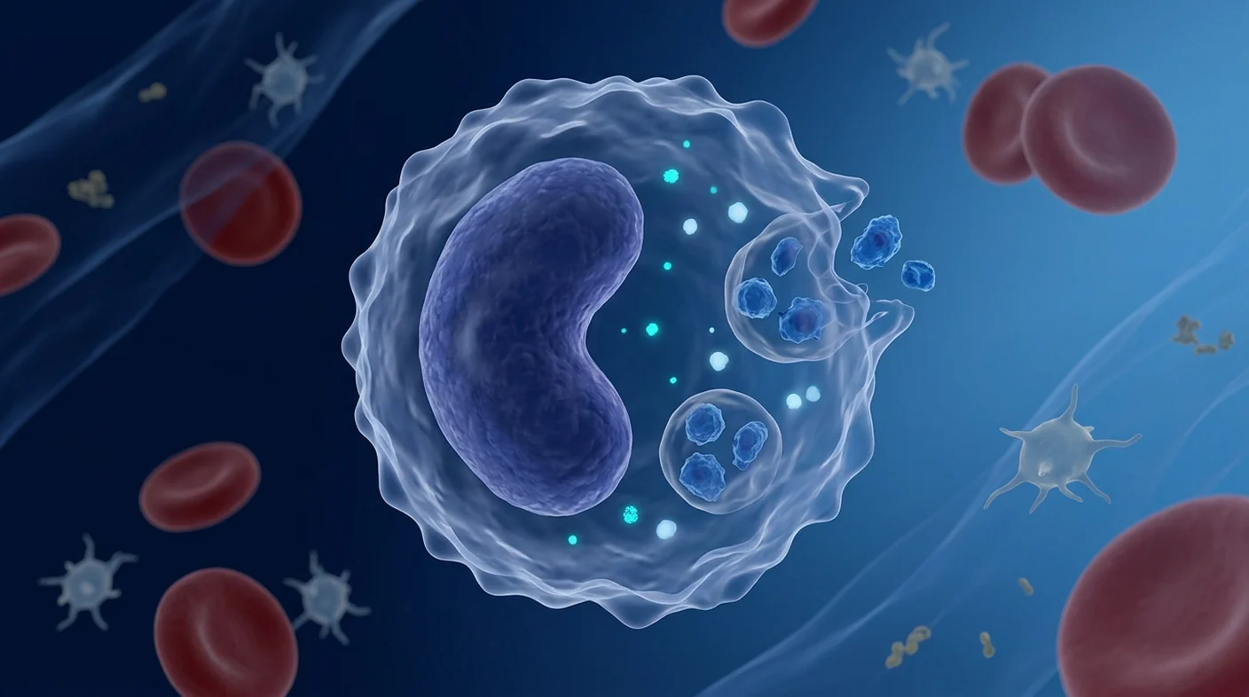
The approach is biologically unusual. Monocytes patrol blood and tissues, ingest cellular material, and carry some of that material intracellularly. EDIM attempts to use these innate immune cells as “reporters” of what they have encountered. This is a plausible sampling concept, but plausible biology is not equivalent to proven screening performance. Published studies are mostly small, selected case-control cohorts or monitoring studies, and several foundational papers include investigators connected to the assay’s intellectual property or development [2].
This review explains what each marker is thought to represent, how phagocytic uptake differs from tumor DNA shedding, what flow cytometry actually measures, and why an EDIM result must be interpreted conservatively alongside validated screening and diagnostic pathways.
What does the EDIM test measure?
EDIM measures the proportion of a gated monocyte population that stains intracellularly for Apo10 and TKTL1 epitopes. In a typical workflow, venous blood is processed, cells are labeled for surface markers used to identify activated or differentiated monocytes, membranes are permeabilized, and antibodies are applied against intracellular Apo10 and TKTL1. Flow cytometry then counts fluorescent events within the predefined gate.
This is different from measuring the concentration of a protein in serum. The reported score combines cell classification, antibody staining and an assay-specific calculation. Pre-analytical handling, time from collection, antibody performance, gating strategy, instrument calibration and threshold selection can all affect the result. A numerical or color-coded category is therefore meaningful only within the validated version of that assay; a cutoff from one study should not be transferred casually to another laboratory.
| Signal | Proposed biological meaning | What it cannot prove alone |
|---|---|---|
| Apo10 | Intracellular epitope linked to altered DNaseX/apoptosis-related biology | That viable cancer is present, where it is located, or whether it is aggressive |
| TKTL1 | Intracellular protein associated in some studies with glucose-carbon handling and tumor metabolic adaptation | A specific cancer type, stage, metabolic flux, or treatment target |
| Combined EDIM score | Assay-defined pattern within selected monocytes | A pathology diagnosis or a replacement for organ-specific screening |
Why monocytes and macrophages can contain tumor-associated material
Monocytes are circulating innate immune cells that can differentiate toward macrophage phenotypes and remove debris through phagocytosis. In inflamed or neoplastic tissue, macrophages encounter apoptotic cells, necrotic fragments, extracellular vesicles and soluble proteins. After engulfment, cargo enters endosomes and phagolysosomes, where it is processed rather than preserved as a complete miniature tumor cell.
The EDIM hypothesis is that monocytes/macrophage-like cells ingest tumor-derived material and later circulate with detectable intracellular epitopes. Co-culture and clinical studies support the possibility that tumor-associated proteins can be detected in CD14+/CD16+ cells [3]. However, an antibody-positive event does not reveal exactly when or where uptake occurred. It may reflect phagocytosed cargo, endogenous monocyte expression, altered activation, nonspecific staining, or a mixture of these sources.
This matters because the phrase “macrophages eat cancer cells” can be misleading. Phagocytes may ingest material from tumors, but they also clear ordinary cell turnover, infection, trauma and inflammation. Tumor-associated macrophages within tissue can even support rather than eliminate cancer under some conditions. EDIM is best understood as an indirect immune-cell readout, not direct visualization of a malignant cell in blood.
Apo10: an epitope linked to DNaseX and disrupted apoptosis
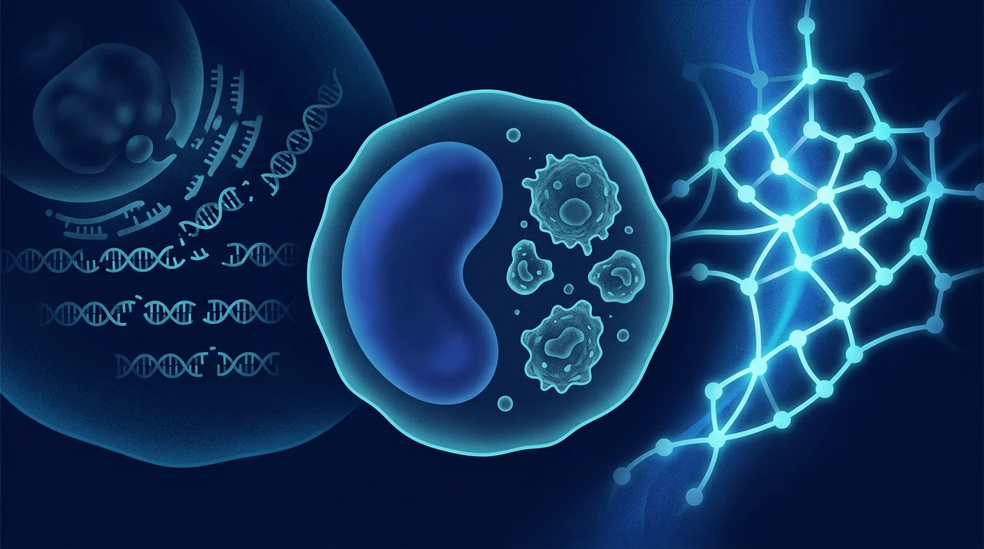
Apo10 is the name used for an antibody-defined epitope associated with a variant or altered form of DNaseX, also known as DNase1-like 1. DNase enzymes participate in breaking down DNA during cell death. The proposed cancer connection is that abnormal DNaseX protein can retain the Apo10 epitope yet fail to complete normal nuclear DNA fragmentation, contributing to a phenotype in which damaged cells evade a terminal step of apoptosis.
That mechanism should be described as a model, not a universal fact across tumors. Apo10 staining has been reported in multiple carcinoma tissues and in monocyte-based assays, but expression across benign disease, inflammatory states, premalignant lesions and diverse healthy populations is not mapped with the breadth expected of an established population-screening marker. Antibody recognition of an epitope also does not by itself demonstrate enzyme function.
Apo10 is therefore a marker of a proposed apoptosis-related abnormality, not a measurement of “how much apoptosis” is occurring. A positive cell does not identify the organ of origin. A low signal does not exclude a small, non-shedding or biologically different cancer. These distinctions are important whenever marketing language compresses a complex protein story into “tumor formation marker.”
TKTL1: metabolic association, not a universal cancer switch
TKTL1 is a transketolase-like protein investigated in glucose metabolism, redox balance and carbon flow related to the pentose phosphate pathway. Cancer cells can reprogram metabolism to support nucleotide synthesis, antioxidant defenses and growth. Experimental work has linked TKTL1 to altered glucose use and malignant behavior in some models [8], while reviews place transketolase biology within a broader network rather than a single pathway controlled by one protein [10].
Two cautions are essential. First, metabolic reprogramming is not exclusive to cancer: immune activation, wound repair and other proliferative states also alter glucose handling. Second, the biochemical role and magnitude of TKTL1’s contribution have been debated. Published experimental evidence has argued against a major role in some hypoxic and normoxic cancer-cell systems [9]. Tumor-tissue associations also vary by histology and method; an ovarian cancer study found correlations with subtype and distant metastasis, but such associations do not validate a blood-screening test [15].
Intracellular TKTL1 staining in a monocyte is therefore not a live measurement of a tumor’s glycolytic rate. It is an assay signal interpreted as evidence that the cell contains or expresses a protein associated with metabolic adaptation. It cannot locate a lesion, name its tissue, establish stage or select a therapy on its own.
Why combine Apo10 and TKTL1?
The conceptual rationale is complementary biology: Apo10 is intended to represent disrupted cell-death control, while TKTL1 is intended to represent metabolic adaptation. Combining two imperfect signals may separate selected cancer cases from controls better than either marker alone in a development cohort. Early studies reported promising classification in mixed carcinomas and elevated scores in oral, prostate, pancreatic, colorectal and biliary cancer settings [3].
Combination does not remove the need for external validation. If thresholds are optimized after looking at the same cases used to estimate performance, sensitivity and specificity can appear better than they will in screening populations. Spectrum bias also matters: distinguishing known, often clinically evident cancer from healthy controls is easier than distinguishing early cancer from benign nodules, inflammatory disease and other real-world reasons for testing.
Biological complementarity is not diagnostic independence
Apo10 and TKTL1 may reflect different processes, but both are measured in the same selected immune-cell compartment with the same specimen and platform. Correlated technical or inflammatory effects can influence both. A combined score still needs prospective calibration and validation.
What have clinical studies reported?
Published studies provide a signal worth investigating, but they do not yet establish EDIM as a population-wide replacement for standard screening. A study in cholangiocellular, pancreatic and colorectal carcinoma reported separation between patients and healthy controls [1]. Oral squamous-cell carcinoma studies reported elevated monocyte signals and declines after surgical resection, supporting investigation as a response or disease-burden marker [4] [5]. A prostate study likewise assessed score changes after radical prostatectomy [6].
Pediatric pilot studies found Apo10/TKTL1 signals in many patients with rhabdomyosarcoma or neuroblastoma and negative results in small healthy control groups [11] [12]. These studies are scientifically interesting because they test the immune-cargo concept across tumor types. They remain pilot evidence: sample sizes were modest, populations selected, and disease prevalence unlike general screening.
A colon-cancer metastasis report and several case-oriented publications describe potential lead-time or monitoring value [7]. Such observations can generate hypotheses but cannot estimate false-positive rates in thousands of asymptomatic people. Screening utility requires prospective cohorts, prespecified thresholds, blinded comparison against a reference standard, representation of benign mimics, and follow-up sufficient to discover cancers initially missed.
Sensitivity, specificity and predictive value are not the same
Even a test with high sensitivity and specificity in a case-control paper may have a low positive predictive value when cancer prevalence is low. Predictive value asks the patient’s real question: given this result, what is the probability that disease is present? It changes with age, symptoms, prior cancer, risk factors and the population being tested.
Imagine applying an exploratory marker to a low-risk population. The number of people without cancer is much larger than the number with cancer, so even a small false-positive percentage can produce more positive results without cancer than with it. Conversely, a negative result cannot erase baseline risk if the studied cancers do not express the markers, release little material, or fall below assay detection.
Multi-cancer early-detection tests also face a localization problem: detecting a general signal is only the first step. A useful pathway must direct efficient confirmatory imaging or organ assessment without excessive incidental findings and invasive procedures. Reviews of blood-based multi-cancer screening emphasize that mortality benefit, diagnostic resolution and downstream harms matter more than headline discrimination statistics [13].
How an EDIM result should—and should not—be used
An EDIM result should be treated as an adjunctive risk signal requiring clinical interpretation, not as a standalone diagnosis. A positive result does not prove cancer. A physician should review symptoms, examination, age-appropriate screening history, personal and family risk, inflammation or infection, previous malignancy, and the exact assay version before deciding whether follow-up is warranted.
- Do not cancel established screening. Mammography, cervical screening, colorectal screening, low-dose CT for eligible smokers and other guideline-based pathways have organ-specific evidence that an exploratory blood marker does not replace.
- Do not start cancer treatment from EDIM alone. Diagnosis generally requires lesion localization and, when appropriate, pathology or another validated reference standard.
- Do not interpret a negative result as “cancer-free.” No biomarker detects every tumor type or biological stage.
- Do investigate red-flag symptoms independently. Unexplained bleeding, a persistent mass, progressive weight loss or focal neurological symptoms need conventional assessment regardless of the score.
- Do document the follow-up plan. Repeat testing, targeted imaging or referral should have a reason and an endpoint, not an open-ended cascade.
Analytical validity comes before clinical promises
A biomarker can have compelling biology yet fail as a reproducible clinical test. Analytical validation asks whether the method accurately and consistently measures its intended signal. Clinical validation asks whether the signal identifies or predicts the relevant condition in the intended population. Clinical utility asks whether using it improves outcomes enough to justify costs and harms.
For a cell-based flow-cytometry assay, essential controls include specimen stability, repeatability, reproducibility across operators and instruments, antibody specificity, compensation, gating rules, limits of blank and detection, interference, lot-to-lot variation and prespecified handling of borderline results. Fit-for-purpose biomarker guidance emphasizes that validation must match the decision the result will support [14].
Clinical utility would require more than finding an occult lesion after a positive result. It should examine whether testing shifts diagnosis to a more treatable stage, reduces cancer mortality or morbidity, and avoids disproportionate false-positive imaging, biopsies, anxiety and overdiagnosis. Those outcomes have not been established for Apo10/TKTL1 EDIM screening in broad asymptomatic populations.
Key limitations in the current evidence
- Many studies are small, single-center, retrospective or case-control rather than prospective screening cohorts.
- Healthy controls may not represent patients with benign tumors, infection, autoimmune disease or chronic inflammation.
- Cancer types and stages are heterogeneous, yet publications sometimes pool them into one performance estimate.
- Thresholds, score definitions and laboratory workflows may not be identical across studies or commercial implementations.
- Independent replication and head-to-head comparison with current standard pathways are limited.
- Conflicts of interest and inventor involvement in foundational literature require transparent consideration, not dismissal of the data.
- Evidence for post-treatment score change does not automatically establish early-detection benefit.
- There is no proof that an EDIM-led screening program reduces cancer mortality.
These limitations do not mean the biomarkers are useless. They define their present status: an innovative, investigational liquid-biopsy approach with preliminary clinical signals and substantial unanswered questions.
Frequently Asked Questions
Are Apo10 and TKTL1 cancer-specific?
No marker should be assumed perfectly cancer-specific. Apo10 and TKTL1 are associated with biological processes reported in cancers, but expression, uptake and assay staining may be affected by tumor heterogeneity, immune-cell biology and non-malignant conditions.
Does a positive EDIM result mean I have cancer?
No. It is an assay-defined biomarker signal, not a pathology diagnosis. The result must be interpreted with clinical risk and, if justified, followed by validated imaging, organ-specific testing or tissue diagnosis.
Can a negative result rule out cancer?
No. Tumors differ in marker expression, size, shedding, immune interaction and location. Continue recommended screening and seek evaluation for concerning symptoms even after a negative result.
How are the markers detected inside monocytes?
Blood cells are identified with surface markers, permeabilized and stained with antibodies against intracellular targets. Flow cytometry counts fluorescent cells inside a specified gate and converts that pattern into assay-specific scores.
Is EDIM the same as circulating tumor DNA testing?
No. Circulating tumor DNA assays analyze tumor-associated genetic or epigenetic fragments in plasma. EDIM analyzes protein epitopes inside selected monocytes/macrophage-like cells after presumed uptake or expression.
Should EDIM replace mammography, colonoscopy or cervical screening?
No. It should not replace evidence-based, age- and risk-appropriate screening. Any adjunctive use should sit within a physician-directed pathway with clear follow-up.
Medical disclaimer
This article is educational and does not diagnose cancer or recommend a test for any individual. EDIM, Apo10 and TKTL1 remain areas of active research; assay availability does not establish population-screening benefit. Decisions should be made with a qualified clinician who can explain alternatives, uncertainty and the consequences of false-positive and false-negative results.
References
Apo10 与 TKTL1 是 EDIM 检测测量的两种细胞内信号,但它们并不是可互换的“癌症基因”,检出这些信号本身也不能诊断癌症。Apo10 是与 DNaseX 蛋白改变及细胞凋亡生物学相关的表位;TKTL1 是一种与代谢重编程有关的转酮醇酶样蛋白。EDIM(单核细胞表位检测)并非测量血清中游离的肿瘤标志物,而是在选定的 CD14+/CD16+ 循环单核细胞/巨噬细胞样群体内寻找这些靶点 [1]。
这种思路具有独特的生物学基础。单核细胞在血液和组织中巡逻,吞噬细胞物质,并可在细胞内携带其中一部分。EDIM 试图把先天免疫细胞当作其所接触物质的“报告者”。这一取样概念具有合理性,但合理的机制不等于已证实的筛查表现。已发表研究多为样本较小、经过选择的病例对照或监测研究,而且若干奠基论文的作者与检测知识产权或研发有关 [2]。
本文解释两个标志物可能代表的含义、吞噬摄取与肿瘤 DNA 脱落的差别、流式细胞术实际测量什么,以及为什么 EDIM 结果必须与已验证的筛查和诊断路径一同谨慎解读。
EDIM 检测测量什么?
EDIM 测量在设定门控的单核细胞群中,Apo10 和 TKTL1 细胞内染色阳性的比例。典型流程包括处理静脉血、以表面标志物识别活化或分化的单核细胞、使细胞膜通透,再使用针对 Apo10 与 TKTL1 的抗体。流式细胞仪随后在预先定义的门内计数荧光事件。
这不同于测量血清蛋白浓度。报告分数结合了细胞分类、抗体染色及检测专用算法。采血后处理、延迟时间、抗体性能、门控策略、仪器校准和阈值选择均可影响结果。因此,数值或颜色分级只能在该检测已验证的版本内解释;不应把一项研究的截点随意移植到另一实验室。
| 信号 | 拟议的生物学含义 | 单独不能证明 |
|---|---|---|
| Apo10 | 与 DNaseX 改变/凋亡相关生物学相联系的细胞内表位 | 存在活癌、病灶位置或侵袭性 |
| TKTL1 | 部分研究中与葡萄糖碳流及肿瘤代谢适应有关的蛋白 | 特定癌种、分期、实时代谢通量或治疗靶点 |
| EDIM 联合分数 | 选定单核细胞内由检测定义的模式 | 病理诊断或可替代器官特异性筛查 |
为什么单核细胞和巨噬细胞会含有肿瘤相关物质?
单核细胞是循环中的先天免疫细胞,可向巨噬细胞表型分化,并通过吞噬作用清除碎片。在炎症或肿瘤组织中,巨噬细胞会接触凋亡细胞、坏死碎片、细胞外囊泡和可溶蛋白。吞噬后,货物进入内体和吞噬溶酶体被加工,而不是作为一枚完整的微型肿瘤细胞保存。
EDIM 假说认为,单核/巨噬细胞样细胞摄取肿瘤来源物质,之后在循环中携带可检出的表位。共培养和临床研究支持在 CD14+/CD16+ 细胞内检测肿瘤相关蛋白的可能性 [3]。然而,抗体阳性事件无法说明摄取发生的确切时间与部位;它可能来自吞噬货物、单核细胞自身表达、活化变化、非特异染色,或多种因素的组合。
因此,“巨噬细胞吞吃癌细胞”这种说法容易过度简化。吞噬细胞也会清除正常细胞更新、感染、创伤及炎症产生的材料;组织中的肿瘤相关巨噬细胞在某些环境下甚至可能支持肿瘤。EDIM 应理解为间接的免疫细胞读数,而不是直接在血中看到恶性细胞。
Apo10:与 DNaseX 和凋亡失调相关的表位
Apo10 是用于描述一种抗体所识别表位的名称,该表位与 DNaseX(亦称 DNase1-like 1)的变异或改变形式有关。DNase 酶参与细胞死亡过程中的 DNA 分解。提出的癌症联系是:异常 DNaseX 可能保留 Apo10 表位,却不能完成正常的核 DNA 断裂,使受损细胞逃避凋亡末端步骤。
这应被视为机制模型,而不是适用于所有肿瘤的事实。多个癌组织及单核细胞检测中曾报告 Apo10 染色,但其在良性病、炎症、癌前病变和多样化健康人群中的分布,尚未达到成熟人群筛查标志物所需的广度。抗体识别表位也不等于证明酶功能。
因此,Apo10 是拟议的凋亡相关异常标志,而非“凋亡量”的测量。阳性细胞不能识别来源器官;低信号也不能排除体积小、不易释放物质或生物学不同的癌症。
TKTL1:代谢关联,而非普遍的癌症开关
TKTL1 是转酮醇酶样蛋白,被研究与葡萄糖代谢、氧化还原平衡及磷酸戊糖途径相关的碳流之间的关系。癌细胞可重编程代谢,以支持核苷酸合成、抗氧化防御与生长。实验研究在部分模型中把 TKTL1 与葡萄糖利用和恶性行为联系起来 [8],而综述则把转酮醇酶置于更广泛的代谢网络中 [10]。
需要两点谨慎。第一,代谢重编程并非癌症独有;免疫活化、伤口修复等增殖状态也会改变葡萄糖处理。第二,TKTL1 的生化作用和贡献大小仍有争议;有实验在缺氧和常氧癌细胞系统中反对其发挥主要作用 [9]。组织关联也因肿瘤类型和方法而异;卵巢癌研究中的亚型和远处转移相关性不能直接验证血液筛查 [15]。
所以,单核细胞内 TKTL1 染色并不是肿瘤糖酵解速率的实时测量,也不能单独定位病灶、确定组织来源或分期、选择治疗。
为什么联合 Apo10 与 TKTL1?
理论依据是互补生物学:Apo10 旨在反映细胞死亡控制异常,TKTL1 旨在反映代谢适应。在开发队列中,联合两个不完美信号可能比单一标志更好地区分部分癌症病例与对照。早期研究在混合癌、口腔癌、前列腺癌、胰腺癌、结直肠癌和胆道癌场景报告了有希望的分类 [3]。
联合并不会取消外部验证的必要。如果在同一批病例上选择最佳阈值并估算性能,敏感度和特异度可能高于真实筛查。谱偏倚也很重要:把已确诊、甚至临床明显的癌症与健康对照区分开,比区分早癌、良性结节、炎症及其他真实受检者容易。
生物学互补不等于诊断独立
两种标志可能反映不同过程,但它们来自同一样本、同一免疫细胞群和同一平台。相关的技术或炎症影响可同时改变两者,因此联合分数仍须前瞻性校准与验证。
临床研究报告了什么?
现有研究提供了值得继续检验的信号,但尚未确立 EDIM 可替代标准人群筛查。胆管、胰腺和结直肠癌研究报告了患者与健康对照之间的差异 [1]。口腔鳞癌研究报告分数升高及术后下降,支持其作为疗效或疾病负荷标志继续研究 [4] [5];前列腺研究也评估了根治术后的变化 [6]。
儿科先导研究在许多横纹肌肉瘤或神经母细胞瘤患者中检出 Apo10/TKTL1,而小型健康对照组为阴性 [11] [12]。这些结果对跨癌种检验免疫货物概念很有意义,但仍是先导证据:样本量有限、受试者经过选择、疾病患病率也不同于普通筛查。
结肠癌转移报告和病例性文献提出可能的提前发现或监测价值 [7],但无法估计数千名无症状人群中的假阳性率。筛查效用需要前瞻队列、预设阈值、盲法参考标准比较、良性类似疾病,以及足够长的随访来发现初次漏检的癌症。
敏感度、特异度与预测值不同
即使病例对照论文中的敏感度和特异度很高,当癌症患病率低时,阳性预测值仍可能很低。预测值回答患者真正关心的问题:得到这个结果后,实际患病概率是多少?它会随年龄、症状、既往癌症、风险因素和受检人群而变化。
在低风险人群中,无癌者远多于有癌者,所以很小的假阳性比例也可能产生大量无癌阳性结果。反之,如果肿瘤不表达标志、释放物质少或低于检测限,阴性结果也不能清除基础风险。
多癌种早筛还面临定位问题:发现通用信号只是第一步。后续路径必须有效指向确认性影像或器官检查,又不造成过多偶然发现与侵入性操作。相关综述强调,死亡率获益、诊断解决率和下游伤害比醒目的区分统计更重要 [13]。
EDIM 结果应该与不应该如何使用?
EDIM 应作为需临床解读的辅助风险信号,而非独立诊断。阳性不证明癌症。医师应审查症状、体检、年龄适宜的筛查史、个人和家族风险、炎症或感染、既往恶性肿瘤及确切检测版本,再决定是否随访。
- 不要停止标准筛查:乳腺 X 线、宫颈与结直肠筛查、适合吸烟者的低剂量 CT 等具有器官特异性证据。
- 不要仅凭 EDIM 开始抗癌治疗:通常需要病灶定位,并在适当时以病理或验证参考标准确诊。
- 不要把阴性理解为“无癌”:没有任何标志可检出所有癌种和阶段。
- 警示症状须独立评估:不明出血、持续肿块、进行性消瘦或局灶神经症状均需常规诊查。
- 记录随访计划:复测、靶向影像或转诊应有明确原因和终点,避免无限检查链。
先有分析有效性,后谈临床承诺
生物学上吸引人的标志物,也可能无法成为可重复的临床检测。分析验证问方法能否准确一致地测量目标;临床验证问它能否在目标人群识别相关疾病;临床效用则问使用它能否改善结局,并足以抵偿成本和伤害。
对细胞流式检测,重要控制包括样本稳定性、重复性、不同操作者和仪器间的再现性、抗体特异性、荧光补偿、门控规则、空白限和检出限、干扰、批间差异及边界结果处理。适用目的的标志物指南强调,验证必须匹配结果要支持的临床决策 [14]。
临床效用不只是阳性后找到隐匿病灶,还应检验是否把诊断提前到可治疗阶段、降低癌症死亡或患病负担,并避免过多影像、活检、焦虑和过度诊断。目前尚未在广泛无症状人群中证实 Apo10/TKTL1 EDIM 筛查具备这些结局。
当前证据的主要局限
- 许多研究规模小、单中心、回顾性或病例对照设计,而非前瞻筛查队列。
- 健康对照不能代表良性肿瘤、感染、自身免疫病或慢性炎症患者。
- 癌种和分期异质,却有时被合并为单一性能估计。
- 不同研究或商业版本的阈值、计分和实验流程可能不一致。
- 独立重复及与标准路径的头对头比较有限。
- 奠基文献中的利益冲突和发明者参与需透明考虑,但也不应因此直接否定数据。
- 治疗后分数变化并不自动证明早筛获益。
- 没有证据显示 EDIM 主导的筛查方案降低癌症死亡率。
这些限制并不表示标志物毫无价值,而是界定其当前地位:具有初步临床信号、但仍有大量未解问题的创新性研究型液体活检方法。
常见问题
Apo10 和 TKTL1 是癌症特异的吗?
不能假定任何标志物完全癌症特异。它们与癌症中报道的过程有关,但肿瘤异质性、免疫细胞生物学及非恶性状态可能影响表达、摄取和染色。
EDIM 阳性表示我患癌吗?
不表示。这是检测定义的标志物信号,不是病理诊断。若有依据,应结合临床风险,并以验证过的影像、器官检查或组织诊断跟进。
阴性能排除癌症吗?
不能。肿瘤在标志表达、大小、释放、免疫相互作用和位置上均不同。阴性后仍应继续推荐筛查,并对可疑症状求医。
如何在单核细胞内检测标志物?
先以表面标志识别血细胞,再使细胞通透并用抗体进行细胞内染色。流式细胞术计数指定门内的荧光细胞,并换算为检测专用分数。
EDIM 与循环肿瘤 DNA 相同吗?
不同。ctDNA 检测分析血浆中的肿瘤相关遗传或表观遗传片段;EDIM 分析选定单核/巨噬细胞样细胞内的蛋白表位。
EDIM 应取代乳腺 X 线、肠镜或宫颈筛查吗?
不应取代。它不能替代基于年龄和风险的循证筛查;任何辅助使用都应在医师指导且随访清晰的路径内进行。
医疗免责声明
本文仅用于健康教育,不为任何个人诊断癌症或推荐检测。EDIM、Apo10 和 TKTL1 仍处于积极研究领域;检测可获得并不等于已证明人群筛查获益。请与合格医师共同决策,并了解替代方案、不确定性及假阳性和假阴性的后果。
参考文献
يمثل Apo10 وTKTL1 الإشارتين داخل الخلية اللتين يقيسهما اختبار EDIM، لكنهما ليسا «جينَي سرطان» قابلين للتبادل، ووجودهما لا يشخّص السرطان بمفرده. Apo10 حاتمة مرتبطة ببروتين DNaseX متغير وببيولوجيا الاستماتة، أما TKTL1 فهو بروتين شبيه بالترانسكيتولاز دُرس في سياق إعادة البرمجة الأيضية. يبحث EDIM، أي كشف الحاتمات في الوحيدات، عن الهدفين داخل مجموعة منتقاة من الوحيدات CD14+/CD16+ والخلايا الشبيهة بالبلاعم، ولا يقيس واسمًا ورميًا حرًا في المصل [1].
الفكرة البيولوجية غير مألوفة: تجول الوحيدات في الدم والأنسجة، وتبتلع مواد خلوية، وقد تحمل جزءًا منها داخلها. يحاول EDIM استخدام خلايا المناعة الفطرية «كمراسل» عما واجهته. هذه فرضية معقولة، لكن المعقولية البيولوجية لا تساوي أداءً مثبتًا في التحري. معظم الدراسات المنشورة صغيرة أو انتقائية من نوع الحالات والشواهد أو المراقبة، كما شارك في أوراق تأسيسية باحثون مرتبطون بالملكية الفكرية أو تطوير الاختبار [2].
تشرح هذه المراجعة معنى كل واسم، والفرق بين التقاط البلاعم وبين طرح DNA الورمي، وما يقيسه التدفق الخلوي فعلًا، ولماذا ينبغي تفسير النتيجة بتحفظ إلى جانب مسارات التحري والتشخيص المثبتة.
ماذا يقيس اختبار EDIM؟
يقيس EDIM نسبة الخلايا التي تصطبغ داخليًا لحاتمتي Apo10 وTKTL1 ضمن بوابة محددة من الوحيدات. تُعالج عينة الدم الوريدي، وتُستخدم واسمات سطحية لتحديد الوحيدات المنشّطة أو المتمايزة، ثم تُنفّذ أغشية الخلايا وتُطبق أضداد للأهداف داخل الخلية. بعد ذلك يعدّ قياس التدفق الخلوي الأحداث الفلورية داخل البوابة المحددة مسبقًا.
يختلف ذلك عن قياس تركيز بروتين في المصل. فالدرجة تجمع تصنيف الخلية وصبغ الأضداد وحسابًا خاصًا بالاختبار. وقد تؤثر معالجة ما قبل التحليل، والزمن بعد السحب، وأداء الأضداد، واستراتيجية البوابات، ومعايرة الجهاز، واختيار العتبة. لذلك لا تُفسَّر الفئة الرقمية أو اللونية إلا ضمن نسخة الاختبار التي تم التحقق منها، ولا ينبغي نقل عتبة دراسة إلى مختبر آخر بلا تحقق.
| الإشارة | المعنى البيولوجي المقترح | ما لا تثبته وحدها |
|---|---|---|
| Apo10 | حاتمة داخلية مرتبطة بتغير DNaseX وبيولوجيا الاستماتة | وجود سرطان حي أو موقعه أو عدوانيته |
| TKTL1 | بروتين مرتبط في بعض الدراسات بتدفق كربون الغلوكوز والتكيف الأيضي الورمي | نوعًا أو مرحلة محددة أو تدفقًا أيضيًا مباشرًا أو هدفًا علاجيًا |
| الدرجة المركبة | نمط يعرّفه الاختبار داخل وحيدات منتقاة | تشخيصًا نسيجيًا أو بديلًا للتحري الخاص بالأعضاء |
لماذا قد تحتوي الوحيدات والبلاعم على مواد مرتبطة بالورم؟
الوحيدات خلايا مناعية فطرية جوالة يمكن أن تتمايز نحو البلاعم وتزيل الحطام بالبلعمة. في النسيج الملتهب أو الورمي، تواجه البلاعم خلايا مستميتة وشظايا نخرية وحويصلات خارج خلوية وبروتينات ذائبة. تدخل الحمولة بعد ابتلاعها إلى الجسيمات الداخلية والبلعمية الحالّة لتُعالج، ولا تبقى كخلية ورمية مصغرة كاملة.
تفترض تقنية EDIM أن الخلايا الشبيهة بالوحيدات والبلاعم تلتقط مادة مشتقة من الورم ثم تدور وهي تحمل حاتمات قابلة للكشف. وتدعم تجارب الزرع المشترك والدراسات السريرية إمكان كشف بروتينات مرتبطة بالورم داخل خلايا CD14+/CD16+ [3]. لكن الحدث الإيجابي بالضد لا يكشف زمن الالتقاط أو مكانه؛ فقد يعكس حمولة مبتلعة، أو تعبيرًا ذاتيًا في الوحيدة، أو تغير التنشيط، أو صبغًا غير نوعي، أو خليطًا منها.
لذلك قد تكون عبارة «البلاعم تأكل الخلايا السرطانية» مضللة. فهي تنظف أيضًا نواتج التجدد الطبيعي والعدوى والرضح والالتهاب، وقد تدعم البلاعم المرتبطة بالورم نمو السرطان في بعض البيئات. EDIM قراءة مناعية غير مباشرة، وليس رؤية مباشرة لخلية خبيثة في الدم.
Apo10: حاتمة مرتبطة بـDNaseX واضطراب الاستماتة
Apo10 اسم لحاتمة يعرّفها ضد وترتبط بشكل متغير أو معدل من DNaseX، المعروف أيضًا بـDNase1-like 1. تشارك إنزيمات DNase في تفكيك DNA أثناء موت الخلية. ويفترض النموذج السرطاني أن DNaseX غير الطبيعي قد يحتفظ بحاتمة Apo10 لكنه يعجز عن استكمال تفتيت DNA النووي، ما يساعد الخلايا المتضررة على تفادي خطوة نهائية من الاستماتة.
يجب وصف ذلك كنموذج، لا كحقيقة عامة في جميع الأورام. أُبلغ عن صبغ Apo10 في سرطانات متعددة وفي اختبارات الوحيدات، لكن انتشاره في الأمراض الحميدة والالتهاب والآفات قبل السرطانية والمجموعات الصحية المتنوعة لم يُرسم بالاتساع المتوقع لواسم تحرٍّ سكاني راسخ. كما أن تعرف الضد إلى حاتمة لا يثبت وظيفة الإنزيم.
وعليه، Apo10 واسم لخلل مقترح مرتبط بالاستماتة، وليس مقياسًا «لكمية الاستماتة». لا تحدد الخلية الإيجابية عضو المنشأ، ولا تستبعد الإشارة المنخفضة سرطانًا صغيرًا أو قليل الطرح أو مختلف البيولوجيا.
TKTL1: ارتباط أيضي وليس مفتاحًا سرطانيًا شاملًا
TKTL1 بروتين شبيه بالترانسكيتولاز يُدرس في استقلاب الغلوكوز وتوازن الأكسدة والاختزال وتدفق الكربون المرتبط بمسار فوسفات البنتوز. تعيد الخلايا السرطانية برمجة الأيض لدعم تصنيع النوكليوتيدات والدفاع المضاد للأكسدة والنمو. ربط عمل تجريبي TKTL1 بتغير استخدام الغلوكوز والسلوك الخبيث في بعض النماذج [8]، بينما تضع المراجعات بيولوجيا الترانسكيتولاز ضمن شبكة أوسع [10].
يلزم تحفّظان. أولًا، إعادة البرمجة الأيضية ليست حصرية للسرطان؛ فتنشيط المناعة والتئام الجروح وحالات التكاثر تغير التعامل مع الغلوكوز أيضًا. ثانيًا، ما زال الدور الكيميائي الحيوي وحجم مساهمة TKTL1 موضع نقاش؛ إذ عارضت تجارب وجود دور رئيسي في بعض خلايا السرطان ناقصة وطبيعية الأكسجة [9]. كما تختلف ارتباطات النسيج باختلاف النوع والطريقة، ولا يثبت ارتباط وُجد في سرطان المبيض صلاحية اختبار دم للتحري [15].
إذن صبغ TKTL1 داخل وحيدة ليس قياسًا مباشرًا لمعدل تحلل الغلوكوز في الورم، ولا يحدد الموقع أو النسيج أو المرحلة أو العلاج بمفرده.
لماذا يُجمع Apo10 وTKTL1؟
المسوغ النظري هو التكامل: يقصد من Apo10 تمثيل اضطراب ضبط موت الخلية، ومن TKTL1 تمثيل التكيف الأيضي. قد يحسن جمع إشارتين غير كاملتين فصل بعض حالات السرطان عن الشواهد في مجموعة التطوير. وقد أبلغت دراسات مبكرة عن تصنيف واعد في سرطانات مختلطة وسياقات الفم والبروستاتا والبنكرياس والقولون والمستقيم والقنوات الصفراوية [3].
لكن الجمع لا يلغي التحقق الخارجي. إذا جرى تحسين العتبة على الحالات نفسها المستخدمة لحساب الأداء، فقد تبدو الحساسية والنوعية أعلى من التحري الواقعي. ويهم تحيز الطيف أيضًا: التفريق بين سرطان معروف وربما واضح وبين أصحاء أسهل من التفريق بين سرطان مبكر وعقدة حميدة والتهاب وبقية أسباب الفحص الحقيقية.
التكامل البيولوجي لا يعني استقلال التشخيص
قد يعكس الواسمان عمليتين مختلفتين، لكنهما يُقاسان في الحجرة المناعية نفسها وبالعينة والمنصة نفسيهما. يمكن لعوامل تقنية أو التهابية مترابطة التأثير فيهما معًا؛ لذا تحتاج الدرجة المركبة إلى معايرة وتحقق مستقبليين.
ماذا أفادت الدراسات السريرية؟
توفر الدراسات المنشورة إشارة تستحق البحث، لكنها لا تثبت EDIM بديلًا للتحري السكاني القياسي. أفادت دراسة لسرطان القنوات الصفراوية والبنكرياس والقولون والمستقيم بفصل المرضى عن الشواهد الأصحاء [1]. وأظهرت دراسات سرطان الخلايا الحرشفية الفموية ارتفاع الإشارات وانخفاضها بعد الاستئصال، ما يدعم بحثها كواسم للاستجابة أو عبء المرض [4] [5]. كما قيّمت دراسة البروستاتا التغير بعد الاستئصال الجذري [6].
وجدت دراسات أطفال استطلاعية Apo10/TKTL1 لدى كثير من مرضى الساركوما العضلية المخططة أو الورم الأرومي العصبي، وكانت مجموعات الشواهد الصحية الصغيرة سلبية [11] [12]. وهي مهمة لاختبار مفهوم الحمولة المناعية عبر الأورام، لكنها تبقى دليلًا استطلاعيًا بعينات محدودة وسكان منتقين وانتشار مرض مختلف عن التحري العام.
تقترح تقارير نقائل القولون ومنشورات الحالات إمكان التبكير أو المراقبة [7]، لكنها لا تقدّر الإيجابيات الكاذبة بين آلاف عديمي الأعراض. يلزم للتحري فوج مستقبلي، وعتبات مسبقة، ومقارنة عمياء بمعيار مرجعي، وتمثيل الحالات الحميدة، ومتابعة تكشف السرطانات التي فاتت أولًا.
الحساسية والنوعية والقيمة التنبؤية ليست شيئًا واحدًا
حتى الاختبار مرتفع الحساسية والنوعية في دراسة حالات وشواهد قد يملك قيمة تنبؤية إيجابية منخفضة عندما يكون انتشار السرطان قليلًا. تسأل القيمة التنبؤية: ما احتمال المرض بعد هذه النتيجة؟ وتتغير بالعمر والأعراض والسرطان السابق والمخاطر والسكان المفحوصين.
في السكان منخفضي الخطورة، عدد غير المصابين أكبر بكثير؛ لذلك قد تنتج نسبة صغيرة من الإيجابيات الكاذبة نتائج إيجابية بلا سرطان أكثر من النتائج المصحوبة به. وبالعكس، لا تمحو النتيجة السلبية الخطر إذا كان الورم لا يعبّر عن الواسم أو يطرح مادة قليلة أو يقع دون حد الكشف.
ويواجه التحري متعدد السرطانات مشكلة تحديد الموقع. فالإشارة العامة ليست إلا الخطوة الأولى، وينبغي أن توجه المتابعة بكفاءة دون إفراط في الموجودات العرضية والإجراءات الغازية. تؤكد المراجعات أن خفض الوفيات وحسم التشخيص والأضرار اللاحقة أهم من إحصاءات التمييز البارزة [13].
كيف تُستخدم نتيجة EDIM وكيف لا تُستخدم؟
ينبغي التعامل معها كإشارة خطر مساعدة تحتاج تفسيرًا سريريًا، لا كتشخيص مستقل. الإيجابية لا تثبت السرطان. يراجع الطبيب الأعراض والفحص والتحري الملائم للعمر والمخاطر الشخصية والعائلية والالتهاب أو العدوى والسرطان السابق ونسخة الاختبار قبل تقرير المتابعة.
- لا توقف التحري المعتمد: تصوير الثدي وتحري عنق الرحم والقولون والتصوير المقطعي منخفض الجرعة للمؤهلين لها أدلة خاصة بالأعضاء.
- لا تبدأ علاج السرطان بناءً على EDIM وحده: يلزم تحديد الآفة، وعند اللزوم التشخيص النسيجي أو معيار مثبت.
- لا تفسر السلبية بأنها «خالٍ من السرطان»: لا يكشف أي واسم جميع الأنواع والمراحل.
- قيّم أعراض الإنذار مستقلًا: النزف غير المفسر والكتلة المستمرة ونقص الوزن المتقدم والأعراض العصبية البؤرية تحتاج تقييمًا تقليديًا.
- وثّق خطة المتابعة: ينبغي أن يكون للتكرار أو التصوير أو الإحالة سبب ونقطة نهاية واضحة.
الصلاحية التحليلية تسبق الوعود السريرية
قد تكون بيولوجيا الواسم مقنعة ويفشل مع ذلك كاختبار سريري قابل للتكرار. يسأل التحقق التحليلي هل تقيس الطريقة الإشارة بدقة واتساق؛ ويسأل التحقق السريري هل تحدد الحالة في السكان المقصودين؛ وتسأل المنفعة السريرية هل يحسن استخدامها النتائج بما يبرر التكلفة والضرر.
تشمل ضوابط اختبار التدفق ثبات العينة والتكرارية وقابلية الإعادة بين الأجهزة والمشغلين ونوعية الأضداد وتعويض الفلورة وقواعد البوابات وحدود الفراغ والكشف والتداخل واختلاف الدفعات ومعالجة النتائج الحدية. وتؤكد إرشادات الواسمات الملائمة للغرض أن التحقق يجب أن يطابق القرار الذي ستدعمه النتيجة [14].
تتطلب المنفعة أكثر من العثور على آفة خفية بعد الإيجابية. ينبغي اختبار تقديم التشخيص إلى مرحلة قابلة للعلاج وخفض الوفيات أو المراضة، مع تجنب الإفراط في التصوير والخزعات والقلق وفرط التشخيص. لم تُثبت هذه النتائج لتحري Apo10/TKTL1 EDIM في السكان الواسعين عديمي الأعراض.
أهم قيود الدليل الحالي
- كثير من الدراسات صغيرة وأحادية المركز أو رجعية أو حالات وشواهد وليست أفواج تحرٍّ مستقبلية.
- الأصحاء لا يمثلون مرضى الأورام الحميدة أو العدوى أو المناعة الذاتية أو الالتهاب المزمن.
- تتباين الأنواع والمراحل، لكنها تُجمع أحيانًا في تقدير أداء واحد.
- قد تختلف العتبات والدرجات وسير المختبر بين الدراسات والنسخ التجارية.
- التكرار المستقل والمقارنة المباشرة بالمسارات القياسية محدودان.
- يجب النظر بشفافية إلى تضارب المصالح ومشاركة المخترعين، من دون رفض البيانات تلقائيًا.
- تغير الدرجة بعد العلاج لا يثبت منفعة الكشف المبكر.
- لا دليل على أن برنامج تحرٍّ يقوده EDIM يخفض وفيات السرطان.
لا تعني هذه القيود أن الواسمات بلا قيمة، بل تحدد وضعها الحالي: خزعة سائلة بحثية مبتكرة ذات إشارات أولية وأسئلة كبيرة لم تُحسم.
الأسئلة الشائعة
هل Apo10 وTKTL1 نوعيان للسرطان؟
لا ينبغي افتراض النوعية الكاملة. يرتبطان بعمليات مذكورة في السرطان، لكن عدم تجانس الورم وبيولوجيا المناعة والحالات غير الخبيثة قد تؤثر في التعبير والالتقاط والصبغ.
هل تعني نتيجة EDIM الإيجابية أنني مصاب بالسرطان؟
لا. إنها إشارة واسم يعرّفها الاختبار وليست تشخيصًا نسيجيًا. تُفسر مع الخطر السريري، وإذا لزم تُتبع بتصوير أو اختبار عضو أو خزعة مثبتة.
هل تستبعد النتيجة السلبية السرطان؟
لا. تختلف الأورام في التعبير والحجم والطرح والتفاعل المناعي والموقع. استمر بالتحري الموصى به واطلب تقييم الأعراض المقلقة.
كيف تُكشف الواسمات داخل الوحيدات؟
تُحدد خلايا الدم بواسمات سطحية، ثم تُنفّذ وتُصبغ بأضداد للأهداف الداخلية. يعدّ التدفق الخلوي الخلايا الفلورية داخل بوابة محددة ويحول النمط إلى درجات خاصة بالاختبار.
هل EDIM هو نفسه اختبار DNA الورمي الجائل؟
لا. يحلل ctDNA شظايا وراثية أو فوق جينية مرتبطة بالورم في البلازما، بينما يحلل EDIM حاتمات بروتينية داخل وحيدات أو خلايا شبيهة بالبلاعم.
هل يحل EDIM محل تصوير الثدي أو تنظير القولون أو تحري عنق الرحم؟
لا. لا يحل محل التحري المبني على الدليل والملائم للعمر والخطر، وأي استخدام مساعد يجب أن يكون ضمن مسار طبي واضح المتابعة.
إخلاء المسؤولية الطبية
هذه المقالة للتثقيف ولا تشخّص السرطان أو توصي باختبار لفرد بعينه. ما زال EDIM وApo10 وTKTL1 مجال بحث نشط؛ وتوافر الاختبار لا يثبت منفعة التحري السكاني. ناقش القرار مع طبيب مؤهل يشرح البدائل وعدم اليقين وعواقب النتائج الكاذبة.
المراجع
- Saman S, Stagno MJ, Warmann SW, et al. Biomarkers Apo10 and TKTL1: Epitope-detection in monocytes (EDIM) as a new diagnostic approach for cholangiocellular, pancreatic and colorectal carcinoma. Cancer Biomarkers. 2020;27(1):129–137. doi:10.3233/CBM-190414
- Coy JF. EDIM-TKTL1/Apo10 Blood Test: An Innate Immune System Based Liquid Biopsy for the Early Detection, Characterization and Targeted Treatment of Cancer. International Journal of Molecular Sciences. 2017;18(4):878. doi:10.3390/ijms18040878
- Grimm M, Schmitt S, Teriete P, et al. A biomarker based detection and characterization of carcinomas exploiting two fundamental biophysical mechanisms in mammalian cells. BMC Cancer. 2013;13:569. doi:10.1186/1471-2407-13-569
- Grimm M, Kraut W, Hoefert S, et al. Evaluation of a biomarker based blood test for monitoring surgical resection of oral squamous cell carcinomas. Clinical Oral Investigations. 2016;20:329–338. doi:10.1007/s00784-015-1518-0
- Grimm M, Kraut W, Hoefert S, et al. Analysis of circulating CD14+/CD16+ monocyte-derived macrophages in the peripheral blood of patients with oral squamous cell carcinoma. Oral Surgery, Oral Medicine, Oral Pathology and Oral Radiology. 2016;121(3):301–306. doi:10.1016/j.oooo.2015.10.024
- Todenhöfer T, Hennenlotter J, Keller G, et al. Effect of radical prostatectomy on levels of cancer related epitopes in circulating macrophages of patients with clinically localized prostate cancer. The Prostate. 2017;77(12):1251–1258. doi:10.1002/pros.23384
- Jansen N, Coy JF. Diagnostic use of epitope detection in monocytes blood test for early detection of colon cancer metastasis. Future Oncology. 2013;9(4):605–609. doi:10.2217/fon.13.8
- Díaz-Moralli S, Aguilar E, Marín S, et al. A key role for transketolase-like 1 in tumor metabolic reprogramming. Oncotarget. 2016;7(32):51875–51897. doi:10.18632/oncotarget.10429
- Mayer A, von Wallbrunn A, Vaupel P. Evidence against a major role for TKTL-1 in hypoxic and normoxic cancer cells. Advances in Experimental Medicine and Biology. 2011;701:123–128. doi:10.1007/978-1-4419-7756-4_17
- Hao S, Meng Q, Sun H, et al. The role of transketolase in human cancer progression and therapy. Biomedicine & Pharmacotherapy. 2022;154:113607. doi:10.1016/j.biopha.2022.113607
- Urla C, Stagno MJ, Schmidt A, et al. Epitope Detection in Monocytes (EDIM) As a New Method of Liquid Biopsy in Pediatric Rhabdomyosarcoma. Biomedicines. 2022;10(8):1812. doi:10.3390/biomedicines10081812
- Stagno MJ, Schmidt A, Bochem J, et al. Epitope detection in monocytes for liquid biopsy including identification of GD2 in childhood neuroblastoma—a pilot study. British Journal of Cancer. 2022;127:1324–1331. doi:10.1038/s41416-022-01855-x
- Brito-Rocha T, Constâncio V, Henrique R, Jerónimo C. Shifting the Cancer Screening Paradigm: The Rising Potential of Blood-Based Multi-Cancer Early Detection Tests. Cells. 2023;12(6):935. doi:10.3390/cells12060935
- Cummings J, Raynaud F, Jones L, Sugar R, Dive C. Fit-for-purpose biomarker method validation for application in clinical trials of anticancer drugs. British Journal of Cancer. 2010;103:1313–1317. doi:10.1038/sj.bjc.6605910
- Krockenberger M, Honig A, Rieger L, et al. Transketolase-like 1 expression correlates with subtypes of ovarian cancer and the presence of distant metastases. International Journal of Gynecological Cancer. 2007;17(1):101–106. doi:10.1111/j.1525-1438.2007.00799.x