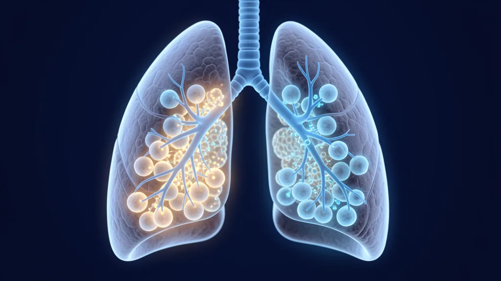
Pulmonary fibrosis is a progressive, irreversible scarring of lung tissue that steadily robs patients of their ability to breathe. Idiopathic pulmonary fibrosis (IPF) — the most common form — carries a median survival of just 3–5 years from diagnosis, worse than many cancers. Current antifibrotic drugs (pirfenidone, nintedanib) slow decline but do not stop or reverse the scarring process. Mesenchymal stem cell (MSC) therapy is being investigated as a way to interrupt the fibrotic cascade at the cellular level — not by replacing scarred tissue, but by altering the signaling environment that drives fibrosis forward.[1][2]
What Is Pulmonary Fibrosis?
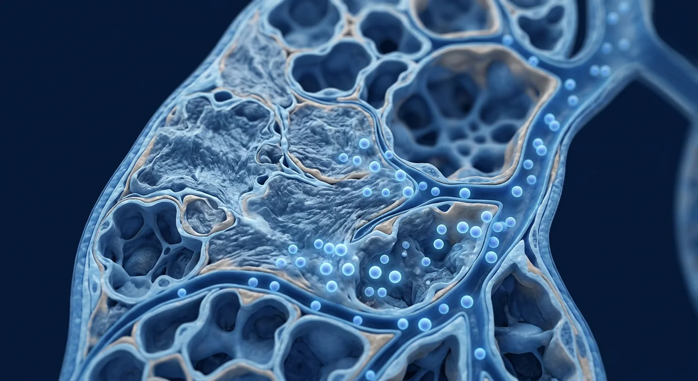
Pulmonary fibrosis is progressive scarring (fibrosis) of the lung interstitium — the delicate connective tissue scaffold that supports the alveoli, where oxygen and carbon dioxide exchange. As scar tissue accumulates, the alveolar walls thicken and stiffen, reducing the lungs' ability to expand and transfer oxygen into the bloodstream.[3]
Where the damage begins. The prevailing model holds that repeated micro-injury to the alveolar epithelium — from environmental exposures, gastroesophageal reflux, viral infections, or genetic predisposition — triggers an aberrant wound-healing response. Instead of resolving, fibroblasts are persistently activated into myofibroblasts that deposit excessive extracellular matrix proteins, particularly collagen. The result is a self-perpetuating cycle: stiff tissue → mechanical stress on adjacent alveoli → further injury → more fibrosis.[4][5]
Why conventional treatments stall. Pirfenidone and nintedanib target specific profibrotic pathways (TGF-β, PDGF, FGF), and clinical trials show they reduce the rate of forced vital capacity (FVC) decline by approximately 50%. But they do not halt progression entirely, carry significant side effects (gastrointestinal distress, photosensitivity, liver toxicity), and do not restore lost lung function. The fundamental barrier remains: once the fibrotic program is locked in, dampening one pathway is not enough to switch it off.[6]
How MSC Therapy Targets Pulmonary Fibrosis
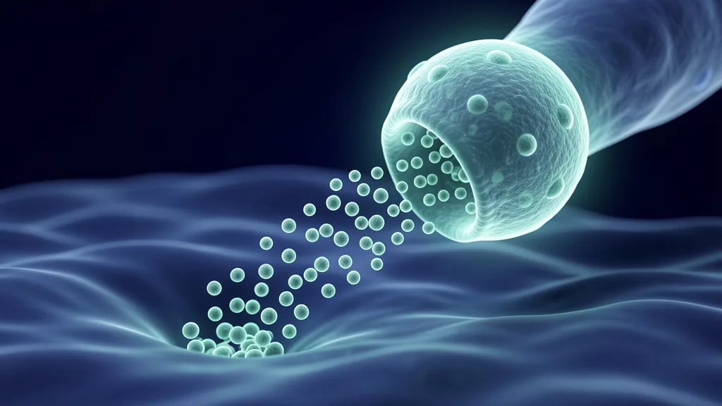
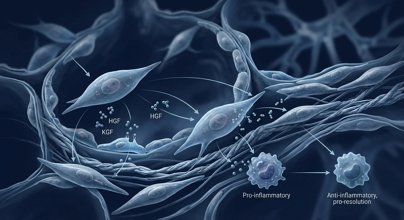
MSCs address fibrosis through multiple mechanisms simultaneously. Unlike single-pathway antifibrotic drugs, MSCs secrete a broad repertoire of bioactive molecules — the "secretome" — that collectively suppress inflammation, induce myofibroblast apoptosis, degrade excess extracellular matrix, and stimulate endogenous repair.[7][8]
- Anti-fibrotic factors: Hepatocyte growth factor (HGF) and keratinocyte growth factor (KGF) directly counter TGF-β-driven myofibroblast activation and collagen synthesis.
- Immunomodulation: MSCs shift macrophages from a profibrotic M2 phenotype toward an antifibrotic M1/M2 balance, reducing IL-4/IL-13-driven collagen deposition. PGE2 secretion suppresses T-cell proliferation.
- Matrix remodeling: MSCs upregulate matrix metalloproteinases (MMPs) that degrade excess collagen while downregulating TIMPs (tissue inhibitors of metalloproteinases), tipping the balance toward scar resolution.
- Mitochondrial transfer: MSCs can transfer healthy mitochondria to injured alveolar epithelial cells via tunneling nanotubes, restoring cellular bioenergetics and reducing apoptosis.
HGF is particularly relevant. In bleomycin-induced mouse models of pulmonary fibrosis, MSC-derived HGF reduced collagen deposition by 40–60% and improved survival. The effect was abolished when HGF was blocked, confirming the centrality of this single factor. Human MSCs produce HGF constitutively, and the levels increase further when the cells are exposed to inflammatory signals — precisely the environment they encounter in a fibrotic lung.[9][10]
Clinical Evidence: What the Trials Show
Clinical data remain early-stage but directionally encouraging. The majority of published studies in pulmonary fibrosis are Phase I safety trials or small Phase I/II pilots. No Phase III registrational trial has been completed — and this honest acknowledgement is essential for patients evaluating their options.
Chambers et al. administered placental MSCs (1–2 × 10⁶ cells/kg IV) to 8 IPF patients. No serious adverse events at 6-month follow-up. FVC and 6MWT remained stable — notable in a population where decline is expected. [11]
Glassberg et al. tested bone marrow MSCs (20–200 × 10⁶ cells IV) in 9 IPF patients. Treatment was well-tolerated. FVC, DLCO, and 6MWT showed trends toward stabilization at 12 months. [12]
Averyanov et al. delivered high-dose bone marrow MSCs (2 × 10⁶ cells/kg IV monthly × 4) to 13 IPF patients. At 12 months, FVC was preserved while matched controls declined. CT fibrosis score was stable in the MSC group. [13]
Interpreting the data honestly. Across these studies, the consistent finding is safety — no ectopic tissue formation, no significant immune reactions, and no tumorigenic events. Efficacy signals (FVC stability, 6MWT maintenance, reduced inflammatory biomarkers) are encouraging but come from trials too small to draw definitive conclusions. Every published paper emphasizes that larger, randomized, placebo-controlled trials are needed before MSC therapy can be considered a standard-of-care option for pulmonary fibrosis.
What Is the Treatment Protocol?
MSC therapy for pulmonary fibrosis is typically delivered intravenously. The IV route allows cells to pass through the pulmonary circulation first — a phenomenon called "first-pass lung trapping" — where MSCs transiently lodge in the lung microvasculature before redistributing. Far from being a limitation, this pulmonary trapping may be therapeutic: MSCs retained in the lung continue secreting paracrine factors at the site of disease.[14]
Umbilical cord-derived MSCs (Wharton's Jelly) — selected for high proliferative capacity, low immunogenicity, and robust HGF/KGF expression profile.
Typically 100–200 million MSCs per IV infusion. Protocols often include 2–4 sessions spaced 4–8 weeks apart, tailored to disease severity and patient response.
Pulmonary function tests (FVC, DLCO), 6-minute walk test, HRCT fibrosis scoring, and quality-of-life questionnaires (SGRQ, UCSD-SOBQ) at baseline and 3–6 month intervals.
Safety and Limitations
MSC therapy for pulmonary fibrosis has a well-documented short-term safety profile. Across all published IPF trials, adverse events have been mild and transient — low-grade fever, transient fatigue, and occasional infusion-related discomfort. No cases of ectopic tissue formation, pulmonary embolism from cell clumping, or tumorigenesis have been reported in the pulmonary fibrosis literature specifically.[15]
- No Phase III trial has demonstrated efficacy — the therapy remains investigational for pulmonary fibrosis.
- The optimal cell source (bone marrow vs. umbilical cord vs. adipose), dose, and dosing frequency are not yet established.
- MSCs are not a cure. The goal is disease modification — slowing or stabilizing fibrosis — not reversal of established scar tissue.
- Patients with advanced disease (FVC < 50% predicted, on supplemental oxygen) have been excluded from most trials; safety and efficacy in this population are even less certain.
- Intravenous delivery results in significant first-pass lung trapping; while this may be therapeutic, the fraction of cells that engraft long-term is negligible.
Why Patients Consider Treatment in Bangkok
Thailand — and Bangkok specifically — has emerged as a destination for patients seeking MSC therapy for pulmonary fibrosis. Several factors contribute: a regulatory framework that permits clinical MSC use under physician supervision, GMP-compliant cell manufacturing facilities, and treatment costs significantly lower than equivalent care in North America or Europe.[16]
The VELAR approach. Patients considering MSC therapy at VELAR Center undergo comprehensive pulmonary evaluation — HRCT imaging, full PFTs including DLCO, 6MWT, and inflammatory biomarker panels — before any treatment decision. Dosing is individualized based on disease severity, and every patient receives honest counsel about what the current evidence does and does not support.
Frequently Asked Questions
Can stem cells cure pulmonary fibrosis?
No. MSCs are not a cure for pulmonary fibrosis. The goal of MSC therapy is disease modification — slowing or stabilizing the progression of fibrosis — not reversal of established scar tissue. Early trials show signals of FVC stabilization, but no study has demonstrated fibrosis reversal.
How much does stem cell therapy for pulmonary fibrosis cost in Thailand?
Treatment costs vary based on cell dose, number of sessions, and individual clinical needs. As a reference, MSC therapy in Thailand typically ranges from $8,000–$18,000 USD per treatment course, substantially less than comparable programs in the United States or Europe.
Is intravenous MSC delivery effective for lung conditions?
IV-delivered MSCs are trapped in the pulmonary microvasculature on first pass — a phenomenon that concentrates the cells in the lungs. While long-term engraftment is minimal, the retained cells secrete antifibrotic and immunomodulatory factors (HGF, KGF, PGE2) that may exert therapeutic effects for 48–72 hours post-infusion.
What is the difference between MSC therapy and antifibrotic drugs?
Antifibrotic drugs (pirfenidone, nintedanib) target single molecular pathways to slow fibrosis. MSCs address multiple pathways simultaneously — suppressing TGF-β, modulating macrophages, degrading excess collagen, and supporting epithelial repair — through their paracrine secretome. Some researchers believe this multi-target approach may offer advantages, but this hypothesis has not been proven in head-to-head trials.
Are there any risks specific to pulmonary fibrosis patients?
Patients with advanced disease (FVC below 50% predicted, significant oxygen dependence) have been excluded from most trials, so the safety profile in this population is less established. Pre-existing pulmonary hypertension — common in advanced IPF — could theoretically increase the risk of microvascular complications from IV cell infusion, though this has not been observed in published studies.
References
- Richeldi L, Collard HR, Jones MG. Idiopathic pulmonary fibrosis. The Lancet. 2017;389(10082):1941-1952. doi:10.1016/S0140-6736(17)30866-8 ↩
- Lederer DJ, Martinez FJ. Idiopathic pulmonary fibrosis. New England Journal of Medicine. 2018;378(19):1811-1823. doi:10.1056/NEJMra1705751 ↩
- Wynn TA. Integrating mechanisms of pulmonary fibrosis. Journal of Experimental Medicine. 2011;208(7):1339-1350. doi:10.1084/jem.20110551 ↩
- Wolters PJ, Collard HR, Jones KD. Pathogenesis of idiopathic pulmonary fibrosis. Annual Review of Pathology. 2014;9:157-179. doi:10.1146/annurev-pathol-012513-104706 ↩
- Chanda D, Otoupalova E, Smith SR, Volckaert T, De Langhe SP, Thannickal VJ. Developmental pathways in the pathogenesis of lung fibrosis. Molecular Aspects of Medicine. 2019;65:56-69. doi:10.1016/j.mam.2018.08.004 ↩
- Flaherty KR, Wells AU, Cottin V, et al. Nintedanib in progressive fibrosing interstitial lung diseases. New England Journal of Medicine. 2019;381(18):1718-1727. doi:10.1056/NEJMoa1908681 ↩
- Tzouvelekis A, Toonkel R, Karampitsakos T, et al. Mesenchymal stem cells for the treatment of idiopathic pulmonary fibrosis. Frontiers in Medicine. 2018;5:142. doi:10.3389/fmed.2018.00142 ↩
- Ntolios P, Janning M, Stathopoulos GT, et al. The role of mesenchymal stem cells in idiopathic pulmonary fibrosis. European Respiratory Review. 2022;31(166):220084. doi:10.1183/16000617.0084-2022 ↩
- Gazdhar A, Grad I, Tamò L, et al. The secretome of induced pluripotent stem cells reduces lung fibrosis in part by hepatocyte growth factor. Stem Cell Research & Therapy. 2014;5(6):123. doi:10.1186/scrt513 ↩
- Cahill EF, Kennelly H, Carty F, Mahon BP, English K. Hepatocyte growth factor is required for mesenchymal stromal cell protection against bleomycin-induced pulmonary fibrosis. Stem Cells Translational Medicine. 2016;5(8):1117-1127. doi:10.5966/sctm.2015-0337 ↩
- Chambers DC, Enever D, Ilic N, et al. A phase 1b study of placenta-derived mesenchymal stromal cells in patients with idiopathic pulmonary fibrosis. Respirology. 2014;19(7):1013-1018. doi:10.1111/resp.12343 ↩
- Glassberg MK, Minkiewicz J, Toonkel RL, et al. Allogeneic human mesenchymal stem cells in patients with idiopathic pulmonary fibrosis via intravenous delivery (AETHER): a phase I safety clinical trial. Chest. 2017;151(5):971-981. doi:10.1016/j.chest.2016.11.050 ↩
- Averyanov A, Koroleva I, Konoplyannikov M, et al. First-in-human high-cumulative-dose stem cell therapy in idiopathic pulmonary fibrosis with rapid lung function decline. Stem Cells Translational Medicine. 2020;9(1):6-16. doi:10.1002/sctm.19-0037 ↩
- Fischer UM, Harting MT, Jimenez F, et al. Pulmonary passage is a major obstacle for intravenous stem cell delivery: the pulmonary first-pass effect. Stem Cells and Development. 2009;18(5):683-692. doi:10.1089/scd.2008.0253 ↩
- Lalu MM, McIntyre L, Pugliese C, et al. Safety of cell therapy with mesenchymal stromal cells (SafeCell): a systematic review and meta-analysis of clinical trials. PLoS ONE. 2012;7(10):e47559. doi:10.1371/journal.pone.0047559 ↩
- Sueblinvong V, Weiss DJ. Stem cells and cell therapy approaches in lung biology and diseases. Translational Research. 2010;156(3):189-204. doi:10.1016/j.trsl.2010.06.006 ↩
肺纤维化(Pulmonary Fibrosis)是一种进行性、不可逆的肺组织瘢痕化疾病,特发性肺纤维化(IPF)的中位生存期仅为3-5年。现有抗纤维化药物(吡非尼酮、尼达尼布)仅能减缓肺功能下降,而间充质干细胞(MSC)疗法正在被研究作为一种从细胞层面中断纤维化级联反应的方法。[1][2]
什么是肺纤维化?
肺纤维化是肺间质的进行性瘢痕化——肺泡周围的支持组织逐渐被胶原蛋白替代,导致肺扩张受限和氧气交换障碍。反复的肺泡上皮微损伤触发异常的伤口愈合反应:成纤维细胞持续活化为肌成纤维细胞,沉积过量的细胞外基质。[3][4]
MSC疗法如何靶向肺纤维化
MSC通过多种机制协同作用于纤维化,而非像抗纤维化药物那样仅阻断单一通路。MSC的"分泌组"包含抗纤维化因子(HGF、KGF)、免疫调节细胞因子(PGE2、IL-10)和基质金属蛋白酶(MMPs),它们共同抑制炎症、诱导肌成纤维细胞凋亡并降解过量胶原蛋白。[7][9]
- 抗纤维化:HGF和KGF直接对抗TGF-β驱动的肌成纤维细胞活化。
- 免疫调节:将巨噬细胞从促纤维化M2表型转向抗纤维化平衡。
- 基质重塑:上调MMPs降解胶原,下调TIMPs。
- 线粒体转移:通过隧道纳米管向受损上皮细胞输送健康线粒体。
临床证据
临床数据仍处于早期阶段。已发表的IPF研究主要是I期安全性试验和小型I/II期试点。Chambers等(2014)对8名IPF患者给予胎盘MSC输注,6个月随访无严重不良事件,FVC和6MWT保持稳定。[11] Glassberg等(2017)在9名IPF患者中测试骨髓MSC,证实安全性并观察到12个月时功能指标趋于稳定。[12] Averyanov等(2020)对13名IPF患者进行高剂量骨髓MSC输注,12个月时FVC保持稳定而对照组下降。[13]
安全性与局限性
- 尚无III期试验证实疗效——该疗法在肺纤维化领域仍属研究性质。
- 最佳细胞来源、剂量和给药频率尚未确立。
- MSC并非治愈手段,目标为疾病修饰(减缓或稳定纤维化),而非逆转已形成的瘢痕。
- 晚期患者(FVC低于50%预计值、依赖吸氧)被排除在大多数试验之外。
常见问题
干细胞能治愈肺纤维化吗?
不能。MSC的目标是疾病修饰——减缓或稳定纤维化进展——而非逆转已有瘢痕组织。
在泰国治疗肺纤维化的干细胞费用是多少?
治疗费用因细胞剂量、治疗次数和个体临床需求而异。泰国的MSC治疗通常在8,000-18,000美元/疗程之间,远低于美国或欧洲的同类项目。
参考资料
- Richeldi L, et al. Idiopathic pulmonary fibrosis. The Lancet. 2017;389:1941-1952. doi:10.1016/S0140-6736(17)30866-8 ↩
- Lederer DJ, Martinez FJ. Idiopathic pulmonary fibrosis. NEJM. 2018;378:1811-1823. doi:10.1056/NEJMra1705751 ↩
- Wynn TA. Integrating mechanisms of pulmonary fibrosis. J Exp Med. 2011;208:1339-1350. doi:10.1084/jem.20110551 ↩
- Wolters PJ, et al. Pathogenesis of IPF. Annu Rev Pathol. 2014;9:157-179. doi:10.1146/annurev-pathol-012513-104706 ↩
- Tzouvelekis A, et al. MSCs for IPF. Front Med. 2018;5:142. doi:10.3389/fmed.2018.00142 ↩
- Gazdhar A, et al. iPSC secretome reduces lung fibrosis via HGF. Stem Cell Res Ther. 2014;5:123. doi:10.1186/scrt513 ↩
- Chambers DC, et al. Placenta-derived MSCs in IPF. Respirology. 2014;19:1013-1018. doi:10.1111/resp.12343 ↩
- Glassberg MK, et al. Allogeneic MSCs in IPF (AETHER). Chest. 2017;151:971-981. doi:10.1016/j.chest.2016.11.050 ↩
- Averyanov A, et al. High-dose MSCs in IPF. Stem Cells Transl Med. 2020;9:6-16. doi:10.1002/sctm.19-0037 ↩
التليف الرئوي هو تندب تدريجي لا رجعة فيه في أنسجة الرئة، حيث يبلغ متوسط البقاء على قيد الحياة في التليف الرئوي مجهول السبب (IPF) 3-5 سنوات فقط — وهو أسوأ من العديد من السرطانات. الأدوية المضادة للتليف الحالية (بيرفينيدون، نينتيدانيب) تبطئ التدهور لكنها لا توقف أو تعكس عملية التندب. يتم دراسة العلاج بالخلايا الجذعية الوسيطة (MSC) كوسيلة لمقاطعة سلسلة التليف على المستوى الخلوي.[1][2]
ما هو التليف الرئوي؟
التليف الرئوي هو تندب تدريجي للنسيج الخلالي للرئة — النسيج الضام الدقيق الذي يدعم الحويصلات الهوائية. مع تراكم النسيج الندبي، تتسمك جدران الحويصلات وتتصلب، مما يقلل من قدرة الرئتين على التمدد ونقل الأكسجين إلى مجرى الدم. تؤدي الإصابات الدقيقة المتكررة للظهارة السنخية إلى استجابة غير طبيعية لالتئام الجروح: يتم تنشيط الخلايا الليفية باستمرار إلى خلايا ليفية عضلية تودع كميات زائدة من الكولاجين.[3][4]
كيف يستهدف علاج MSC التليف الرئوي
تعالج الخلايا الجذعية الوسيطة التليف من خلال آليات متعددة في وقت واحد. على عكس الأدوية المضادة للتليف أحادية المسار، تفرز MSCs مجموعة واسعة من الجزيئات النشطة بيولوجيًا — "السيكريتوم" — التي تثبط الالتهاب بشكل جماعي، وتحفز موت الخلايا الليفية العضلية، وتحلل المصفوفة خارج الخلية الزائدة، وتحفز الإصلاح الداخلي.[7][9]
- مضاد للتليف: HGF و KGF يتصدان مباشرة لتنشيط الخلايا الليفية العضلية المدفوع بـ TGF-β.
- تعديل المناعة: تحويل البلاعم من النمط M2 المحفز للتليف نحو توازن مضاد للتليف.
- إعادة تشكيل المصفوفة: زيادة MMPs لتحلل الكولاجين، وتقليل TIMPs.
- نقل الميتوكوندريا: نقل ميتوكوندريا سليمة إلى الخلايا الظهارية التالفة عبر أنابيب نانوية نفقية.
الأدلة السريرية
لا تزال البيانات السريرية في مرحلة مبكرة. معظم الدراسات المنشورة هي تجارب سلامة من المرحلة الأولى. أظهرت دراسة Chambers (2014) على 8 مرضى IPF عدم وجود أحداث سلبية خطيرة مع استقرار FVC و 6MWT.[11] أكدت دراسة Glassberg (2017) على 9 مرضى السلامة مع اتجاهات نحو الاستقرار في 12 شهرًا.[12] أظهر Averyanov (2020) على 13 مريضًا استقرار FVC مقارنة بانخفاض المجموعة الضابطة.[13]
السلامة والقيود
- لم تثبت أي تجربة من المرحلة الثالثة الفعالية — لا يزال العلاج قيد البحث.
- لم يتم تحديد المصدر الأمثل للخلايا والجرعة وتكرار الجرعات بعد.
- MSCs ليست علاجًا شافيًا. الهدف هو تعديل المرض — إبطاء أو تثبيت التليف — وليس عكس النسيج الندبي الموجود.
- تم استبعاد المرضى في المراحل المتقدمة من معظم التجارب.
الأسئلة الشائعة
هل يمكن للخلايا الجذعية علاج التليف الرئوي؟
لا. الهدف من علاج MSC هو تعديل المرض — إبطاء أو تثبيت تقدم التليف — وليس عكس النسيج الندبي الموجود.
كم تكلفة علاج الخلايا الجذعية للتليف الرئوي في تايلاند؟
تختلف تكاليف العلاج بناءً على جرعة الخلايا وعدد الجلسات والاحتياجات السريرية الفردية. كمرجع، يتراوح علاج MSC في تايلاند عادةً بين 8,000-18,000 دولار أمريكي لكل دورة علاجية.
المراجع
- Richeldi L, et al. Idiopathic pulmonary fibrosis. The Lancet. 2017;389:1941-1952. doi:10.1016/S0140-6736(17)30866-8 ↩
- Lederer DJ, Martinez FJ. Idiopathic pulmonary fibrosis. NEJM. 2018;378:1811-1823. doi:10.1056/NEJMra1705751 ↩
- Wynn TA. Integrating mechanisms of pulmonary fibrosis. J Exp Med. 2011;208:1339-1350. doi:10.1084/jem.20110551 ↩
- Wolters PJ, et al. Pathogenesis of IPF. Annu Rev Pathol. 2014;9:157-179. doi:10.1146/annurev-pathol-012513-104706 ↩
- Tzouvelekis A, et al. MSCs for IPF. Front Med. 2018;5:142. doi:10.3389/fmed.2018.00142 ↩
- Gazdhar A, et al. iPSC secretome reduces lung fibrosis via HGF. Stem Cell Res Ther. 2014;5:123. doi:10.1186/scrt513 ↩
- Chambers DC, et al. Placenta-derived MSCs in IPF. Respirology. 2014;19:1013-1018. doi:10.1111/resp.12343 ↩
- Glassberg MK, et al. Allogeneic MSCs in IPF (AETHER). Chest. 2017;151:971-981. doi:10.1016/j.chest.2016.11.050 ↩
- Averyanov A, et al. High-dose MSCs in IPF. Stem Cells Transl Med. 2020;9:6-16. doi:10.1002/sctm.19-0037 ↩