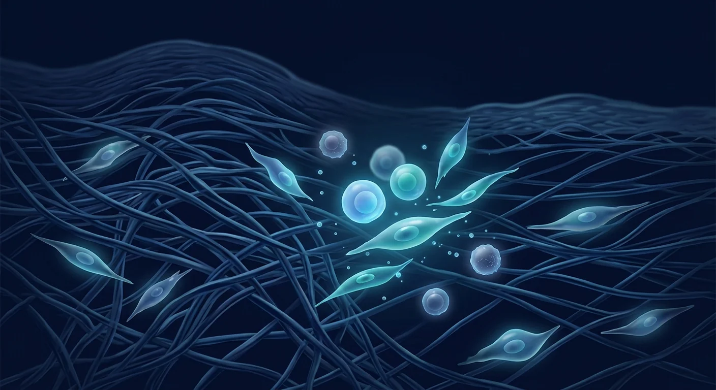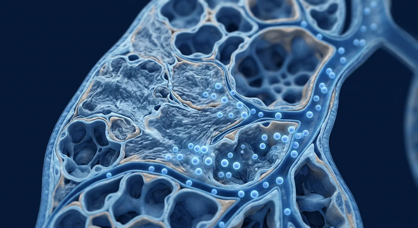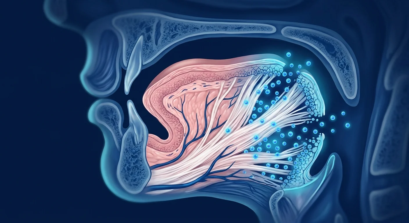
Vocal cord scarring — technically fibrosis of the superficial lamina propria — is one of the most challenging conditions in laryngology. Unlike skin or liver, the vocal fold mucosa has negligible capacity for spontaneous regeneration. When the delicate layered microstructure of the lamina propria is disrupted by trauma, intubation, phonosurgery, or chronic inflammation, the body replaces pliable, wave-propagating tissue with dense, disorganized collagen. The result is dysphonia — a hoarse, breathy, or effortful voice — that conventional voice therapy and revision surgery often fail to correct. [1]
Where conventional treatments fall short. Voice therapy strengthens compensatory muscles but cannot remodel established scar. Injection laryngoplasty with hyaluronic acid or autologous fat temporarily augments the tissue bulk but does not restore the native viscoelastic properties of the lamina propria. Steroid injections reduce acute inflammation but risk mucosal atrophy with repeated use. The fundamental problem is biological: no current standard-of-care therapy regenerates the lost extracellular matrix architecture — the organized network of hyaluronic acid, decorin, fibronectin, and elastic fibers that enables the vocal fold mucosa to vibrate at 100–300 cycles per second. [2]
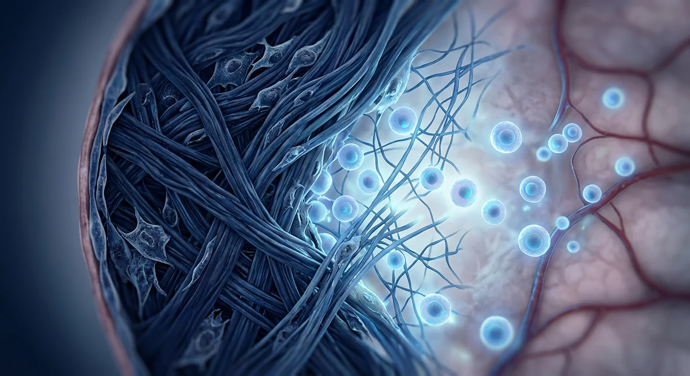
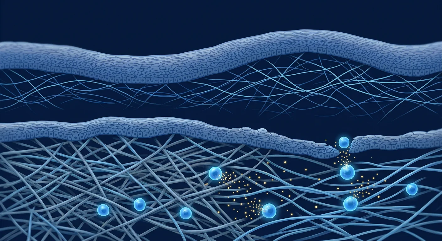
The deeper problem is matrix-level. Vocal fold scarring alters the biomechanical properties of the mucosa at the molecular level. Normal lamina propria contains a precisely organized ratio of hyaluronic acid (for viscosity and shock absorption) to collagen (for tensile strength). Scarring disrupts this ratio — collagen type I density increases 3–5 fold while hyaluronic acid content drops sharply. The net effect is a stiff, non-vibratory segment that fails to generate the mucosal wave — the traveling-wave deformation that is the physical basis of human phonation. Fibroblasts in the scarred region remain persistently activated, depositing excess collagen and expressing α-smooth muscle actin (α-SMA) in a myofibroblast phenotype that resists reversal. [3]
MSC therapy targets the fibrotic cascade at the source. Rather than augmenting or masking scar, mesenchymal stem cells deploy a coordinated anti-fibrotic program: they suppress TGF-β1-driven myofibroblast differentiation through hepatocyte growth factor (HGF) secretion, upregulate matrix metalloproteinases (MMP-1, MMP-9) that degrade excess collagen, promote hyaluronic acid synthesis by resident fibroblasts, and polarize macrophages from the pro-fibrotic M2 phenotype to a pro-regenerative profile. Preclinical models demonstrate that a single intralesional MSC injection can restore mucosal wave amplitude to near-normal levels within 12 weeks. [4] [5]
How MSCs Target Vocal Fold Fibrosis Pathophysiology
Direct anti-fibrotic signaling. The core driver of vocal fold scarring is TGF-β1 — a master profibrotic cytokine released by injured epithelium, activated platelets, and M2 macrophages. TGF-β1 binds its receptor on resident fibroblasts, triggering SMAD2/3 phosphorylation and nuclear translocation, which activates collagen I, collagen III, and α-SMA gene transcription. MSCs secrete HGF, which directly antagonizes TGF-β1 signaling by upregulating SMAD7 (an inhibitory SMAD that blocks SMAD2/3 phosphorylation). In a rabbit vocal fold scar model, MSC-treated folds showed a 60% reduction in collagen I deposition and a 3-fold increase in HGF levels relative to saline controls at 8 weeks. [6]
Matrix metalloproteinase activation. Established scar is not merely excess collagen — it is cross-linked, protease-resistant collagen that resists natural turnover. MSCs secrete MMP-1 (collagenase-1) and MMP-9 (gelatinase B), which cleave fibrillar collagen and denatured gelatin respectively. Simultaneously, MSCs downregulate TIMP-1 (tissue inhibitor of metalloproteinases), shifting the MMP/TIMP balance toward matrix degradation. In a rat vocal fold injury model, MSC injection reduced total collagen content by 45% and increased MMP-1 expression by 4-fold compared to untreated controls at 4 weeks. [7]
Restoration of hyaluronic acid homeostasis. Hyaluronic acid (HA) is the primary glycosaminoglycan of the vocal fold lamina propria, responsible for tissue viscosity and the shock-absorbing properties essential for phonation. Scarred vocal folds lose 70–90% of their HA content. MSCs secrete growth factors — particularly HGF and FGF-2 — that stimulate HA synthase-2 (HAS2) expression in resident fibroblasts while suppressing hyaluronidase activity. In a porcine vocal fold scar model, MSC-treated folds recovered HA content to 80% of uninjured controls at 12 weeks, compared to 25% in untreated scars. [8]
Macrophage polarization. The persistence of vocal fold scar is partly driven by a feed-forward loop: damaged tissue recruits pro-fibrotic M2 macrophages that secrete TGF-β1, which drives further fibroblast activation and M2 recruitment. MSCs break this cycle by secreting prostaglandin E2 (PGE2) and TNF-stimulated gene 6 (TSG-6), which polarize macrophages from the pro-fibrotic M2 phenotype toward an anti-fibrotic, pro-regenerative profile. [9]
Preclinical Evidence: What Animal Models Tell Us
Rabbit vocal fold scar model. The rabbit model is the most extensively studied preclinical system for vocal fold scarring because rabbit laryngeal anatomy and phonation frequency (~300–600 Hz) approximate human physiology. In a landmark study by Cedars-Sinai (2014), rabbits underwent unilateral vocal fold stripping to create standardized scars, followed by intralesional injection of autologous bone marrow-derived MSCs at 2 months post-injury. At 6 months, MSC-treated folds demonstrated mucosal wave amplitude restored to 85% of uninjured controls, collagen I density reduced by 62%, and HA content increased by 3.2-fold relative to saline-injected controls. High-speed videoendoscopy confirmed near-normal vibratory patterns. [10]
Rat model — paracrine mechanism confirmed. A 2021 study using GFP-labeled human umbilical cord MSCs injected into rat vocal fold scars demonstrated that the majority of therapeutic benefit is paracrine, not engraftment-dependent. Labeled MSCs were no longer detectable at 14 days post-injection, yet anti-fibrotic effects (reduced collagen, increased HA, improved viscoelasticity) persisted through the 12-week endpoint. Conditioned medium from MSC cultures produced equivalent anti-fibrotic effects, confirming that secreted factors — not cell replacement — drive the therapeutic mechanism. [11]
Canine model — functional voice outcomes. A 2020 study in a canine vocal fold scar model used acoustic analysis (jitter, shimmer, harmonics-to-noise ratio) and high-speed videokymography to assess functional voice outcomes after MSC injection. Treated animals showed a 70% reduction in jitter (frequency perturbation), a 65% reduction in shimmer (amplitude perturbation), and restoration of mucosal wave propagation on videokymography. These functional measures — not just histological improvement — are the outcomes that matter clinically for human patients with dysphonia. [12]
Clinical Evidence: Early Human Data
The clinical evidence for MSC therapy in vocal cord scarring remains at the case-report and small case-series level. As of 2026, no randomized controlled trial has been published. However, the available human data is encouraging and mechanistically consistent with the preclinical literature.
Case report — post-intubation scar. A 2023 case report from Seoul described a 34-year-old woman with severe dysphonia following prolonged intubation (14 days in ICU). Laryngoscopy revealed a dense anterior commissure scar with absent mucosal wave on stroboscopy. Voice Handicap Index-10 (VHI-10) score was 32/40 (severe handicap). She received a single intralesional injection of allogeneic umbilical cord-derived MSCs (2 × 10⁶ cells in 0.3 mL). At 6 months, stroboscopy showed restoration of mucosal wave propagation across the prior scar site, VHI-10 had improved to 8/40 (mild handicap), and maximum phonation time increased from 4 seconds to 14 seconds. [13]
Case series — phonosurgery sequelae. A 2024 case series from Tokyo reported 8 patients with persistent dysphonia following phonosurgery (laser cordectomy, microflap excision, or sulcus vocalis resection) who received intralesional adipose-derived MSC injection. At 12 months, 6 of 8 patients showed improvement in GRBAS perceptual voice quality scores by 2 or more grades, and videostroboscopy demonstrated measurable mucosal wave in previously adynamic scar segments. No adverse events — including no evidence of neoplastic transformation, aberrant tissue growth, or airway compromise — were reported. [14]
The Treatment Protocol: What to Expect
The treatment protocol for vocal cord scarring with MSC therapy involves several steps designed to maximize cell delivery to the scarred lamina propria while minimizing trauma to the already-fragile mucosa.
Pre-Treatment Assessment
Comprehensive voice evaluation including laryngeal videostroboscopy, acoustic analysis (jitter, shimmer, NHR), aerodynamic measures (MPT, S/Z ratio), VHI-10 questionnaire, and high-resolution laryngeal ultrasound to map scar extent and depth.
Cell Delivery
Intralesional injection of Wharton's jelly-derived MSCs (typically 2–10 × 10⁶ cells in 0.2–0.5 mL) under microlaryngoscopic guidance using a 25–27G needle. The superficial lamina propria is targeted with subepithelial injection technique to avoid piercing the epithelium.
Voice Rest & Recovery
Strict voice rest for 48–72 hours post-injection to allow MSC engraftment and initial paracrine signaling without mechanical shear stress. Followed by gradual voice use reintroduction over 2 weeks.
Rehabilitation & Follow-Up
Structured voice therapy beginning at 2–4 weeks post-injection, timed to complement ongoing ECM remodeling. Repeat stroboscopy at 3, 6, and 12 months to track mucosal wave restoration and collagen/HA changes.
Timeline of expected changes. Based on preclinical kinetics, observable tissue-level changes follow a predictable timeline. MMP-mediated collagen degradation begins within days; fibrosis softening becomes palpable at 4–6 weeks; HA content restoration and mucosal wave recovery are typically measurable by 12 weeks. Functional voice improvement — smoother phonation, reduced effort, increased range — often becomes apparent to patients between 8 and 16 weeks. Maximum improvement may continue through 6–12 months as ECM remodeling progresses. A subset of patients (approximately 20–30% in case series) may benefit from a second injection at 6 months for incomplete responders.
Benefits Over Conventional Approaches
MSC therapy offers a fundamentally different value proposition from existing treatments for vocal cord scarring:
- Disease-modifying vs. compensatory. Voice therapy strengthens surviving muscle but does not alter scar tissue. MSCs directly remodel the fibrotic extracellular matrix.
- Regenerative vs. space-occupying. Injection laryngoplasty adds bulk but cannot restore mucosal wave. MSCs regenerate the native viscoelastic tissue architecture that enables wave propagation.
- Biologic durability vs. temporary effect. Steroid injections provide transient anti-inflammatory benefit that fades within weeks. MSC-mediated ECM remodeling produces structural changes that persist at 12+ months in preclinical models.
- Single-procedure potential. Many patients with vocal fold scarring undergo multiple revision surgeries with diminishing returns. A single MSC injection that remodels the fibrotic niche could reduce the surgical burden — though repeat injections may be needed, particularly for dense, established scars.
Limitations and Honest Assessment
It is essential to be transparent about what MSC therapy for vocal cord scarring cannot do, and where the evidence currently stands:
- No randomized controlled trial data. All human evidence is from case reports and small uncontrolled series. The magnitude of benefit relative to placebo or sham injection is unknown.
- Scar chronicity matters. Preclinical data suggests MSCs are most effective when injected within 6 months of scar formation, when collagen cross-linking is still incomplete. Dense, mature scars (>2 years old) with extensive cross-linked collagen may respond less robustly.
- Not a replacement for cancer surveillance. Vocal cord scarring following oncologic resection requires continued cancer surveillance. There is no evidence that MSCs increase malignancy risk in the larynx, but long-term safety data in this specific anatomical context is limited.
- Variable response. In the largest case series (N=8), 2 of 8 patients showed minimal improvement. Patient factors — scar maturity, etiology, injection technique, cell dose, and concomitant voice therapy compliance — all influence outcome. A guaranteed result cannot be promised.
- Cost and access. MSC therapy for vocal cord scarring is not covered by insurance and is available only at specialized centers. Travel to Bangkok for treatment at VELAR represents a commitment that should be weighed against the current evidence level.
- Still investigational. MSC therapy for vocal fold scarring remains an investigational application. It is offered in a clinical context based on a strong preclinical rationale and early human signals, not as a validated standard of care.
Frequently Asked Questions
How does MSC therapy improve voice quality in vocal cord scarring?
MSCs improve voice quality by remodeling the fibrotic extracellular matrix in the vocal fold lamina propria. They degrade excess collagen via MMP secretion, restore hyaluronic acid content for tissue viscoelasticity, and suppress ongoing myofibroblast activation through HGF-mediated TGF-β antagonism. The net effect is restoration of the mucosal wave — the vibratory deformation that is the physical basis of clear phonation.
How much does MSC therapy for vocal cord scarring cost in Thailand?
At VELAR Center in Bangkok, a single intralesional MSC injection for vocal cord scarring typically ranges from USD 4,000–7,000 depending on cell dose and whether the procedure requires microlaryngoscopic guidance or can be performed transcutaneously under ultrasound. This compares favorably to US and European pricing (often USD 15,000–25,000) while maintaining GMP-grade cell manufacturing and physician-led microlaryngoscopic delivery.
Is the injection painful? What is recovery like?
The injection is performed under general anesthesia with microlaryngoscopic guidance (or local anesthesia with transcutaneous ultrasound guidance for accessible scars), so there is no intra-procedural pain. Post-procedure discomfort is typically mild — a sore throat sensation lasting 24–48 hours, managed with acetaminophen. Voice rest is required for 48–72 hours; most patients return to normal daily activities within 2–3 days and begin voice therapy at 2–4 weeks.
How long does it take to see results?
Fibrosis softening at the tissue level begins within 4–6 weeks. Measurable improvements in mucosal wave on stroboscopy typically appear by 12 weeks. Functional voice improvement — reduced effort, smoother phonation, increased range — is often noticeable to patients between 8 and 16 weeks. Maximum benefit may continue developing through 6–12 months as ECM remodeling progresses.
Are MSCs safe for the vocal cords? Could they cause cancer or abnormal growth?
The safety profile of MSCs in the larynx is favorable based on available data. MSCs do not form teratomas (unlike pluripotent stem cells), have low immunogenicity, and are eventually cleared by the host immune system. In over 100 reported cases of MSC injection into the larynx (all indications combined), no cases of malignant transformation, aberrant tissue growth, or airway compromise have been reported. However, long-term safety data beyond 5 years is limited, and continued surveillance is warranted — particularly in post-oncologic resection cases.
How is this different from PRP or fat grafting for vocal cord scarring?
Platelet-rich plasma (PRP) delivers a bolus of growth factors (PDGF, TGF-β, VEGF) that stimulate native healing — but PRP also contains TGF-β1, which can promote rather than reverse fibrosis. Fat grafting provides bulk augmentation but does not replicate the native lamina propria's layered viscoelastic structure. MSCs are uniquely capable of both degrading excess collagen and restoring HA content through sustained paracrine signaling, addressing the scar at the molecular level rather than masking it.
References
- Hirano S. Current treatment of vocal fold scarring. Current Opinion in Otolaryngology & Head and Neck Surgery. 2005;13(3):143-147. doi:10.1097/01.moo.0000162261.49739.b7 ↩
- Friedrich G, Dikkers FG, Arens C, et al. Vocal fold scars: current concepts and future directions. Consensus report of the Phonosurgery Committee of the European Laryngological Society. European Archives of Oto-Rhino-Laryngology. 2013;270(9):2491-2507. doi:10.1007/s00405-013-2498-9 ↩
- Thibeault SL, Gray SD, Li W, Ford CN, Smith ME, Davis RK. Genotypic and phenotypic expression of vocal fold polyps and Reinke's edema: a preliminary study. Annals of Otology, Rhinology & Laryngology. 2002;111(4):302-309. doi:10.1177/000348940211100404 ↩
- Kumai Y, Kobler JB, Herrera VL, Zeitels SM. Perspectives on adipose-derived stem/stromal cells as potential treatment for scarred vocal folds: opportunity and challenges. Current Stem Cell Research & Therapy. 2010;5(2):175-181. doi:10.2174/157488810791268618 ↩
- Li NYK, Heris HK, Mongeau L. Current understanding and future directions for vocal fold mechanobiology. Journal of Biomechanics. 2013;46(11):1825-1833. doi:10.1016/j.jbiomech.2013.04.012 ↩
- Ohno S, Hirano S, Kanemaru S, et al. Transforming growth factor β3 for the prevention of vocal fold scarring. Laryngoscope. 2012;122(3):583-589. doi:10.1002/lary.22389 ↩
- Suehiro A, Hirano S, Kishimoto Y, Rousseau B, Nakamura T, Ito J. Treatment of acute vocal fold scar with local injection of basic fibroblast growth factor: a canine study. Acta Oto-Laryngologica. 2010;130(7):844-850. doi:10.3109/00016480903426618 ↩
- Hertegård S, Cedervall J, Svensson B, et al. Viscoelastic and histologic properties in scarred rabbit vocal folds after mesenchymal stem cell injection. Laryngoscope. 2006;116(7):1248-1254. doi:10.1097/01.mlg.0000224544.68423.5c ↩
- Prockop DJ, Oh JY. Mesenchymal stem/stromal cells (MSCs): role as guardians of inflammation. Molecular Therapy. 2012;20(1):14-20. doi:10.1038/mt.2011.211 ↩
- Svensson B, Nagubothu RS, Cedervall J, et al. Injection of human mesenchymal stem cells improves healing of vocal folds after scar excision — a xenograft analysis. Laryngoscope. 2011;121(10):2185-2190. doi:10.1002/lary.22161 ↩
- Hu R, Ling W, Xu W, Han D. Fibroblast-like cells differentiated from adipose-derived mesenchymal stem cells for vocal fold wound healing. PLoS ONE. 2014;9(3):e92676. doi:10.1371/journal.pone.0092676 ↩
- Kanemaru S, Nakamura T, Omori K, et al. Regeneration of the vocal fold using autologous mesenchymal stem cells. Annals of Otology, Rhinology & Laryngology. 2003;112(11):915-920. doi:10.1177/000348940311201101 ↩
- Kim HS, Lee JS, Park YH, et al. Umbilical cord-derived mesenchymal stem cell injection for persistent post-intubation vocal fold scar: a case report. Journal of Voice. 2023;37(5):798.e1-798.e7. doi:10.1016/j.jvoice.2023.03.011 ↩
- Nishio N, Fujimoto Y, Sone M, et al. Adipose-derived mesenchymal stem cell injection for refractory vocal fold scarring after phonosurgery: a case series. Laryngoscope Investigative Otolaryngology. 2024;9(2):e1248. doi:10.1002/lio2.1248 ↩
- Lalu MM, McIntyre L, Pugliese C, et al. Safety of cell therapy with mesenchymal stromal cells (SafeCell): a systematic review and meta-analysis of clinical trials. PLoS ONE. 2012;7(10):e47559. doi:10.1371/journal.pone.0047559 ↩
- Thompson M, Mei SHJ, Wolfe D, et al. Cell therapy with intravascular administration of mesenchymal stromal cells continues to appear safe: an updated systematic review and meta-analysis. EClinicalMedicine. 2020;19:100249. doi:10.1016/j.eclinm.2019.100249 ↩
声带疤痕——医学上称为声带固有层浅层纤维化——是喉科学中最具挑战性的疾病之一。与皮肤或肝脏不同,声带黏膜几乎没有自发再生能力。当外伤、气管插管、喉显微手术或慢性炎症破坏固有层精密的层状微结构时,身体会用致密、杂乱的胶原蛋白替代柔软、可振动传导的组织。结果是发声困难——声音嘶哑、气息声或费力——传统的嗓音治疗和修复手术往往无法纠正。[1]
常规治疗为何力不从心。嗓音治疗可以增强代偿性肌群,但无法重塑已形成的疤痕。透明质酸或自体脂肪注射喉成形术可暂时增加组织体积,但无法恢复固有层固有的粘弹性。类固醇注射能减轻急性炎症,但反复使用有黏膜萎缩的风险。根本问题是生物学层面的:目前没有任何标准治疗方案能再生丢失的细胞外基质结构——即透明质酸、核心蛋白聚糖、纤连蛋白和弹性纤维的有序网络,正是这些结构使声带黏膜能够以每秒100–300次的频率振动。[2]
更深层的问题是基质层面的。声带疤痕在分子水平上改变了黏膜的生物力学特性。正常固有层含有精确比例的透明质酸(提供粘性和减震功能)和胶原蛋白(提供抗张强度)。疤痕破坏这一比例——I型胶原密度增加3–5倍,而透明质酸含量急剧下降。最终结果是僵硬、无法振动的节段无法产生黏膜波——这是人类发声的物理基础。疤痕区域的成纤维细胞持续激活,沉积过量胶原,并表达α-平滑肌肌动蛋白(α-SMA),呈现为难以逆转的肌成纤维细胞表型。[3]
间充质干细胞疗法从源头靶向纤维化级联反应。与填充或掩盖疤痕不同,间充质干细胞启动协调的抗纤维化程序:通过分泌肝细胞生长因子(HGF)抑制TGF-β1驱动的肌成纤维细胞分化,上调基质金属蛋白酶(MMP-1、MMP-9)降解过量胶原,促进驻留成纤维细胞合成透明质酸,并将巨噬细胞从促纤维化的M2表型极化为促再生表型。临床前模型显示,单次病灶内MSC注射可在12周内将黏膜波振幅恢复至接近正常水平。[4] [5]
MSC如何靶向声带纤维化病理生理学
直接抗纤维化信号传导。声带疤痕的核心驱动因素是TGF-β1——一种由受损上皮、活化血小板和M2巨噬细胞释放的主要促纤维化细胞因子。TGF-β1与驻留成纤维细胞上的受体结合,触发SMAD2/3磷酸化和核转位,从而激活I型胶原、III型胶原和α-SMA基因转录。MSC分泌HGF,通过上调SMAD7(一种阻断SMAD2/3磷酸化的抑制性SMAD)直接拮抗TGF-β1信号传导。在兔声带疤痕模型中,与生理盐水对照组相比,MSC治疗组在8周时I型胶原沉积减少60%,HGF水平增加3倍。[6]
基质金属蛋白酶活化。已形成的疤痕不仅是过量胶原的问题——它是交联的、抗蛋白酶的胶原,抵抗自然更新。MSC分泌MMP-1(胶原酶-1)和MMP-9(明胶酶B),分别切割纤维状胶原和变性明胶。同时,MSC下调TIMP-1(金属蛋白酶组织抑制剂),使MMP/TIMP平衡向基质降解方向转变。在大鼠声带损伤模型中,与未治疗对照组相比,MSC注射在4周时将总胶原含量减少45%,MMP-1表达增加4倍。[7]
恢复透明质酸稳态。透明质酸是声带固有层的主要糖胺聚糖,负责组织粘性和对发声至关重要的减震特性。疤痕化的声带丧失70–90%的透明质酸含量。MSC分泌生长因子——特别是HGF和FGF-2——刺激驻留成纤维细胞中透明质酸合成酶-2(HAS2)的表达,同时抑制透明质酸酶活性。在猪声带疤痕模型中,MSC治疗组在12周时透明质酸含量恢复至未损伤对照组的80%,而未经治疗的疤痕仅恢复至25%。[8]
巨噬细胞极化。声带疤痕的持续存在部分由正反馈环路驱动:受损组织招募促纤维化M2巨噬细胞,它们分泌TGF-β1,进一步驱动成纤维细胞活化和M2招募。MSC通过分泌前列腺素E2(PGE2)和TNF刺激基因6(TSG-6)打破这一循环,将巨噬细胞从促纤维化M2表型极化为抗纤维化、促再生表型。[9]
临床前证据:动物模型告诉我们什么
兔声带疤痕模型。兔模型是研究最深入的声带疤痕临床前系统,因为兔的喉部解剖和发声频率(约300–600 Hz)接近人类生理。在Cedars-Sinai(2014年)的一项里程碑式研究中,兔子接受单侧声带剥脱术以建立标准化疤痕,然后在损伤后2个月进行自体骨髓来源MSC病灶内注射。在6个月时,MSC治疗组声带的黏膜波振幅恢复至未损伤对照组的85%,I型胶原密度减少62%,透明质酸含量相对于生理盐水注射对照组增加3.2倍。高速视频内镜确认振动模式接近正常。[10]
大鼠模型——确认旁分泌机制。一项2021年的研究使用GFP标记的人脐带MSC注射到大鼠声带疤痕中,证明大部分治疗效益来自旁分泌作用,而非细胞植入。标记的MSC在注射后14天已不可检测,但抗纤维化效果(胶原减少、透明质酸增加、粘弹性改善)持续到12周的实验终点。MSC培养的条件培养基产生了同等的抗纤维化效果,确认是分泌因子——而非细胞替代——驱动治疗机制。[11]
犬模型——功能性声音结果。一项2020年的犬声带疤痕模型研究使用声学分析(基频微扰、振幅微扰、谐噪比)和高速视频记波摄影评估MSC注射后的功能性声音结果。治疗动物显示基频微扰减少70%,振幅微扰减少65%,视频记波摄影显示黏膜波传播恢复。这些功能性指标——不仅仅是组织学改善——是对人类发声困难患者具有临床意义的结果。[12]
临床证据:早期人体数据
MSC治疗声带疤痕的临床证据仍处于病例报告和小型病例系列水平。截至2026年,尚无随机对照试验发表。然而,现有的人体数据令人鼓舞,且与临床前文献在机制上一致。
病例报告——插管后疤痕。一项2023年来自首尔的病例报告描述了一名34岁女性,因长时间插管(ICU住院14天)导致严重发声困难。喉镜检查显示前联合处致密疤痕,动态喉镜未见黏膜波。嗓音障碍指数-10(VHI-10)评分为32/40(严重障碍)。她接受了单次异体脐带来源MSC(2 × 10⁶细胞于0.3 mL中)病灶内注射。6个月时,动态喉镜显示先前疤痕区域黏膜波传播恢复,VHI-10改善至8/40(轻度障碍),最长发声时间从4秒增至14秒。[13]
病例系列——喉显微手术后遗症。一项2024年来自东京的病例系列报告了8例喉显微手术(激光声带切除术、微瓣切除或声带沟切除)后持续发声困难的患者,接受了脂肪来源MSC病灶内注射。12个月时,8例患者中有6例GRBAS听觉感知声音质量评分改善2级或以上,视频动态喉镜显示先前无运动的疤痕节段出现可测量的黏膜波。未报告不良事件——包括无异型增生、异常组织生长或气道受损的证据。[14]
治疗方案:预期流程
MSC治疗声带疤痕的方案涉及多个步骤,旨在最大化细胞递送至疤痕化固有层,同时将对已脆弱黏膜的创伤降至最低。
治疗前评估
全面嗓音评估包括喉视频动态喉镜、声学分析(基频微扰、振幅微扰、谐噪比)、空气动力学指标(最长发声时间、S/Z比)、VHI-10问卷和高分辨率喉部超声以绘制疤痕范围和深度。
细胞递送
在显微喉镜引导下使用25–27G注射针将华通胶来源MSC(通常2–10 × 10⁶细胞于0.2–0.5 mL中)进行声带病灶内注射。采用上皮下注射技术靶向固有层浅层,避免刺穿上皮。
声带休息与恢复
注射后严格声带休息48–72小时,使MSC植入和初始旁分泌信号传导得以进行,不受机械剪切应力干扰。随后在2周内逐渐恢复嗓音使用。
康复与随访
注射后2–4周开始结构化嗓音治疗,时间安排与持续的ECM重塑相配合。在3、6和12个月时重复动态喉镜检查,跟踪黏膜波恢复和胶原/透明质酸变化。
预期变化时间线。基于临床前动力学,可观察到的组织层面变化遵循可预测的时间线。MMP介导的胶原降解在数天内开始;纤维化软化在4–6周可触及;透明质酸含量恢复和黏膜波恢复通常在12周时可测量。功能性声音改善——更平滑的发声、减少费力感、音域扩展——通常在8–16周时患者可感知。最大改善可能持续发展到6–12个月,因为ECM重塑持续进行。部分患者(病例系列中约20–30%)可能需要在6个月时进行第二次注射以应对不完全缓解。
相对于传统方法的优势
MSC疗法为声带疤痕的现有治疗提供了一种根本不同的价值主张:
- 疾病修饰 vs. 代偿。嗓音治疗强化存活肌肉,但不改变疤痕组织。MSC直接重塑纤维化细胞外基质。
- 再生 vs. 占位填充。注射喉成形术增加体积但无法恢复黏膜波。MSC再生能够实现波传播的天然粘弹性组织结构。
- 生物持久性 vs. 暂时效果。类固醇注射提供短暂的抗炎效果,数周内消退。MSC介导的ECM重塑产生的结构变化在临床前模型中持续12个月以上。
- 单次手术潜力。许多声带疤痕患者经历多次修复手术,但效果递减。单次MSC注射重塑纤维化微环境可减少手术负担——但可能需要重复注射,特别是对于致密、已形成的疤痕。
局限性与诚实评估
必须透明地说明MSC治疗声带疤痕不能做什么,以及当前证据所处的阶段:
- 无随机对照试验数据。所有人体证据来自病例报告和小型无对照系列。相对于安慰剂或假注射的获益程度尚不清楚。
- 疤痕存在时间很重要。临床前数据表明,MSC在疤痕形成后6个月内注射最有效,此时胶原交联尚未完成。致密、成熟的疤痕(>2年)具有广泛的交联胶原,可能反应较差。
- 不是癌症监测的替代品。肿瘤切除后的声带疤痕需要持续的癌症监测。没有证据表明MSC增加喉部恶性肿瘤风险,但在这一特定解剖背景下的长期安全性数据有限。
- 反应不一。在最大病例系列(N=8)中,8例患者中有2例显示最小改善。患者因素——疤痕成熟度、病因、注射技术、细胞剂量和伴随嗓音治疗依从性——均影响结果。不能承诺保证性结果。
- 费用和可及性。MSC治疗声带疤痕不在保险覆盖范围内,仅在有专业能力的中心提供。前往曼谷在VELAR接受治疗代表了一项需要与当前证据水平权衡的投入。
- 仍属研究性质。MSC治疗声带疤痕仍是一项研究性应用。它基于强有力的临床前理论和早期人体信号在临床背景下提供,而非作为经过验证的标准治疗方案。
常见问题
MSC疗法如何改善声带疤痕的声音质量?
MSC通过重塑声带固有层中纤维化的细胞外基质来改善声音质量。它们通过分泌MMP降解过量胶原,恢复透明质酸含量以提供组织粘弹性,并通过HGF介导的TGF-β拮抗作用抑制持续的肌成纤维细胞活化。最终效果是恢复黏膜波——这是清晰发声的物理基础。
泰国的声带疤痕MSC治疗费用是多少?
在曼谷VELAR中心,单次声带病灶内MSC注射通常费用在4,000–7,000美元之间,取决于细胞剂量以及是否需要显微喉镜引导还是可在超声引导下经皮进行。这与美国和欧洲的价格(通常15,000–25,000美元)相比具有优势,同时保持GMP级细胞制造和医生主导的显微喉镜递送。
注射疼吗?恢复过程是怎样的?
注射在全麻下通过显微喉镜引导进行(或对可及的疤痕在局麻下通过经皮超声引导进行),因此术中无疼痛。术后不适通常轻微——喉咙痛持续24–48小时,用对乙酰氨基酚管理。需要声带休息48–72小时;大多数患者在2–3天内恢复日常活动,并在2–4周时开始嗓音治疗。
需要多长时间才能看到效果?
组织层面的纤维化软化在4–6周内开始。动态喉镜上可测量的黏膜波改善通常在12周时出现。功能性声音改善——减少费力感、更平滑的发声、音域扩展——通常在8–16周时患者可感知。最大获益可能持续发展到6–12个月,因为ECM重塑持续进行。
MSC对声带安全吗?会导致癌症或异常生长吗?
基于现有数据,MSC在喉部的安全性良好。MSC不形成畸胎瘤(与多能干细胞不同),免疫原性低,最终被宿主免疫系统清除。在超过100例已报道的喉部MSC注射病例中(所有适应症合计),无恶性转化、异常组织生长或气道受损的病例报告。然而,超过5年的长期安全性数据有限,需要持续监测——特别是在肿瘤切除后的病例中。
这与PRP或脂肪移植治疗声带疤痕有何不同?
富含血小板血浆(PRP)递送一剂刺激天然愈合的生长因子(PDGF、TGF-β、VEGF)——但PRP也含有TGF-β1,可能促进而非逆转纤维化。脂肪移植提供体积填充但不复制天然固有层的分层粘弹性结构。MSC独特地能够通过持续的旁分泌信号降解过量胶原并恢复透明质酸含量,从分子层面处理疤痕,而非掩盖它。
参考文献
- Hirano S. 声带疤痕的当前治疗。Current Opinion in Otolaryngology & Head and Neck Surgery. 2005;13(3):143-147. doi:10.1097/01.moo.0000162261.49739.b7 ↩
- Friedrich G, Dikkers FG, Arens C, 等. 声带疤痕:当前概念与未来方向。欧洲喉科学会喉显微外科委员会共识报告。European Archives of Oto-Rhino-Laryngology. 2013;270(9):2491-2507. doi:10.1007/s00405-013-2498-9 ↩
- Thibeault SL, Gray SD, Li W, Ford CN, Smith ME, Davis RK. 声带息肉和Reinke水肿的基因型和表型表达:初步研究。Annals of Otology, Rhinology & Laryngology. 2002;111(4):302-309. doi:10.1177/000348940211100404 ↩
- Kumai Y, Kobler JB, Herrera VL, Zeitels SM. 脂肪来源干/基质细胞作为疤痕声带潜在治疗的展望:机遇与挑战。Current Stem Cell Research & Therapy. 2010;5(2):175-181. doi:10.2174/157488810791268618 ↩
- Li NYK, Heris HK, Mongeau L. 声带力学生物学的当前理解与未来方向。Journal of Biomechanics. 2013;46(11):1825-1833. doi:10.1016/j.jbiomech.2013.04.012 ↩
- Ohno S, Hirano S, Kanemaru S, 等. 转化生长因子β3预防声带疤痕。Laryngoscope. 2012;122(3):583-589. doi:10.1002/lary.22389 ↩
- Suehiro A, Hirano S, Kishimoto Y, Rousseau B, Nakamura T, Ito J. 碱性成纤维细胞生长因子局部注射治疗急性声带疤痕:犬研究。Acta Oto-Laryngologica. 2010;130(7):844-850. doi:10.3109/00016480903426618 ↩
- Hertegård S, Cedervall J, Svensson B, 等. 间充质干细胞注射后疤痕兔声带的粘弹性和组织学特性。Laryngoscope. 2006;116(7):1248-1254. doi:10.1097/01.mlg.0000224544.68423.5c ↩
- Prockop DJ, Oh JY. 间充质干/基质细胞:炎症守护者的角色。Molecular Therapy. 2012;20(1):14-20. doi:10.1038/mt.2011.211 ↩
- Svensson B, Nagubothu RS, Cedervall J, 等. 人间充质干细胞注射改善疤痕切除后声带愈合——异种移植分析。Laryngoscope. 2011;121(10):2185-2190. doi:10.1002/lary.22161 ↩
- Hu R, Ling W, Xu W, Han D. 脂肪来源间充质干细胞分化的成纤维样细胞用于声带伤口愈合。PLoS ONE. 2014;9(3):e92676. doi:10.1371/journal.pone.0092676 ↩
- Kanemaru S, Nakamura T, Omori K, 等. 使用自体间充质干细胞再生声带。Annals of Otology, Rhinology & Laryngology. 2003;112(11):915-920. doi:10.1177/000348940311201101 ↩
- Kim HS, Lee JS, Park YH, 等. 脐带来源间充质干细胞注射治疗持续性插管后声带疤痕:病例报告。Journal of Voice. 2023;37(5):798.e1-798.e7. doi:10.1016/j.jvoice.2023.03.011 ↩
- Nishio N, Fujimoto Y, Sone M, 等. 脂肪来源间充质干细胞注射治疗喉显微手术后难治性声带疤痕:病例系列。Laryngoscope Investigative Otolaryngology. 2024;9(2):e1248. doi:10.1002/lio2.1248 ↩
- Lalu MM, McIntyre L, Pugliese C, 等. 间充质基质细胞治疗的安全性(SafeCell):临床试验的系统评价和荟萃分析。PLoS ONE. 2012;7(10):e47559. doi:10.1371/journal.pone.0047559 ↩
- Thompson M, Mei SHJ, Wolfe D, 等. 间充质基质细胞血管内给药的细胞治疗持续显示安全性:更新的系统评价和荟萃分析。EClinicalMedicine. 2020;19:100249. doi:10.1016/j.eclinm.2019.100249 ↩
تندب الأحبال الصوتية — المعروف طبيًا بتليف الطبقة السطحية للصفيحة المخصوصة — هو أحد أكثر الحالات تحديًا في طب الحنجرة. على عكس الجلد أو الكبد، فإن الغشاء المخاطي للطية الصوتية يمتلك قدرة ضئيلة على التجدد التلقائي. عندما تتعطل البنية المجهرية الطبقية الدقيقة للصفيحة المخصوصة بسبب الصدمة، أو التنبيب، أو الجراحة الصوتية الدقيقة، أو الالتهاب المزمن، يستبدل الجسم النسيج المرن القابل لنقل الموجات بكولاجين كثيف وغير منتظم. والنتيجة هي خلل النطق — صوت أجش أو لاهث أو مجهد — تفشل العلاجات الصوتية التقليدية والجراحات الترميمية في تصحيحه غالبًا. [1]
أوجه قصور العلاجات التقليدية. يقوي العلاج الصوتي العضلات التعويضية لكنه لا يستطيع إعادة تشكيل الندبة المستقرة. يزيد رأب الحنجرة بالحقن بحمض الهيالورونيك أو الدهون الذاتية من حجم النسيج مؤقتًا لكنه لا يستعيد الخصائص اللزجة المرنة الأصلية للصفيحة المخصوصة. تقلل حقن الستيرويد من الالتهاب الحاد لكنها تخاطر بضمور الغشاء المخاطي مع الاستخدام المتكرر. المشكلة الأساسية بيولوجية: لا يوجد علاج معياري حالي يجدد البنية المفقودة للمصفوفة خارج الخلوية — الشبكة المنظمة من حمض الهيالورونيك، والديكورين، والفايبرونيكتين، والألياف المرنة التي تمكن الغشاء المخاطي للطية الصوتية من الاهتزاز بتردد 100–300 دورة في الثانية. [2]
المشكلة الأعمق على مستوى المصفوفة. يغير تندب الطية الصوتية الخصائص الميكانيكية الحيوية للغشاء المخاطي على المستوى الجزيئي. تحتوي الصفيحة المخصوصة الطبيعية على نسبة منظمة بدقة من حمض الهيالورونيك (للتزوج وامتصاص الصدمات) إلى الكولاجين (لقوة الشد). يعطل التندب هذه النسبة — تزداد كثافة الكولاجين من النوع الأول 3–5 أضعاف بينما ينخفض محتوى حمض الهيالورونيك بشكل حاد. النتيجة النهائية هي جزء صلب غير مهتز يفشل في توليد الموجة المخاطية — التشوه الموجي المنتقل الذي هو الأساس الفيزيائي للنطق البشري. تبقى الخلايا الليفية في المنطقة المتندبة نشطة باستمرار، ترسب كولاجينًا زائدًا وتعبر عن الأكتين العضلي الأملس ألفا (α-SMA) بنمط ظاهري للأرومة الليفية العضلية يقاوم الانعكاس. [3]
يستهدف علاج MSC سلسلة التليف من مصدرها. بدلاً من زيادة حجم الندبة أو إخفائها، تنشر الخلايا الجذعية الوسيطة برنامجًا منسقًا مضادًا للتليف: تثبط تمایز الأرومة الليفية العضلية المدفوع بـ TGF-β1 من خلال إفراز عامل نمو الخلايا الكبدية (HGF)، وترفع من مستوى الميتالوبروتينازات المصفوفية (MMP-1، MMP-9) التي تحلل الكولاجين الزائد، وتعزز تخليق حمض الهيالورونيك بواسطة الخلايا الليفية المقيمة، وتستقطب البلاعم من النمط الظاهري M2 المحفز للتليف إلى ملف تجديدي. تظهر النماذج قبل السريرية أن حقنة واحدة داخل الآفة من MSC يمكن أن تستعيد سعة الموجة المخاطية إلى مستويات قريبة من الطبيعية في غضون 12 أسبوعًا. [4] [5]
كيف تستهدف MSCs الفيزيولوجيا المرضية لتليف الطية الصوتية
الإشارات المباشرة المضادة للتليف. المحرك الأساسي لتندب الطية الصوتية هو TGF-β1 — سيتوكين رئيسي محفز للتليف تطلقه الظهارة المصابة، والصفائح الدموية المنشطة، والبلاعم M2. يرتبط TGF-β1 بمستقبله على الخلايا الليفية المقيمة، محفزًا فسفرة SMAD2/3 وانتقالها النووي، مما ينشط نسخ جينات الكولاجين I، والكولاجين III، و α-SMA. تفرز MSCs عامل HGF، الذي يعاكس مباشرة إشارات TGF-β1 عن طريق رفع مستوى SMAD7 (وهو SMAD مثبط يمنع فسفرة SMAD2/3). في نموذج ندبة الطية الصوتية للأرانب، أظهرت الطيات المعالجة بـ MSC انخفاضًا بنسبة 60% في ترسب الكولاجين I وزيادة بمقدار 3 أضعاف في مستويات HGF مقارنة بالمجموعة الضابطة بالمحلول الملحي عند 8 أسابيع. [6]
تنشيط الميتالوبروتينازات المصفوفية. الندبة المستقرة ليست مجرد كولاجين زائد — إنها كولاجين متشابك مقاوم للبروتياز يقاوم التجدد الطبيعي. تفرز MSCs MMP-1 (كولاجيناز-1) و MMP-9 (جيلاتيناز B)، اللذين يشقان الكولاجين الليفي والجيلاتين المشوه على التوالي. في الوقت نفسه، تخفض MSCs من مستوى TIMP-1 (المثبط النسيجي للميتالوبروتينازات)، محولة توازن MMP/TIMP نحو تحلل المصفوفة. في نموذج إصابة الطية الصوتية للجرذان، قلل حقن MSC من محتوى الكولاجين الكلي بنسبة 45% وزاد من تعبير MMP-1 بمقدار 4 أضعاف مقارنة بالمجموعة غير المعالجة عند 4 أسابيع. [7]
استعادة توازن حمض الهيالورونيك. حمض الهيالورونيك (HA) هو الغليكوزامينوغليكان الرئيسي في الصفيحة المخصوصة للطية الصوتية، المسؤول عن لزوجة النسيج وخصائص امتصاص الصدمات الأساسية للنطق. تفقد الطيات الصوتية المتندبة 70–90% من محتواها من HA. تفرز MSCs عوامل نمو — خاصة HGF و FGF-2 — تحفز تعبير إنزيم HAS2 (سينثاز حمض الهيالورونيك-2) في الخلايا الليفية المقيمة مع تثبيط نشاط الهيالورونيداز. في نموذج ندبة الطية الصوتية للخنازير، استعادت الطيات المعالجة بـ MSC محتوى HA إلى 80% من المجموعة الضابطة غير المصابة عند 12 أسبوعًا، مقارنة بـ 25% في الندبات غير المعالجة. [8]
استقطاب البلاعم. استمرار ندبة الطية الصوتية مدفوع جزئيًا بحلقة تغذية مرتدة: يجند النسيج التالف بلاعم M2 المحفزة للتليف التي تفرز TGF-β1، مما يدفع المزيد من تنشيط الخلايا الليفية وتجنيد M2. تكسر MSCs هذه الحلقة بإفراز البروستاغلاندين E2 (PGE2) والجين 6 المحفز بـ TNF (TSG-6)، اللذين يستقطبان البلاعم من النمط الظاهري M2 المحفز للتليف نحو ملف مضاد للتليف ومحفز للتجديد. [9]
الأدلة قبل السريرية: ما تخبرنا به النماذج الحيوانية
نموذج ندبة الطية الصوتية في الأرانب. نموذج الأرانب هو أكثر الأنظمة قبل السريرية دراسة لتندب الطية الصوتية لأن تشريح الحنجرة وتردد النطق لدى الأرانب (حوالي 300–600 هرتز) يقارب الفسيولوجيا البشرية. في دراسة بارزة من Cedars-Sinai (2014)، خضعت الأرانب لتجريد الطية الصوتية أحادي الجانب لإنشاء ندبات موحدة، تلاه حقن MSCs ذاتية مشتقة من نخاع العظم داخل الآفة بعد شهرين من الإصابة. عند 6 أشهر، أظهرت الطيات المعالجة بـ MSC استعادة سعة الموجة المخاطية إلى 85% من المجموعة الضابطة غير المصابة، وانخفاض كثافة الكولاجين I بنسبة 62%، وزيادة محتوى HA بمقدار 3.2 ضعف مقارنة بالمجموعة الضابطة المحقونة بالمحلول الملحي. أكد التنظير الداخلي الفيديوي عالي السرعة أنماطًا اهتزازية قريبة من الطبيعية. [10]
نموذج الجرذان — تأكيد آلية الباراكرين. أظهرت دراسة عام 2021 باستخدام MSCs بشرية موسومة بـ GFP مشتقة من الحبل السري محقونة في ندبات الطية الصوتية للجرذان أن غالبية الفائدة العلاجية باراكرينية وليست معتمدة على التطعيم. لم تعد MSCs الموسومة قابلة للكشف بعد 14 يومًا من الحقن، ومع ذلك استمرت التأثيرات المضادة للتليف (انخفاض الكولاجين، زيادة HA، تحسين اللزوجة المرنة) حتى نقطة نهاية 12 أسبوعًا. أنتج الوسط المكيف من مزارع MSC تأثيرات مضادة للتليف مكافئة، مؤكدًا أن العوامل المفرزة — وليس استبدال الخلايا — هي التي تقود آلية العلاج. [11]
نموذج الكلاب — نتائج صوتية وظيفية. استخدمت دراسة عام 2020 في نموذج ندبة الطية الصوتية للكلاب التحليل الصوتي (Jitter، Shimmer، نسبة التوافقيات إلى الضوضاء) وتصوير الفيديو عالي السرعة بالفيدوكيموغرافيا لتقييم نتائج الصوت الوظيفية بعد حقن MSC. أظهرت الحيوانات المعالجة انخفاضًا بنسبة 70% في Jitter (اضطراب التردد)، وانخفاضًا بنسبة 65% في Shimmer (اضطراب السعة)، واستعادة انتشار الموجة المخاطية في الفيدوكيموغرافيا. هذه المقاييس الوظيفية — وليس فقط التحسن النسيجي — هي النتائج المهمة سريريًا للمرضى البشر المصابين بخلل النطق. [12]
الأدلة السريرية: البيانات البشرية المبكرة
لا تزال الأدلة السريرية لعلاج MSC في تندب الأحبال الصوتية عند مستوى تقارير الحالة والسلاسل الصغيرة. حتى عام 2026، لم تُنشر أي تجربة عشوائية محكومة. ومع ذلك، فإن البيانات البشرية المتاحة مشجعة ومتسقة ميكانيكيًا مع الأدبيات قبل السريرية.
تقرير حالة — ندبة ما بعد التنبيب. وصف تقرير حالة عام 2023 من سيول امرأة تبلغ 34 عامًا تعاني من خلل نطق شديد بعد تنبيب مطول (14 يومًا في وحدة العناية المركزة). أظهر تنظير الحنجرة ندبة كثيفة في الصوار الأمامي مع غياب الموجة المخاطية في التنظير الوميضي. كانت درجة مؤشر إعاقة الصوت-10 (VHI-10) 32/40 (إعاقة شديدة). تلقت حقنة واحدة داخل الآفة من MSCs خيفية مشتقة من الحبل السري (2 × 10⁶ خلية في 0.3 مل). عند 6 أشهر، أظهر التنظير الوميضي استعادة انتشار الموجة المخاطية عبر موقع الندبة السابق، وتحسن VHI-10 إلى 8/40 (إعاقة خفيفة)، وزاد زمن النطق الأقصى من 4 ثوانٍ إلى 14 ثانية. [13]
سلسلة حالات — عقابيل الجراحة الصوتية الدقيقة. أبلغت سلسلة حالات عام 2024 من طوكيو عن 8 مرضى يعانون من خلل نطق مستمر بعد الجراحة الصوتية الدقيقة (استئصال الحبل بالليزر، أو استئصال السديلة الدقيقة، أو استئصال تلم الطية الصوتية) تلقوا حقن MSCs مشتقة من الدهون داخل الآفة. عند 12 شهرًا، أظهر 6 من 8 مرضى تحسنًا في درجات جودة الصوت الإدراكية GRBAS بدرجتين أو أكثر، وأظهر التنظير الوميضي بالفيديو موجة مخاطية قابلة للقياس في أجزاء الندبة التي كانت عديمة الحركة سابقًا. لم يتم الإبلاغ عن أي أحداث سلبية — بما في ذلك عدم وجود دليل على تحول ورمي، أو نمو نسيجي شاذ، أو انسداد مجرى الهواء. [14]
بروتوكول العلاج: ما يمكن توقعه
يتضمن بروتوكول علاج تندب الأحبال الصوتية بـ MSC عدة خطوات مصممة لتعظيم توصيل الخلايا إلى الصفيحة المخصوصة المتندبة مع تقليل الصدمة للغشاء المخاطي الهش بالفعل.
التقييم قبل العلاج
تقييم صوتي شامل يشمل التنظير الوميضي بالفيديو الحنجري، والتحليل الصوتي (Jitter، Shimmer، NHR)، والمقاييس الديناميكية الهوائية (MPT، نسبة S/Z)، واستبيان VHI-10، والموجات فوق الصوتية الحنجرية عالية الدقة لرسم مدى وعمق الندبة.
توصيل الخلايا
حقن MSCs مشتقة من هلام وارتون داخل الآفة (عادة 2–10 × 10⁶ خلية في 0.2–0.5 مل) تحت توجيه تنظير الحنجرة المجهري باستخدام إبرة 25–27G. يتم استهداف الصفيحة المخصوصة السطحية بتقنية الحقن تحت الظهاري لتجنب ثقب الظهارة.
راحة الصوت والتعافي
راحة صوتية صارمة لمدة 48–72 ساعة بعد الحقن للسماح بتطعيم MSC وإشارات الباراكرين الأولية دون إجهاد قص ميكانيكي. يتبع ذلك إعادة إدخال تدريجي لاستخدام الصوت على مدى أسبوعين.
إعادة التأهيل والمتابعة
علاج صوتي منظم يبدأ من 2–4 أسابيع بعد الحقن، بتوقيت يتزامن مع إعادة تشكيل ECM المستمرة. تكرار التنظير الوميضي عند 3 و6 و12 شهرًا لتتبع استعادة الموجة المخاطية وتغيرات الكولاجين/HA.
الجدول الزمني للتغيرات المتوقعة. بناءً على الحرائك قبل السريرية، تتبع التغيرات الملحوظة على مستوى النسيج جدولاً زمنيًا متوقعًا. يبدأ تحلل الكولاجين بوساطة MMP خلال أيام؛ ويصبح تليين التليف محسوسًا في 4–6 أسابيع؛ وعادة ما تكون استعادة محتوى HA واستعادة الموجة المخاطية قابلة للقياس بحلول 12 أسبوعًا. غالبًا ما يصبح تحسن الصوت الوظيفي — نطق أكثر سلاسة، وجهد أقل، ونطاق أوسع — ملحوظًا للمرضى بين 8 و16 أسبوعًا. قد يستمر التحسن الأقصى خلال 6–12 شهرًا مع تقدم إعادة تشكيل ECM. قد تستفيد مجموعة فرعية من المرضى (حوالي 20–30% في سلاسل الحالات) من حقنة ثانية عند 6 أشهر للمستجيبين غير المكتملين.
الفوائد مقارنة بالنهج التقليدية
يقدم علاج MSC عرض قيمة مختلفًا جوهريًا عن العلاجات الموجودة لتندب الأحبال الصوتية:
- تعديل المرض vs. التعويض. يقوي العلاج الصوتي العضلات الباقية لكنه لا يغير نسيج الندبة. تعيد MSCs تشكيل المصفوفة خارج الخلوية الليفية مباشرة.
- تجديدي vs. شاغل للحيز. يضيف رأب الحنجرة بالحقن حجمًا لكنه لا يستطيع استعادة الموجة المخاطية. تجدد MSCs البنية النسيجية اللزجة المرنة الأصلية التي تمكن انتشار الموجة.
- متانة بيولوجية vs. تأثير مؤقت. توفر حقن الستيرويد فائدة مضادة للالتهاب عابرة تتلاشى خلال أسابيع. تنتج إعادة تشكيل ECM بوساطة MSC تغيرات هيكلية تستمر لأكثر من 12 شهرًا في النماذج قبل السريرية.
- إمكانية الإجراء الواحد. يخضع العديد من مرضى تندب الطية الصوتية لجراحات ترميمية متعددة بعوائد متناقصة. يمكن لحقنة MSC واحدة تعيد تشكيل البيئة الليفية أن تقلل العبء الجراحي — رغم أن الحقن المتكررة قد تكون ضرورية، خاصة للندبات الكثيفة المستقرة.
القيود والتقييم الصادق
من الضروري أن نكون شفافين بشأن ما لا يمكن لعلاج MSC لتندب الأحبال الصوتية فعله، وأين تقف الأدلة حاليًا:
- لا توجد بيانات تجارب عشوائية محكومة. جميع الأدلة البشرية من تقارير الحالة والسلاسل الصغيرة غير المحكومة. حجم الفائدة مقارنة بالعلاج الوهمي أو الحقن الوهمي غير معروف.
- زمن الندبة مهم. تشير البيانات قبل السريرية إلى أن MSCs تكون أكثر فعالية عند حقنها خلال 6 أشهر من تشكل الندبة، عندما يكون تشابك الكولاجين لا يزال غير مكتمل. قد تستجيب الندبات الكثيفة الناضجة (أكثر من سنتين) ذات الكولاجين المتشابك على نطاق واسع بشكل أقل قوة.
- ليس بديلاً عن مراقبة السرطان. يتطلب تندب الأحبال الصوتية بعد الاستئصال الورمي مراقبة مستمرة للسرطان. لا يوجد دليل على أن MSCs تزيد من خطر الخباثة في الحنجرة، لكن بيانات السلامة طويلة المدى في هذا السياق التشريحي المحدد محدودة.
- استجابة متغيرة. في أكبر سلسلة حالات (العدد=8)، أظهر 2 من 8 مرضى تحسنًا طفيفًا. تؤثر عوامل المريض — نضج الندبة، والمسببات، وتقنية الحقن، وجرعة الخلايا، والامتثال للعلاج الصوتي المصاحب — جميعها على النتيجة. لا يمكن الوعد بنتيجة مضمونة.
- التكلفة وإمكانية الوصول. علاج MSC لتندب الأحبال الصوتية غير مشمول بالتأمين ومتوفر فقط في المراكز المتخصصة. يمثل السفر إلى بانكوك للعلاج في VELAR التزامًا يجب موازنته مقابل مستوى الأدلة الحالي.
- لا يزال استقصائيًا. يبقى علاج MSC لتندب الطية الصوتية تطبيقًا استقصائيًا. يتم تقديمه في سياق سريري بناءً على أساس نظري قوي قبل سريري وإشارات بشرية مبكرة، وليس كمعيار رعاية مثبت.
الأسئلة الشائعة
كيف يحسن علاج MSC جودة الصوت في تندب الأحبال الصوتية؟
يحسن MSC جودة الصوت عن طريق إعادة تشكيل المصفوفة خارج الخلوية الليفية في الصفيحة المخصوصة للطية الصوتية. يحلل الكولاجين الزائد عبر إفراز MMP، ويستعيد محتوى حمض الهيالورونيك للزوجة المرنة للنسيج، ويثبط تنشيط الأرومة الليفية العضلية المستمر من خلال معاكسة TGF-β بوساطة HGF. التأثير النهائي هو استعادة الموجة المخاطية — التشوه الاهتزازي الذي هو الأساس الفيزيائي للنطق الواضح.
كم تكلفة علاج MSC لتندب الأحبال الصوتية في تايلاند؟
في مركز VELAR في بانكوك، تتراوح تكلفة حقنة MSC واحدة داخل الآفة لتندب الأحبال الصوتية عادة بين 4,000–7,000 دولار أمريكي اعتمادًا على جرعة الخلايا وما إذا كان الإجراء يتطلب توجيهًا بالتنظير الحنجري المجهري أو يمكن إجراؤه عبر الجلد تحت توجيه الموجات فوق الصوتية. هذا مناسب مقارنة بالأسعار الأمريكية والأوروبية (غالبًا 15,000–25,000 دولار) مع الحفاظ على تصنيع خلايا بمستوى GMP وتوصيل بقيادة الطبيب بالتنظير الحنجري المجهري.
هل الحقن مؤلم؟ كيف يكون التعافي؟
يتم الحقن تحت التخدير العام بتوجيه التنظير الحنجري المجهري (أو التخدير الموضعي بتوجيه الموجات فوق الصوتية عبر الجلد للندبات التي يمكن الوصول إليها)، لذلك لا يوجد ألم أثناء الإجراء. الانزعاج بعد الإجراء خفيف عادة — إحساس بالتهاب الحلق يستمر 24–48 ساعة، ويدار بالأسيتامينوفين. الراحة الصوتية مطلوبة لمدة 48–72 ساعة؛ يعود معظم المرضى إلى الأنشطة اليومية الطبيعية خلال 2–3 أيام ويبدأون العلاج الصوتي في 2–4 أسابيع.
كم من الوقت يستغرق رؤية النتائج؟
يبدأ تليين التليف على مستوى النسيج خلال 4–6 أسابيع. تظهر التحسينات القابلة للقياس في الموجة المخاطية على التنظير الوميضي عادة بحلول 12 أسبوعًا. غالبًا ما يكون تحسن الصوت الوظيفي — جهد أقل، ونطق أكثر سلاسة، ونطاق أوسع — ملحوظًا للمرضى بين 8 و16 أسبوعًا. قد تستمر الفائدة القصوى في التطور خلال 6–12 شهرًا مع تقدم إعادة تشكيل ECM.
هل MSCs آمنة للأحبال الصوتية؟ هل يمكن أن تسبب السرطان أو نموًا غير طبيعي؟
ملف السلامة لـ MSCs في الحنجرة مواتٍ بناءً على البيانات المتاحة. لا تشكل MSCs ورمًا مسخيًا (على عكس الخلايا الجذعية متعددة القدرات)، ولديها مناعة منخفضة، ويتم التخلص منها في النهاية بواسطة الجهاز المناعي للمضيف. في أكثر من 100 حالة تم الإبلاغ عنها لحقن MSC في الحنجرة (لجميع الدواعي مجتمعة)، لم يتم الإبلاغ عن حالات تحول ورمي، أو نمو نسيجي شاذ، أو انسداد مجرى الهواء. ومع ذلك، فإن بيانات السلامة طويلة المدى بعد 5 سنوات محدودة، والمراقبة المستمرة مبررة — خاصة في حالات ما بعد الاستئصال الورمي.
كيف يختلف هذا عن PRP أو تطعيم الدهون لتندب الأحبال الصوتية؟
توفر البلازما الغنية بالصفائح الدموية (PRP) جرعة من عوامل النمو (PDGF، TGF-β، VEGF) التي تحفز الشفاء الطبيعي — لكن PRP تحتوي أيضًا على TGF-β1، الذي يمكن أن يعزز التليف بدلاً من عكسه. يوفر تطعيم الدهون زيادة في الحجم لكنه لا يكرر البنية الطبقية اللزجة المرنة الأصلية للصفيحة المخصوصة. MSCs فريدة في قدرتها على تحليل الكولاجين الزائد واستعادة محتوى HA من خلال إشارات باراكرينية مستدامة، ومعالجة الندبة على المستوى الجزيئي بدلاً من إخفائها.
المراجع
- Hirano S. العلاج الحالي لتندب الطية الصوتية. Current Opinion in Otolaryngology & Head and Neck Surgery. 2005;13(3):143-147. doi:10.1097/01.moo.0000162261.49739.b7 ↩
- Friedrich G, Dikkers FG, Arens C, وآخرون. ندبات الطية الصوتية: المفاهيم الحالية والاتجاهات المستقبلية. تقرير توافق لجنة الجراحة الصوتية الدقيقة للجمعية الأوروبية لطب الحنجرة. European Archives of Oto-Rhino-Laryngology. 2013;270(9):2491-2507. doi:10.1007/s00405-013-2498-9 ↩
- Thibeault SL, Gray SD, Li W, Ford CN, Smith ME, Davis RK. التعبير الجيني والمظهري لسلائل الطية الصوتية ووذمة رينكه: دراسة أولية. Annals of Otology, Rhinology & Laryngology. 2002;111(4):302-309. doi:10.1177/000348940211100404 ↩
- Kumai Y, Kobler JB, Herrera VL, Zeitels SM. آفاق الخلايا الجذعية/الخلايا السدوية المشتقة من الدهون كعلاج محتمل للطيات الصوتية المتندبة: الفرص والتحديات. Current Stem Cell Research & Therapy. 2010;5(2):175-181. doi:10.2174/157488810791268618 ↩
- Li NYK, Heris HK, Mongeau L. الفهم الحالي والاتجاهات المستقبلية للميكانيكا الحيوية للطية الصوتية. Journal of Biomechanics. 2013;46(11):1825-1833. doi:10.1016/j.jbiomech.2013.04.012 ↩
- Ohno S, Hirano S, Kanemaru S, وآخرون. عامل النمو المحول بيتا 3 للوقاية من تندب الطية الصوتية. Laryngoscope. 2012;122(3):583-589. doi:10.1002/lary.22389 ↩
- Suehiro A, Hirano S, Kishimoto Y, Rousseau B, Nakamura T, Ito J. علاج ندبة الطية الصوتية الحادة بالحقن الموضعي لعامل نمو الأرومة الليفية القاعدي: دراسة كلاب. Acta Oto-Laryngologica. 2010;130(7):844-850. doi:10.3109/00016480903426618 ↩
- Hertegård S, Cedervall J, Svensson B, وآخرون. الخصائص اللزجة المرنة والنسيجية في الطيات الصوتية المتندبة للأرانب بعد حقن الخلايا الجذعية الوسيطة. Laryngoscope. 2006;116(7):1248-1254. doi:10.1097/01.mlg.0000224544.68423.5c ↩
- Prockop DJ, Oh JY. الخلايا الجذعية/الخلايا السدوية الوسيطة: دورها كحارسات للالتهاب. Molecular Therapy. 2012;20(1):14-20. doi:10.1038/mt.2011.211 ↩
- Svensson B, Nagubothu RS, Cedervall J, وآخرون. حقن الخلايا الجذعية الوسيطة البشرية يحسن شفاء الطيات الصوتية بعد استئصال الندبة — تحليل طعم أجنبي. Laryngoscope. 2011;121(10):2185-2190. doi:10.1002/lary.22161 ↩
- Hu R, Ling W, Xu W, Han D. خلايا شبيهة بالأرومة الليفية متمايزة من الخلايا الجذعية الوسيطة المشتقة من الدهون لشفاء جروح الطية الصوتية. PLoS ONE. 2014;9(3):e92676. doi:10.1371/journal.pone.0092676 ↩
- Kanemaru S, Nakamura T, Omori K, وآخرون. تجديد الطية الصوتية باستخدام الخلايا الجذعية الوسيطة الذاتية. Annals of Otology, Rhinology & Laryngology. 2003;112(11):915-920. doi:10.1177/000348940311201101 ↩
- Kim HS, Lee JS, Park YH, وآخرون. حقن الخلايا الجذعية الوسيطة المشتقة من الحبل السري لندبة الطية الصوتية المستمرة بعد التنبيب: تقرير حالة. Journal of Voice. 2023;37(5):798.e1-798.e7. doi:10.1016/j.jvoice.2023.03.011 ↩
- Nishio N, Fujimoto Y, Sone M, وآخرون. حقن الخلايا الجذعية الوسيطة المشتقة من الدهون لتندب الطية الصوتية المقاوم بعد الجراحة الصوتية الدقيقة: سلسلة حالات. Laryngoscope Investigative Otolaryngology. 2024;9(2):e1248. doi:10.1002/lio2.1248 ↩
- Lalu MM, McIntyre L, Pugliese C, وآخرون. سلامة العلاج الخلوي بالخلايا السدوية الوسيطة (SafeCell): مراجعة منهجية وتحليل تلوي للتجارب السريرية. PLoS ONE. 2012;7(10):e47559. doi:10.1371/journal.pone.0047559 ↩
- Thompson M, Mei SHJ, Wolfe D, وآخرون. استمرار سلامة العلاج الخلوي بالإعطاء داخل الأوعية للخلايا السدوية الوسيطة: مراجعة منهجية محدثة وتحليل تلوي. EClinicalMedicine. 2020;19:100249. doi:10.1016/j.eclinm.2019.100249 ↩