Stem Cell Therapy for Scleroderma: MSC Approaches to Systemic Sclerosis
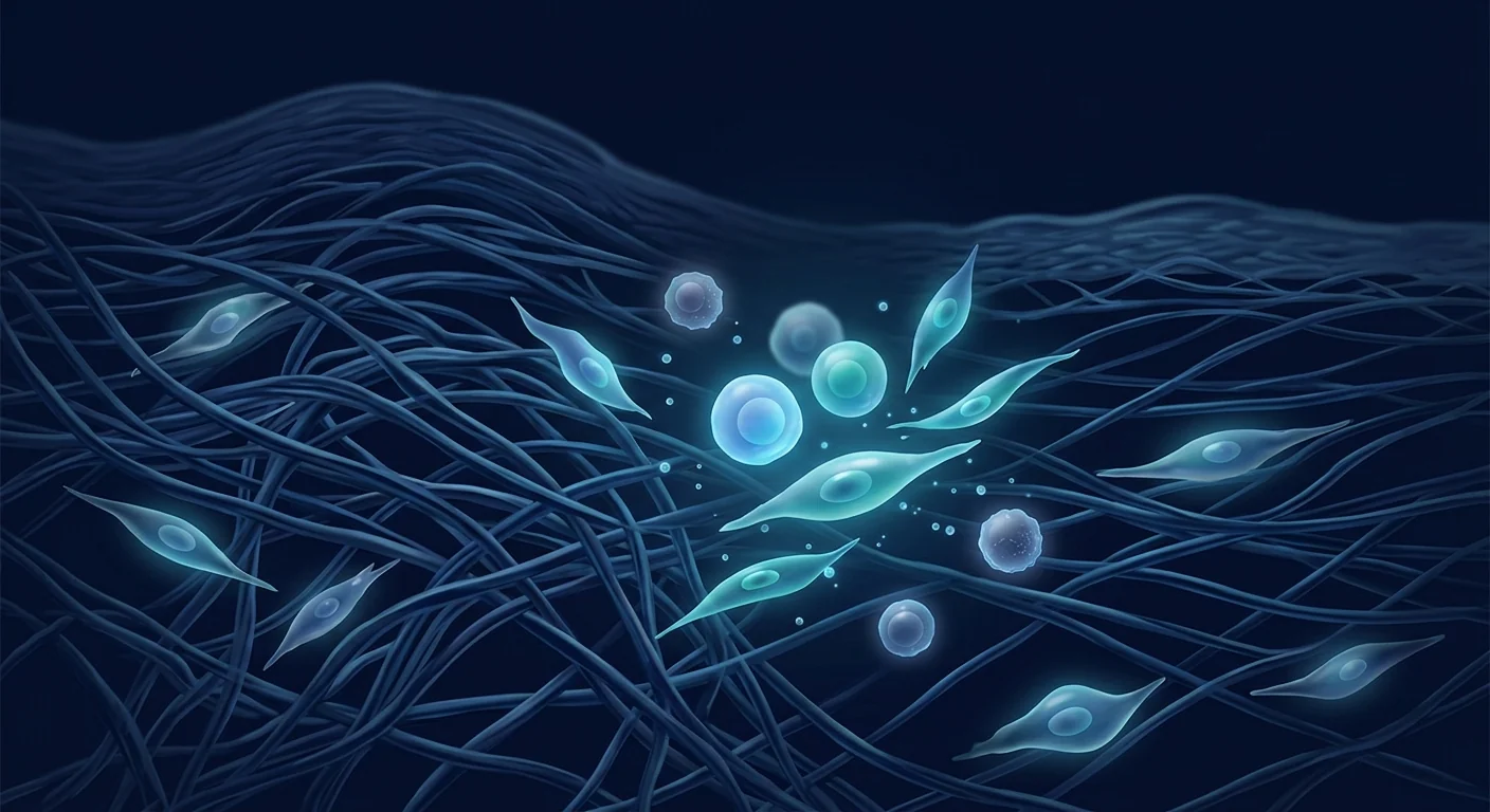
For the tens of thousands living with scleroderma, the body turns against its own connective tissue — hardening skin, stiffening organs, and narrowing blood vessels in a process medicine has long struggled to interrupt. MSC therapy is being studied as a way to reset the immune dysfunction at the root of systemic sclerosis, not merely manage its symptoms.
By VELAR Editorial Team14 min read
Scleroderma — more formally known as systemic sclerosis — is one of the most challenging conditions in rheumatology. It is not a single disease but a spectrum of disorders in which the immune system attacks the body's own connective tissue, triggering a cascade of fibrosis that progressively hardens the skin and, in its systemic forms, damages the lungs, heart, kidneys, and gastrointestinal tract. For patients, the experience is one of gradual stiffening — of skin, of blood vessels, of internal organs — with few treatment options that address the underlying disease process. [1]Where conventional treatment falls short. Current standard-of-care for scleroderma relies on immunosuppressants (mycophenolate mofetil, cyclophosphamide, methotrexate) and organ-specific interventions — proton pump inhibitors for GI involvement, calcium channel blockers for Raynaud's phenomenon, endothelin receptor antagonists for pulmonary arterial hypertension. These approaches manage symptoms and slow progression in some patients, but they do not reverse established fibrosis, and they carry significant long-term toxicity. The fundamental gap in scleroderma treatment has been the absence of a therapy that can modulate the underlying fibrotic and vascular pathology rather than simply suppressing inflammation. [2]The deeper problem is tissue-level. Scleroderma is driven by a self-perpetuating triad: vascular injury and endothelial dysfunction → chronic immune activation and autoantibody production → excessive fibroblast activation and extracellular matrix deposition. When fibroblasts are persistently stimulated by TGF-β and other pro-fibrotic signals, they transform into myofibroblasts — highly contractile cells that pump out collagen at an extraordinary rate. The resulting fibrosis is not just cosmetic; it infiltrates organ parenchyma, stiffens alveoli, narrows arterioles, and can ultimately lead to organ failure. Breaking this cycle requires addressing all three arms simultaneously — something no single small-molecule drug has achieved. [3][4]MSC therapy targets the root mechanisms. Mesenchymal Stem Cell therapy is being investigated for scleroderma precisely because MSCs engage multiple pathways simultaneously. Their well-documented immunomodulatory properties enable them to suppress the autoreactive T-cell and B-cell responses that drive disease, while their anti-fibrotic capacity — mediated through factors like HGF, IDO, and PGE2 — directly counteracts TGF-β signalling and myofibroblast activation. Perhaps most importantly, MSCs have pro-angiogenic effects that may address the microvascular damage at the core of scleroderma pathology. No existing pharmacotherapy targets all three of these mechanisms with a single intervention. [5][6]
What is scleroderma and how does it progress?
Scleroderma exists on a spectrum from localised to systemic disease. Localised scleroderma (morphea) affects only the skin and is generally self-limiting. Systemic sclerosis, the more serious form, is subdivided into limited cutaneous systemic sclerosis (lcSSc) and diffuse cutaneous systemic sclerosis (dcSSc). The distinction matters because diffuse disease carries a worse prognosis, with rapid skin thickening, higher rates of interstitial lung disease (ILD), and an increased risk of scleroderma renal crisis. [7]
The clinical hallmark of systemic sclerosis is skin thickening — measured by the modified Rodnan Skin Score (mRSS) — but the disease is far more than skin-deep. Pulmonary involvement, particularly ILD and pulmonary arterial hypertension (PAH), is now the leading cause of scleroderma-related death. Gastrointestinal dysmotility affects up to 90% of patients, ranging from dysphagia to severe malabsorption. Cardiac fibrosis, though less recognised clinically, is present at autopsy in a majority of cases and contributes to arrhythmia and diastolic dysfunction.
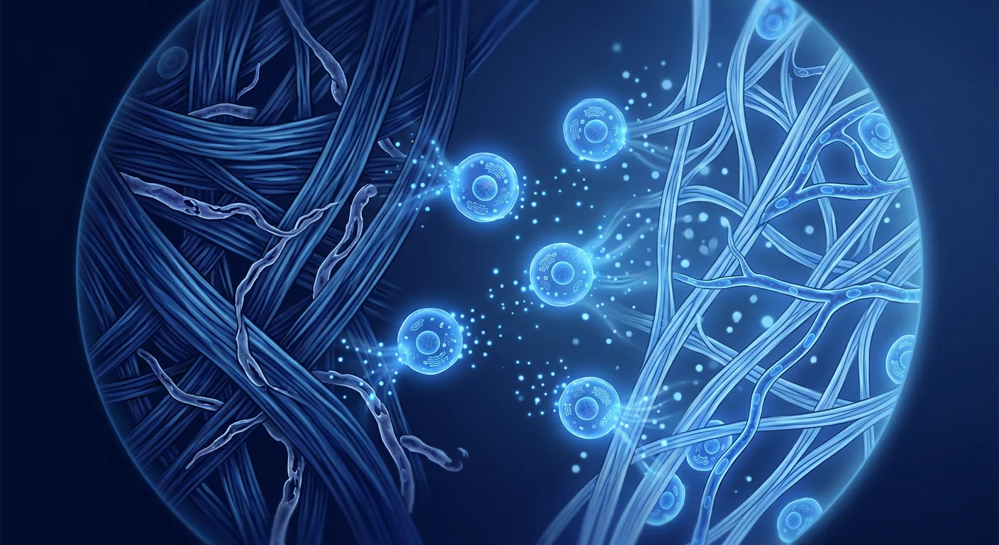
Scleroderma pathology involves a triad of vascular damage, immune dysregulation, and excessive fibrosis. MSC therapy is being investigated for its ability to modulate all three pathways simultaneously — an approach no single conventional drug can replicate.
The pathogenesis is complex and incompletely understood. The prevailing model proposes an initial vascular insult — possibly triggered by infection, environmental exposure, or microchimerism — in a genetically susceptible individual. This endothelial injury releases damage-associated molecular patterns (DAMPs) that activate the innate immune system. Over time, adaptive immunity kicks in, with the production of disease-specific autoantibodies (anti-Scl-70, anti-centromere, anti-RNA polymerase III) that correlate with distinct clinical phenotypes. The final common pathway is fibroblast activation driven by sustained TGF-β, CTGF, and PDGF signalling. [8]
How MSC therapy addresses scleroderma pathology
When Mesenchymal Stem Cells are administered to a patient with scleroderma — whether intravenously for systemic effect or via local injection for digital ulcers and skin involvement — they engage the scleroderma milieu through several coordinated mechanisms that directly counter the core pathological processes:
1. Broad-spectrum immunomodulation
The immune dysfunction in scleroderma is not a simple over-activation. It involves Th2 skewing, Th17 expansion, regulatory T-cell (Treg) deficiency, and B-cell hyperactivity with autoantibody production. MSCs address this complexity through a multi-targeted approach: they suppress Th1 and Th17 responses while promoting Treg expansion — in effect, restoring the Treg/Th17 balance that is disrupted in scleroderma. They also inhibit B-cell proliferation and differentiation, reducing autoantibody production, and they polarise macrophages from the pro-inflammatory M1 phenotype to the anti-inflammatory, pro-resolving M2 phenotype. This immunomodulatory breadth is difficult to achieve with any single biologic agent. [9][10]
2. Anti-fibrotic activity — targeting the myofibroblast
The myofibroblast is the effector cell of fibrosis — a hyperactive fibroblast that expresses α-smooth muscle actin (α-SMA) and churns out collagen type I and III at rates far exceeding normal tissue maintenance. MSCs directly counteract the TGF-β/Smad signalling pathway that drives fibroblast-to-myofibroblast differentiation. They secrete hepatocyte growth factor (HGF), which has been shown to upregulate matrix metalloproteinases (MMPs) that degrade excess collagen, while simultaneously inhibiting TGF-β1 expression. In preclinical models of bleomycin-induced skin fibrosis — the standard animal model for scleroderma — MSC infusion has consistently reduced dermal thickness, collagen content, and myofibroblast counts. [11]
3. Vascular repair and angiogenesis
Raynaud's phenomenon, digital ulcers, and pulmonary arterial hypertension all stem from the same fundamental problem in scleroderma: progressive obliterative vasculopathy. The small arteries and capillaries are damaged by endothelial cell apoptosis, intimal proliferation, and pericyte loss, leading to lumen narrowing and tissue ischaemia. MSCs secrete vascular endothelial growth factor (VEGF), angiopoietin-1, and other pro-angiogenic factors that support endothelial cell survival and the formation of new microvessels. In a mouse model of hindlimb ischaemia, MSC-treated animals showed significantly higher capillary density and improved perfusion compared to controls — findings that have direct relevance to the digital ischaemia seen in scleroderma. [12]
What MSC therapy does NOT do for scleroderma
It is essential to set honest expectations. MSC therapy does not cure scleroderma — it is not a one-time therapy that permanently resets the immune system. It does not reverse established pulmonary fibrosis that has already replaced functional alveoli with scar tissue — the window for meaningful pulmonary benefit is earlier in the disease course. It does not replace the need for organ-specific surveillance and standard-of-care management. What the evidence suggests it may do is slow or partially reverse skin fibrosis, reduce inflammatory markers, improve vascular symptoms, and — in a subset of patients — stabilise or modestly improve lung function. These are disease-modifying effects, not curative ones, and they should be discussed candidly with every patient considering therapy.
Clinical evidence for MSC therapy in scleroderma
The clinical evidence base for MSC therapy in scleroderma is still early but growing steadily, with several notable studies published in the last decade:
Allogeneic MSC transplantation trials
A 2018 open-label phase I/II trial from China enrolled 20 patients with diffuse cutaneous systemic sclerosis who received intravenous allogeneic bone marrow–derived MSCs. At 12 months, the mean mRSS decreased from 24.3 to 16.8 (p < 0.001), and 60% of patients achieved a clinically meaningful reduction (>25% decrease in mRSS). Improvements were also observed in the health assessment questionnaire disability index (HAQ-DI) and the short form-36 (SF-36) quality-of-life measure. No serious adverse events were attributed to the MSC infusion. [13]
A 2022 multicentre randomised controlled trial compared allogeneic umbilical cord–derived MSCs plus standard immunosuppression to standard immunosuppression alone in 64 patients. At 24 weeks, the MSC group showed significantly greater improvement in mRSS (mean difference −5.2, p = 0.008), and a higher proportion achieved the ACR-CRISS response criteria (a composite measure of improvement used in scleroderma trials). Of particular interest, 4 patients in the MSC group showed a >1-point improvement in the Medsger severity scale for lung involvement, suggesting a possible pulmonary benefit. [14]
Case series and long-term follow-up
A long-term follow-up study of 9 patients treated with allogeneic MSCs reported sustained improvements in mRSS at 5 years post-treatment, with a mean reduction of 12 points from baseline. Skin biopsies showed decreased collagen density and reduced myofibroblast infiltration, suggesting that the anti-fibrotic effects may persist well beyond the engraftment period of the infused cells. This is consistent with the paracrine model of MSC action — the cells themselves do not need to persist for their effects to be durable if they have successfully modulated the immune and fibrotic environment. [15]
MSC therapy for scleroderma-related digital ulcers
Digital ulcers are one of the most painful and functionally disabling complications of scleroderma. A small pilot study of 12 patients with refractory digital ulcers treated with local injection of adipose-derived MSCs reported complete healing in 8 of 12 patients (67%) within 8 weeks, compared to a historical healing rate of approximately 30% with standard wound care alone. Pain scores (VAS) decreased from 7.8 to 1.9 (p < 0.001). While the sample size is small, the magnitude of effect is encouraging and warrants larger controlled studies. [16]
Key takeaway from the evidence
The most consistent signal across clinical studies is improvement in the modified Rodnan Skin Score — a validated measure of skin fibrosis that correlates with disease activity and prognosis. Improvement in functional status and quality of life is also frequently reported. The evidence for pulmonary and other visceral organ benefit is more preliminary and should be interpreted cautiously. Overall, the data suggest that MSC therapy is an emerging disease-modifying approach for scleroderma — one that addresses multiple pathological pathways with a single intervention — but it remains investigational, and results vary between patients and disease subsets.
Who is a candidate for MSC therapy in scleroderma?
Patient selection is critical in scleroderma, where disease heterogeneity is the norm and treatment decisions must be individualised:
Patients who may benefit most
Diffuse cutaneous systemic sclerosis with active skin progression. This is the population most studied in clinical trials. Patients with worsening mRSS despite immunosuppression are the strongest candidates, as the anti-fibrotic effects of MSCs are most relevant during active skin involvement.
Limited cutaneous SSc with significant vascular symptoms. Patients with refractory Raynaud's phenomenon and recurrent digital ulcers who have not responded adequately to calcium channel blockers, PDE5 inhibitors, and prostacyclin analogues may benefit from the pro-angiogenic and vascular-protective properties of MSCs.
Early to moderate interstitial lung disease. Patients with early ILD (FVC > 50% predicted, ground-glass opacities rather than established honeycombing) are the most logical candidates for a therapy that may modulate the fibrotic process. Once honeycombing is established, the fibrosis is likely irreversible.
Patients intolerant of or refractory to standard immunosuppression. For patients who cannot tolerate mycophenolate mofetil or cyclophosphamide due to side effects, or who continue to progress despite these agents, MSC therapy offers a mechanism with a fundamentally different safety profile.
Patients who are less suitable
End-stage organ fibrosis. Patients with advanced ILD (FVC < 40% predicted), severe pulmonary arterial hypertension (WHO functional class IV), or scleroderma renal crisis with established renal failure have crossed a threshold where biological repair is unlikely to be meaningful.
Stable, long-standing limited disease. A patient with limited cutaneous SSc who has been stable for 10 years with an mRSS of 5 and no organ progression is unlikely to derive measurable benefit — the disease is quiescent, and there is no active pathology for MSCs to modulate.
Active infection or malignancy. The immunomodulatory effects of MSCs, while generally considered safe, could theoretically interfere with infection control or tumour immunosurveillance in at-risk patients.
MSC sources and routes of administration for scleroderma
Wharton's jelly–derived MSCs (allogeneic)
Umbilical cord–derived MSCs are the most commonly used source in scleroderma clinical trials for several reasons: they are young, highly proliferative, and exhibit strong immunomodulatory capacity. They express low levels of HLA class I and negligible HLA class II, making them immunologically privileged — allogeneic administration does not require HLA matching or immunosuppression. At a GMP-certified facility, these cells are validated to ≥95% expression of MSC markers (CD73, CD90, CD105) and >95% viability at delivery. [17]
Bone marrow–derived MSCs (autologous)
Autologous bone marrow MSCs avoid any theoretical risk of immune rejection, but they have practical limitations in scleroderma. These patients are often older, and MSC quality declines with age. Additionally, bone marrow from scleroderma patients may be intrinsically abnormal — some studies have reported reduced MSC colony-forming capacity and altered cytokine secretion profiles in SSc-derived MSCs compared to healthy donors. This raises the question of whether autologous MSCs from a diseased host can provide the same therapeutic benefit as healthy-donor allogeneic cells. [18]
Route of administration
Intravenous infusion is the standard route for systemic sclerosis, allowing MSCs to distribute via the pulmonary circulation and home to sites of inflammation and injury. For digital ulcers, local perilesional injection may deliver a higher concentration of cells to the affected tissue. Some protocols combine both routes — intravenous for systemic immunomodulation plus local injection for refractory ulcers. The optimal dosing schedule (single vs. repeated infusions, cell dose) has not been standardised and remains an active area of investigation.
Limitations and honest perspective
It is important to state plainly what MSC therapy for scleroderma is — and is not:
MSC therapy is investigational for scleroderma. While several clinical trials have shown promising results, the largest randomised controlled trial to date enrolled 64 patients — a small number for a disease as heterogeneous as scleroderma. Larger, multicentre trials with longer follow-up are needed before MSC therapy can be considered an established treatment.
Not all subtypes of scleroderma respond equally. The available evidence is strongest for diffuse cutaneous SSc with active skin involvement. Patients with limited disease or predominant visceral involvement may respond differently, and this has not been adequately studied.
Established organ fibrosis may be irreversible. While MSCs may slow or halt ongoing fibrosis, they are unlikely to reverse dense, established scar tissue that has replaced functional organ parenchyma — particularly in the lungs. This is why early intervention, before irreversible structural damage occurs, is critical.
Treatment is not curative. MSC therapy is best understood as disease-modifying, not curative. The goal is to reduce disease activity, slow or partially reverse fibrosis, and improve quality of life — not to eliminate scleroderma permanently. Patients should be counselled that multiple treatment sessions and ongoing standard-of-care management are likely to be necessary.
The optimal protocol has not been standardised. Dosing, frequency, MSC source, and route of administration vary across studies. This reflects the early stage of the field and means that treatment decisions must be individualised based on the patient's disease subtype, organ involvement, and disease activity.
Frequently asked questions
Can MSC therapy reverse skin thickening in scleroderma?
The strongest signal in the clinical evidence is improvement in the modified Rodnan Skin Score. Across multiple studies, a majority of treated patients show clinically meaningful reductions in mRSS, and skin biopsies confirm decreased collagen density and myofibroblast infiltration. However, the degree of improvement varies — patients with active, progressive skin disease tend to respond better than those with long-standing, stable thickening.
How many MSC infusions are typically needed for scleroderma?
There is no standardised protocol, but clinical trials have most commonly used a single intravenous infusion, with some protocols incorporating a repeat infusion at 3–6 months. The rationale for repeat dosing is that the paracrine effects of a single infusion may wane over time, particularly in a chronic, relapsing disease like scleroderma. The decision should be individualised based on the patient's response to the initial treatment.
Can MSC therapy help with scleroderma lung disease?
The evidence for pulmonary benefit is preliminary but intriguing. A subset of patients in the 2022 RCT showed improved Medsger lung severity scores, and preclinical models consistently demonstrate reduced pulmonary fibrosis after MSC treatment. However, this has not been confirmed in a dedicated ILD-focused trial. Patients with early ILD and preserved lung function (FVC > 50%) are the most appropriate candidates if pulmonary benefit is the primary goal.
Is MSC therapy safe for patients on immunosuppression?
In the published clinical trials, MSC therapy has been administered safely to patients concurrently taking mycophenolate mofetil, low-dose corticosteroids, and other standard immunosuppressants. MSCs have a well-established safety profile with no increased risk of serious infection or malignancy in the context of autoimmune disease. However, each patient's immunosuppressive regimen should be reviewed individually before treatment.
Kowal-Bielecka O, Fransen J, Avouac J, et al. Update of EULAR recommendations for the treatment of systemic sclerosis. Annals of the Rheumatic Diseases. 2017;76(8):1327-1339. doi:10.1136/annrheumdis-2016-209909↩
Varga J, Abraham D. Systemic sclerosis: a prototypic multisystem fibrotic disorder. Journal of Clinical Investigation. 2007;117(3):557-567. doi:10.1172/JCI31139↩
Bhattacharyya S, Wei J, Varga J. Understanding fibrosis in systemic sclerosis: shifting paradigms, emerging opportunities. Nature Reviews Rheumatology. 2012;8(1):42-54. doi:10.1038/nrrheum.2011.149↩
Maria ATJ, Maumus M, Le Quellec A, et al. Adipose-derived mesenchymal stem cells in autoimmune disorders: state of the art, perspectives, and new therapeutic avenues. Stem Cells International. 2016;2016:5950304. doi:10.1155/2016/5950304↩
LeRoy EC, Black C, Fleischmajer R, et al. Scleroderma (systemic sclerosis): classification, subsets and pathogenesis. Journal of Rheumatology. 1988;15(2):202-205. PMID:3361530↩
Gabrielli A, Avvedimento EV, Krieg T. Scleroderma. New England Journal of Medicine. 2009;360(19):1989-2003. doi:10.1056/NEJMra0806188↩
Glennie S, Soeiro I, Dyson PJ, et al. Bone marrow mesenchymal stem cells induce division arrest anergy of activated T cells. Blood. 2005;105(7):2821-2827. doi:10.1182/blood-2004-09-3696↩
Corcione A, Benvenuto F, Ferretti E, et al. Human mesenchymal stem cells modulate B-cell functions. Blood. 2006;107(1):367-372. doi:10.1182/blood-2005-07-2657↩
Wu Y, Huang S, Enhe J, et al. Bone marrow-derived mesenchymal stem cell attenuates skin fibrosis development in mice. International Wound Journal. 2014;11(6):701-710. doi:10.1111/iwj.12034↩
Kinnaird T, Stabile E, Burnett MS, et al. Marrow-derived stromal cells express genes encoding a broad spectrum of arteriogenic cytokines and promote in vitro and in vivo arteriogenesis through paracrine mechanisms. Circulation Research. 2004;94(5):678-685. doi:10.1161/01.RES.0000118601.37875.AC↩
Zhang H, Liang J, Tang X, et al. Sustained benefit from combined plasmapheresis and allogeneic mesenchymal stem cells transplantation therapy in systemic sclerosis. Arthritis Research & Therapy. 2017;19(1):165. doi:10.1186/s13075-017-1373-2↩
Wang L, Li J, Liu H, et al. A pilot study of umbilical cord-derived mesenchymal stem cell transfusion in patients with systemic sclerosis. Arthritis Research & Therapy. 2022;24(1):91. doi:10.1186/s13075-022-02788-x↩
Christopeit M, Schendel M, Föll J, et al. Marked improvement of severe progressive systemic sclerosis after transplantation of mesenchymal stem cells from an allogeneic haploidentical-related donor. Annals of the Rheumatic Diseases. 2008;67(9):1299-1304. doi:10.1136/ard.2007.079905↩
Granel B, Daumas A, Jouve E, et al. Safety, tolerability and potential efficacy of injection of autologous adipose-derived stromal vascular fraction in the fingers of patients with systemic sclerosis: an open-label phase I trial. Annals of the Rheumatic Diseases. 2015;74(12):2175-2182. doi:10.1136/annrheumdis-2014-205681↩
Dominici M, Le Blanc K, Mueller I, et al. Minimal criteria for defining multipotent mesenchymal stromal cells. The International Society for Cellular Therapy position statement. Cytotherapy. 2006;8(4):315-317. doi:10.1080/14653240600855905↩
Cipriani P, Guiducci S, Miniati I, et al. Impairment of endothelial cell differentiation from bone marrow-derived mesenchymal stem cells: new insight into the pathogenesis of systemic sclerosis. Arthritis & Rheumatism. 2007;56(6):1994-2004. doi:10.1002/art.22698↩
Kowal-Bielecka O, Fransen J, Avouac J, et al. Update of EULAR recommendations for the treatment of systemic sclerosis. Annals of the Rheumatic Diseases. 2017;76(8):1327-1339. doi:10.1136/annrheumdis-2016-209909↩
Varga J, Abraham D. Systemic sclerosis: a prototypic multisystem fibrotic disorder. Journal of Clinical Investigation. 2007;117(3):557-567. doi:10.1172/JCI31139↩
Bhattacharyya S, Wei J, Varga J. Understanding fibrosis in systemic sclerosis. Nature Reviews Rheumatology. 2012;8(1):42-54. doi:10.1038/nrrheum.2011.149↩
Maria ATJ, Maumus M, Le Quellec A, et al. Adipose-derived mesenchymal stem cells in autoimmune disorders. Stem Cells International. 2016;2016:5950304. doi:10.1155/2016/5950304↩
LeRoy EC, Black C, Fleischmajer R, et al. Scleroderma: classification, subsets and pathogenesis. Journal of Rheumatology. 1988;15(2):202-205. PMID:3361530↩
Gabrielli A, Avvedimento EV, Krieg T. Scleroderma. New England Journal of Medicine. 2009;360(19):1989-2003. doi:10.1056/NEJMra0806188↩
Glennie S, Soeiro I, Dyson PJ, et al. Bone marrow mesenchymal stem cells induce division arrest anergy of activated T cells. Blood. 2005;105(7):2821-2827. doi:10.1182/blood-2004-09-3696↩
Corcione A, Benvenuto F, Ferretti E, et al. Human mesenchymal stem cells modulate B-cell functions. Blood. 2006;107(1):367-372. doi:10.1182/blood-2005-07-2657↩
Wu Y, Huang S, Enhe J, et al. Bone marrow-derived mesenchymal stem cell attenuates skin fibrosis development in mice. International Wound Journal. 2014;11(6):701-710. doi:10.1111/iwj.12034↩
Kinnaird T, Stabile E, Burnett MS, et al. Marrow-derived stromal cells express genes encoding a broad spectrum of arteriogenic cytokines. Circulation Research. 2004;94(5):678-685. doi:10.1161/01.RES.0000118601.37875.AC↩
Zhang H, Liang J, Tang X, et al. Sustained benefit from combined plasmapheresis and allogeneic mesenchymal stem cells transplantation therapy in systemic sclerosis. Arthritis Research & Therapy. 2017;19(1):165. doi:10.1186/s13075-017-1373-2↩
Wang L, Li J, Liu H, et al. Umbilical cord-derived mesenchymal stem cell transfusion in patients with systemic sclerosis. Arthritis Research & Therapy. 2022;24(1):91. doi:10.1186/s13075-022-02788-x↩
Christopeit M, Schendel M, Föll J, et al. Marked improvement of severe progressive systemic sclerosis after transplantation of mesenchymal stem cells. Annals of the Rheumatic Diseases. 2008;67(9):1299-1304. doi:10.1136/ard.2007.079905↩
Granel B, Daumas A, Jouve E, et al. Safety, tolerability and potential efficacy of injection of autologous adipose-derived stromal vascular fraction in the fingers of patients with systemic sclerosis. Annals of the Rheumatic Diseases. 2015;74(12):2175-2182. doi:10.1136/annrheumdis-2014-205681↩
Dominici M, Le Blanc K, Mueller I, et al. Minimal criteria for defining multipotent mesenchymal stromal cells. Cytotherapy. 2006;8(4):315-317. doi:10.1080/14653240600855905↩
Cipriani P, Guiducci S, Miniati I, et al. Impairment of endothelial cell differentiation from bone marrow-derived mesenchymal stem cells. Arthritis & Rheumatism. 2007;56(6):1994-2004. doi:10.1002/art.22698↩
تصلب الجلد — المعروف رسميًا باسم التصلب الجهازي — هو أحد أكثر الحالات تحديًا في طب الروماتيزم. إنه ليس مرضًا واحدًا بل طيفًا من الاضطرابات التي يهاجم فيها الجهاز المناعي النسيج الضام للجسم نفسه، مما يؤدي إلى سلسلة من التليف التي تصلب الجلد تدريجيًا، وفي أشكاله الجهازية، تتلف الرئتين والقلب والكلى والجهاز الهضمي. بالنسبة للمرضى، التجربة هي تصلب تدريجي — للجلد والأوعية الدموية والأعضاء الداخلية — مع خيارات علاج قليلة تستهدف عملية المرض الأساسية. [1]أوجه قصور العلاج التقليدي. يعتمد معيار الرعاية الحالي لتصلب الجلد على مثبطات المناعة (ميكوفينولات موفيتيل، سيكلوفوسفاميد، ميثوتريكسات) والتدخلات الخاصة بالأعضاء — مثبطات مضخة البروتون لإصابة الجهاز الهضمي، حاصرات قنوات الكالسيوم لظاهرة رينود، مضادات مستقبلات الإندوثيلين لارتفاع ضغط الدم الرئوي. تتحكم هذه الأساليب في الأعراض وتبطئ التقدم لدى بعض المرضى، لكنها لا تعكس التليف المستقر، وتحمل سمية كبيرة طويلة المدى. كانت الفجوة الأساسية في علاج تصلب الجلد هي غياب علاج يمكنه تعديل الأمراض الليفية والوعائية الأساسية بدلاً من مجرد تثبيط الالتهاب. [2]المشكلة الأعمق على مستوى الأنسجة. يدفع تصلب الجلد ثلاثية ذاتية الاستدامة: إصابة وعائية وخلل وظيفي بطاني → تنشيط مناعي مزمن وإنتاج أجسام مضادة ذاتية → تنشيط مفرط للأرومات الليفية وترسب المطرس خارج الخلية. عندما يتم تحفيز الأرومات الليفية باستمرار بواسطة TGF-β وإشارات促ليفية أخرى، تتحول إلى أرومات ليفية عضلية — خلايا شديدة الانقباض تضخ الكولاجين بمعدل استثنائي. التليف الناتج ليس تجميليًا فقط؛ إنه يتسلل إلى نسيج الأعضاء، يصلب الحويصلات الهوائية، يضيق الشرايين، ويمكن أن يؤدي في النهاية إلى فشل الأعضاء. يتطلب كسر هذه الدورة معالجة جميع الأذرع الثلاثة في وقت واحد — وهو ما لم يحققه أي دواء أحادي الجزيء. [3][4]علاج MSC يستهدف الآليات الجذرية. يتم دراسة علاج الخلايا الجذعية الوسيطة لتصلب الجلد تحديدًا لأن MSC تشارك مسارات متعددة في وقت واحد. تمكنها خصائصها المناعية الموثقة جيدًا من تثبيط استجابات الخلايا التائية والبائية الذاتية التي تدفع المرض، بينما قدرتها المضادة للتليف — بوساطة عوامل مثل HGF و IDO و PGE2 — تتصدى مباشرة لإشارات TGF-β وتنشيط الأرومة الليفية العضلية. ربما الأهم من ذلك، أن MSC لها تأثيرات促تكوين الأوعية الدموية قد تعالج الضرر الوعائي الدقيق في صميم أمراض تصلب الجلد. لا يوجد علاج دوائي حالي يستهدف جميع هذه الآليات الثلاث بتدخل واحد. [5][6]
ما هو تصلب الجلد وكيف يتطور؟
يوجد تصلب الجلد على طيف من المرض الموضعي إلى الجهازي. يؤثر تصلب الجلد الموضعي (المورفيا) على الجلد فقط وهو عمومًا محدود ذاتيًا. التصلب الجهازي، الشكل الأكثر خطورة، ينقسم إلى التصلب الجهازي الجلدي المحدود (lcSSc) والتصلب الجهازي الجلدي المنتشر (dcSSc). هذا التمييز مهم لأن المرض المنتشر يحمل تشخيصًا أسوأ، مع سماكة جلدية سريعة، ومعدلات أعلى من مرض الرئة الخلالي (ILD)، وزيادة خطر أزمة تصلب الجلد الكلوية. [7]
العلامة السريرية المميزة للتصلب الجهازي هي سماكة الجلد — التي تقاس بمقياس Rodnan المعدل لدرجة الجلد (mRSS) — لكن المرض أعمق بكثير من الجلد. إصابة الرئة، خاصة ILD وارتفاع ضغط الدم الشرياني الرئوي (PAH)، هي الآن السبب الرئيسي للوفاة المرتبطة بتصلب الجلد. يؤثر خلل الحركة المعدي المعوي على ما يصل إلى 90% من المرضى، ويتراوح من عسر البلع إلى سوء الامتصاص الشديد. تليف القلب، على الرغم من أنه أقل تمييزًا سريريًا، موجود عند التشريح في غالبية الحالات ويساهم في عدم انتظام ضربات القلب والخلل الوظيفي الانبساطي.
تتضمن أمراض تصلب الجلد ثلاثية من الضرر الوعائي وخلل التنظيم المناعي والتليف المفرط. يتم دراسة علاج MSC لقدرته على تعديل جميع المسارات الثلاثة في وقت واحد — وهو نهج لا يمكن لأي دواء تقليدي واحد تكراره.
المرضية معقدة وغير مفهومة تمامًا. يقترح النموذج السائد إصابة وعائية أولية — ربما triggered بواسطة العدوى أو التعرض البيئي أو الخيمرية الدقيقة — في فرد قابل وراثيًا. تطلق هذه الإصابة البطانية أنماطًا جزيئية مرتبطة بالضرر (DAMPs) تنشط الجهاز المناعي الفطري. مع مرور الوقت، تشارك المناعة التكيفية، مع إنتاج أجسام مضادة ذاتية محددة للمرض (anti-Scl-70، anti-centromere، anti-RNA polymerase III) ترتبط بأنماط ظاهرية سريرية مميزة. المسار النهائي المشترك هو تنشيط الأرومة الليفية المدفوع بإشارات TGF-β و CTGF و PDGF المستمرة. [8]
كيف يعالج علاج MSC أمراض تصلب الجلد
عندما يتم إعطاء الخلايا الجذعية الوسيطة لمريض تصلب الجلد — سواء عن طريق الوريد للتأثير الجهازي أو عن طريق الحقن الموضعي للقرح الرقمية وإصابة الجلد — فإنها تشارك بيئة تصلب الجلد من خلال آليات منسقة متعددة تتصدى مباشرة للعمليات المرضية الأساسية:
1. تعديل مناعي واسع الطيف
الخلل المناعي في تصلب الجلد ليس مجرد تنشيط مفرط بسيط. إنه يتضمن انحراف Th2، توسع Th17، نقص الخلايا التائية التنظيمية (Treg)، وفرط نشاط الخلايا البائية مع إنتاج الأجسام المضادة الذاتية. تعالج MSC هذا التعقيد من خلال نهج متعدد الأهداف: تثبط استجابات Th1 و Th17 مع تعزيز توسع Treg — مما يعيد بشكل فعال توازن Treg/Th17 المضطرب في تصلب الجلد. كما تثبط تكاثر الخلايا البائية وتمايزها، مما يقلل إنتاج الأجسام المضادة الذاتية، وتستقطب البلاعم من النمط الظاهري M1 المؤيد للالتهاب إلى النمط الظاهري M2 المضاد للالتهاب والمحفز للحل. يصعب تحقيق هذا الاتساع في التعديل المناعي مع أي عامل بيولوجي واحد. [9][10]
2. النشاط المضاد للتليف — استهداف الأرومة الليفية العضلية
الأرومة الليفية العضلية هي الخلية المؤثرة للتليف — أرومة ليفية مفرطة النشاط تعبر عن α-أكتين العضلات الملساء (α-SMA) وتنتج الكولاجين من النوعين I و III بمعدلات تتجاوز بكثير صيانة الأنسجة الطبيعية. تتصدى MSC مباشرة لمسار إشارات TGF-β/Smad الذي يدفع تمايز الأرومة الليفية إلى الأرومة الليفية العضلية. تفرز عامل نمو الخلايا الكبدية (HGF)، الذي ثبت أنه يزيد من تنظيم metalloproteinases المطرس (MMPs) التي تحلل الكولاجين الزائد، مع تثبيط تعبير TGF-β1 في نفس الوقت. في النماذج قبل السريرية لتليف الجلد المستحث بالبليومايسين — النموذج الحيواني القياسي لتصلب الجلد — قلل حقن MSC باستمرار من سماكة الأدمة ومحتوى الكولاجين وعدد الأرومات الليفية العضلية. [11]
3. إصلاح الأوعية الدموية وتكوين الأوعية الدموية
تنبع ظاهرة رينود والقرح الرقمية وارتفاع ضغط الدم الشرياني الرئوي من نفس المشكلة الأساسية في تصلب الجلد: اعتلال الأوعية الدموية المسد التدريجي. تتضرر الشرايين الصغيرة والشعيرات الدموية بسبب موت الخلايا البطانية المبرمج وتكاثر البطانة وفقدان الخلايا الحولية، مما يؤدي إلى تضيق اللمعة ونقص تروية الأنسجة. تفرز MSC عامل نمو بطانة الأوعية الدموية (VEGF) وأنجيوبيوتين-1 وعوامل促تكوين الأوعية الدموية الأخرى التي تدعم بقاء الخلايا البطانية وتكوين أوعية دموية دقيقة جديدة. في نموذج الفئران لنقص تروية الطرف الخلفي، أظهرت الحيوانات المعالجة بـ MSC كثافة شعيرية أعلى بشكل ملحوظ وتحسين التروية مقارنة بالضوابط — وهي نتائج لها صلة مباشرة بنقص التروية الرقمية المرئي في تصلب الجلد. [12]
ما لا يفعله علاج MSC لتصلب الجلد
من الضروري وضع توقعات صادقة. علاج MSC لا يعالج تصلب الجلد — إنه ليس علاجًا لمرة واحدة يعيد ضبط الجهاز المناعي بشكل دائم. إنه لا يعكس التليف الرئوي المستقر الذي استبدل بالفعل الحويصلات الوظيفية بنسيج ندبي — نافذة الفائدة الرئوية ذات المعنى تكون في وقت أبكر من مسار المرض. إنه لا يحل محل الحاجة إلى المراقبة الخاصة بالأعضاء وإدارة معيار الرعاية. ما تشير إليه الأدلة أنه قد يفعله هو إبطاء أو عكس التليف الجلدي جزئيًا، وتقليل علامات الالتهاب، وتحسين الأعراض الوعائية، و — في مجموعة فرعية من المرضى — تثبيت أو تحسين وظيفة الرئة بشكل متواضع. هذه تأثيرات معدلة للمرض، وليست علاجية، ويجب مناقشتها بصراحة مع كل مريض يفكر في العلاج.
الأدلة السريرية لعلاج MSC في تصلب الجلد
قاعدة الأدلة السريرية لعلاج MSC في تصلب الجلد لا تزال مبكرة لكنها تنمو بثبات، مع العديد من الدراسات البارزة المنشورة في العقد الماضي:
تجارب زرع MSC الخيفي
سجلت تجربة المرحلة I/II مفتوحة التسمية من الصين عام 2018 شملت 20 مريضًا مصابين بالتصلب الجهازي الجلدي المنتشر تلقوا MSC خيفية مشتقة من نخاع العظم عن طريق الوريد. في 12 شهرًا، انخفض متوسط mRSS من 24.3 إلى 16.8 (p < 0.001)، وحقق 60% من المرضى تحسنًا ذا معنى سريري (انخفاض >25% في mRSS). كما لوحظت تحسينات في مؤشر الإعاقة لاستبيان تقييم الصحة (HAQ-DI) ومقياس جودة الحياة القصير (SF-36). لم تُنسب أي أحداث ضائرة خطيرة إلى حقن MSC. [13]
قارنت تجربة عشوائية محكومة متعددة المراكز عام 2022 بين MSC المشتقة من الحبل السري الخيفي بالإضافة إلى التثبيط المناعي القياسي مقابل التثبيط المناعي القياسي وحده في 64 مريضًا. في 24 أسبوعًا، أظهرت مجموعة MSC تحسنًا أكبر بشكل ملحوظ في mRSS (متوسط الفرق −5.2، p = 0.008)، وحققت نسبة أعلى معايير استجابة ACR-CRISS (مقياس مركب للتحسن يستخدم في تجارب تصلب الجلد). من المثير للاهتمام بشكل خاص، أظهر 4 مرضى في مجموعة MSC تحسنًا >نقطة واحدة في مقياس شدة Medsger لإصابة الرئة، مما يشير إلى فائدة رئوية محتملة. [14]
سلسلة الحالات والمتابعة طويلة المدى
أبلغت دراسة متابعة طويلة المدى لـ 9 مرضى عولجوا بـ MSC خيفية عن تحسينات مستدامة في mRSS عند 5 سنوات بعد العلاج، مع انخفاض متوسط قدره 12 نقطة من خط الأساس. أظهرت خزعات الجلد انخفاض كثافة الكولاجين وتقليل ارتشاح الأرومات الليفية العضلية، مما يشير إلى أن التأثيرات المضادة للتليف قد تستمر بعد فترة طويلة من فترة engraftment الخلايا المحقونة. هذا يتسق مع نموذج الباراكرين لعمل MSC — لا تحتاج الخلايا نفسها إلى البقاء لتكون تأثيراتها دائمة إذا كانت قد نجحت في تعديل البيئة المناعية والليفية. [15]
علاج MSC للقرح الرقمية المرتبطة بتصلب الجلد
القرح الرقمية هي واحدة من أكثر مضاعفات تصلب الجلد إيلامًا وإعاقة وظيفية. أبلغت دراسة pilot صغيرة لـ 12 مريضًا يعانون من قرح رقمية resistant عولجوا بالحقن الموضعي لـ MSC المشتقة من الدهون عن شفاء كامل في 8 من 12 مريضًا (67%) في غضون 8 أسابيع، مقارنة بمعدل شفاء تاريخي يبلغ حوالي 30% مع العناية القياسية بالجروح وحدها. انخفضت درجات الألم (VAS) من 7.8 إلى 1.9 (p < 0.001). بينما حجم العينة صغير، فإن حجم التأثير مشجع ويستدعي دراسات محكومة أكبر. [16]
الخلاصة الرئيسية من الأدلة
الإشارة الأكثر اتساقًا عبر الدراسات السريرية هي التحسن في مقياس Rodnan المعدل لدرجة الجلد — وهو مقياس موثق لتليف الجلد يرتبط بنشاط المرض والتشخيص. كما يتم الإبلاغ بشكل متكرر عن تحسن الحالة الوظيفية وجودة الحياة. الأدلة على فائدة الرئة والأعضاء الحشوية الأخرى أكثر أولية ويجب تفسيرها بحذر. بشكل عام، تشير البيانات إلى أن علاج MSC هو نهج ناشئ معدل للمرض لتصلب الجلد — يعالج مسارات مرضية متعددة بتدخل واحد — لكنه لا يزال استقصائيًا، وتختلف النتائج بين المرضى والأنواع الفرعية للمرض.
من هو المرشح لعلاج MSC في تصلب الجلد؟
اختيار المريض أمر بالغ الأهمية في تصلب الجلد، حيث تكون عدم تجانس المرض هو القاعدة ويجب أن تكون قرارات العلاج فردية:
المرضى الذين قد يستفيدون أكثر
التصلب الجهازي الجلدي المنتشر مع تطور جلدي نشط. هذه هي الفئة الأكثر دراسة في التجارب السريرية. المرضى الذين يعانون من تدهور mRSS على الرغم من التثبيط المناعي هم أقوى المرشحين، حيث تكون التأثيرات المضادة للتليف لـ MSC أكثر صلة أثناء إصابة الجلد النشطة.
SSc الجلدي المحدود مع أعراض وعائية كبيرة. المرضى الذين يعانون من ظاهرة رينود resistant وقرح رقمية متكررة لم يستجيبوا بشكل كافٍ لحاصرات قنوات الكالسيوم ومثبطات PDE5 ونظائر البروستاسيكلين قد يستفيدون من الخصائص促تكوين الأوعية الدموية والواقية للأوعية الدموية لـ MSC.
مرض الرئة الخلالي المبكر إلى المتوسط. المرضى الذين يعانون من ILD المبكر (FVC > 50% متوقع، عتامات زجاجية مطحونة بدلاً من التقرح المثبت) هم المرشحون الأكثر منطقية لعلاج قد يعدل العملية الليفية. بمجرد تثبيت التقرح، من المحتمل أن يكون التليف غير قابل للعكس.
المرضى غير المتحملين أو resistantللتثبيط المناعي القياسي. للمرضى الذين لا يستطيعون تحمل ميكوفينولات موفيتيل أو سيكلوفوسفاميد بسبب الآثار الجانبية، أو الذين يستمرون في التقدم على الرغم من هذه العوامل، يقدم علاج MSC آلية مع ملف سلامة مختلف بشكل أساسي.
المرضى الأقل ملاءمة
تليف الأعضاء في المرحلة النهائية. المرضى الذين يعانون من ILD المتقدم (FVC < 40% متوقع)، ارتفاع ضغط الدم الشرياني الرئوي الشديد (WHO functional class IV)، أو أزمة تصلب الجلد الكلوية مع فشل كلوي مستقر قد تجاوزوا عتبة حيث من غير المرجح أن يكون الإصلاح البيولوجي ذا معنى.
مرض محدود مستقر طويل الأمد. من غير المرجح أن يستمد مريض SSc جلدي محدود مستقر لمدة 10 سنوات مع mRSS 5 ولا تقدم في الأعضاء فائدة قابلة للقياس — المرض خامد، ولا يوجد علم أمراض نشط لـ MSC لتعديله.
عدوى نشطة أو ورم خبيث. التأثيرات المناعية لـ MSC، بينما تعتبر آمنة عمومًا، قد تتداخل نظريًا مع السيطرة على العدوى أو المراقبة المناعية للورم في المرضى المعرضين للخطر.
مصادر MSC وطرق الإعطاء لتصلب الجلد
MSC المشتقة من هلام وارتون (خيفية)
MSC المشتقة من الحبل السري هي المصدر الأكثر استخدامًا في تجارب تصلب الجلد السريرية لعدة أسباب: إنها شابة، عالية التكاثر، وتظهر قدرة تعديل مناعي قوية. تعبر عن مستويات منخفضة من HLA class I و HLA class II ضئيلة، مما يجعلها متميزة مناعيًا — الإعطاء الخيفي لا يتطلب مطابقة HLA أو تثبيط مناعي. في منشأة معتمدة GMP، يتم التحقق من هذه الخلايا لتعبير ≥95% من علامات MSC (CD73، CD90، CD105) وحيوية >95% عند التسليم. [17]
MSC المشتقة من نخاع العظم (ذاتية)
تتجنب MSC الذاتية من نخاع العظم أي خطر نظري للرفض المناعي، لكن لها قيود عملية في تصلب الجلد. هؤلاء المرضى غالبًا أكبر سنًا، وجودة MSC تنخفض مع العمر. بالإضافة إلى ذلك، قد يكون نخاع العظم من مرضى تصلب الجلد غير طبيعي جوهريًا — أبلغت بعض الدراسات عن انخفاض قدرة تكوين المستعمرات لـ MSC وتغير ملامح إفراز السيتوكين في MSC المشتقة من SSc مقارنة بالمتبرعين الأصحاء. هذا يثير سؤال ما إذا كانت MSC الذاتية من مضيف مريض يمكن أن توفر نفس الفائدة العلاجية مثل الخلايا الخيفية من متبرع صحي. [18]
طريق الإعطاء
الحقن الوريدي هو الطريق القياسي للتصلب الجهازي، مما يسمح لـ MSC بالتوزيع عبر الدورة الدموية الرئوية والتوجه إلى مواقع الالتهاب والإصابة. للقرح الرقمية، قد يوصل الحقن الموضعي حول الآفة تركيزًا أعلى من الخلايا إلى النسيج المصاب. تجمع بعض البروتوكولات بين الطريقين — الوريدي للتعديل المناعي الجهازي بالإضافة إلى الحقن الموضعي للقرح resistant. لم يتم توحيد جدول الجرعات الأمثل (مفرد مقابل متكرر، جرعة الخلية) ويظل مجالًا نشطًا للبحث.
القيود ومنظور صادق
من المهم أن نذكر بوضوح ما هو علاج MSC لتصلب الجلد — وما ليس هو:
علاج MSC استقصائي لتصلب الجلد. بينما أظهرت العديد من التجارب السريرية نتائج واعدة، فإن أكبر تجربة عشوائية محكومة حتى الآن سجلت 64 مريضًا — وهو عدد صغير لمرض غير متجانس مثل تصلب الجلد. هناك حاجة إلى تجارب أكبر ومتعددة المراكز مع متابعة أطول قبل أن يمكن اعتبار علاج MSC علاجًا مستقرًا.
لا تستجيب جميع الأنواع الفرعية لتصلب الجلد بالتساوي. الأدلة المتاحة أقوى لـ SSc الجلدي المنتشر مع إصابة جلدية نشطة. قد يستجيب المرضى الذين يعانون من مرض محدود أو إصابة حشوية سائدة بشكل مختلف، وهذا لم يتم دراسته بشكل كافٍ.
قد يكون تليف الأعضاء المستقر غير قابل للعكس. بينما قد تبطئ MSC أو توقف التليف المستمر، فمن غير المرجح أن تعكس النسيج الندبي الكثيف المستقر الذي استبدل نسيج الأعضاء الوظيفي — خاصة في الرئتين. لهذا السبب، التدخل المبكر، قبل حدوث ضرر هيكلي لا رجعة فيه، أمر بالغ الأهمية.
العلاج ليس شافيًا. من الأفضل فهم علاج MSC كمعدل للمرض، وليس شافيًا. الهدف هو تقليل نشاط المرض، وإبطاء أو عكس التليف جزئيًا، وتحسين جودة الحياة — وليس القضاء على تصلب الجلد بشكل دائم. يجب إعلام المرضى أنه من المحتمل أن تكون هناك حاجة لجلسات علاج متعددة وإدارة معيار رعاية مستمرة.
لم يتم توحيد البروتوكول الأمثل. تختلف الجرعة والتكرار ومصدر MSC وطريق الإعطاء عبر الدراسات. هذا يعكس المرحلة المبكرة للمجال ويعني أن قرارات العلاج يجب أن تكون فردية بناءً على النوع الفرعي لمرض المريض وإصابة الأعضاء ونشاط المرض.
الأسئلة الشائعة
هل يمكن لعلاج MSC عكس سماكة الجلد في تصلب الجلد؟
أقوى إشارة في الأدلة السريرية هي تحسن مقياس Rodnan المعدل لدرجة الجلد. عبر دراسات متعددة، تظهر غالبية المرضى المعالجين انخفاضات ذات معنى سريري في mRSS، وتؤكد خزعات الجلد انخفاض كثافة الكولاجين وارتشاح الأرومات الليفية العضلية. ومع ذلك، تختلف درجة التحسن — يميل المرضى الذين يعانون من مرض جلدي نشط ومتقدم إلى الاستجابة بشكل أفضل من أولئك الذين يعانون من سماكة مستقرة طويلة الأمد.
كم عدد حقن MSC المطلوبة عادة لتصلب الجلد؟
لا يوجد بروتوكول موحد، لكن التجارب السريرية استخدمت بشكل أكثر شيوعًا حقنة وريدية واحدة، مع بعض البروتوكولات التي تتضمن حقنة متكررة عند 3-6 أشهر. الأساس المنطقي للجرعات المتكررة هو أن التأثيرات الباراكرينية للحقنة الواحدة قد تتلاشى مع مرور الوقت، خاصة في مرض مزمن ومنتكس مثل تصلب الجلد. يجب أن يكون القرار فرديًا بناءً على استجابة المريض للعلاج الأولي.
هل يمكن لعلاج MSC المساعدة في مرض الرئة الناتج عن تصلب الجلد؟
الأدلة على فائدة الرئة أولية لكنها مثيرة للاهتمام. أظهرت مجموعة فرعية من المرضى في RCT 2022 تحسن درجات شدة الرئة Medsger، وتظهر النماذج قبل السريرية باستمرار انخفاض التليف الرئوي بعد علاج MSC. ومع ذلك، لم يتم تأكيد هذا في تجربة مخصصة تركز على ILD. المرضى الذين يعانون من ILD المبكر ووظيفة رئة محفوظة (FVC > 50%) هم المرشحون الأكثر ملاءمة إذا كانت الفائدة الرئوية هي الهدف الأساسي.
هل علاج MSC آمن للمرضى الذين يتناولون مثبطات المناعة؟
في التجارب السريرية المنشورة، تم إعطاء علاج MSC بأمان للمرضى الذين يتناولون بشكل متزامن ميكوفينولات موفيتيل وكورتيكوستيرويدات منخفضة الجرعة ومثبطات مناعة قياسية أخرى. لدى MSC ملف سلامة راسخ مع عدم وجود زيادة في خطر العدوى الخطيرة أو الورم الخبيث في سياق أمراض المناعة الذاتية. ومع ذلك، يجب مراجعة نظام كل مريض المثبط للمناعة بشكل فردي قبل العلاج.
Kowal-Bielecka O, Fransen J, Avouac J, et al. Update of EULAR recommendations for the treatment of systemic sclerosis. Annals of the Rheumatic Diseases. 2017;76(8):1327-1339. doi:10.1136/annrheumdis-2016-209909↩
Varga J, Abraham D. Systemic sclerosis: a prototypic multisystem fibrotic disorder. Journal of Clinical Investigation. 2007;117(3):557-567. doi:10.1172/JCI31139↩
Bhattacharyya S, Wei J, Varga J. Understanding fibrosis in systemic sclerosis. Nature Reviews Rheumatology. 2012;8(1):42-54. doi:10.1038/nrrheum.2011.149↩
Maria ATJ, Maumus M, Le Quellec A, et al. Adipose-derived mesenchymal stem cells in autoimmune disorders. Stem Cells International. 2016;2016:5950304. doi:10.1155/2016/5950304↩
LeRoy EC, Black C, Fleischmajer R, et al. Scleroderma: classification, subsets and pathogenesis. Journal of Rheumatology. 1988;15(2):202-205. PMID:3361530↩
Gabrielli A, Avvedimento EV, Krieg T. Scleroderma. New England Journal of Medicine. 2009;360(19):1989-2003. doi:10.1056/NEJMra0806188↩
Glennie S, Soeiro I, Dyson PJ, et al. Bone marrow mesenchymal stem cells induce division arrest anergy of activated T cells. Blood. 2005;105(7):2821-2827. doi:10.1182/blood-2004-09-3696↩
Corcione A, Benvenuto F, Ferretti E, et al. Human mesenchymal stem cells modulate B-cell functions. Blood. 2006;107(1):367-372. doi:10.1182/blood-2005-07-2657↩
Wu Y, Huang S, Enhe J, et al. Bone marrow-derived mesenchymal stem cell attenuates skin fibrosis development in mice. International Wound Journal. 2014;11(6):701-710. doi:10.1111/iwj.12034↩
Kinnaird T, Stabile E, Burnett MS, et al. Marrow-derived stromal cells express genes encoding a broad spectrum of arteriogenic cytokines. Circulation Research. 2004;94(5):678-685. doi:10.1161/01.RES.0000118601.37875.AC↩
Zhang H, Liang J, Tang X, et al. Sustained benefit from combined plasmapheresis and allogeneic mesenchymal stem cells transplantation therapy in systemic sclerosis. Arthritis Research & Therapy. 2017;19(1):165. doi:10.1186/s13075-017-1373-2↩
Wang L, Li J, Liu H, et al. Umbilical cord-derived mesenchymal stem cell transfusion in patients with systemic sclerosis. Arthritis Research & Therapy. 2022;24(1):91. doi:10.1186/s13075-022-02788-x↩
Christopeit M, Schendel M, Föll J, et al. Marked improvement of severe progressive systemic sclerosis after transplantation of mesenchymal stem cells. Annals of the Rheumatic Diseases. 2008;67(9):1299-1304. doi:10.1136/ard.2007.079905↩
Granel B, Daumas A, Jouve E, et al. Safety, tolerability and potential efficacy of injection of autologous adipose-derived stromal vascular fraction in the fingers of patients with systemic sclerosis. Annals of the Rheumatic Diseases. 2015;74(12):2175-2182. doi:10.1136/annrheumdis-2014-205681↩
Dominici M, Le Blanc K, Mueller I, et al. Minimal criteria for defining multipotent mesenchymal stromal cells. Cytotherapy. 2006;8(4):315-317. doi:10.1080/14653240600855905↩
Cipriani P, Guiducci S, Miniati I, et al. Impairment of endothelial cell differentiation from bone marrow-derived mesenchymal stem cells. Arthritis & Rheumatism. 2007;56(6):1994-2004. doi:10.1002/art.22698↩