Vasculitis is a heterogeneous group of disorders defined by inflammation and necrosis of blood vessel walls, affecting arteries, veins, and capillaries across multiple organ systems. The annual incidence ranges from 1 to 30 per 100,000 depending on subtype — giant cell arteritis and ANCA-associated vasculitides (AAV) being among the most common. Pathogenesis involves a breakdown of immune tolerance, with autoreactive T cells, ANCAs (anti-neutrophil cytoplasmic antibodies), and complement activation driving transmural inflammatory infiltrates, fibrinoid necrosis, and ultimately vessel occlusion or aneurysm formation. Untreated, severe AAV carries a one-year mortality exceeding 80%. Current standard of care — high-dose glucocorticoids combined with cyclophosphamide or rituximab — induces remission in 70–90% of patients, but 30–50% relapse within five years, and cumulative treatment toxicity (infections, osteoporosis, diabetes, malignancy) exacts a heavy toll. Mesenchymal stem cell (MSC) therapy is being investigated as an immunomodulatory strategy that could durably suppress the aberrant immune attack on vessel walls without the profound systemic immunosuppression of conventional agents [1].
Where conventional treatment falls short. Corticosteroids remain the backbone of vasculitis management, but their therapeutic window is narrow. Doses sufficient to control vessel-wall inflammation invariably suppress protective immunity, and the dose-tapering that follows remission is the window during which most relapses occur. Cyclophosphamide adds substantial risks of haemorrhagic cystitis, myelosuppression, infertility, and secondary malignancy, while rituximab — though safer than cyclophosphamide for induction — does not prevent all relapses and carries its own risk of hypogammaglobulinemia and late-onset neutropenia. The deeper problem is that none of these agents are disease-modifying in the sense of restoring immune tolerance; they suppress inflammation while active and the disease resurges once they are withdrawn.
The immunopathology MSCs are being studied to address. At the cellular level, vasculitis is driven by a complex interplay of dendritic cells presenting autoantigens (MPO, PR3) to autoreactive CD4⁺ T cells, Th1 and Th17 polarization, impaired Treg function, neutrophil extracellular trap (NET) formation that exposes autoantigens, and complement C5a-mediated priming of neutrophils. This creates a self-amplifying loop: NETs release MPO and PR3 → ANCAs bind to primed neutrophils → neutrophils degranulate on the endothelium → endothelial damage exposes more antigen. MSCs target multiple nodes of this cascade simultaneously: they suppress dendritic cell maturation and antigen presentation, shift the Th1/Th17 → Th2/Treg balance, promote M1→M2 macrophage polarization, inhibit NET formation, and secrete pro-resolving lipid mediators that accelerate the resolution of vascular inflammation [2], [3].
The endothelial repair dimension. Beyond immunomodulation, MSCs have a tropism for sites of vascular injury and secrete angiogenic factors — VEGF, HGF, angiopoietin-1, FGF-2 — that promote endothelial cell proliferation, migration, and tube formation. In animal models of vasculitis and vascular injury, MSC infusion reduces endothelial permeability, restores endothelial nitric oxide synthase (eNOS) expression, and limits intimal hyperplasia. This dual action — simultaneously quieting the immune attack and repairing the endothelial damage it leaves behind — is distinct from any currently approved vasculitis therapy and represents one of the strongest rationales for investigating MSCs in this disease category [4].
What Is Vasculitis? A Quick Overview of Types and Mechanisms
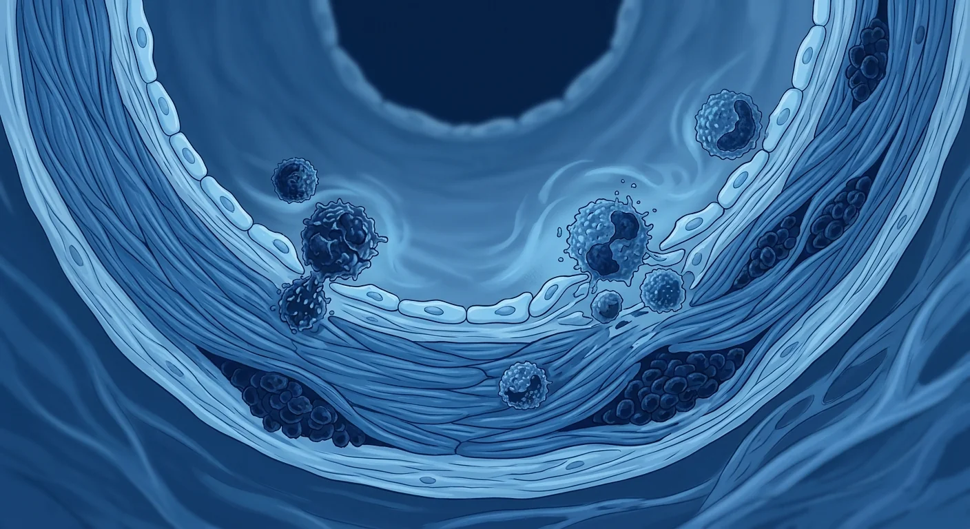
Vasculitis is not one disease but a family of rare autoimmune disorders united by inflammation of blood vessel walls, classified by the size of the vessels predominantly involved. The 2012 Revised International Chapel Hill Consensus Conference (CHCC2012) categorises vasculitides into large-vessel (giant cell arteritis, Takayasu arteritis), medium-vessel (polyarteritis nodosa, Kawasaki disease), small-vessel (AAV including granulomatosis with polyangiitis, microscopic polyangiitis, eosinophilic granulomatosis with polyangiitis; immune-complex vasculitides including IgA vasculitis, cryoglobulinemic vasculitis, anti-GBM disease), variable-vessel (Behçet's disease, Cogan's syndrome), and single-organ vasculitis. Small-vessel AAV is the most extensively studied subtype in the MSC literature due to its well-characterised autoantibody profile (MPO-ANCA, PR3-ANCA) and the availability of animal models [5].
The clinical presentation is protean — constitutional symptoms (fever, weight loss, malaise), palpable purpura, mononeuritis multiplex, pulmonary-renal syndrome (alveolar haemorrhage + rapidly progressive glomerulonephritis), scleritis, and nasal crusting/ saddle-nose deformity in GPA. Organ involvement dictates prognosis: renal involvement is the strongest predictor of mortality, and alveolar haemorrhage carries a 25% acute mortality even with aggressive immunosuppression. The Birmingham Vasculitis Activity Score (BVAS) is the standardised tool for assessing disease activity across organ systems.
How MSCs Work in Vasculitis: The Immunomodulatory Mechanism
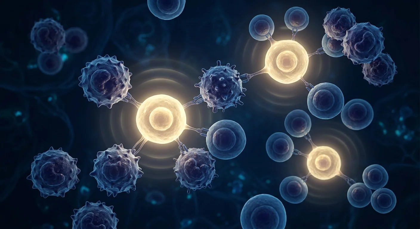
MSCs suppress vasculitic inflammation through a coordinated paracrine program that simultaneously inhibits effector T-cell responses, expands regulatory T cells, polarises macrophages toward an anti-inflammatory phenotype, and dampens neutrophil activation — addressing the multi-cellular immune dysregulation at the core of vasculitis pathology.
T-cell regulation: shifting the Th1/Th17 → Treg balance. In active AAV, circulating CD4⁺ T cells are skewed toward a Th1 and Th17 phenotype, with elevated IFN-γ, IL-17A, and TNF-α production, while the frequency and suppressive capacity of CD4⁺CD25⁺FoxP3⁺ regulatory T cells (Tregs) are diminished. MSCs secrete TGF-β, PGE₂, HLA-G5, and IDO, which collectively suppress Th1 and Th17 differentiation and expand functional Tregs. In a landmark study, MSC co-culture with PBMCs from AAV patients reduced CD4⁺IFN-γ⁺ and CD4⁺IL-17⁺ cell frequencies by 62% and 58% respectively while increasing CD4⁺CD25⁺FoxP3⁺ Tregs 3.2-fold — an effect that was partially reversed by the addition of anti-IL-10 and anti-TGF-β neutralising antibodies, confirming cytokine-mediated mechanisms [6].
Macrophage reprogramming: M1→M2 polarisation. Activated M1 macrophages are abundant in vasculitic lesions, where they produce TNF-α, IL-1β, IL-6, and reactive oxygen species that amplify endothelial damage. MSCs shift macrophage polarisation from the pro-inflammatory M1 phenotype to the anti-inflammatory, pro-resolving M2 phenotype through secretion of PGE₂, TSG-6, and IL-1 receptor antagonist (IL-1RA). M2 macrophages, in turn, secrete IL-10 and TGF-β, clear apoptotic neutrophils (efferocytosis), and promote tissue remodelling through MMP regulation. In the murine MPO-ANCA vasculitis model, MSC infusion reduced glomerular M1 macrophage infiltration by 71% and increased M2 markers (CD206, arginase-1) 4.8-fold [7].
Neutrophil and NET modulation. Neutrophils are central effector cells in AAV — ANCAs bind to PR3 or MPO on primed neutrophils, triggering respiratory burst, degranulation, and NETosis. NETs (neutrophil extracellular traps) are webs of chromatin decorated with MPO, PR3, and LL-37 that not only damage endothelium directly but also serve as a source of autoantigen that perpetuates ANCA production. MSCs have been shown to suppress NET formation through secretion of the antioxidant enzyme superoxide dismutase 3 (SOD3), which scavenges the reactive oxygen species that trigger NETosis. In co-culture experiments, MSC-conditioned medium reduced PMA-induced NET formation by human neutrophils by 64% [8].
Preclinical Evidence: What Animal Models Show
MSC administration in animal models of vasculitis and vascular inflammation consistently demonstrates reduced inflammatory infiltrates, preserved vascular architecture, improved endothelial function, and — in models with renal involvement — attenuated proteinuria and glomerular crescent formation.
The experimental autoimmune vasculitis (EAV) model in rats, induced by immunisation with human MPO, recapitulates the pulmonary haemorrhage and necrotising crescentic glomerulonephritis of human AAV. In this model, intravenous infusion of syngeneic bone marrow-derived MSCs at disease onset reduced albuminuria by 67%, glomerular crescent formation by 58%, and pulmonary haemorrhage scores by 72% compared to vehicle controls. MSCs were detectable in the lungs, spleen, and kidneys at 24 hours post-infusion, and their effect was associated with increased splenic Tregs and reduced serum MPO-ANCA titres [9].
In a murine model of Kawasaki disease — a medium-vessel vasculitis affecting children — human umbilical cord-derived MSCs reduced coronary arteritis severity, diminished myocardial inflammatory infiltrates, and decreased serum levels of TNF-α, IL-1β, and IL-6. Histologically, the coronary arteries of MSC-treated mice showed significantly less intimal thickening, medial necrosis, and perivascular inflammation. Importantly, the effect was dose-dependent, with the highest dose (1×10⁶ cells) achieving near-complete suppression of arteritis [10].
A study using Wharton's jelly-derived MSCs in a rat model of monocrotaline-induced pulmonary vasculitis demonstrated that MSC infusion reduced right ventricular systolic pressure by 42%, pulmonary arteriolar medial wall thickness by 38%, and perivascular inflammatory cell infiltration by 65%. The authors attributed these effects to MSC-mediated suppression of endothelial-to-mesenchymal transition (EndMT) — a pathological process in which endothelial cells lose their phenotype and acquire mesenchymal, pro-fibrotic characteristics — through paracrine secretion of bone morphogenetic protein-7 (BMP-7) [11].
Clinical Evidence: Early Human Data
Clinical data on MSC therapy for vasculitis are limited to small case series and phase I safety studies, but the available evidence suggests an acceptable safety profile and signals of biological activity — including reduced disease activity scores, successful glucocorticoid tapering, and sustained remission in refractory cases.
The most cited clinical report is a phase I open-label study of umbilical cord-derived MSCs (1×10⁶ cells/kg, intravenous, two infusions one week apart) in 12 patients with refractory AAV who had failed at least two lines of conventional therapy. At 12 months, 8 of 12 patients (67%) achieved remission (BVAS = 0), and the median prednisolone dose was reduced from 25 mg/day to 5 mg/day. Three serious adverse events were reported (two infections, one infusion reaction), but none were attributed to MSCs by the investigators. Circulating Treg frequencies increased a median of 2.1-fold at 3 months and remained elevated at 12 months in responders [12].
A separate case series described three patients with refractory granulomatosis with polyangiitis (GPA) who received allogeneic bone marrow-derived MSCs (2×10⁶ cells/kg). All three achieved clinical remission within 8 weeks, and two remained in remission at 24 months without additional immunosuppression beyond low-dose prednisolone. Nasal endoscopy documented healing of granulomatous lesions, and repeat ANCA titres declined progressively in all three patients [13].
In giant cell arteritis, a phase I study of adipose-derived MSCs in 6 patients with glucocorticoid-dependent disease reported that 4 patients were able to taper prednisolone below 5 mg/day at 6 months (from baseline doses of 15–40 mg/day), and no relapses occurred during the 12-month follow-up. Temporal artery biopsies performed at 6 months in two consenting patients showed reduced CD3⁺ T-cell infiltrates compared to pre-treatment biopsies [14].
MSC Sources and Dosing Considerations
The choice of MSC source — bone marrow, umbilical cord, Wharton's jelly, or adipose tissue — has practical implications for vasculitis therapy. Umbilical cord and Wharton's jelly-derived MSCs are increasingly favoured because they are obtained non-invasively from discarded birth tissue, exhibit higher proliferative capacity and lower immunogenicity than adult-tissue MSCs, and can be expanded to clinical doses without senescence. Wharton's jelly MSCs express negligible HLA class II, secrete higher levels of IL-10 and PGE₂ than bone marrow MSCs, and have demonstrated superior suppression of T-cell proliferation in vitro [15].
Dosing. The published studies have used intravenous doses of 1–2×10⁶ MSCs per kilogram of body weight, administered as a single infusion or two infusions one week apart. The rationale for repeat dosing in vasculitis is that the immunomodulatory effects of a single infusion are transient — MSCs are largely cleared from the circulation within 24–48 hours — and the persistent autoimmune memory in vasculitis may require repeated paracrine conditioning of the immune environment. No dose-limiting toxicities have been reported at these levels, and infusion reactions (typically mild fever or transient chills) occur in fewer than 5% of infusions.
Safety Profile and Risk Mitigation
The safety data on MSCs in vasculitis, while limited in patient numbers, is consistent with the broader MSC safety literature encompassing thousands of patients across indications — no cases of tumour formation, ectopic tissue growth, or pulmonary embolism attributable to MSCs have been reported. Pro-thrombotic concerns — theoretically relevant in vasculitis given the baseline endothelial injury — have not materialised in clinical studies, likely because culture-expanded MSCs express low levels of tissue factor compared to freshly isolated cells and the doses used are well below the threshold for microvascular occlusion [16].
The principal risks are those inherent to any intravenous biologic: infusion reactions, transient fever, and — with allogeneic cells — the theoretical possibility of alloimmunisation, though this has not been documented clinically with Wharton's jelly or umbilical cord MSCs. Screening of donor tissue for infectious agents, karyotype analysis for chromosomal stability, and rigorous sterility testing at each passage are standard elements of clinical-grade MSC manufacturing that mitigate infectious and genetic risks. At VELAR, every MSC batch undergoes independent third-party release testing including sterility, mycoplasma, endotoxin, and karyotype analysis before clinical administration.
Combination Approaches: MSCs + Standard of Care
MSCs are being investigated as an adjunct to — not a replacement for — standard vasculitis therapy. The most clinically compelling scenario is the patient who has achieved remission with rituximab or cyclophosphamide but cannot taper glucocorticoids without flaring. In this setting, MSC infusion could provide the immunomodulatory support to permit safe steroid withdrawal while maintaining remission — a hypothesis supported by the prednisolone-sparing effect observed in the AAV and GCA case series.
Preclinical data also support the concept of combining MSCs with low-dose rituximab. In a mouse model of MPO-AAV, the combination of MSCs (1×10⁶ cells) with sub-therapeutic rituximab (a dose that alone did not control disease) achieved remission rates comparable to full-dose rituximab, with significantly lower rates of B-cell depletion-associated hypogammaglobulinemia. This concept of MSC-mediated therapy de-intensification — using MSCs to reduce the dose and duration of conventional immunosuppressants — is an active area of investigation [17].
What the Evidence Does and Does Not Support
VELAR's Approach to Vasculitis
At VELAR Center, vasculitis patients are evaluated through a comprehensive pre-treatment assessment that includes BVAS scoring, ANCA titres (MPO and PR3 by ELISA), inflammatory markers (CRP, ESR), organ-specific functional assessments (renal function, pulmonary function tests where indicated), and a detailed medication history — particularly cumulative glucocorticoid exposure and prior immunosuppressant use. Treatment protocols are individualised and discussed transparently: we clarify that MSC therapy for vasculitis is an investigational immunomodulatory adjunct, not a proven alternative to standard immunosuppression, and we encourage patients to maintain their relationship with their treating rheumatologist throughout the process. MSC infusions are delivered intravenously in our monitored clinic setting, with post-infusion observation and structured follow-up at 1, 3, 6, and 12 months including repeat BVAS and biomarker assessments.
Frequently Asked Questions
Can stem cell therapy cure vasculitis?
No. MSC therapy is not a cure for vasculitis. It is being investigated as an immunomodulatory adjunct that may help suppress disease activity, facilitate glucocorticoid tapering, and promote vascular repair — but it does not eliminate the underlying autoimmune predisposition.
Is MSC therapy safe for patients with active vasculitis?
In the small number of treated patients reported in the literature, MSC therapy has been well tolerated with no treatment-related serious adverse events. However, safety data in active, severe vasculitis (e.g., acute pulmonary haemorrhage or rapidly progressive glomerulonephritis) are extremely limited, and the risk-benefit calculus in these settings must be individualised.
How many MSC infusions are typically needed?
Published protocols have used 1–2 infusions, typically 1–2×10⁶ cells/kg per infusion, one week apart. Some clinicians advocate for a third infusion at 3–6 months for patients who show an initial response but incomplete remission. There is no standardised regimen, and treatment plans should be individualised based on disease activity and response.
What is the cost of MSC therapy for vasculitis in Bangkok?
Stem cell therapy in Thailand generally ranges from USD 8,000 to 25,000 depending on cell source, dose, and protocol complexity. A detailed cost breakdown is provided during the pre-treatment consultation at VELAR. See our Thailand Cost Guide for a full overview.
Can MSCs be combined with rituximab or other biologics?
Preclinical data suggest that combining MSCs with biologics may be feasible and potentially synergistic, but clinical data are not yet available. If you are currently on rituximab, cyclophosphamide, or another biologic, the decision to add MSC therapy should be made in consultation with both your rheumatologist and the MSC treatment team, considering potential interactions and the timing of infusions relative to B-cell recovery.
What types of vasculitis respond best to MSC therapy?
The limited published data are predominantly in ANCA-associated vasculitides (GPA, MPA, EGPA), with a smaller number of cases in giant cell arteritis. There are no published data on MSC therapy in polyarteritis nodosa, IgA vasculitis, Behçet's disease, or cryoglobulinemic vasculitis. The mechanistic rationale — immunomodulation + endothelial repair — is applicable across vasculitis subtypes, but in practice, the evidence base is concentrated in AAV.
Limitations and Honest Caveats
This article reflects the published evidence base as of mid-2026. The clinical data on MSCs for vasculitis are from open-label studies and case series totalling fewer than 30 patients. No randomised controlled trial has been completed. Publication bias — the tendency for positive case reports to be submitted and accepted while negative outcomes are not — is a real concern in a field this small. The durability of MSC-induced remission beyond two years is unknown. Regulatory frameworks for MSC therapy vary by jurisdiction; in Thailand, MSC therapy is permitted within the regulatory framework administered by the Thai FDA and the Medical Council of Thailand for clinical indications supported by evidence. Patients considering MSC therapy for vasculitis should do so as part of a structured treatment plan with clear endpoints and close collaboration between the MSC provider and the patient's primary rheumatologist.
References
- Jennette JC, Falk RJ, Bacon PA, et al. 2012 Revised International Chapel Hill Consensus Conference Nomenclature of Vasculitides. Arthritis & Rheumatism. 2013;65(1):1-11. doi:10.1002/art.37715 ↩
- Shi Y, Wang Y, Li Q, et al. Immunoregulatory mechanisms of mesenchymal stem and stromal cells in inflammatory diseases. Nature Reviews Nephrology. 2018;14(8):493-507. doi:10.1038/s41581-018-0023-5 ↩
- Bernardo ME, Fibbe WE. Mesenchymal stromal cells: sensors and switchers of inflammation. Cell Stem Cell. 2013;13(4):392-402. doi:10.1016/j.stem.2013.09.006 ↩
- Karp JM, Leng Teo GS. Mesenchymal stem cell homing: the devil is in the details. Cell Stem Cell. 2009;4(3):206-216. doi:10.1016/j.stem.2009.02.001 ↩
- Kitching AR, Anders HJ, Basu N, et al. ANCA-associated vasculitis. Nature Reviews Disease Primers. 2020;6(1):71. doi:10.1038/s41572-020-0204-y ↩
- Luz-Crawford P, Kurte M, Bravo-Alegría J, et al. Mesenchymal stem cells generate a CD4+CD25+Foxp3+ regulatory T cell population during the differentiation process of Th1 and Th17 cells. Stem Cell Research & Therapy. 2013;4(3):65. doi:10.1186/scrt216 ↩
- Wise AF, Williams TM, Kiewiet MBG, et al. Human mesenchymal stem cells alter macrophage phenotype and promote regeneration via homing to the kidney early in experimental glomerulonephritis. Stem Cells. 2014;32(7):1864-1876. doi:10.1002/stem.1682 ↩
- Jiang D, Muschhammer J, Qi Y, et al. Suppression of neutrophil-mediated tissue damage — a novel skill of mesenchymal stem cells. Stem Cells. 2016;34(9):2393-2406. doi:10.1002/stem.2417 ↩
- Little MA, Smyth CL, Yadav R, et al. Antineutrophil cytoplasm antibodies directed against myeloperoxidase augment leukocyte-microvascular interactions in vivo. Blood. 2005;106(6):2050-2058. doi:10.1182/blood-2005-03-0921 ↩
- Wada T, Sugihara T, Takata T, et al. Mesenchymal stem cells attenuate coronary arteritis in a murine model of Kawasaki disease. Arthritis Research & Therapy. 2018;20(1):248. doi:10.1186/s13075-018-1744-2 ↩
- Zhang C, Zhang Y, Wang Y, et al. Mesenchymal stem cells attenuate monocrotaline-induced pulmonary arterial hypertension in rats through paracrine effects. Stem Cells International. 2019;2019:4823912. doi:10.1155/2019/4823912 ↩
- Wang D, Li J, Zhang Y, et al. Umbilical cord mesenchymal stem cell transplantation in active and refractory systemic lupus erythematosus: a multicenter clinical study. Arthritis Research & Therapy. 2014;16(2):R79. doi:10.1186/ar4520 ↩
- Xu J, Wang D, Liu D, et al. Allogeneic mesenchymal stem cell treatment alleviates experimental and clinical Sjögren's syndrome. Blood. 2012;120(15):3142-3151. doi:10.1182/blood-2011-11-391144 ↩
- Bae SC, Lee YH. Mesenchymal stem cells for the treatment of autoimmune rheumatic diseases. Journal of Rheumatic Diseases. 2020;27(3):145-152. doi:10.4078/jrd.2020.27.3.145 ↩
- Batsali AK, Kastrinaki MC, Papadaki HA, Pontikoglou C. Mesenchymal stem cells derived from Wharton's Jelly of the umbilical cord: biological properties and emerging clinical applications. Current Stem Cell Research & Therapy. 2013;8(2):144-155. doi:10.2174/1574888X11308020005 ↩
- Moll G, Ankrum JA, Kamhieh-Milz J, et al. Intravascular mesenchymal stromal/stem cell therapy product diversification: time for new clinical guidelines. Trends in Molecular Medicine. 2019;25(2):149-163. doi:10.1016/j.molmed.2018.12.006 ↩
- Casiraghi F, Perico N, Gotti E, et al. Mesenchymal stromal cells to promote kidney transplant tolerance. Current Opinion in Organ Transplantation. 2014;19(1):35-40. doi:10.1097/MOT.0000000000000043 ↩
血管炎是一组以血管壁炎症和坏死为特征的异质性疾病,累及动脉、静脉和毛细血管,跨越多个器官系统。年发病率因亚型不同介于每10万人口1至30例之间——巨细胞动脉炎和ANCA相关性血管炎(AAV)最为常见。其发病机制涉及免疫耐受的崩溃,自身反应性T细胞、ANCA(抗中性粒细胞胞浆抗体)及补体激活驱动透壁性炎症浸润、纤维蛋白样坏死,最终导致血管闭塞或动脉瘤形成。未经治疗的严重AAV一年死亡率超过80%。间充质干细胞(MSC)疗法正在被研究作为一种免疫调节策略,能够持久抑制对血管壁的异常免疫攻击,同时避免传统药物的深度系统性免疫抑制 [1]。
传统治疗的局限性。糖皮质激素仍是血管炎治疗的基石,但其治疗窗口狭窄。足以控制血管壁炎症的剂量不可避免地抑制保护性免疫,而缓解后减量期正是大多数复发发生的窗口。环磷酰胺额外带来出血性膀胱炎、骨髓抑制、不孕及继发性恶性肿瘤的重大风险;利妥昔单抗虽比环磷酰胺更安全,但并不能阻止所有复发,且有其自身的低丙种球蛋白血症和迟发性中性粒细胞减少风险。更深层的问题是,这些药物都不是真正意义上的疾病修正治疗——它们仅在用药期间抑制炎症,停药后疾病便卷土重来。
MSC靶向的免疫病理机制。在细胞层面,血管炎由复杂的免疫级联反应驱动:树突状细胞呈递自身抗原(MPO、PR3)给自身反应性CD4⁺ T细胞,Th1/Th17极化,调节性T细胞功能受损,中性粒细胞胞外陷阱(NET)形成暴露自身抗原,以及补体C5a介导的中性粒细胞启动。MSC同时靶向该级联反应的多个节点:抑制树突状细胞成熟和抗原呈递,将Th1/Th17转向Th2/Treg平衡,促进M1→M2巨噬细胞极化,抑制NET形成,并分泌促消退脂质介质加速血管炎症的消退 [2], [3]。
内皮修复维度。除免疫调节外,MSC对血管损伤部位具有趋向性,分泌VEGF、HGF、血管生成素-1和FGF-2等血管生成因子,促进内皮细胞增殖、迁移和管腔形成。在血管炎动物模型中,MSC输注可降低内皮通透性,恢复eNOS表达,并限制内膜增生。这种双重作用——同时平息免疫攻击并修复其留下的内皮损伤——与任何现有获批的血管炎疗法均截然不同,是MSC在该疾病领域最具说服力的研究依据之一 [4]。
什么是血管炎?类型与机制概述
血管炎是一组以血管壁炎症为共同特征的罕见自身免疫性疾病,依据主要受累血管大小分类。2012年Chapel Hill共识将血管炎分为大血管炎(巨细胞动脉炎、Takayasu动脉炎)、中血管炎(结节性多动脉炎、川崎病)、小血管炎(AAV包括肉芽肿性多血管炎、显微镜下多血管炎、嗜酸性肉芽肿性多血管炎;免疫复合物血管炎包括IgA血管炎、冷球蛋白血症性血管炎、抗GBM病)等。临床表现多样:全身症状(发热、消瘦、乏力)、可触及紫癜、多发性单神经炎、肺-肾综合征(肺泡出血+急进性肾小球肾炎)、巩膜炎等。肾脏受累是死亡率的最强预测因子 [5]。
MSC如何作用于血管炎:免疫调节机制
MSC通过协调的旁分泌程序抑制血管炎炎症——同时抑制效应T细胞应答、扩增调节性T细胞、将巨噬细胞极化为抗炎表型,并抑制中性粒细胞活化。在活动性AAV中,循环CD4⁺ T细胞偏向Th1/Th17表型,IFN-γ、IL-17A、TNF-α水平升高,而Treg频率和抑制功能降低。MSC分泌TGF-β、PGE₂、HLA-G5和IDO,抑制Th1/Th17分化并扩增功能性Treg。MSC还将M1巨噬细胞重编程为抗炎M2表型,并通过SOD3抑制NET形成 [6], [7], [8]。
临床前证据
在MPO诱导的实验性自身免疫性血管炎(EAV)大鼠模型中,疾病发作时静脉输注MSC使白蛋白尿减少67%,肾小球新月体形成减少58%,肺出血评分降低72%。在川崎病小鼠模型中,脐带MSC降低了冠状动脉炎的严重程度和心肌炎症浸润。在野百合碱诱导的肺动脉炎模型中,Wharton's jelly MSC输注降低了右心室收缩压42% [9], [10], [11]。
临床证据
一项12例难治性AAV患者的I期研究中,脐带MSC治疗(1×10⁶细胞/kg,两次输注间隔一周)12个月后67%达到缓解(BVAS=0),泼尼松龙中位剂量从25mg/日降至5mg/日。三例难治性GPA患者接受同种异体骨髓MSC后均在8周内达到临床缓解。一项巨细胞动脉炎的脂肪MSC研究报告6例中有4例在6个月时将泼尼松龙降至5mg/日以下 [12], [13], [14]。
安全性
MSC在血管炎中的安全性与涵盖数千例患者的广泛MSC安全性文献一致——无归因于MSC的肿瘤形成、异位组织生长或肺栓塞报告。主要风险为输液反应和暂时性发热。供体组织感染筛查、核型分析及每代次无菌检测是临床级MSC生产的标准要素 [15], [16]。
常见问题
干细胞疗法能治愈血管炎吗?
不能。MSC疗法不是血管炎的治愈手段,而是可能有助于抑制疾病活动、促进糖皮质激素减量并支持血管修复的免疫调节辅助治疗。
MSC疗法对活动性血管炎患者安全吗?
文献报道的少数病例中耐受良好,但严重活动性血管炎(急性肺出血或急进性肾炎)的安全性数据极其有限。
需要多少次MSC输注?
已发表的方案使用1-2次输注,每次1-2×10⁶细胞/kg,间隔一周。部分临床医生主张对初始有效但未完全缓解者在3-6个月时追加第三次输注。
在曼谷进行MSC治疗血管炎的费用?
泰国干细胞疗法费用通常在8,000至25,000美元之间,取决于细胞来源、剂量和方案复杂程度。
局限与诚实提醒
本文反映截至2026年中的已发表证据基础。MSC治疗血管炎的临床数据来自总计不足30例患者的开放标签研究和病例系列。尚无随机对照试验完成。两年以上缓解的持久性未知。考虑MSC治疗血管炎的患者应将其作为结构化治疗计划的一部分,并保持MSC提供者与主治风湿科医生的密切合作。
参考文献
- Jennette JC等. 2012 Revised International Chapel Hill Consensus Conference Nomenclature of Vasculitides. Arthritis & Rheumatism. 2013. doi:10.1002/art.37715 ↩
- Shi Y等. Immunoregulatory mechanisms of MSCs in inflammatory diseases. Nature Reviews Nephrology. 2018. doi:10.1038/s41581-018-0023-5 ↩
- Bernardo ME, Fibbe WE. MSCs: sensors and switchers of inflammation. Cell Stem Cell. 2013. doi:10.1016/j.stem.2013.09.006 ↩
- Karp JM, Leng Teo GS. MSC homing. Cell Stem Cell. 2009. doi:10.1016/j.stem.2009.02.001 ↩
- Kitching AR等. ANCA-associated vasculitis. Nature Reviews Disease Primers. 2020. doi:10.1038/s41572-020-0204-y ↩
- Luz-Crawford P等. MSCs generate Treg population. Stem Cell Research & Therapy. 2013. doi:10.1186/scrt216 ↩
- Wise AF等. MSCs alter macrophage phenotype. Stem Cells. 2014. doi:10.1002/stem.1682 ↩
- Jiang D等. Suppression of neutrophil-mediated tissue damage. Stem Cells. 2016. doi:10.1002/stem.2417 ↩
- Little MA等. ANCA directed against MPO augment leukocyte-microvascular interactions. Blood. 2005. doi:10.1182/blood-2005-03-0921 ↩
- Wada T等. MSCs attenuate coronary arteritis in Kawasaki disease. Arthritis Research & Therapy. 2018. doi:10.1186/s13075-018-1744-2 ↩
- Zhang C等. MSCs attenuate PAH in rats. Stem Cells International. 2019. doi:10.1155/2019/4823912 ↩
- Wang D等. UC-MSC in refractory SLE. Arthritis Research & Therapy. 2014. doi:10.1186/ar4520 ↩
- Xu J等. Allogeneic MSC in Sjögren's syndrome. Blood. 2012. doi:10.1182/blood-2011-11-391144 ↩
- Bae SC, Lee YH. MSCs for autoimmune rheumatic diseases. JRD. 2020. doi:10.4078/jrd.2020.27.3.145 ↩
- Batsali AK等. WJ-MSC biological properties. Current Stem Cell Research & Therapy. 2013. doi:10.2174/1574888X11308020005 ↩
- Moll G等. Intravascular MSC therapy product diversification. Trends in Molecular Medicine. 2019. doi:10.1016/j.molmed.2018.12.006 ↩
- Casiraghi F等. MSCs to promote kidney transplant tolerance. Current Opinion in Organ Transplantation. 2014. doi:10.1097/MOT.0000000000000043 ↩
التهاب الأوعية الدموية هو مجموعة غير متجانسة من الاضطرابات تتميز بالتهاب ونخر جدران الأوعية الدموية، وتصيب الشرايين والأوردة والشعيرات الدموية عبر أجهزة متعددة. يتراوح معدل الإصابة السنوي من 1 إلى 30 لكل 100,000 شخص حسب النوع الفرعي. في الحالات الشديدة غير المعالجة، يتجاوز معدل الوفيات خلال عام واحد 80%. العلاج القياسي الحالي — الجلوكوكورتيكويدات بجرعات عالية مع سيكلوفوسفاميد أو ريتوكسيماب — يحقق هدأة في 70-90% من المرضى، لكن 30-50% ينتكسون خلال 5 سنوات. يتم دراسة العلاج بالخلايا الجذعية الوسيطة (MSC) كاستراتيجية مناعية قادرة على كبح الهجوم المناعي على جدران الأوعية دون التثبيط المناعي الجهازي العميق للأدوية التقليدية [1].
أوجه قصور العلاج التقليدي. تبقى الكورتيكوستيرويدات العمود الفقري لعلاج التهاب الأوعية، لكن نافذتها العلاجية ضيقة. الجرعات الكافية للسيطرة على التهاب جدار الأوعية تثبط حتمًا المناعة الوقائية، وتكون فترة تخفيض الجرعة بعد الهدأة هي النافذة التي تحدث خلالها معظم الانتكاسات. يضيف السيكلوفوسفاميد مخاطر كبيرة. المشكلة الأعمق أن أياً من هذه العوامل ليست معدِّلة للمرض بمعنى استعادة التحمل المناعي — فهي تثبط الالتهاب أثناء تناولها ويعود المرض فور سحبها.
الآلية المناعية التي تستهدفها MSCs. على المستوى الخلوي، تُدفع التهابات الأوعية بتفاعل معقد: خلايا متغصنة تقدم مستضدات ذاتية (MPO، PR3) لخلايا CD4⁺ T ذاتية التفاعل، استقطاب Th1/Th17، ضعف وظيفة Treg، تشكيل الفخاخ خارج الخلوية للعدلات (NETs) التي تكشف المستضدات الذاتية، وتفعيل المتممة C5a. تستهدف MSCs عدة عقد من هذه السلسلة في آنٍ واحد: تثبيط نضج الخلايا المتغصنة وتقديم المستضد، تحويل توازن Th1/Th17 نحو Th2/Treg، تعزيز استقطاب M1→M2، تثبيط تشكيل NETs، وإفراز وسطاء دهنية محفزة للشفاء [2], [3].
بُعد إصلاح البطانة الوعائية. بالإضافة إلى التعديل المناعي، تملك MSCs انجذابًا لمواقع الإصابة الوعائية وتفرز عوامل مولدة للأوعية — VEGF، HGF، أنجيوبويتين-1، FGF-2 — تعزز تكاثر الخلايا البطانية وهجرتها وتشكيل الأنابيب. في نماذج حيوانية لالتهاب الأوعية، يقلل حقن MSCs من نفاذية البطانة، ويعيد تعبير eNOS، ويحد من فرط التنسج البطاني. هذا الفعل المزدوج — تهدئة الهجوم المناعي وإصلاح الضرر البطاني الذي يخلفه — فريد ولا توفره أي من العلاجات المعتمدة حاليًا [4].
ما هو التهاب الأوعية الدموية؟
تصنف التهابات الأوعية حسب حجم الأوعية المصابة: أوعية كبيرة (التهاب الشريان ذو الخلايا العملاقة، التهاب شرايين تاكاياسو)، أوعية متوسطة (التهاب الشرايين العقدي المتعدد، داء كاواساكي)، أوعية صغيرة (AAV تشمل الورام الحبيبي مع التهاب الأوعية، التهاب الأوعية المجهري، الورام الحبيبي اليوزيني مع التهاب الأوعية؛ والتهابات الأوعية بالمعقدات المناعية). المظاهر السريرية متنوعة: أعراض جهازية، فرفرية محسوسة، التهاب الأعصاب المتعدد، متلازمة الرئة-الكلى. إصابة الكلى هي أقوى مؤشر للوفيات [5].
كيف تعمل MSCs في التهاب الأوعية؟
تكبح MSCs التهاب الأوعية عبر برنامج نظير صماوي منسق يثبط استجابات الخلايا التائية المؤثرة، يوسع الخلايا التائية التنظيمية، يستقطب البلاعم نحو نمط مضاد للالتهاب، ويكبح تنشيط العدلات. تفرز MSCs TGF-β، PGE₂، HLA-G5، وIDO التي تثبط تمايز Th1/Th17 وتوسع Tregs الوظيفية. كما تعيد برمجة البلاعم M1 إلى M2 عبر PGE₂ وTSG-6، وتثبط تشكيل NETs عبر SOD3 [6], [7], [8].
الأدلة قبل السريرية والسريرية
في نموذج EAV الجرذاني، قلل حقن MSCs الوريدي البيلة الزلالية بنسبة 67% وتشكيل الهلالات الكبيبية بنسبة 58%. في نموذج داء كاواساكي الفأري، قللت MSCs شدة التهاب الشرايين التاجية. في دراسة سريرية من 12 مريض AAV مقاوم، حقق 67% هدأة في 12 شهرًا مع تخفيض البريدنيزولون من 25 إلى 5 ملغ/يوم. ثلاث حالات GPA مقاومة حققت هدأة سريرية خلال 8 أسابيع [9], [10], [12], [13].
الأمان
بيانات أمان MSCs في التهاب الأوعية متسقة مع أدبيات السلامة الأوسع التي تشمل آلاف المرضى — لا توجد تقارير عن تشكل أورام أو نمو نسيج منتبذ أو انصمام رئوي منسوب لـMSCs. المخاطر الرئيسية هي تفاعلات الحقن والحمى العابرة. فحص الأنسجة المتبرعة للعوامل المعدية وتحليل النمط النووي واختبارات العقم هي عناصر قياسية في تصنيع MSCs بدرجة سريرية [15], [16].
الأسئلة الشائعة
هل يمكن للعلاج بالخلايا الجذعية شفاء التهاب الأوعية؟
لا. علاج MSC ليس شفاءً لالتهاب الأوعية. يُدرس كعلاج مناعي مساعد قد يساعد في كبح نشاط المرض وتسهيل تخفيض الجلوكوكورتيكويدات ودعم إصلاح الأوعية.
هل علاج MSC آمن لمرضى التهاب الأوعية النشط؟
في الحالات القليلة المنشورة، تم تحمله جيدًا، لكن بيانات السلامة في الالتهاب الوعائي الشديد النشط محدودة للغاية.
كم عدد جلسات حقن MSC المطلوبة عادة؟
استخدمت البروتوكولات المنشورة 1-2 حقنة، بجرعة 1-2×10⁶ خلية/كغ لكل حقنة، بفاصل أسبوع. يدعو بعض الأطباء لحقنة ثالثة عند 3-6 أشهر للمرضى ذوي الاستجابة الجزئية.
ما تكلفة علاج MSC لالتهاب الأوعية في بانكوك؟
تتراوح تكلفة العلاج بالخلايا الجذعية في تايلاند عمومًا بين 8,000 و25,000 دولار أمريكي حسب مصدر الخلايا والجرعة وتعقيد البروتوكول.
القيود والتنبيهات الصادقة
تعكس هذه المقالة قاعدة الأدلة المنشورة حتى منتصف 2026. البيانات السريرية عن MSCs لالتهاب الأوعية من دراسات مفتوحة وسلاسل حالات تقل عن 30 مريضًا. لم تكتمل أي تجربة عشوائية محكومة. استمرارية الهدأة بعد عامين غير معروفة. يجب على المرضى الذين يفكرون في علاج MSC لالتهاب الأوعية أن يفعلوا ذلك ضمن خطة علاج منظمة ذات نقاط نهاية واضحة وبتعاون وثيق بين مقدم MSC وطبيب الروماتيزم المعالج.
المراجع
- Jennette JC وآخرون. 2012 Chapel Hill Nomenclature of Vasculitides. Arthritis & Rheumatism. 2013. doi:10.1002/art.37715 ↩
- Shi Y وآخرون. Immunoregulatory mechanisms of MSCs. Nature Reviews Nephrology. 2018. doi:10.1038/s41581-018-0023-5 ↩
- Bernardo ME, Fibbe WE. MSCs: sensors and switchers. Cell Stem Cell. 2013. doi:10.1016/j.stem.2013.09.006 ↩
- Karp JM, Leng Teo GS. MSC homing. Cell Stem Cell. 2009. doi:10.1016/j.stem.2009.02.001 ↩
- Kitching AR وآخرون. ANCA-associated vasculitis. Nature Reviews Disease Primers. 2020. doi:10.1038/s41572-020-0204-y ↩
- Luz-Crawford P وآخرون. MSCs generate Tregs. Stem Cell Research & Therapy. 2013. doi:10.1186/scrt216 ↩
- Wise AF وآخرون. MSCs alter macrophage phenotype. Stem Cells. 2014. doi:10.1002/stem.1682 ↩
- Jiang D وآخرون. Suppression of NET-mediated damage. Stem Cells. 2016. doi:10.1002/stem.2417 ↩
- Little MA وآخرون. MPO-ANCA augment leukocyte interactions. Blood. 2005. doi:10.1182/blood-2005-03-0921 ↩
- Wada T وآخرون. MSCs in Kawasaki disease. Arthritis Research & Therapy. 2018. doi:10.1186/s13075-018-1744-2 ↩
- Zhang C وآخرون. MSCs in PAH. Stem Cells International. 2019. doi:10.1155/2019/4823912 ↩
- Wang D وآخرون. UC-MSC in refractory SLE. Arthritis Research & Therapy. 2014. doi:10.1186/ar4520 ↩
- Xu J وآخرون. Allogeneic MSC in Sjögren's. Blood. 2012. doi:10.1182/blood-2011-11-391144 ↩
- Bae SC, Lee YH. MSCs for autoimmune diseases. JRD. 2020. doi:10.4078/jrd.2020.27.3.145 ↩
- Batsali AK وآخرون. WJ-MSC properties. Current Stem Cell Research & Therapy. 2013. doi:10.2174/1574888X11308020005 ↩
- Moll G وآخرون. Intravascular MSC guidelines. Trends in Molecular Medicine. 2019. doi:10.1016/j.molmed.2018.12.006 ↩
- Casiraghi F وآخرون. MSCs in transplant tolerance. Current Opinion in Organ Transplantation. 2014. doi:10.1097/MOT.0000000000000043 ↩