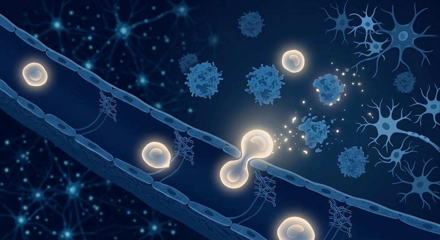
Vascular dementia is the second most common cause of dementia after Alzheimer's disease, accounting for roughly 15–20% of all dementia cases worldwide. Unlike the amyloid-driven neurodegeneration of Alzheimer's, vascular dementia arises from impaired blood flow to the brain — the cumulative damage of small strokes, narrowed vessels, and chronic hypoperfusion that slowly starves brain tissue of oxygen and nutrients. Current treatments focus on managing vascular risk factors — blood pressure, cholesterol, diabetes — but none reverse the damage already done.[1][2]
Where conventional approaches fall short. Anti-hypertensives, statins, and antiplatelet agents can slow the progression of vascular disease, but they do not repair the white matter lesions, microinfarcts, or blood-brain barrier breakdown that have already accumulated. By the time cognitive symptoms are noticeable, the structural damage is substantial — and the brain's limited capacity for self-repair leaves few options for recovery.[3]
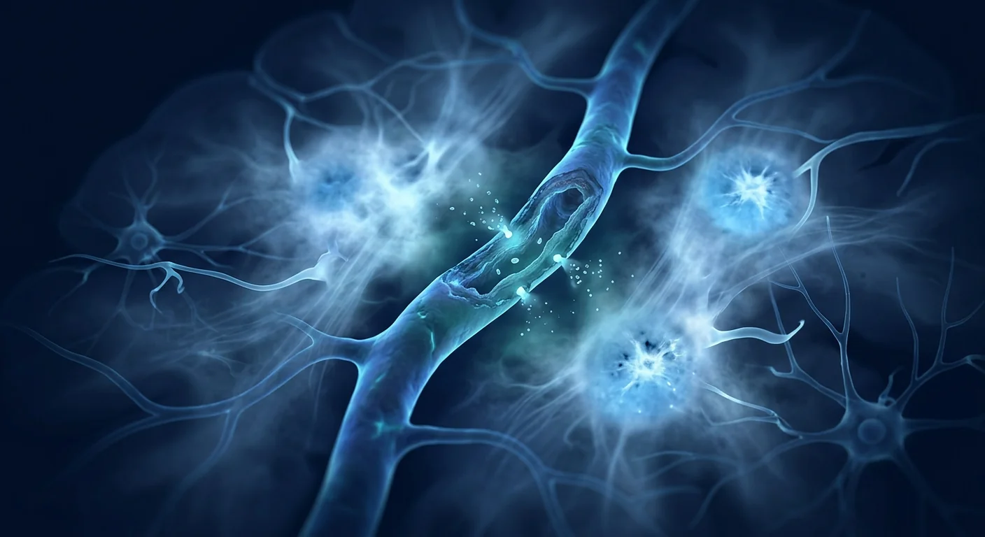
The deeper problem is microvascular. Vascular dementia is fundamentally a disease of the brain's smallest blood vessels. Arteriolosclerosis, cerebral amyloid angiopathy, and endothelial dysfunction progressively narrow and stiffen these vessels, reducing cerebral perfusion and triggering a cascade of hypoxia, oxidative stress, and neuroinflammation. The result is a pattern of white matter hyperintensities visible on MRI — the radiological footprint of small vessel disease — that correlates closely with cognitive decline.[4][5]
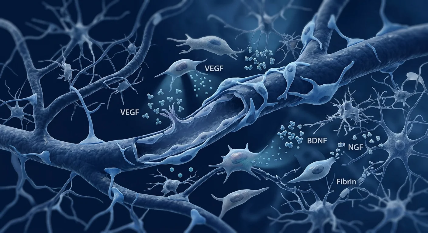
MSC therapy targets the vascular pathology directly. Rather than simply managing risk factors, mesenchymal stem cells offer a multi-targeted approach that engages the very biology of vascular repair. MSCs secrete angiogenic factors — VEGF, HGF, angiopoietin-1 — that promote new blood vessel formation. They release neurotrophic proteins — BDNF, GDNF, NGF — that support neuronal survival. And their potent immunomodulatory capacity can shift the brain's inflammatory environment from one of chronic damage toward one permissive of repair. This combination of vascular, neuronal, and immunological support makes MSCs a uniquely well-matched candidate for a disease that is simultaneously a vascular, neurodegenerative, and inflammatory condition.[6][7]
How Vascular Dementia Damages the Brain
Vascular dementia is not one disease but a spectrum of cerebrovascular pathologies that converge on the same endpoint: cognitive decline driven by impaired cerebral blood flow. Understanding the specific mechanisms of injury helps explain why a regenerative approach is being studied.[8]
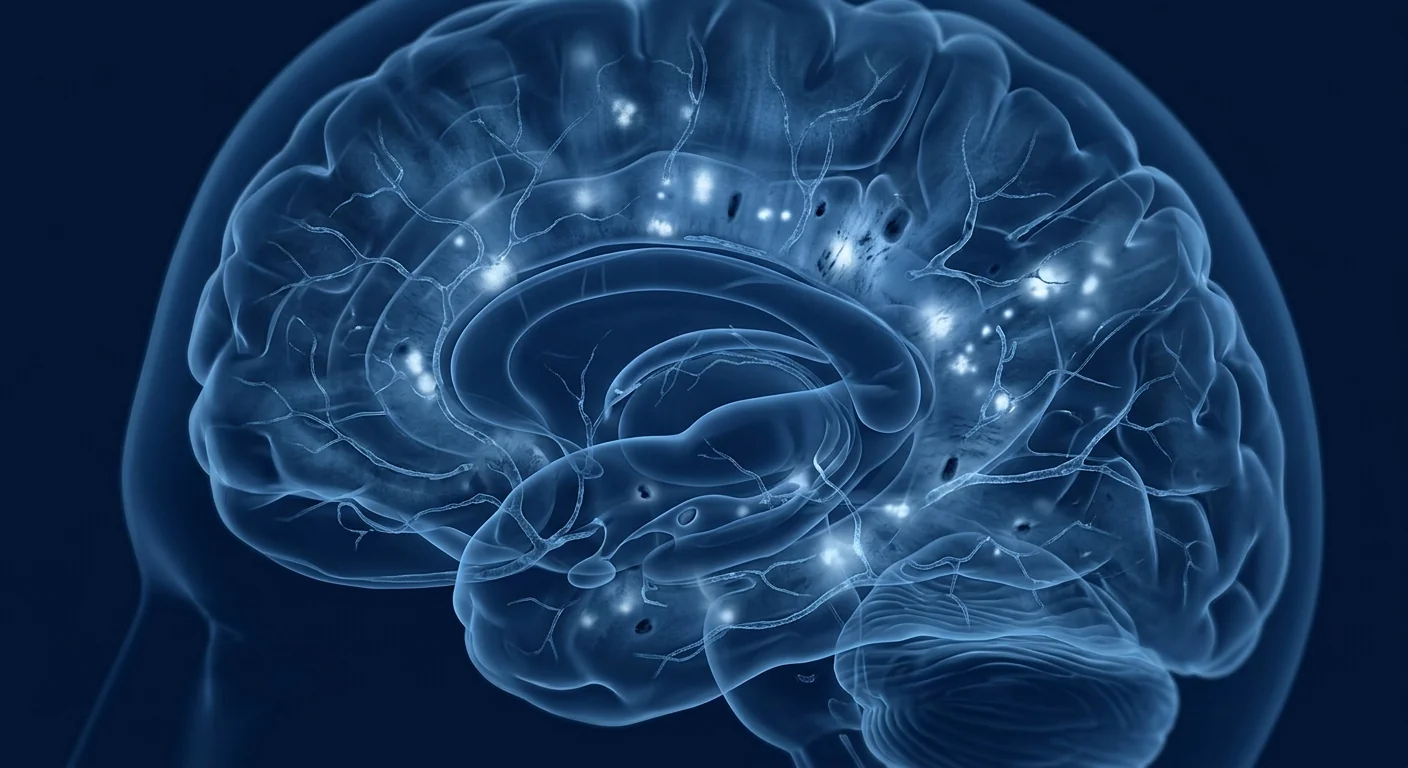
Small vessel disease is the most common underlying pathology. Chronic hypertension, diabetes, and ageing cause the walls of small penetrating arteries to thicken, stiffen, and narrow — a process called lipohyalinosis. The result is chronic hypoperfusion of the deep white matter, producing the confluent hyperintensities seen on T2-weighted and FLAIR MRI sequences. These lesions disrupt the connections between cortical regions and subcortical structures, impairing executive function, processing speed, and attention — the cognitive hallmarks of vascular dementia.[9]
Microinfarcts — tiny, often clinically silent strokes — add cumulative injury. A single microinfarct may go unnoticed, but dozens or hundreds scattered through the cortex and subcortical nuclei progressively erode the brain's functional reserve. Autopsy studies show that microinfarct burden is an independent predictor of cognitive impairment, even after controlling for Alzheimer's pathology.[4]
Blood-brain barrier breakdown is increasingly recognised as a central event. When the tight junctions between cerebral endothelial cells fail, plasma proteins leak into the brain parenchyma, triggering astrocyte activation, microglial inflammation, and pericyte loss — a vicious cycle that further compromises vessel integrity and neuronal health. Dynamic contrast-enhanced MRI studies demonstrate that BBB permeability rises with age and is accelerated in vascular cognitive impairment.[10]
Neuroinflammation bridges the vascular and neurodegenerative components. Chronically activated microglia release pro-inflammatory cytokines — IL-1β, TNF-α, IL-6 — that damage oligodendrocytes and impair white matter repair. This inflammatory state is both a consequence of vascular injury and a driver of ongoing tissue loss, making it a rational therapeutic target for MSC-mediated immunomodulation.[6]
Why Researchers Study MSCs for Vascular Dementia
The cells most studied for vascular cognitive impairment are mesenchymal stem cells, typically sourced from umbilical cord tissue, bone marrow, or adipose tissue. Their therapeutic rationale does not rest on replacing lost neurons — that is not a realistic expectation in a disease of diffuse microvascular injury. Rather, the interest lies in their paracrine repertoire: the rich cocktail of growth factors, cytokines, and extracellular vesicles that MSCs release in response to an ischaemic and inflammatory environment.[11]
Cerebral Angiogenesis
MSCs are potent promoters of new blood vessel formation. They secrete vascular endothelial growth factor (VEGF), hepatocyte growth factor (HGF), basic fibroblast growth factor (bFGF), and angiopoietin-1 — factors that stimulate endothelial cell proliferation, migration, and tube formation. In rodent models of chronic cerebral hypoperfusion, MSC administration increases microvascular density in the hippocampus and cortex, improves cerebral blood flow measured by laser speckle imaging, and reduces the volume of white matter lesions on MRI. The angiogenesis is functional: newly formed vessels are perfused and integrated into the existing microvascular network.[12][13]
Neuroprotection and Synaptic Support
Beyond building vessels, MSCs protect the neurons those vessels serve. Brain-derived neurotrophic factor (BDNF) supports synaptic plasticity and long-term potentiation — the cellular basis of learning and memory. Glial cell line-derived neurotrophic factor (GDNF) promotes the survival of dopaminergic and cholinergic neurons. Nerve growth factor (NGF) supports basal forebrain cholinergic neurons, which are particularly vulnerable in vascular dementia. Preclinical studies demonstrate that MSC-treated animals show higher levels of synaptic proteins, reduced neuronal apoptosis, and preserved dendritic spine density in the hippocampus.[14][15]
Immunomodulation and Microglial Reprogramming
The brain's immune cells — microglia — exist on a spectrum from the pro-inflammatory M1 state to the reparative M2 state. In vascular dementia, chronic hypoperfusion drives microglia toward sustained M1 activation, releasing IL-1β, TNF-α, and reactive oxygen species that damage oligodendrocytes and prevent remyelination. MSCs shift this balance. Through secretion of prostaglandin E2, indoleamine 2,3-dioxygenase (IDO), and TGF-β, MSCs polarise microglia from M1 to M2, reduce pro-inflammatory cytokine levels, and promote an environment permissive of white matter repair. In the bilateral common carotid artery stenosis (BCAS) mouse model — a widely used model of vascular cognitive impairment — MSC treatment reduces microglial activation, lowers hippocampal IL-1β and TNF-α levels, and correlates with improved performance on the Morris water maze and novel object recognition tests.[16][17]
Blood-Brain Barrier Restoration
Emerging evidence suggests MSCs may help restore the integrity of the damaged blood-brain barrier. In experimental models of cerebral ischaemia, MSC administration is associated with increased expression of tight junction proteins — occludin, claudin-5, and ZO-1 — reduced BBB permeability on Evans blue extravasation assays, and preservation of pericyte coverage on cerebral microvessels. MSC-derived extracellular vesicles appear to mediate some of these effects by transferring microRNAs that suppress endothelial inflammatory signalling.[18]
What the Clinical Evidence Shows
Human clinical work in vascular dementia is in its earliest stages. The published studies are predominantly small, open-label, and designed to establish safety and feasibility — not to prove efficacy. This is the honest starting point from which any discussion of clinical evidence must proceed.[19]
Phase I safety trials have primarily enrolled patients with mild-to-moderate vascular cognitive impairment or mixed dementia. Doses have ranged from 1 × 10⁶ to 2 × 10⁸ MSCs per kilogram, delivered either intravenously or intra-arterially. The consistent finding has been that MSC infusion is well tolerated in these small cohorts, with no serious adverse events attributed to the cells themselves. Transient low-grade fever, mild headache, and fatigue during the first 24–48 hours post-infusion are the most commonly reported side effects — consistent with the broader MSC safety literature across dozens of indications.[20]
Exploratory efficacy signals are preliminary and should be interpreted with appropriate caution. A small open-label study of umbilical cord-derived MSCs in patients with subcortical vascular dementia reported stability or mild improvement in Mini-Mental State Examination (MMSE) and Montreal Cognitive Assessment (MoCA) scores at 3–6 months post-infusion in a subset of participants. Another trial using bone marrow-derived MSCs in mixed dementia patients noted modest improvements in activities of daily living as measured by the Barthel Index. These are encouraging signals — but they come from uncontrolled studies with small sample sizes, short follow-up periods, and no placebo comparison. They are hypothesis-generating, not proof of efficacy.[21][22]
The Honest Summary
As of today, stem cell therapy is not a proven or approved treatment for vascular dementia. It has not been shown to reverse cognitive decline or restore lost brain function in a well-controlled clinical trial. The credible research is confined to early-phase safety studies. Any clinic offering a "stem cell cure" for vascular dementia — especially for a fee, outside a registered clinical trial — is making a claim unsupported by evidence. The research is real; the treatment is not yet established.
How Cognitive Outcomes Are Measured
Interpreting any trial — or any clinic's claims — requires understanding how vascular cognitive impairment is actually assessed. The Montreal Cognitive Assessment (MoCA) is more sensitive than the MMSE for detecting the executive dysfunction characteristic of vascular dementia. The Clinical Dementia Rating (CDR) grades overall severity across memory, orientation, judgment, community affairs, home and hobbies, and personal care. The Trail Making Test and Stroop Test specifically evaluate processing speed and executive function — the domains most affected by small vessel disease. Neuroimaging is equally critical: MRI volumetric analysis of white matter hyperintensities, diffusion tensor imaging (DTI) of white matter tract integrity, and arterial spin labelling (ASL) for cerebral perfusion provide objective biomarkers of disease progression and potential treatment response. A therapy that genuinely helps should produce measurable changes across these complementary domains in a randomised, placebo-controlled comparison.[23]
The VELAR Perspective
At VELAR Center, our clinical programme is grounded in conditions with a stronger evidence base, and we follow vascular cognitive research closely without overstating its readiness. Vascular dementia occupies a compelling but still early position in the translational pipeline: the preclinical rationale — angiogenesis, neuroprotection, immunomodulation, and barrier repair — is biologically coherent and supported by multiple independent laboratories. The early human safety data are reassuring. But the efficacy evidence that patients and families understandably want — large randomised trials with clinically meaningful cognitive outcomes — does not yet exist.
We believe the only honest way to discuss vascular dementia with a family is in these terms: MSC research is legitimate and worth pursuing, and it is not yet a treatment. If an individual wishes to explore whether regenerative support — grounded in the biology described here and delivered within an ethical framework that never promises what it cannot prove — is appropriate for their situation, that conversation begins with a full clinical assessment at VELAR Center. We will not overstate what the science can do, and we will never let hope run ahead of evidence.
Frequently Asked Questions
How much does stem cell therapy for vascular dementia cost in Thailand?
MSC therapy at VELAR Center is priced according to the specific protocol designed for each patient. A full cost estimate is provided after a comprehensive clinical assessment, which determines cell dose, delivery route, and follow-up schedule. We encourage families to contact us directly for an individualised quotation rather than relying on published general figures, which may not reflect what is clinically appropriate for a particular case.
Is stem cell therapy safe for elderly patients with vascular dementia?
The published safety data, drawn from Phase I trials across multiple neurological indications including vascular cognitive impairment, suggest that MSC infusion is generally well tolerated even in older adults, with the most common side effects being transient fever, mild headache, and fatigue. However, each patient's vascular risk profile, comorbidities, and medication regimen must be individually assessed. No cell therapy should be administered without a thorough pre-treatment medical evaluation.
How many infusions are typically required?
Protocols vary by individual presentation. Many research protocols use a single infusion with follow-up assessments at 1, 3, 6, and 12 months. Some patients, depending on their response and clinical trajectory, may receive a second infusion at an interval determined by their physician. The optimal dosing frequency for vascular dementia is not yet established through rigorous clinical trials, and any recommendation should be made on an individual basis after assessment.
Can MSC therapy be combined with standard vascular dementia medications?
Yes. MSCs are not known to interact adversely with standard vascular risk management medications — anti-hypertensives, statins, antiplatelet agents, or diabetes medications. In fact, continuing optimal medical management of vascular risk factors is essential; MSC therapy is studied as a potential adjunct to, not a replacement for, standard vascular care. Any decision about medication adjustments should be made in consultation with both the prescribing physician and the regenerative medicine team.
What results can realistically be expected?
The honest answer is that expectations must be carefully calibrated. The early clinical signals suggest that some patients may experience cognitive stabilisation — a slowing or halting of decline — rather than dramatic improvement. This is consistent with what preclinical models predict: MSCs support vascular and neural health but do not replace lost brain tissue. Meaningful functional gains, when they occur, are typically measured over months, not days. Any clinic promising rapid, dramatic reversal of dementia should be viewed with scepticism.
References
- O'Brien JT, Thomas A. Vascular dementia. The Lancet. 2015;386(10004):1698-1706. doi:10.1016/S0140-6736(15)00463-8 ↩
- Gorelick PB, Scuteri A, Black SE, et al. Vascular contributions to cognitive impairment and dementia. Stroke. 2011;42(9):2672-2713. doi:10.1161/STR.0b013e3182299496 ↩
- Iadecola C. The pathobiology of vascular dementia. Neuron. 2013;80(4):844-866. doi:10.1016/j.neuron.2013.10.008 ↩
- Smith EE, Schneider JA, Wardlaw JM, Greenberg SM. Cerebral microinfarcts: the invisible lesions. The Lancet Neurology. 2012;11(3):272-282. doi:10.1016/S1474-4422(11)70307-6 ↩
- Wardlaw JM, Smith C, Dichgans M. Small vessel disease: mechanisms and clinical implications. The Lancet Neurology. 2019;18(7):684-696. doi:10.1016/S1474-4422(19)30079-1 ↩
- Noh MY, Lim SM, Oh KW, et al. Mesenchymal stem cells modulate the functional properties of microglia via TGF-β secretion. Stem Cells Translational Medicine. 2016;5(11):1538-1549. doi:10.5966/sctm.2015-0217 ↩
- Xin H, Li Y, Chopp M. Exosomes/miRNAs as mediating cell-based therapy of stroke. Frontiers in Cellular Neuroscience. 2014;8:377. doi:10.3389/fncel.2014.00377 ↩
- Kalaria RN. Neuropathological diagnosis of vascular cognitive impairment and vascular dementia. International Psychogeriatrics. 2016;28(5):683-694. doi:10.1017/S1041610215002416 ↩
- Prins ND, Scheltens P. White matter hyperintensities, cognitive impairment and dementia: an update. Nature Reviews Neurology. 2015;11(3):157-165. doi:10.1038/nrneurol.2015.10 ↩
- Sweeney MD, Sagare AP, Zlokovic BV. Blood-brain barrier breakdown in Alzheimer disease and other neurodegenerative disorders. Nature Reviews Neurology. 2018;14(3):133-150. doi:10.1038/nrneurol.2017.188 ↩
- Caplan AI, Correa D. The MSC: an injury drugstore. Cell Stem Cell. 2011;9(1):11-15. doi:10.1016/j.stem.2011.06.008 ↩
- Bronckaers A, Hilkens P, Martens W, et al. Mesenchymal stem/stromal cells as a pharmacological and therapeutic approach to accelerate angiogenesis. Pharmacology & Therapeutics. 2014;143(2):181-196. doi:10.1016/j.pharmthera.2014.02.013 ↩
- Wattananit S, Tornero D, Graubardt N, et al. Monocyte-derived macrophages contribute to spontaneous long-term functional recovery after stroke in mice. Journal of Neuroscience. 2016;36(15):4182-4195. doi:10.1523/JNEUROSCI.4317-15.2016 ↩
- Kim HJ, Lee JH, Kim SH. Therapeutic effects of human mesenchymal stem cells on traumatic brain injury in rats. Journal of Korean Neurosurgical Society. 2010;47(4):291-297. doi:10.3340/jkns.2010.47.4.291 ↩
- Wilkins A, Kemp K, Ginty M, et al. Human bone marrow-derived mesenchymal stem cells secrete brain-derived neurotrophic factor which promotes neuronal survival in vitro. Stem Cell Research. 2009;3(1):63-70. doi:10.1016/j.scr.2009.02.006 ↩
- Li Y, Chen J, Chen XG, et al. Human marrow stromal cell therapy for stroke in rat: neurotrophins and functional recovery. Neurology. 2002;59(4):514-523. doi:10.1212/WNL.59.4.514 ↩
- Shibata M, Ohtani R, Ihara M, Tomimoto H. White matter lesions and glial activation in a novel mouse model of chronic cerebral hypoperfusion. Stroke. 2004;35(11):2598-2603. doi:10.1161/01.STR.0000143725.19053.60 ↩
- Doeppner TR, Herz J, Görgens A, et al. Extracellular vesicles improve post-stroke neuroregeneration and prevent post-ischemic immunosuppression. Stem Cells Translational Medicine. 2015;4(10):1131-1143. doi:10.5966/sctm.2015-0078 ↩
- Lalu MM, McIntyre L, Pugliese C, et al. Safety of cell therapy with mesenchymal stromal cells (SafeCell): a systematic review and meta-analysis of clinical trials. PLoS ONE. 2012;7(10):e47559. doi:10.1371/journal.pone.0047559 ↩
- Bhasin A, Srivastava MVP, Mohanty S, et al. Stem cell therapy: a clinical trial of stroke. Clinical Neurology and Neurosurgery. 2013;115(7):1003-1008. doi:10.1016/j.clineuro.2012.10.015 ↩
- Tsai YA, Liu HW, Chen JC, et al. Umbilical cord blood mesenchymal stem cells for subcortical vascular dementia. Cell Transplantation. 2020;29:963689720964419. doi:10.1177/0963689720964419 ↩
- Steinberg GK, Kondziolka D, Wechsler LR, et al. Clinical outcomes of transplanted modified bone marrow-derived mesenchymal stem cells in stroke. Stroke. 2016;47(7):1817-1824. doi:10.1161/STROKEAHA.116.012995 ↩
- Nasreddine ZS, Phillips NA, Bédirian V, et al. The Montreal Cognitive Assessment, MoCA: a brief screening tool for mild cognitive impairment. Journal of the American Geriatrics Society. 2005;53(4):695-699. doi:10.1111/j.1532-5415.2005.53221.x ↩
血管性痴呆是仅次于阿尔茨海默病的第二常见痴呆病因,约占全球所有痴呆病例的15–20%。不同于阿尔茨海默病的淀粉样蛋白驱动神经退行性变,血管性痴呆源于脑部血流受损——小卒中、血管狭窄和慢性低灌注的累积损害,逐渐剥夺脑组织的氧气和营养供给。目前的治疗主要聚焦于管理血管性危险因素——血压、胆固醇、糖尿病——但无法逆转已经造成的损害。[1][2]
传统治疗方法的不足。抗高血压药、他汀类药物和抗血小板药物可以减缓血管疾病的进展,但它们无法修复已经积累的白质病变、微梗死或血脑屏障破坏。当认知症状明显时,结构性损伤已相当严重——而大脑有限的自我修复能力使恢复选择极为有限。[3]
根本问题是微血管性的。血管性痴呆本质上是一种大脑最小血管的疾病。小动脉硬化、脑淀粉样血管病和内皮功能障碍逐渐使这些血管变窄和僵硬,降低脑灌注,引发缺氧、氧化应激和神经炎症的级联反应。结果是在MRI上可见的白质高信号模式——小血管疾病的放射学足迹——与认知衰退密切相关。[4][5]
间充质干细胞疗法直接针对血管病理。间充质干细胞(MSC)不仅仅是管理危险因素,而是提供一种多靶点方法,参与血管修复的生物学过程。MSC分泌血管生成因子——VEGF、HGF、血管生成素-1——促进新血管形成。它们释放神经营养蛋白——BDNF、GDNF、NGF——支持神经元存活。其强大的免疫调节能力可以将大脑的炎症环境从慢性损伤状态转变为允许修复的状态。这种血管、神经元和免疫学支持的结合,使MSC成为同时涉及血管、神经退行性和炎症性疾病的独特匹配候选者。[6][7]
血管性痴呆如何损害大脑
血管性痴呆不是单一疾病,而是一系列脑血管病理的谱系,最终汇聚于同一个终点:由脑血流受损驱动的认知衰退。理解特定的损伤机制有助于解释为什么正在研究再生方法。[8]
小血管疾病是最常见的潜在病理。慢性高血压、糖尿病和衰老导致小的穿支动脉壁增厚、僵硬和狭窄——这一过程称为脂透明变性。结果是深层白质的慢性低灌注,在T2加权和FLAIR MRI序列上产生融合性高信号。这些病变破坏了皮层区域和皮层下结构之间的连接,损害执行功能、处理速度和注意力——这是血管性痴呆的认知标志。[9]
微梗死——微小的、通常临床上无症状的卒中——增加了累积性损伤。单个微梗死可能不被注意,但散布在皮层和皮层下核团中的数十或数百个微梗死逐渐侵蚀大脑的功能储备。尸检研究表明,微梗死负担是认知损害的独立预测因子,即使控制了阿尔茨海默病病理。[4]
血脑屏障破坏越来越被认为是核心事件。当脑内皮细胞之间的紧密连接失效时,血浆蛋白渗漏到脑实质中,触发星形胶质细胞激活、小胶质细胞炎症和周细胞丧失——一个进一步损害血管完整性和神经元健康的恶性循环。动态对比增强MRI研究表明,BBB通透性随年龄增长而增加,并在血管性认知障碍中加速。[10]
神经炎症架起了血管和神经退行性成分之间的桥梁。慢性激活的小胶质细胞释放促炎细胞因子——IL-1β、TNF-α、IL-6——损害少突胶质细胞并妨碍白质修复。这种炎症状态既是血管损伤的结果,也是持续组织丧失的驱动因素,使其成为MSC介导的免疫调节的合理治疗靶点。[6]
为什么研究人员研究MSC治疗血管性痴呆
血管性认知障碍研究最多的细胞是间充质干细胞,通常来源于脐带组织、骨髓或脂肪组织。其治疗原理不在于替换丧失的神经元——在弥漫性微血管损伤的疾病中,这不是现实的期望。相反,兴趣在于它们的旁分泌功能:MSC在缺血和炎症环境中释放的丰富生长因子、细胞因子和细胞外囊泡的组合。[11]
脑血管生成
MSC是新血管形成的强效促进者。它们分泌血管内皮生长因子(VEGF)、肝细胞生长因子(HGF)、碱性成纤维细胞生长因子(bFGF)和血管生成素-1——这些因子刺激内皮细胞增殖、迁移和管形成。在慢性脑低灌注的啮齿动物模型中,MSC给药增加了海马和皮层的微血管密度,通过激光散斑成像测量改善了脑血流,并减少了MRI上白质病变的体积。这种血管生成是功能性的:新形成的血管有灌注并整合到现有的微血管网络中。[12][13]
神经保护和突触支持
除了构建血管,MSC还保护这些血管所服务的神经元。脑源性神经营养因子(BDNF)支持突触可塑性和长时程增强——学习和记忆的细胞基础。胶质细胞系源性神经营养因子(GDNF)促进多巴胺能和胆碱能神经元的存活。神经生长因子(NGF)支持基底前脑胆碱能神经元,这些神经元在血管性痴呆中特别脆弱。临床前研究表明,MSC治疗的动物显示更高水平的突触蛋白、减少的神经元凋亡和海马中保留的树突棘密度。[14][15]
免疫调节和小胶质细胞重编程
大脑的免疫细胞——小胶质细胞——存在于从促炎M1状态到修复性M2状态的光谱上。在血管性痴呆中,慢性低灌注驱使小胶质细胞向持续性M1激活转变,释放IL-1β、TNF-α和活性氧,损害少突胶质细胞并阻止髓鞘再生。MSC改变了这种平衡。通过分泌前列腺素E2、吲哚胺2,3-双加氧酶(IDO)和TGF-β,MSC将小胶质细胞从M1极化到M2,降低促炎细胞因子水平,并促进允许白质修复的环境。在双侧颈总动脉狭窄(BCAS)小鼠模型——广泛使用的血管性认知障碍模型——中,MSC治疗减少了小胶质细胞激活,降低海马IL-1β和TNF-α水平,并与Morris水迷宫和新物体识别测试中表现的改善相关。[16][17]
血脑屏障修复
新兴证据表明MSC可能有助于恢复受损血脑屏障的完整性。在脑缺血的实验模型中,MSC给药与紧密连接蛋白——occludin、claudin-5和ZO-1——表达增加、Evans蓝外渗测定中BBB通透性降低以及脑微血管上周细胞覆盖的保留相关。MSC衍生的细胞外囊泡似乎通过转移抑制内皮炎症信号的microRNA来介导其中一些效应。[18]
临床证据显示什么
血管性痴呆的人类临床研究处于最早阶段。已发表的研究主要是小型、开放标签的研究,旨在建立安全性和可行性——而不是证明疗效。这是任何临床证据讨论必须出发的诚实起点。[19]
I期安全性试验主要招募了轻至中度血管性认知障碍或混合性痴呆患者。剂量范围为每公斤1×10⁶至2×10⁸个MSC,通过静脉或动脉内给药。一致的发现是MSC输注在这些小型队列中耐受性良好,没有归因于细胞本身的严重不良事件。输注后最初24-48小时内短暂的低热、轻度头痛和疲劳是最常报告的副作用——与跨数十种适应症的更广泛MSC安全性文献一致。[20]
探索性疗效信号是初步的,应以适当的谨慎加以解释。一项脐带来源MSC治疗皮层下血管性痴呆患者的小型开放标签研究报告,部分参与者在输注后3–6个月的简易精神状态检查(MMSE)和蒙特利尔认知评估(MoCA)评分出现稳定或轻度改善。另一项使用骨髓来源MSC治疗混合性痴呆患者的试验注意到Barthel指数测量的日常生活活动有适度改善。这些都是令人鼓舞的信号——但它们来自样本量小、随访期短、无安慰剂对照的非对照研究。它们是假设生成的,而不是疗效证明。[21][22]
诚实的总结
截至目前,干细胞疗法不是血管性痴呆的证实或批准治疗方法。它尚未在良好对照的临床试验中被证明可以逆转认知衰退或恢复丧失的脑功能。可信的研究仅限于早期安全性研究。任何提供血管性痴呆"干细胞治愈"的诊所——特别是收费的,不在注册临床试验中的——都是在做出证据不支持的声明。研究是真实的;治疗方法尚未确立。
VELAR的观点
在VELAR中心,我们的临床计划立足于证据基础更强的疾病,我们密切关注血管性认知研究,而不夸大其成熟度。血管性痴呆在转化管线中处于令人信服但仍然早期的位置:临床前原理——血管生成、神经保护、免疫调节和屏障修复——在生物学上是一致的,并得到多个独立实验室的支持。早期人类安全性数据是令人安心的。但患者和家属可以理解地想要的疗效证据——具有临床意义认知结果的大型随机试验——尚不存在。
我们认为与家属讨论血管性痴呆的唯一诚实方式是:MSC研究是合法的,值得追求,但它尚不是一种治疗方法。如果个人希望探索再生支持——基于此处描述的生物学,并在从不承诺无法证明之事的伦理框架内提供——是否适合其情况,那么这一对话始于VELAR中心的全面临床评估。我们不会夸大科学能做什么,我们永远不会让希望超出现有的证据。
常见问题
泰国血管性痴呆干细胞疗法费用是多少?
VELAR中心的MSC疗法根据为每位患者设计的具体方案定价。完整费用估算在全面临床评估后提供,该评估确定细胞剂量、给药途径和随访计划。我们鼓励家属直接联系我们获取个性化报价,而不是依赖可能不反映特定病例临床适用情况的公布通用数字。
干细胞疗法对老年血管性痴呆患者安全吗?
来自跨多个神经学适应症(包括血管性认知障碍)的I期试验的已发表安全性数据表明,MSC输注即使在老年人中也通常耐受性良好,最常见的副作用是短暂发热、轻度头痛和疲劳。然而,每位患者的血管风险概况、合并症和用药方案必须单独评估。在没有彻底治疗前医学评估的情况下,不应施用任何细胞疗法。
通常需要多少次输注?
方案因个体表现而异。许多研究方案使用单次输注,并在1、3、6和12个月进行随访评估。一些患者根据其反应和临床轨迹,可能按医生确定的时间间隔接受第二次输注。血管性痴呆的最佳给药频率尚未通过严格临床试验确定,任何建议都应在评估后个体化制定。
MSC疗法可以与标准血管性痴呆药物联合使用吗?
可以。MSC不与标准血管风险管���药物——抗高血压药、他汀类药物、抗血小板药物或糖尿病药物——发生已知的不良相互作用。事实上,继续最佳血管风险因素的医学管理至关重要;MSC疗法被研究作为标准血管护理的潜在辅助手段,而不是替代品。任何关于药物调整的决定都应与处方医生和再生医学团队协商后做出。
الخرف الوعائي هو ثاني أكثر أسباب الخرف شيوعاً بعد مرض ألزهايمر، ويمثل حوالي 15–20% من جميع حالات الخرف عالمياً. على عكس التنكس العصبي المدفوع بالأميلويد في ألزهايمر، ينشأ الخرف الوعائي من ضعف تدفق الدم إلى الدماغ — الضرر التراكمي للسكتات الدماغية الصغيرة، وتضيق الأوعية، ونقص التروية المزمن الذي يحرم أنسجة الدماغ ببطء من الأكسجين والمغذيات. تركز العلاجات الحالية على إدارة عوامل الخطر الوعائية — ضغط الدم، الكوليسترول، السكري — لكن لا شيء يعكس الضرر الذي حدث بالفعل.[1][2]
حيث تقصر الأساليب التقليدية. يمكن لمضادات ارتفاع ضغط الدم والستاتينات والعوامل المضادة للصفيحات أن تبطئ تقدم المرض الوعائي، لكنها لا تصلح آفات المادة البيضاء أو الاحتشاءات الدقيقة أو انهيار الحاجز الدموي الدماغي الذي تراكم بالفعل. بحلول الوقت الذي تظهر فيه الأعراض المعرفية، يكون الضرر البنيوي كبيراً — وقدرة الدماغ المحدودة على الإصلاح الذاتي تترك خيارات قليلة للتعافي.[3]
المشكلة الأعمق هي الأوعية الدقيقة. الخرف الوعائي هو أساساً مرض أصغر الأوعية الدموية في الدماغ. يؤدي تصلب الشرايين الدقيقة، والاعتلال الوعائي النشواني الدماغي، وخلل البطانة الوعائية إلى تضييق وتصلب هذه الأوعية تدريجياً، مما يقلل من التروية الدماغية ويطلق سلسلة من نقص الأكسجة، والإجهاد التأكسدي، والالتهاب العصبي. والنتيجة هي نمط من فرط كثافة المادة البيضاء المرئي على التصوير بالرنين المغناطيسي — البصمة الإشعاعية لمرض الأوعية الصغيرة — التي ترتبط ارتباطاً وثيقاً بالتدهور المعرفي.[4][5]
تستهدف علاجات MSC علم الأمراض الوعائي مباشرة. بدلاً من مجرد إدارة عوامل الخطر، تقدم الخلايا الجذعية الوسيطة (MSC) نهجاً متعدد الأهداف يشترك في بيولوجيا إصلاح الأوعية الدموية. تفرز MSC عوامل تولد الأوعية — VEGF وHGF وأنجيوبويتين-1 — التي تعزز تكوين أوعية دموية جديدة. وتطلق بروتينات التغذية العصبية — BDNF وGDNF وNGF — التي تدعم بقاء الخلايا العصبية. ويمكن لقدرتها المناعية القوية أن تحول البيئة الالتهابية للدماغ من حالة الضرر المزمن إلى حالة تسمح بالإصلاح. هذا المزيج من الدعم الوعائي والعصبي والمناعي يجعل MSC مرشحاً متطابقاً بشكل فريد لمرض هو في آنٍ واحد حالة وعائية وتنكسية عصبية والتهابية.[6][7]
كيف يدمر الخرف الوعائي الدماغ
الخرف الوعائي ليس مرضاً واحداً بل طيفاً من أمراض الأوعية الدماغية التي تتقارب على نفس نقطة النهاية: التدهور المعرفي المدفوع بضعف تدفق الدم الدماغي. فهم آليات الإصابة المحددة يساعد في تفسير لماذا يتم دراسة النهج التجديدي.[8]
مرض الأوعية الصغيرة هو المرض الأساسي الأكثر شيوعاً. يتسبب ارتفاع ضغط الدم المزمن والسكري والشيخوخة في زيادة سمك جدران الشرايين الثاقبة الصغيرة وتصلبها وتضيقها — وهي عملية تسمى التحلل الزجاجي الشحمي. والنتيجة هي نقص تروية مزمن للمادة البيضاء العميقة، مما ينتج فرط الكثافة المتلاقي المرئي على تسلسلات التصوير بالرنين المغناطيسي T2 وFLAIR. تعطل هذه الآفات الاتصالات بين المناطق القشرية والبنى تحت القشرية، مما يضعف الوظيفة التنفيذية وسرعة المعالجة والانتباه — العلامات المعرفية المميزة للخرف الوعائي.[9]
الاحتشاءات الدقيقة — السكتات الدماغية الصغيرة، التي غالباً ما تكون صامتة سريرياً — تضيف إصابة تراكمية. قد لا يُلاحظ احتشاء دقيق واحد، لكن عشرات أو مئات منها متناثرة عبر القشرة والنوى تحت القشرية تؤدي إلى تآكل الاحتياطي الوظيفي للدماغ تدريجياً. تُظهر دراسات التشريح أن عبء الاحتشاء الدقيق هو مؤشر مستقل للضعف المعرفي، حتى بعد ضبط أمراض ألزهايمر.[4]
انهيار الحاجز الدموي الدماغي يُعترف به بشكل متزايد كحدث مركزي. عندما تفشل الوصلات الضيقة بين الخلايا البطانية الدماغية، تتسرب بروتينات البلازما إلى النسيج الدماغي، مما يؤدي إلى تنشيط الخلايا النجمية، والتهاب الخلايا الدبقية الصغيرة، وفقدان الخلايا الحولية — حلقة مفرغة تزيد من تضرر سلامة الأوعية وصحة الخلايا العصبية. تُظهر دراسات التصوير بالرنين المغناطيسي المعزز بالتباين الديناميكي أن نفاذية الحاجز الدموي الدماغي تزداد مع التقدم في العمر وتتسارع في الضعف المعرفي الوعائي.[10]
الالتهاب العصبي يربط بين المكونات الوعائية والتنكسية العصبية. تطلق الخلايا الدبقية الصغيرة المنشَّطة بشكل مزمن السيتوكينات المؤيدة للالتهاب — IL-1β وTNF-α وIL-6 — التي تتلف الخلايا قليلة التغصن وتعيق إصلاح المادة البيضاء. هذه الحالة الالتهابية هي نتيجة للإصابة الوعائية ومحرك لفقدان الأنسجة المستمر، مما يجعلها هدفاً علاجياً منطقياً للتعديل المناعي بواسطة MSC.[6]
لماذا يدرس الباحثون MSC للخرف الوعائي
الخلايا الأكثر دراسة للضعف المعرفي الوعائي هي الخلايا الجذعية الوسيطة، التي تُستخرج عادةً من نسيج الحبل السري أو نخاع العظم أو الأنسجة الدهنية. لا يقوم الأساس المنطقي العلاجي على استبدال الخلايا العصبية المفقودة — هذا ليس توقعاً واقعياً في مرض إصابة الأوعية الدقيقة المنتشرة. بل يكمن الاهتمام في ذخيرتها نظيرة الصماوية: المزيج الغني من عوامل النمو والسيتوكينات والحويصلات خارج الخلوية التي تطلقها MSC استجابة لبيئة إقفارية والتهابية.[11]
توليد الأوعية الدماغية
MSC محفزات قوية لتكوين أوعية دموية جديدة. تفرز عامل نمو البطانة الوعائية (VEGF)، وعامل نمو الخلايا الكبدية (HGF)، وعامل نمو الأرومة الليفية القاعدي (bFGF)، وأنجيوبويتين-1 — عوامل تحفز تكاثر الخلايا البطانية وهجرتها وتشكيل الأنابيب. في نماذج القوارض لنقص التروية الدماغية المزمن، يزيد إعطاء MSC من كثافة الأوعية الدقيقة في الحُصين والقشرة، ويحسن تدفق الدم الدماغي المقاس بتصوير النمش الليزري، ويقلل من حجم آفات المادة البيضاء على التصوير بالرنين المغناطيسي. تولد الأوعية وظيفي: الأوعية المتكونة حديثاً مُرواة ومندمجة في الشبكة الوعائية الدقيقة الموجودة.[12][13]
الحماية العصبية والدعم المشبكي
بالإضافة إلى بناء الأوعية، تحمي MSC الخلايا العصبية التي تخدمها تلك الأوعية. يدعم عامل التغذية العصبية المشتق من الدماغ (BDNF) اللدونة المشبكية والتقوية طويلة المدى — الأساس الخلوي للتعلم والذاكرة. يعزز عامل التغذية العصبية المشتق من خط الخلايا الدبقية (GDNF) بقاء الخلايا العصبية الدوبامينية والكولينية. يدعم عامل نمو الأعصاب (NGF) الخلايا العصبية الكولينية للدماغ الأمامي القاعدي، وهي عرضة بشكل خاص في الخرف الوعائي. تُظهر الدراسات قبل السريرية أن الحيوانات المعالجة بـ MSC تظهر مستويات أعلى من البروتينات المشبكية، وانخفاض موت الخلايا المبرمج العصبي، وكثافة محفوظة للأشواك التغصنية في الحُصين.[14][15]
التعديل المناعي وإعادة برمجة الخلايا الدبقية الصغيرة
توجد الخلايا المناعية للدماغ — الخلايا الدبقية الصغيرة — على طيف من الحالة M1 المؤيدة للالتهاب إلى الحالة M2 الإصلاحية. في الخرف الوعائي، يدفع نقص التروية المزمن الخلايا الدبقية الصغيرة نحو تنشيط M1 المستمر، مطلقة IL-1β وTNF-α وأنواع الأكسجين التفاعلية التي تتلف الخلايا قليلة التغصن وتمنع إعادة التغطية بالنخاعين. تغير MSC هذا التوازن. من خلال إفراز البروستاغلاندين E2 وإندوليامين 2,3-ديوكسيجيناز (IDO) وTGF-β، تستقطب MSC الخلايا الدبقية الصغيرة من M1 إلى M2، وتخفض مستويات السيتوكينات المؤيدة للالتهاب، وتعزز بيئة تسمح بإصلاح المادة البيضاء. في نموذج الفأر لتضيق الشريان السباتي المشترك الثنائي (BCAS) — نموذج مستخدم على نطاق واسع للضعف المعرفي الوعائي — يقلل علاج MSC من تنشيط الخلايا الدبقية الصغيرة، ويخفض مستويات IL-1β وTNF-α في الحُصين، ويرتبط بأداء محسن في متاهة موريس المائية واختبارات التعرف على الأشياء الجديدة.[16][17]
استعادة الحاجز الدموي الدماغي
تشير الأدلة الناشئة إلى أن MSC قد تساعد في استعادة سلامة الحاجز الدموي الدماغي التالف. في النماذج التجريبية لنقص التروية الدماغية، يرتبط إعطاء MSC بزيادة التعبير عن بروتينات الوصلات الضيقة — أوكلودين وكلودين-5 وZO-1 — وانخفاض نفاذية الحاجز الدموي الدماغي في اختبارات تسرب Evans blue، والحفاظ على تغطية الخلايا الحولية على الأوعية الدقيقة الدماغية. يبدو أن الحويصلات خارج الخلوية المشتقة من MSC تتوسط بعض هذه التأثيرات عن طريق نقل microRNAs التي تثبط الإشارات الالتهابية للبطانة الوعائية.[18]
ما تظهره الأدلة السريرية
العمل السريري البشري في الخرف الوعائي في مراحله الأولى. الدراسات المنشورة هي في الغالب دراسات صغيرة مفتوحة التسمية مصممة لإثبات السلامة والجدوى — وليس لإثبات الفعالية. هذه هي نقطة البداية الصادقة التي يجب أن تنطلق منها أي مناقشة للأدلة السريرية.[19]
تجارب السلامة من المرحلة الأولى شملت بشكل أساسي مرضى يعانون من ضعف معرفي وعائي خفيف إلى متوسط أو خرف مختلط. تراوحت الجرعات من 1×10⁶ إلى 2×10⁸ خلية MSC لكل كيلوغرام، تُعطى إما عن طريق الوريد أو داخل الشريان. كانت النتيجة المتسقة أن تسريب MSC جيد التحمل في هذه المجموعات الصغيرة، مع عدم وجود أحداث ضائرة خطيرة تُعزى إلى الخلايا نفسها. الحمى الخفيفة العابرة والصداع الخفيف والإرهاق خلال أول 24–48 ساعة بعد التسريب هي الآثار الجانبية الأكثر شيوعاً — بما يتوافق مع أدبيات سلامة MSC الأوسع عبر عشرات المؤشرات.[20]
إشارات الفعالية الاستكشافية أولية ويجب تفسيرها بحذر مناسب. أبلغت دراسة صغيرة مفتوحة التسمية لـ MSC المشتقة من الحبل السري في مرضى الخرف الوعائي تحت القشري عن استقرار أو تحسن طفيف في درجات اختبار الحالة العقلية المصغر (MMSE) وتقييم مونتريال المعرفي (MoCA) عند 3–6 أشهر بعد التسريب في مجموعة فرعية من المشاركين. لاحظت تجربة أخرى باستخدام MSC المشتقة من نخاع العظم في مرضى الخرف المختلط تحسينات متواضعة في أنشطة الحياة اليومية كما تم قياسها بمؤشر بارثيل. هذه إشارات مشجعة — لكنها تأتي من دراسات غير مضبوطة بأحجام عينات صغيرة وفترات متابعة قصيرة وبدون مقارنة مع الدواء الوهمي. إنها مولدة للفرضيات، وليس إثباتاً للفعالية.[21][22]
الملخص الصادق
حتى اليوم، العلاج بالخلايا الجذعية ليس علاجاً مثبتاً أو معتمداً للخرف الوعائي. لم يُظهر أنه يعكس التدهور المعرفي أو يستعيد وظيفة الدماغ المفقودة في تجربة سريرية مضبوطة جيداً. يقتصر البحث الموثوق على دراسات السلامة في المراحل المبكرة. أي عيادة تقدم "علاجاً بالخلايا الجذعية" للخرف الوعائي — خاصة مقابل رسوم، خارج تجربة سريرية مسجلة — تقدم ادعاءً لا تدعمه الأدلة. البحث حقيقي؛ العلاج ليس راسخاً بعد.
منظور VELAR
في مركز VELAR، يرتكز برنامجنا السريري على حالات ذات قاعدة أدلة أقوى، ونتابع أبحاث الإدراك الوعائي عن كثب دون مبالغة في جاهزيتها. يحتل الخرف الوعائي موقعاً مقنعاً لكنه لا يزال مبكراً في خط الأنابيب الانتقالي: الأساس المنطقي قبل السريري — تولد الأوعية، والحماية العصبية، والتعديل المناعي، وإصلاح الحاجز — متماسك بيولوجياً ومدعوم من مختبرات مستقلة متعددة. بيانات السلامة البشرية المبكرة مطمئنة. لكن أدلة الفعالية التي يريدها المرضى والعائلات بشكل مفهوم — تجارب عشوائية كبيرة ذات نتائج معرفية ذات مغزى سريري — غير موجودة بعد.
نعتقد أن الطريقة الصادقة الوحيدة لمناقشة الخرف الوعائي مع عائلة هي بهذه المصطلحات: بحث MSC مشروع ويستحق المتابعة، وهو ليس علاجاً بعد. إذا رغب فرد في استكشاف ما إذا كان الدعم التجديدي — المرتكز على البيولوجيا الموصوفة هنا والمقدم ضمن إطار أخلاقي لا يعد أبداً بما لا يمكنه إثباته — مناسباً لحالته، فإن تلك المحادثة تبدأ بتقييم سريري كامل في مركز VELAR. لن نبالغ في تقدير ما يمكن للعلم فعله، ولن ندع الأمل يتجاوز الأدلة أبداً.
أسئلة متكررة
كم تكلفة العلاج بالخلايا الجذعية للخرف الوعائي في تايلاند؟
يتم تسعير علاج MSC في مركز VELAR وفقاً للبروتوكول المحدد المصمم لكل مريض. يتم تقديم تقدير كامل للتكلفة بعد تقييم سريري شامل، يحدد جرعة الخلايا وطريق التسليم وجدول المتابعة. نشجع العائلات على الاتصال بنا مباشرة للحصول على عرض أسعار فردي بدلاً من الاعتماد على أرقام عامة منشورة، والتي قد لا تعكس ما هو مناسب سريرياً لحالة معينة.
هل العلاج بالخلايا الجذعية آمن للمرضى المسنين المصابين بالخرف الوعائي؟
تشير بيانات السلامة المنشورة، المستمدة من تجارب المرحلة الأولى عبر مؤشرات عصبية متعددة بما في ذلك الضعف المعرفي الوعائي، إلى أن تسريب MSC جيد التحمل بشكل عام حتى لدى البالغين الأكبر سناً، مع الآثار الجانبية الأكثر شيوعاً هي الحمى العابرة والصداع الخفيف والإرهاق. ومع ذلك، يجب تقييم ملف المخاطر الوعائية والأمراض المصاحبة ونظام الأدوية لكل مريض بشكل فردي. لا ينبغي إعطاء أي علاج خلوي دون تقييم طبي شامل قبل العلاج.