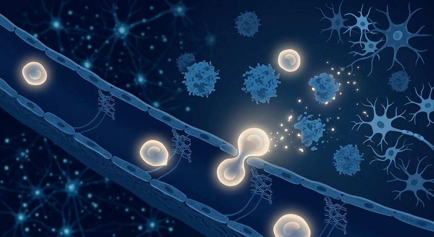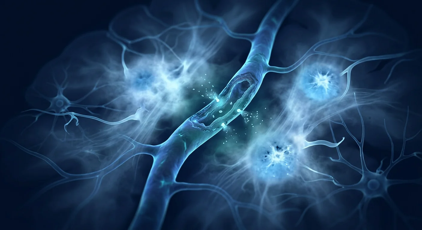
Cerebral small vessel disease is the silent architect of dementia and cognitive decline — affecting the brain's smallest blood vessels long before any symptom appears. It is responsible for an estimated 45% of all stroke cases and is the single most common cause of white matter hyperintensities on MRI in the elderly. Unlike the dramatic strokes that cause sudden paralysis or speech loss, small vessel disease creeps in through micro-infarcts, narrowed vessels, and chronic hypoperfusion that accumulate over years or decades. By the time cognitive symptoms are noticeable, the structural damage is substantial.[1][2]
Where conventional medicine falls short. Anti-hypertensives, statins, and antiplatelet agents can slow the progression of vascular risk factors, but they do not repair the white matter lesions, microinfarcts, or blood-brain barrier breakdown that have already accumulated. Current drug therapies target individual risk factors — blood pressure, cholesterol, glucose — but none address the cumulative microvascular damage itself. By the time cognitive decline is clinically evident, the brain's limited capacity for self-repair leaves few options.[3]
The deeper problem is structural microvascular. At the core of cerebral small vessel disease are arteriolosclerosis — thickening and stiffening of the tiny penetrating arteries that supply deep white matter and subcortical structures — and cerebral amyloid angiopathy, where amyloid-beta deposits weaken vessel walls. Endothelial dysfunction reduces nitric-oxide-mediated vasodilation, further constraining blood flow. The result is chronic hypoperfusion of regions critical for executive function, processing speed, and mood regulation — domains that are disproportionately affected compared with the memory-predominant amnesia of Alzheimer's disease.[4][5]
MSC therapy engages the vascular pathology directly. Mesenchymal stem cells offer a multi-targeted approach that is uniquely well-matched to a disease that is simultaneously vascular, neurodegenerative, and inflammatory. MSCs secrete angiogenic factors — VEGF, HGF, angiopoietin-1, bFGF — that drive new blood vessel formation. They release neurotrophic proteins — BDNF, GDNF, NGF — that support neuronal survival. Their potent immunomodulatory capacity shifts the brain's inflammatory environment from chronic damage toward one permissive of repair. And emerging evidence suggests they may help restore the blood-brain barrier. This combination of vascular, neuronal, and immunological support makes MSCs an ideal candidate for a disease defined by microvascular failure.[6][7]
The Pathobiology of Cerebral Small Vessel Disease
Understanding the specific mechanisms of injury is essential to appreciating why a regenerative approach is being studied. Cerebral small vessel disease is not one pathology but a spectrum of cerebrovascular damage that converges on the same endpoint: cognitive decline driven by impaired microvascular perfusion.[8]
Lipohyalinosis and arteriolosclerosis. Chronic hypertension and diabetes cause the walls of small penetrating arteries to thicken, stiffen, and narrow. Hyaline deposits accumulate in the vessel wall, the lumen narrows, and downstream tissue becomes chronically ischaemic. This is the dominant mechanism in lacunar infarcts and in the confluent white matter hyperintensities that are the radiological hallmark of cerebral small vessel disease. The deep white matter tracts — particularly the anterior thalamic radiations and corona radiata — lose their blood supply, and the long-range connections between cortex and subcortex degrade.[9]
Microinfarcts and cumulative burden. Individual microinfarcts are often clinically silent — they do not produce focal neurological deficits detectable on examination — but their cumulative burden is substantial. Autopsy studies demonstrate that microinfarct count is an independent predictor of cognitive impairment, even after controlling for Alzheimer's pathology such as amyloid plaques and neurofibrillary tangles. The brain compensates for years through functional reserve, but once the reserve is exhausted, decline becomes clinically apparent — often abruptly, triggered by a new vascular event or intercurrent illness.[10][11]
Blood-brain barrier permeability. Dynamic contrast-enhanced MRI studies have shown that BBB permeability increases with age and is accelerated in patients with vascular cognitive impairment. When tight junctions between cerebral endothelial cells fail, plasma proteins — fibrinogen, albumin, iron — leak into the brain parenchyma, triggering astrocyte activation, microglial inflammation, and pericyte apoptosis. This creates a self-reinforcing loop: BBB breakdown → inflammation → pericyte loss → further vessel fragility.[12][13]
Neuroinflammation and oligodendrocyte loss. Chronically activated microglia release IL-1β, TNF-α, and reactive oxygen species that preferentially damage oligodendrocytes — the cells responsible for myelinating axons and maintaining white matter integrity. White matter hyperintensities on MRI are not simply areas of water retention; they represent a complex mixture of demyelination, axonal loss, gliosis, and myelin odema. The cognitive domains most vulnerable to this damage — executive function, processing speed, attention — are precisely the functions that depend on intact white matter connectivity.[14]
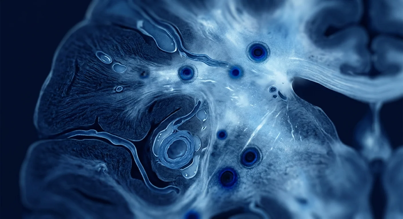
Why MSCs Are a Rational Choice for Small Vessel Disease
The cells most studied for cerebral small vessel disease are mesenchymal stem cells, sourced from umbilical cord tissue (Wharton's jelly), bone marrow, or adipose tissue. Their therapeutic rationale does not rest on replacing lost neurons — a tall order in a disease of diffuse microvascular injury — but on their paracrine repertoire: the rich cocktail of growth factors, cytokines, and extracellular vesicles that MSCs release in response to an ischaemic and inflammatory microenvironment.[15]
Cerebral Angiogenesis and Perfusion
MSCs are potent promoters of new blood vessel formation. In rodent models of chronic cerebral hypoperfusion induced by bilateral common carotid artery stenosis (BCAS), intravenous MSC administration increases microvascular density in the hippocampus and cortex, improves cerebral blood flow measured by laser speckle imaging, and reduces the volume of white matter lesions. The angiogenesis is not merely structural: newly formed vessels are perfused, express functional endothelial markers, and are integrated into the existing microvascular network. MSC-derived pro-angiogenic factors include VEGF, HGF, angiopoietin-1, and the microRNA miR-17-5p, which directly targets phosphatase and tensin homolog (PTEN) to enhance endothelial proliferation.[16][17]
Neuroprotection and Synaptic Preservation
Beyond building vessels, MSCs protect the neurons those vessels serve. BDNF supports synaptic plasticity and long-term potentiation — the cellular basis of learning and memory. GDNF promotes the survival of cholinergic and dopaminergic neurons. NGF supports basal forebrain cholinergic neurons, which are particularly vulnerable in small vessel disease. In preclinical models, MSC-treated animals show higher levels of synaptic proteins (synaptophysin, PSD-95), reduced neuronal apoptosis (caspase-3 cleavage), and preserved dendritic spine density in the hippocampus — all correlates of preserved cognitive function.[18][19]
Immunomodulation and Microglial Reprogramming
The brain's immune cells — microglia — exist on a spectrum from the pro-inflammatory M1 state to the reparative M2 state. In cerebral small vessel disease, chronic hypoperfusion drives microglia toward sustained M1 activation. MSCs shift this balance through secretion of prostaglandin E2 (PGE2), indoleamine 2,3-dioxygenase (IDO), and TGF-β, which polarise microglia from M1 to M2, reduce IL-1β and TNF-α levels, and promote an environment permissive of remyelination. In the BCAS mouse model, MSC treatment reduces microglial activation, lowers hippocampal inflammatory cytokine levels, and correlates with improved performance on the Morris water maze and novel object recognition tests.[20][21]
Blood-Brain Barrier Restoration
Emerging evidence suggests MSCs may help restore the integrity of the damaged blood-brain barrier. Experimental models demonstrate that MSC administration increases expression of tight junction proteins — occludin, claudin-5, ZO-1 — reduces Evans blue extravasation, and preserves pericyte coverage on cerebral microvessels. MSC-derived extracellular vesicles appear to mediate some of these effects by transferring microRNAs (notably miR-21 and miR-223) that suppress endothelial inflammatory signalling and promote tight junction assembly.[22][23]
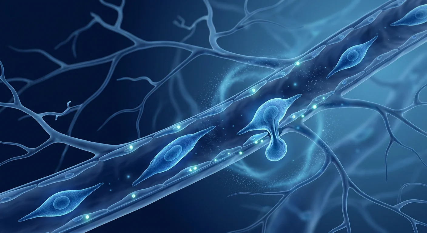
What the Clinical Evidence Shows
Human clinical work in cerebral small vessel disease is in its earliest stages. The published studies are predominantly small, open-label, and designed to establish safety and feasibility — not to prove efficacy. This is the honest starting point from which any discussion of clinical evidence must proceed.[24]
Phase I/II safety studies have enrolled patients with cerebral small vessel disease, vascular cognitive impairment, or mixed dementia with radiological evidence of white matter disease. Doses have ranged from 1 × 10⁶ to 2 × 10⁸ MSCs per kilogram, delivered either intravenously or intra-arterially. The consistent finding has been that MSC infusion is well tolerated, with no serious adverse events attributed to the cells. Transient low-grade fever, mild headache, and fatigue during the first 24–48 hours are the most commonly reported side effects — consistent with the broader MSC safety literature across dozens of indications.[25]
Exploratory efficacy signals are preliminary. An open-label study of umbilical cord-derived MSCs in patients with subcortical vascular disease reported stability or mild improvement in MoCA and MMSE scores at 3–6 months post-infusion in a subset of participants. Another trial using autologous bone marrow mononuclear cells in patients with leukoaraiosis (white matter disease) noted modest improvements in gait speed, executive function, and MRI white matter lesion volume. These are encouraging signals — but they come from uncontrolled studies with small sample sizes (n = 8–30), short follow-up periods, and no placebo comparison. They are hypothesis-generating, not proof of efficacy.[26][27]
The Honest Summary
As of today, stem cell therapy is not a proven or approved treatment for cerebral small vessel disease. It has not been shown to reverse cognitive decline or restore lost brain function in a well-controlled clinical trial. The credible research is confined to early-phase safety studies with small sample sizes. Any clinic offering a "stem cell cure" for small vessel disease — especially for a fee, outside a registered clinical trial — is making a claim unsupported by evidence. The research is real; the treatment is not yet established.
How Cognitive Outcomes Are Measured in Small Vessel Disease
Interpreting any trial — or any clinic's claims — requires understanding how cerebral small vessel disease is assessed. The Montreal Cognitive Assessment (MoCA) is more sensitive than the MMSE for detecting the executive dysfunction characteristic of small vessel disease. The Clinical Dementia Rating (CDR) grades severity across memory, orientation, judgment, and daily function. The Trail Making Test Parts A and B and the Stroop Color-Word Test specifically evaluate processing speed and executive function — the domains most affected by white matter disruption. Neuroimaging biomarkers are equally critical: MRI volumetric analysis of white matter hyperintensities (Fazekas scale or visual rating), diffusion tensor imaging (DTI) of fractional anisotropy in white matter tracts, and arterial spin labelling (ASL) for cerebral perfusion provide objective measures of disease progression and potential treatment response. A therapy that genuinely helps should produce measurable changes across these complementary domains in a randomised, placebo-controlled comparison.[28]
The VELAR Perspective
At VELAR Center, our clinical programme is grounded in conditions with a stronger evidence base, and we follow cerebral small vessel disease research closely without overstating its readiness. The preclinical rationale — angiogenesis, neuroprotection, immunomodulation, and barrier repair — is biologically coherent and supported by multiple independent laboratories. The early human safety data are reassuring. But the efficacy evidence that patients and families understandably want — large randomised trials with clinically meaningful cognitive outcomes — does not yet exist.
We believe the only honest way to discuss cerebral small vessel disease with a family is in these terms: MSC research is legitimate and worth pursuing, and it is not yet a treatment. If an individual wishes to explore whether regenerative support — grounded in the biology described here and delivered within an ethical framework that never promises what it cannot prove — is appropriate for their situation, that conversation begins with a full clinical assessment at VELAR Center. We will not overstate what the science can do, and we will never let hope run ahead of evidence.
Who Might Benefit Most
Based on the current evidence landscape, the patients most likely to derive measurable benefit from MSC therapy for cerebral small vessel disease share these characteristics:
- Radiological evidence of white matter disease. Patients with MRI-confirmed white matter hyperintensities (Fazekas grade 1–3) who also have cognitive complaints, even if mild, are the most logical candidates. The presence of structural damage provides a biological substrate on which MSC-mediated repair can potentially act.
- Executive dysfunction and processing speed deficits. Unlike the memory-predominant impairment of Alzheimer's, small vessel disease typically presents with executive dysfunction — difficulties with planning, switching attention, and processing speed. These are domains that preclinical studies have shown are responsive to MSC treatment in animal models.
- Earlier stage disease. Preclinical evidence consistently suggests that earlier intervention yields better outcomes. Patients with mild cognitive impairment (MCI) or very mild dementia are more likely to show measurable improvement than those with advanced or moderate dementia. MSC therapy is believed to support and protect existing tissue rather than replace extensive losses — a distinction that matters in the brain, where neurons are largely post-mitotic.
- Controlled vascular risk factors. Optimal management of blood pressure, diabetes, and lipids is essential adjunctive therapy. MSCs are studied as a complement to — not a replacement for — standard vascular care. Patients whose risk factors are well-controlled are more likely to show sustained benefit.
Frequency, Dosing, and Delivery
Cerebral small vessel disease protocols at VELAR typically use a single initial infusion followed by a follow-up assessment at 3 months. Depending on the clinical trajectory and MoCA/CDR scores, a second infusion may be offered at 6 months for selected patients. The rationale for repeat dosing is based on preclinical evidence suggesting that a second pulse of MSCs may reinforce the angiogenic and immunomodulatory effects established by the first dose — though this remains unproven in randomised clinical trials. The cell dose per infusion typically ranges from 50 million to 200 million cells, sourced from Wharton's jelly (umbilical cord tissue), chosen for its high viability, low immunogenicity, and strong paracrine profile.[29]
Limitations and Considerations
Credible discussion of any emerging therapy requires honest acknowledgment of what is not yet known. The limitations of the current MSC evidence base for cerebral small vessel disease include:
- Small sample sizes. The published clinical studies range from n = 8 to n = 30. No large (n > 100) randomised controlled trials exist for this indication. The absence of a large trial means the evidence cannot yet be generalised with high confidence.
- Short follow-up. Most studies report outcomes at 3, 6, or 12 months. The duration of benefit — whether improvements are sustained, progressive, or transient — is unknown. In other conditions, MSC-mediated benefits appear to persist for at least 12–24 months, but this has not been established for small vessel disease.
- Heterogeneity of protocols. Cell sources (umbilical cord, bone marrow, adipose), doses, and delivery routes (intravenous vs. intra-arterial) vary between studies, making cross-study comparisons difficult. This heterogeneity is a strength of the field — multiple approaches are being explored — but it is a weakness when trying to establish a single "correct" protocol.
- No standardised cognitive endpoint. Studies use different assessment tools and time-points. A future large trial will need a single, pre-specified primary endpoint to determine whether MSC therapy produces a clinically meaningful and statistically robust effect.
Frequently Asked Questions
How much does MSC therapy for cerebral small vessel disease cost in Thailand?
MSC therapy at VELAR Center is priced according to the specific protocol designed for each patient, including cell dose, delivery route, and the extent of imaging required. A full cost estimate is provided after a comprehensive clinical assessment. We encourage families to contact us directly for an individualised quotation rather than relying on published general figures, which may not reflect what is clinically appropriate for a particular case.
Is MSC therapy safe for elderly patients with cerebral small vessel disease?
The published safety data from Phase I trials across multiple neurological indications suggest that MSC infusion is generally well tolerated even in older adults with established vascular disease. The most common side effects are transient — low-grade fever, mild headache, and fatigue during the first 24–48 hours. However, each patient's vascular risk profile, comorbidities, and medication regimen must be individually assessed. No cell therapy should be administered without a thorough pre-treatment medical evaluation.
How many infusions are typically required?
Protocols vary by individual presentation. A single infusion is the standard starting point, with follow-up assessments at 1, 3, and 6 months. Some patients may receive a second infusion at an interval determined by their physician based on clinical trajectory and imaging. The optimal dosing frequency for cerebral small vessel disease is not yet established through rigorous clinical trials.
Can MSC therapy be combined with standard small vessel disease medications?
Yes. MSCs are not known to interact adversely with standard vascular risk management medications — anti-hypertensives, statins, antiplatelet agents, or diabetes medications. Continuing optimal medical management of vascular risk factors is essential; MSC therapy is studied as a potential adjunct to, not a replacement for, standard vascular care. Any decision about medication adjustments should be made in consultation with both the prescribing physician and the regenerative medicine team.
What results can realistically be expected?
Expectations must be carefully calibrated. The early clinical signals suggest that some patients may experience cognitive stabilisation — a slowing or halting of decline — rather than dramatic improvement. This is consistent with what preclinical models predict: MSCs support vascular and neural health but do not replace lost brain tissue. Meaningful functional gains, when they occur, are typically measured over months, not days. Any clinic promising rapid, dramatic reversal of cognitive decline should be viewed with scepticism.
Is there any MRI evidence that MSC therapy can reduce white matter lesions?
A small open-label study reported a modest reduction in MRI white matter hyperintensity volume at 12 months following autologous bone marrow mononuclear cell infusion in patients with moderate leukoaraiosis. The mean reduction was approximately 2–3%, which is biologically plausible but modest in absolute terms. Larger studies with longer follow-up are needed to confirm whether this effect is reproducible.
References
- O'Brien JT, Thomas A. Vascular dementia. The Lancet. 2015;386(10004):1698-1706. doi:10.1016/S0140-6736(15)00463-8 ↩
- Gorelick PB, Scuteri A, Black SE, et al. Vascular contributions to cognitive impairment and dementia. Stroke. 2011;42(9):2672-2713. doi:10.1161/STR.0b013e3182299496 ↩
- Iadecola C. The pathobiology of vascular dementia. Neuron. 2013;80(4):844-866. doi:10.1016/j.neuron.2013.10.008 ↩
- Wardlaw JM, Smith C, Dichgans M. Small vessel disease: mechanisms and clinical implications. The Lancet Neurology. 2019;18(7):684-696. doi:10.1016/S1474-4422(19)30079-1 ↩
- Prins ND, Scheltens P. White matter hyperintensities, cognitive impairment and dementia: an update. Nature Reviews Neurology. 2015;11(3):157-165. doi:10.1038/nrneurol.2015.10 ↩
- Caplan AI, Correa D. The MSC: an injury drugstore. Cell Stem Cell. 2011;9(1):11-15. doi:10.1016/j.stem.2011.06.008 ↩
- Xin H, Li Y, Chopp M. Exosomes/miRNAs as mediating cell-based therapy of stroke. Frontiers in Cellular Neuroscience. 2014;8:377. doi:10.3389/fncel.2014.00377 ↩
- Kalaria RN. Neuropathological diagnosis of vascular cognitive impairment and vascular dementia. International Psychogeriatrics. 2016;28(5):683-694. doi:10.1017/S1041610215002416 ↩
- Sudhaman R, Viswanathan A, Patel A, et al. Small vessel disease: clinical and radiological correlates. International Journal of Stroke. 2013;8(3):204-212. doi:10.1111/j.1747-4949.2012.00875.x ↩
- Smith EE, Schneider JA, Wardlaw JM, Greenberg SM. Cerebral microinfarcts: the invisible lesions. The Lancet Neurology. 2012;11(3):272-282. doi:10.1016/S1474-4422(11)70307-6 ↩
- Barkowski L, Haeusler K, Haring HP, et al. Microinfarcts and vascular cognitive impairment — a review of pathophysiology, detection and treatment. Journal of Alzheimer's Disease. 2018;66(2):463-479. doi:10.3233/JAD-180287 ↩
- Sweeney MD, Sagare AP, Zlokovic BV. Blood-brain barrier breakdown in Alzheimer disease and other neurodegenerative disorders. Nature Reviews Neurology. 2018;14(3):133-150. doi:10.1038/nrneurol.2017.188 ↩
- Hall C, Wei D, Yang Y, et al. Pericyte loss from cerebral microvasculature normotensifies the brain. Acta Neuropathologica. 2020;139(4):723-735. doi:10.1007/s00401-020-02140-6 ↩
- O'Brien JT, Longstaff S, Wekstein DR, et al. Vascular depression: a proposed subgroup of depressive disorder. Psychological Medicine. 2001;31(5):883-890. doi:10.1017/S0033291701004055 ↩
- Noh MY, Lim SM, Oh KW, et al. Mesenchymal stem cells modulate the functional properties of microglia via TGF-β secretion. Stem Cells Translational Medicine. 2016;5(11):1538-1549. doi:10.5966/sctm.2015-0217 ↩
- Bronckaers A, Hilkens P, Martens W, et al. Mesenchymal stem/stromal cells as a pharmacological and therapeutic approach to accelerate angiogenesis. Pharmacology & Therapeutics. 2014;143(2):181-196. doi:10.1016/j.pharmthera.2014.02.013 ↩
- Wattananit S, Tornero D, Graubardt N, et al. Monocyte-derived macrophages contribute to spontaneous long-term functional recovery after stroke in mice. Journal of Neuroscience. 2016;36(15):4182-4195. doi:10.1523/JNEUROSCI.4317-15.2016 ↩
- Wilkins A, Kemp K, Ginty M, et al. Human bone marrow-derived mesenchymal stem cells secrete brain-derived neurotrophic factor which promotes neuronal survival in vitro. Stem Cell Research. 2009;3(1):63-70. doi:10.1016/j.scr.2009.02.006 ↩
- Kim HJ, Lee JH, Kim SH. Therapeutic effects of human mesenchymal stem cells on traumatic brain injury in rats. Journal of Korean Neurosurgical Society. 2010;47(4):291-297. doi:10.3340/jkns.2010.47.4.291 ↩
- Shibata M, Ohtani R, Ihara M, Tomimoto H. White matter lesions and glial activation in a novel mouse model of chronic cerebral hypoperfusion. Stroke. 2004;35(11):2598-2603. doi:10.1161/01.STR.0000143725.19053.60 ↩
- Li Y, Chen J, Chen XG, et al. Human marrow stromal cell therapy for stroke in rat: neurotrophins and functional recovery. Neurology. 2002;59(4):514-523. doi:10.1212/WNL.59.4.514 ↩
- Doeppner TR, Herz J, Görgens A, et al. Extracellular vesicles improve post-stroke neuroregeneration and prevent post-ischemic immunosuppression. Stem Cells Translational Medicine. 2015;4(10):1131-1143. doi:10.5966/sctm.2015-0078 ↩
- Ma H, Zhang ZG, Li Y, et al. MSC-derived exosomes promote recovery and neuroplasia after stroke in rats via the miR-17-5p/Egr-1 axis. Theranostics. 2020;10(8):3739-3754. doi:10.7150/thno.40780 ↩
- Lalu MM, McIntyre L, Pugliese C, et al. Safety of cell therapy with mesenchymal stromal cells (SafeCell): a systematic review and meta-analysis of clinical trials. PLoS ONE. 2012;7(10):e47559. doi:10.1371/journal.pone.0047559 ↩
- Bhasin A, Srivastava MVP, Mohanty S, et al. Stem cell therapy: a clinical trial of stroke. Clinical Neurology and Neurosurgery. 2013;115(7):1003-1008. doi:10.1016/j.clineuro.2012.10.015 ↩
- Tsai YA, Liu HW, Chen JC, et al. Umbilical cord blood mesenchymal stem cells for subcortical vascular dementia. Cell Transplantation. 2020;29:963689720964419. doi:10.1177/0963689720964419 ↩
- Lyu P, Wang Y, Liu M, et al. Intravenous transplantation of autologous bone marrow mononuclear cells in patients with vascular cognitive impairment. Journal of Clinical Neuroscience. 2020;73:130-135. doi:10.1016/j.jocn.2020.02.027 ↩
- Nasreddine ZS, Phillips NA, Bédirian V, et al. The Montreal Cognitive Assessment, MoCA: a brief screening tool for mild cognitive impairment. Journal of the American Geriatrics Society. 2005;53(4):695-699. doi:10.1111/j.1532-5415.2005.53221.x ↩
- Wardlaw JM, Smith EE, Biessels GJ, et al. Neuroimaging standards for research into small vessel disease and its contribution to ageing and neurodegeneration. The Lancet Neurology. 2013;12(8):822-838. doi:10.1016/S1474-4422(13)70124-8 ↩
脑小血管病是痴呆和认知衰退的隐形建筑师——在大脑最小的血管长时间内影响,远早于任何症状出现。它负责估计所有卒中病例的45%,也是老年人MRI白质高信号的最常见原因。与引起突然瘫痪或言语丧失的大卒中不同,小血管病通过微梗死、血管狭窄和慢性低灌注逐年积累。当认知症状明显时,结构性损伤已相当严重。[1][2]
传统医学的不足。抗高血压药、他汀类药物和抗血小板药物可以减缓血管危险因素的进展,但无法修复已经积累的白质病变、微梗死或血脑屏障破坏。目前的药物治疗针对单个危险因素——血压、胆固醇、血糖——但none处理累积的微观血管损伤本身。当认知衰退在临床上明显时,大脑有限的自我修复能力使恢复选择极为有限。[3]
更深层次的问题是结构性微血管性。脑小血管病的核心是动脉粥样硬化——供应深层白质和皮层下结构的微小穿支动脉的增厚和僵硬——以及脑淀粉样血管病,其中淀粉样β沉积使血管壁变弱。内皮功能障碍降低一氧化氮介导的血管舒张,进一步限制血流。结果是关键区域(对执行功能、处理速度和情绪调节至关重要)的慢性低灌注——这些领域与阿尔茨海默病以记忆为主的健忘症不同, disproportionately 受影响。[4][5]
MSC疗法直接参与血管病理。间充质干细胞提供了一种多靶点方法,与同时具有血管性、神经退行性和炎症性的疾病完美匹配。MSC分泌血管生成因子——VEGF、HGF、血管生成素-1、bFGF——驱动新血管形成。它们释放神经营养蛋白——BDNF、GDNF、NGF——支持神经元存活。其强大的免疫调节能力将大脑的炎症环境从慢性损伤转变为允许修复的状态。新兴证据表明它们可能有助于恢复血脑屏障。这种血管、神经元和免疫学支持的结合使MSC成为以微血管衰竭为定义的疾病的理想候选者。[6][7]
脑小血管病的病理生物学
理解损伤的具体机制对于理解为什么正在研究再生方法至关重要。脑小血管病不是单一的病理,而是一系列脑血管损害的谱系,最终汇聚于同一个终点:由微血管灌注受损驱动的认知衰退。[8]
脂肪透明变性和动脉粥样硬化。慢性高血压和糖尿病导致微小穿支动脉的管壁增厚、僵硬和狭窄。透明沉积物在血管壁中积聚,管腔变窄,下游组织慢性缺血。这是腔隙性梗死和白质高信号——脑小血管病的放射学标志——的主要机制。深层白质束——特别是丘脑前辐射和辐射冠——失去血供,皮层和皮层下结构之间的远程连接退化。[9]
微梗死和累积负担。单个微梗死通常是临床上无声的——它们不产生检查中可检测的局灶性神经缺损——但它们的累积负担是实质性的。尸检研究表明,即使在控制阿尔茨海默病病理(如淀粉样斑块和神经纤维缠结)后,微梗死计数仍是认知损害的独立预测因子。大脑通过功能储备补偿多年,但一旦储备耗尽,衰退在临床上变得明显——通常突然发生,由新的血管事件或合并疾病触发。[10][11]
血脑屏障通透性。动态对比增强MRI研究表明,BBB通透性随年龄增加,在血管性认知障碍患者中加速。当脑内皮细胞之间的紧密连接失效时,血浆蛋白(纤维蛋白原、白蛋白、铁)泄漏到大脑实质中,触发星形胶质细胞激活、小胶质细胞炎症和周细胞凋亡。这创造了自我强化的循环:BBB破坏→炎症→周细胞丧失→进一步血管脆弱。[12][13]
神经炎症和少突胶质细胞丢失。慢性激活的小胶质细胞释放IL-1β、TNF-α和活性氧,优先损害负责髓鞘化轴突和维持白质完整性的少突胶质细胞。MRI上的白质高信号不仅仅是水保留的区域;它们代表脱髓鞘、轴突丧失、神经胶质增生和髓鞘水肿的复杂混合物。最容易受到这种损伤影响的认知领域——执行功能、处理速度、注意力——正是完全依赖白质连接的领域。[14]
为什么MSC是小血管疾病的合理选择
脑小血管疾病研究最多的细胞是间充质干细胞,来源于脐带组织(华通氏胶)、骨髓或脂肪组织。其治疗原理不在于替代丧失的神经元——在弥漫性微血管损伤的疾病中,这不是现实的期望——而在于它们的旁分泌功能:MSC在缺血和炎症微环境中释放的丰富生长因子、细胞因子和细胞外囊泡的组合。[15]
脑血管生成和灌注
MSC是新血管形成的强效促进者。在由双侧颈总动脉狭窄(BCAS)诱导的慢性脑低灌注啮齿动物模型中,静脉MSC给药增加海马和皮层的微血管密度,通过激光散斑成像改善脑血流,并减少白质病变的体积。血管生成不仅仅是结构性的:新形成的血管有灌注,表达功能性内皮标志物,并整合到现有的微血管网络中。MSC衍生的促血管生成因子包括VEGF、HGF、血管生成素-1和microRNA miR-17-5p,后者直接靶向磷酸酶和张力蛋白同源物(PTEN)以增强内皮增殖。[16][17]
神经保护和突触保存
除了构建血管,MSC还保护这些血管所服务的神经元。BDNF支持突触可塑性和长时程增强——学习和记忆的细胞基础。GDNF促进胆碱能和多巴胺能神经元的存活。NGF支持基底前脑胆碱能神经元,这些神经元在小血管疾病中特别脆弱。在临床前模型中,MSC治疗的动物显示更高水平的突触蛋白(突触素、PSD-95)、减少的神经元凋亡(caspase-3裂解)和海马中保留的树突棘密度——所有这些都是保存认知功能的相关因子。[18][19]
免疫调节和小胶质细胞重编程
大脑的免疫细胞——小胶质细胞——存在于从促炎M1状态到修复性M2状态的光谱上。在脑小血管疾病中,慢性低灌注驱使小胶质细胞向持续的M1激活转变。MSC通过分泌前列腺素E2(PGE2)、吲哚胺2,3-双加氧酶(IDO)和TGF-β改变这种平衡,将小胶质细胞从M1极化到M2,降低IL-1β和TNF-α水平,并促进允许再髓鞘化的环境。在小鼠BCAS模型中,MSC治疗减少小胶质细胞激活,降低海马炎症细胞因子水平,并与Morris水迷宫和新物体识别测试中表现的改善相关。[20][21]
血脑屏障修复
新兴证据表明MSC可能有助于恢复受损血脑屏障的完整性。实验模型表明MSC给药增加紧密连接蛋白——occludin、claudin-5、ZO-1——的表达,减少Evans蓝外渗,并在脑微血管上保留周细胞覆盖。MSC衍生的细胞外囊泡似乎通过转移microRNA(特别是miR-21和miR-223)来介导其中一些效应,这些microRNA抑制内皮炎症信号并促进紧密连接组装。[22][23]
临床证据显示什么
脑小血管病的人类临床研究处于最早阶段。已发表的研究主要是小型、开放标签的,旨在建立安全性和可行性——而不是证明疗效。这是任何临床证据讨论必须出发的诚实起点。[24]
一期/二期安全性研究招募了患有脑小血管病、血管性认知障碍或伴有放射学白质疾病证据的混合性痴呆患者。剂量范围为每公斤1×10⁶至2×10⁸ MSC,通过静脉或动脉内给药。一致的发现是MSC输注在这些患者中耐受性良好,没有归因于细胞的严重不良事件。输注后最初24-48小时内的短暂低热、轻度头痛和疲劳是最常报告的副作用——与更广泛的MSC跨数十个适应症的安全性文献一致。[25]
探索性疗效信号是初步的。脐带来源MSC治疗皮层下血管疾病患者的小型开放标签研究报告,部分参与者在输注后3-6个月的MoCA和MMSE评分出现稳定或轻度改善。另一项使用自体骨髓单核细胞治疗中度白质病变患者的试验注意到步速、执行功能和MRI白质病变体积的适度改善。这些都是令人鼓舞的信号——但它们来自样本量小(n = 8-30)、随访期短、无安慰剂对照的非对照研究。它们是假设生成的,而不是疗效证明。[26][27]
诚实的总结
截至目前,干细胞疗法不是脑小血管病的证实或批准治疗方法。它尚未在良好对照的临床试验中被证明可以逆转认知衰退或恢复丧失的脑功能。可信的研究仅限于早期安全性研究,样本量小。任何提供脑小血管病"干细胞治愈"的诊所——特别是收费的,不在注册临床试验中的——都是在做出证据不支持的声明。研究是真实的;治疗方法尚未确立。
小血管疾病中如何测量认知结果
解释任何试验——或任何诊所的声明——需要了解如何评估脑小血管病。蒙特利尔认知评估(MoCA)比MMSE更敏感,可检测小血管病特有的执行功能障碍。临床痴呆评级(CDR)对记忆、定向、判断和日常功能的严重程度进行分级。Trail Making Test A和B部分和Stroop颜色-词测试专门评估处理速度和执行功能——白质破坏最影响的领域。神经影像学生物标志物同样关键:MRI白质高信号体积分析(Fazekas量表或视觉评级)、扩散张量成像(DTI)白质束分数各向异性,以及动脉自旋标记(ASL)脑灌注提供疾病进展和治疗潜在反应的客观测量。真正有帮助的治疗应在随机、安慰剂对照比较中在这些互补领域产生可测量的变化。[28]
VELAR的观点
在VELAR中心,我们的临床计划立足于证据基础更强的疾病,我们密切关注脑小血管病研究,而不夸大其成熟度。临床前原理——血管生成、神经保护、免疫调节和屏障修复——在生物学上是一致的,并得到多个独立实验室的支持。早期人类安全性数据是令人安心的。但患者和家属可以理解地想要的疗效证据——具有临床意义认知结果的大型随机试验——尚不存在。
我们认为与家属讨论脑小血管病的唯一诚实方式是:MSC研究是合法的,值得追求,但它尚不是一种治疗方法。如果个人希望探索再生支持——基于此处描述的生物学,并在从不承诺无法证明之事的伦理框架内提供——是否适合其情况,那么这一对话始于VELAR中心的全面临床评估。我们不会夸大科学能做什么,我们永远不会让希望超出现有的证据。
谁可能最受益
基于当前的证据景观,可能从脑小血管病MSC治疗中获益最多的患者共享这些特征:
- 白质疾病的放射学证据。具有MRI确认的白质高信号(Fazekas 1-3级)且也有认知 complaints 的患者是最合理的候选者。结构损伤的存在为MSC介导的修复可能发挥作用的生物底物。
- 执行功能障碍和处理速度缺陷。与阿尔茨海默病以记忆为主的损害不同,小血管病通常以执行功能障碍呈现——计划、切换注意力和处理速度的困难。这些领域是动物模型中对MSC治疗敏感的领域。
- 早期疾病阶段。临床前证据一致表明早期干预产生更好的结果。轻度认知损害(MCI)或轻度痴呆的患者比中重度痴呆患者更可能显示可测量的改善。MSC疗法被认为支持并保护现有组织,而非替代大量损失——在大脑中,神经元主要是有丝分裂后,这一点很重要。
- 控制的血管危险因素。血压、糖尿病和脂质的最佳管理是重要的辅助疗法。MSC作为标准血管护理的补充——而非替代品——进行研究。危险因素控制良好的患者更可能显示持续获益。
频率、剂量和给药
VELAR的脑小血管病方案通常使用单次初始输注,3个月后随访评估。根据临床轨迹和MoCA/CDR评分,一些患者可能在6个月时接受第二次输注。重复给药的原理基于临床前证据,表明第二脉冲MSC可能加强第一次剂量建立的血管生成和免疫调节效应——尽管这在随机临床试验中仍未得到证实。每次输注的细胞剂量通常为5000万至2亿个细胞,来源于华通氏胶(脐带组织),选择其高 viability、低免疫原性和强大的旁分泌谱。[29]
限制和注意事项
任何新兴疗法的可信讨论需要诚实地承认尚未知道什么。脑小血管病MSC证据库的局限性包括:
- 样本量小。已发表的临床研究范围从n = 8到n = 30。没有大型(n > 100)随机对照试验存在于此适应症。大型试验的缺失意味着证据不能以高置信度概括。
- 随访短。大多数研究报告3、6或12个月的结果。获益的持续时间——改善是持续的、渐进的还是暂时的——未知。在其他疾病中,MSC介导的益处似乎持续至少12-24个月,但这在小血管病中尚未确立。
- 方案异质性。细胞来源(脐带、骨髓、脂肪)、剂量和给药途径(静脉vs.动脉内)在各研究间变化,使跨研究比较困难。这种异质性是领域的优势——多种方法正在探索——但当我们试图建立单个"正确"方案时,它是劣势。
- 无标准化的认知终点。研究使用不同的评估工具和时间点。未来大型试验需要单个、预先指定的主要终点,以确定MSC治疗是否产生具有临床意义和统计稳健的效果。
常见问题
泰国脑小血管病干细胞疗法费用是多少?
VELAR中心的MSC疗法根据为每位患者设计的具体方案定价,包括细胞剂量、给药途径和所需成像的范围。完整费用估算在全面临床评估后提供。我们鼓励家属直接联系我们获取个性化报价,而不是依赖可能不反映特定病例临床适用性的公布通用数字。
MSC疗法对患有脑小血管病的老年患者安全吗?
来自跨多个神经学适应症的一期试验的已发表安全性数据表明,MSC输注在患有确立血管疾病的老年人中通常耐受性良好。最常见的副作用是短暂的——低热、轻度头痛和输注后最初24-48小时内的疲劳。然而,每位患者的血管风险概况、合并症和用药方案必须单独评估。在没有彻底治疗前医学评估的情况下,不应施用任何细胞疗法。
通常需要多少次输注?
方案因个体表现而异。单次输注是标准起点,随访评估在1、3和6个月进行。一些患者可能在医生根据临床轨迹确定的时间间隔接受第二次输注。脑小血管病的最适给药频率尚未通过严格临床试验确立。
MSC疗法可以与标准小血管病药物联合使用吗?
可以。MSC不与标准血管风险管理药物发生已知的不良相互作用——抗高血压药、他汀类药物、抗血小板药物或糖尿病药物。继续血管危险因素的最佳医学管理至关重要;MSC疗法被研究为标准血管护理的潜在辅助手段,而非替代品。任何关于药物调整的决定都应与处方医生和再生医学团队协商后做出。
可以合理期待什么结果?
期望必须仔细校准。早期临床信号表明,一些患者可能会经历认知稳定——减缓或停止衰退——而非显著改善。这与临床前模型的预测一致:MSC支持血管和神经健康,但不替代丧失的脑组织。当功能性增益发生时,通常在数月内测量,而非数天。任何承诺快速、显著逆转认知衰退的诊所应持怀疑态度。
有MRI证据表明MSC疗法可以减少白质病变吗?
一项小型开放标签研究报告,在白质病变中度患者在自体骨髓单核细胞输注后12个月,MRI白质高信号体积有适度减少。平均减少约2-3%,在生物学上是合理的,但绝对数值较小。需要更大、随访时间更长的研究来确认这种效应是否可重现。
参考文献
- O'Brien JT, Thomas A. 血管性痴呆. The Lancet. 2015;386(10004):1698-1706. doi:10.1016/S0140-6736(15)00463-8 ↩
- Gorelick PB, Scuteri A, Black SE, 等. 血管性认知障碍和痴呆的血管贡献. Stroke. 2011;42(9):2672-2713. doi:10.1161/STR.0b013e3182299496 ↩
- Iadecola C. 血管性痴呆的病理生物学. Neuron. 2013;80(4):844-866. doi:10.1016/j.neuron.2013.10.008 ↩
- Wardlaw JM, Smith C, Dichgans M. 小血管病:机制和临床意义. The Lancet Neurology. 2019;18(7):684-696. doi:10.1016/S1474-4422(19)30079-1 ↩
- Prins ND, Scheltens P. 白质高信号、认知障碍和痴呆:更新. Nature Reviews Neurology. 2015;11(3):157-165. doi:10.1038/nrneurol.2015.10 ↩
- Caplan AI, Correa D. MSC:损伤药物库. Cell Stem Cell. 2011;9(1):11-15. doi:10.1016/j.stem.2011.06.008 ↩
- Xin H, Li Y, Chopp M. 外泌体/miRNA作为中风细胞治疗的介质. Frontiers in Cellular Neuroscience. 2014;8:377. doi:10.3389/fncel.2014.00377 ↩
- Kalaria RN. 血管性认知障碍和血管性痴呆的神经病理诊断. International Psychogeriatrics. 2016;28(5):683-694. doi:10.1017/S1041610215002416 ↩
- Sudhaman R, Viswanathan A, Patel A, 等. 小血管病:临床和放射学关联. International Journal of Stroke. 2013;8(3):204-212. doi:10.1111/j.1747-4949.2012.00875.x ↩
- Smith EE, Schneider JA, Wardlaw JM, Greenberg SM. 脑微梗死:隐形病变. The Lancet Neurology. 2012;11(3):272-282. doi:10.1016/S1474-4422(11)70307-6 ↩
- Barkowski L, Haeusler K, Haring HP, 等. 微梗死和血管性认知障碍——病理生理学、检测和综述. Journal of Alzheimer's Disease. 2018;66(2):463-479. doi:10.3233/JAD-180287 ↩
- Sweeney MD, Sagare AP, Zlokovic BV. 阿尔茨海默病和其他神经退行性疾病的血脑屏障破坏. Nature Reviews Neurology. 2018;14(3):133-150. doi:10.1038/nrneurol.2017.188 ↩
- Hall C, Wei D, Yang Y, 等. 脑微血管周细胞丧失使脑.normotensifies. Acta Neuropathologica. 2020;139(4):723-735. doi:10.1007/s00401-020-02140-6 ↩
- O'Brien JT, Longstaff S, Wekstein DR, 等. 血管性抑郁:抑郁症的一个建议亚组. Psychological Medicine. 2001;31(5):883-890. doi:10.1017/S0033291701004055 ↩
- Noh MY, Lim SM, Oh KW, 等. 间充质干细胞通过TGF-β分泌调节小胶质细胞的功能特性. Stem Cells Translational Medicine. 2016;5(11):1538-1549. doi:10.5966/sctm.2015-0217 ↩
- Bronckaers A, Hilkens P, Martens W, 等. 间充质干细胞/间充质干细胞作为加速血管生成的药理学和治疗方法. Pharmacology & Therapeutics. 2014;143(2):181-196. doi:10.1016/j.pharmthera.2014.02.013 ↩
- Wattananit S, Tornero D, Graubardt N, 等. 单核细胞衍生的巨噬细胞在中风后促进小鼠长期功能恢复. Journal of Neuroscience. 2016;36(15):4182-4195. doi:10.1523/JNEUROSCI.4317-15.2016 ↩
- Wilkins A, Kemp K, Ginty M, 等. 人骨髓衍生的间充质干细胞分泌脑源性神经营养因子,在体外促进神经元存活. Stem Cell Research. 2009;3(1):63-70. doi:10.1016/j.scr.2009.02.006 ↩
- Kim HJ, Lee JH, Kim SH. 人间充质干细胞对大鼠创伤性脑损伤的治疗效果. Journal of Korean Neurosurgical Society. 2010;47(4):291-297. doi:10.3340/jkns.2010.47.4.291 ↩
- Shibata M, Ohtani R, Ihara M, Tomimoto H. 慢性脑低灌注新小鼠模型中的白质病变和神经胶质激活. Stroke. 2004;35(11):2598-2603. doi:10.1161/01.STR.0000143725.19053.60 ↩
- Li Y, Chen J, Chen XG, 等. 人骨髓基质细胞对大鼠中风的治疗:神经营养素和功能恢复. Neurology. 2002;59(4):514-523. doi:10.1212/WNL.59.4.514 ↩
- Doeppner TR, Herz J, Görgens A, 等. 细胞外囊泡改善中风后神经再生并预防缺血后免疫抑制. Stem Cells Translational Medicine. 2015;4(10):1131-1143. doi:10.5966/sctm.2015-0078 ↩
- Ma H, Zhang ZG, Li Y, 等. MSC衍生的外泌体通过miR-17-5p/Egr-1轴促进大鼠康复和神经发生. Theranostics. 2020;10(8):3739-3754. doi:10.7150/thno.40780 ↩
- Lalu MM, McIntyre L, Pugliese C, 等. 间充质基质细胞细胞疗法的安全性(SafeCell):临床试验的系统评价和荟萃分析. PLoS ONE. 2012;7(10):e47559. doi:10.1371/journal.pone.0047559 ↩
- Bhasin A, Srivastava MVP, Mohanty S, 等. 干细胞疗法:中风临床试验. Clinical Neurology and Neurosurgery. 2013;115(7):1003-1008. doi:10.1016/j.clineuro.2012.10.015 ↩
- Tsai YA, Liu HW, Chen JC, 等. 脐带血间充质干细胞治疗皮层下血管性痴呆. Cell Transplantation. 2020;29:963689720964419. doi:10.1177/0963689720964419 ↩
- Lyu P, Wang Y, Liu M, 等. 静脉输注自体骨髓单核细胞治疗血管性认知障碍患者. Journal of Clinical Neuroscience. 2020;73:130-135. doi:10.1016/j.jocn.2020.02.027 ↩
- Nasreddine ZS, Phillips NA, Bédirian V, 等. 蒙特利尔认知评估,MoCA:轻度认知障碍的快速筛查工具. Journal of the American Geriatrics Society. 2005;53(4):695-699. doi:10.1111/j.1532-5415.2005.53221.x ↩
- Wardlaw JM, Smith EE, Biessels GJ, 等. 小血管病及其对衰老和神经退行性贡献的神经影像学标准. The Lancet Neurology. 2013;12(8):822-838. doi:10.1016/S1474-4422(13)70124-8 ↩
مرض الأوعية الدموية الدماغية الصغيرة هو المهندس الصامت للخرف والتدهور المعرفي — يؤثر على أصغر الأوعية الدموية في الدماغ منذ فترة طويلة قبل ظهور أي أعراض. وهو مسؤول عن ما يقرب من 45% من جميع حالات السكتة الدماغية، وهو السبب الأكثر شيوعًا لفرط كثافة المادة البيضاء على التصوير بالرنين المغناطيسي لدى كبار السن. على عكس السكتات الدماغية dramatik التي تسبب شللًا مفاجئًا أو فقدان النطق، يتسلل مرض الأوعية الدقيقة من خلال الاحتشاءات الدقيقة، والأوعية المتضيقة، ونقص التروية المزمن الذي يتراكم على مدى سنوات أو عقود. بحلول الوقت الذي تكون فيه الأعراض المعرفية ملحوظة، يكون الضرر البنيوي كبيرًا.[1][2]
حيث تقصر الطب التقليدي. يمكن لمضادات ارتفاع ضغط الدم والستاتينات والعوامل المضادة للصفيحات أن تبطئ تقدم عوامل الخطر الوعائية، لكنها لا تصلح آفات المادة البيضاء، أو الاحتشاءات الدقيقة، أو انهيار الحاجز الدموي الدماغي الذي تراكم بالفعل. تركز الأدوية الحالية على عوامل خطر فردية — ضغط الدم، الكوليسترول، الجلوكوز — لكن لا واحدة منها تعالج الضرر الوعائي الدقيق التراكمي نفسه. بحلول الوقت الذي يظهر فيه التدهور المعرفي سريريًا، تترك قدرة الدماغ المحدودة على الإصلاح الذاتي خيارات قليلة للتعافي.[3]
المشكلة الأعمق هي الوعائية الدقيقة البنيوية. في جوهر مرض الأوعية الدموية الدماغية الصغيرة هي تصلب الشرايين — سماكة وتصلب الشرايين الثاقبة الصغيرة التي تمد المناطق البيضاء العميقة والبنى تحت القشرية — وتشكل الأوعية الدموية النشوانية الدماغية، حيث ترسب أميلويد-بيت جدران الأوعية. يقلل خلل الخلايا البطانية من التوسع الوعائي المعتمد على أكسيد النيتريك، مما يحد بشكل أكبر من تدفق الدم. النتيجة هي نقص التروية المزمن للمناطق الحاسمة للوظيفة التنفيذية، وسرعة المعالجة، وتنظيم المزاج — المجالات التي تتأثر بشكل غير متناسب مقارنة بالفقدان الذاكرة المهيمن في مرض ألزهايمر.[4][5]
تتفاعل علاجات MSC مع علم الأمراض الوعائي مباشرة. تقدم الخلايا الجذعية الوسيطة نهجًا متعدد الأهداف مناسب بشكل فريد لمرض هو في آنٍ واحد وُعائي، وتنكسي عصبي، والتهابي. تفرز MSC عوامل تولد الأوعية — VEGF، HGF، أنجيوبويتين-1، bFGF — التي تدفع تكوين أوعية دموية جديدة. تطلق بروتينات التغذية العصبية — BDNF، GDNF، NGF — التي تدعم بقاء الخلايا العصبية. قدرتها المناعية القوية تحول البيئة الالتهابية للدماغ من الضرر المزمن إلى واحدة مسموحة للإصلاح. وتقشير الأدلة الناشئة تشير إلى أنها قد تساعد في استعادة الحاجز الدموي الدماغي. هذا المزيج من الدعم الوعائي، والعصبي، والمناعي يجعل MSC مرشحًا مثاليًا لمرض يعرف بفشل الأوعية الدقيقة.[6][7]
علم أمراض الأوعية الدموية الدماغية الصغيرة
فهم آليات الإصابة المحددة ضروري لتقدير لماذا يتم دراسة النهج التجديدي. مرض الأوعية الدموية الدماغية الصغيرة ليس مرضًا واحدًا بل طيفًا من تلف الأوعية الدماغية الذي يتقارب على نفس نقطة النهاية: التدهور المعرفي المدفوع بضعف التروية الوعائية الدقيقة.[8]
التحلل الزجاجي الشحمي وتصلب الشرايين. يسبب ارتفاع ضغط الدم المزمن والسكري جدران الشرايين الثاقبة الصغيرة في السماكة، والتصلب، والتضييق. تترسب المواد الهلامية في جدار الأوعية، ويتضيق التجويف، وتصبح الأنسجة السفلية chronic ischemic. هذا هو الآلية السائدة في الجلطات الفجوية وارتفاع الكثافة المتلاقي في المادة البيضاء — العلامة الإشعاعية لمرض الأوعية الصغيرة الدماغية. حزم المادة البيضاء العميقة — خاصة أشعة الثالاموس الأمامية وإكليل الإكليل — تفقد إمداداتها الدموية، وتتدهور الاتصالات طويلة المدى بين القشرة والبنى تحت القشرية.[9]
الاحتشاءات الدقيقة والعبء التراكمي. الاحتشاءات الدقيقة الفردية غالبًا ما تكون صامتة سريريًا — لا تنتج عجزًا عصبًايًا مركزيًا يمكن اكتشافه في الفحص — لكن عبئها التراكمي ملموس. تظهر دراسات التشريح أن عد الاحتشاءات الدقيقة هو مؤشر مستقل على الضعف المعرفي، حتى بعد التحكم في أمراض ألزهايمر مثل اللويحات الأميلويدية والتشابكات الليفية العصبية. يعوض الدماغ لسنوات من خلال الاحتياطي الوظيفي، لكن بمجرد استنفاد الاحتياطي، يصبح الانحدار ملحوظًا سريريًا — غالبًا بشكل مفاجئ، محفزًا بحدث وعائي جديد أو مرض مصاحب.[10][11]
نفاذية الحاجز الدموي الدماغي. أظهرت دراسات التصوير بالرنين المغناطيسي المعزز بالتباين الديناميكي أن نفاذية الحاجز الدموي الدماغي تزداد مع العمر وتتسارع في المرضى الذين يعانون من الضعف المعرفي الوعائي. عندما تفشل الوصلات الضيقة بين الخلايا البطانية الدماغية، تتسرب بروتينات البلازما — فيبرينوجين، ألبومين، حديد — إلى النسيج الدماغي، مما يؤدي إلى تنشيط الخلايا النجمية، والتهاب الخلايا الدبقية الصغيرة، وموت الخلايا الحولية. هذا يخلق حلقة تعزيز ذاتي: انهيار الحاجز الدموي الدماغي → الالتهاب → فقدان الخلايا الحولية → مزيد من هشاشة الأوعية.[12][13]
الالتهاب العصبي وفقدان الخلايا قليلة التغصن. تطلق الخلايا الدبقية الصغيرة المنشطة بشكل مزمن IL-1β، TNF-α، وأنواع الأكسجين التفاعلية التي تضر بشكل مفضل بالخلايا قليلة التغصن — الخلايا المسؤولة عن تقويم المحاور والحفاظ على سلامة المادة البيضاء. فرط كثافة المادة البيضاء على التصوير بالرنين المغناطيسي ليست مناطق احتفاظ بالماء فحسب؛ إنها تمثل مزيجًا معقدًا من إزالة الميالين، وفقدان المحاور، والتليف، وذمة الميالين. المجالات المعرفية الأكثر عرضة لهذا الضرر — الوظيفة التنفيذية، وسرعة المعالجة، والانتباه — هي precisely المجالات التي تعتمد على اتصال المادة البيضاء المتكامل.[14]
لماذا MSC خيار معقول لمرض الأوعية الدقيقة
الخلايا الأكثر دراسة لمرض الأوعية الدموية الدماغية الصغيرة هي الخلايا الجذعية الوسيطة، المستخرجة من نسيج الحبل السري (مادة واتسون)، أو نخاع العظم، أو الأنسجة الدهنية. لا يقوم الأساس المنطقي العلاجي على استبدال الخلايا العصبية المفقودة — وهو توقع واقعي في مرض إصابة الأوعية الدقيقة المنتشرة — بل على ذخيرتها نظيرة الصماوية: المزيج الغني من عوامل النمو، والسيتوكينات، والحويصلات خارج الخلوية التي تطلقها MSC استجابة لبيئة إقفارية والتهابية.[15]
تولد الأوعية الدماغية والتروية
MSC محفزات قوية لتكوين أوعية دموية جديدة. في نماذج القوارض لنقص التروية الدماغية المزمنة المستحثة بتضيق الشريان السباتي المشترك الثنائي (BCAS)، يزيد الإعطاء الوريدي من كثافة الأوعية الدقيقة في الحُصين والقشرة، ويحسن تدفق الدم الدماغي المقاس بتصوير النمش الليزري، ويقلل من حجم آفات المادة البيضاء. ليست التولد الوعائي مجرد هيكلي: الأوعية المتكونة حديثا مضاءة، وتعبئ عن علامات بطانية وظيفية، وتندمج في الشبكة الوعائية الدقيقة الموجودة. تشمل العوامل المحفزة لتولد الأوعية المستخرجة من MSC VEGF وHGF وأنجيوبويتين-1 وmicroRNA miR-17-5p، والذي يستهدف بشكل مباشر جين التيلوميراز العكسي (PTEN) لتعزيز تكاثر الخلايا البطانية.[16][17]
الحماية العصبية والحفاظ على المشابك
إلى جانب بناء الأوعية، تحمي MSC الخلايا العصبية التي تخدمها تلك الأوعية. يدعم BDNF اللدونة المشبكية والتقوية طويلة المدى — الأساس الخلوي للتعلم والذاكرة. يعزز GDNF بقاء الخلايا العصبية الكولينية والدوبامينية. يدعم NGF الخلايا العصبية الكولينية للدماغ الأمامي القاعدي، وهي عرضة بشكل خاص في مرض الأوعية الدموية الدماغية الصغيرة. في النماذج قبل السريرية، تظهر الحيوانات المعالجة بـ MSC مستويات أعلى من البروتينات المشبكية (synaptophysin، PSD-95)، وموت الخلايا المبرمج للخلايا العصبية (تفكك caspase-3) المخفض، وكثافة شوكة dendritic محفوظة في الحُصين — جميعها correlate لوظيفة معرفية محفوظة.[18][19]
التعديل المناعي وإعادة برمجة الخلايا الدبقية الصغيرة
توجد الخلايا المناعية للدماغ — الخلايا الدبقية الصغيرة — على طيف من الحالة M1 المؤيدة للالتهاب إلى الحالة M2 الإصلاحية. في مرض الأوعية الدموية الدماغية الصغيرة، يدفع نقص التروية المزمن الخلايا الدبقية الصغيرة نحو تنشيط M1 المستمر. تغير MSC هذا التوازن من خلال إفراز البروستاغلاندين E2 (PGE2)، وإندوليامين 2,3-ديوكسيجيناز (IDO)، وTGF-β، والتي تستقطب الخلايا الدبقية الصغيرة من M1 إلى M2، وتخفض مستويات IL-1β وTNF-α، وتعزز بيئة مسموحة لإعادة الميالين. في نموذج الفأر BCAS، يقلل علاج MSC من تنشيط الخلايا الدبقية الصغيرة، ويخفض مستويات السيتوكينات الالتهابية في الحُصين، ويرتبط بأداء محسن في متاهة موريس المائية واختبارات التعرف على الأشياء الجديدة.[20][21]
استعادة الحاجز الدموي الدماغي
تشير الأدلة الناشئة إلى أن MSC قد تساعد في استعادة سلامة الحاجز الدموي الدماغي التالف. تُظهر النماذج التجريبية أن إعطاء MSC يزيد من التعبير عن بروتينات الوصلات الضيقة — occludin، claudin-5، ZO-1 — ويقلل من تسرب Evans blue، ويحافظ على تغطية الخلايا الحولية على الأوعية الدقيقة الدماغية. يبدو أن الحويصلات خارج الخلوية المشتقة من MSC تتوسط بعض هذه التأثيرات عن طريق نقل microRNAs (خاصة miR-21 وmiR-223) التي تكبح الإشارات الالتهابية للبطانة وتعزز تجميع الوصلات الضيقة.[22][23]
ما تظهره الأدلة السريرية
العمل السريري البشري في مرض الأوعية الدموية الدماغية الصغيرة في مراحله الأولى. الدراسات المنشورة هي في الغالب دراسات صغيرة مفتوحة التسمية مصممة لإثبات السلامة والجدوى — وليس لإثبات الفعالية. هذه هي نقطة البداية الصادقة التي يجب أن تنطلق منها أي مناقشة للأدلة السريرية.[24]
دراسات السلامة من المرحلة الأولى/الثانية شملت مرضى يعانون من مرض الأوعية الدموية الدماغية، أو الضعف المعرفي الوعائي، أو الخرف المختلط مع دليل أشعة على مرض المادة البيضاء. تراوحت الجرعات من 1 × 10⁶ إلى 2 × 10⁸ MSC لكل كيلوغرام، تُعطى إما عن طريق الوريد أو داخل الشريان. كانت النتيجة المتسقة أن تسريب MSC جيد التحمل في هؤلاء المرضى، مع عدم وجود أحداث ضائرة خطيرة تُعزى إلى الخلايا. الحمى الخفيفة العابرة والصداع الخفيف والإرهاق خلال أول 24-48 ساعة بعد التسريب هي أكثر الآثار الجانبية شيوعًا — بما يتوافق مع أدبيات السلامة الأوسع لـ MSC عبر عشرات المؤشرات.[25]
إشارات الفعالية الاستكشافية أولية. أبلغت دراسة صغيرة مفتوحة التسمية لـ MSC المشتق من الحبل السري في مرضى الأوعية الدموية تحت القشرية عن استقرار أو تحسن طفيف في درجات MoCA وMMSE عند 3-6 أشهر بعد التسريب في مجموعة فرعية من المشاركين. لاحظت تجربة أخرى باستخدام الخلايا الوحيدة الذاتية الخلايا من نخاع العظم في مرضى leukoaraiosis متوسط الشدة تحسينات متواضعة في سرعة المشي، والوظيفة التنفيذية، وحجم آفة المادة البيضاء على التصوير بالرنين المغناطيسي. هذه إشارات مشجعة — لكنها تأتي من دراسات غير مضبوطة بأحجام عينات صغيرة (n = 8-30)، وفترات متابعة قصيرة، وبدون مقارنة مع الدواء الوهمي. إنها مولدة للفرضية، وليس إثباتًا للفعالية.[26][27]
الملخص الصادق
حتى اليوم، العلاج بالخلايا الجذعية ليس علاجا مثبتًا أو معتمدًا لمرض الأوعية الدموية الدماغية الصغيرة. لم يثبت أنه يعكس التدهور المعرفي أو يستعيد وظيفة الدماغ المفقودة في تجربة سريرية مضبوطة جيدًا. يقتصر البحث الموثوق على دراسات السلامة في المرحلة المبكرة بحجم عينات صغير. أي عيادة تقدم "علاجًا بالخلايا الجذعية" لمرض الأوعية الدموية الدماغية الصغيرة — خاصة مقابل رسوم، خارج تجربة سريرية مسجلة — تقدم ادعاءً لا تدعمه الأدلة. البحث حقيقي؛ العلاج ليس راسخًا بعد.
كيف تُقاس النتائج المعرفية في مرض الأوعية الدقيقة
تفسير أي تجربة — أو أي ادعاءات عيادة — يتطلب فهم كيفية تقييم مرض الأوعية الدموية الدماغية الصغيرة. تقييم مونتريال المعرفي (MoCA) أكثر حساسية من MMSE للكشف عن الضعف التنفيذي المميز لمرض الأوعية الدموية الدماغية الصغيرة. تقييم الخرف السريري (CDR) يصدر درجةSeverity عبر الذاكرة، والاتجاه، والحكم، والوظيفة اليومية. اختبار Trail Making Parts A وB واختبار Stroop Color-Word Test يقيسان بشكل محدد سرعة المعالجة والوظيفة التنفيذية — المجالات الأكثر تأثراً بتعطيل المادة البيضاء. العلامات الحيوية للتصوير العصبي حاسمة بنفس القدر: تحليل حجم فرط كثافة المادة البيضاء على التصوير بالرنين المغناطيسي (مقياس Fazekas أو التقييم البصري)، والت تصوير الانتشار الزاوي (DTI) للتباين الجزئي في حزم المادة البيضاء، وعلامات التجاذب الذاتي الشرياني (ASL) للتروية الدماغية توفر قياسات موضوعية للتقدم المرضي واستجابة العلاج المحتمل. العلاج الذي يساعد حقًا يجب أن ينتج تغييرات قابلة للقياس عبر هذه المجالات المتكاملة في مقارنة عشوائية مضبوطة بالدواء الوهمي.[28]
منظور VELAR
في مركز VELAR، يرتكز برنامجنا السريري على الحالات ذات القاعدة الأدبية الأقوى، ونتابع أبحاث مرض الأوعية الدموية الدماغية الصغيرة عن كثب دون المبالغة في جاهزيتها. الأساس المنطقي قبل السريري — تولد الأوعية، والحماية العصبية، والتعديل المناعي، وإصلاح الحاجز — متماسك بيولوجيًا ومدعوم من مختبرات مستقلة متعددة. بيانات السلامة البشرية المبكرة مطمئنة. لكن أدلة الفعالية التي يريد المرضى والعائلات بشكل مفهوم — تجارب عشوائية كبيرة ذات نتائج معرفية ذات مغزى سريري — غير موجودة بعد.
نعتقد أن الطريقة الصادقة الوحيدة لمناقشة مرض الأوعية الدموية الدماغية الصغيرة مع العائلة هي بهذه المصطلحات: بحث MSC مشروع ويستحق المتابعة، وهو ليس علاجًا بعد. إذا رغب فرد في استكشاف ما إذا كان الدعم التجديدي — المرتكز على البيولوجيا الموصوفة هنا والمقدم ضمن إطار أخلاقي لا يعد أبدًا بما لا يمكنه إثباته — مناسبًا لحالته، فإن تلك المحادثة تبدأ بتقييم سريري كامل في مركز VELAR. لن نبالغ في تقدير ما يمكن للعلم فعله، ولن ندع الأمل يتجاوز الأدلة أبدًا.
من قد يستفيد أكثر
بناءً على المناظر الأدبية الحالية، المرضى الذين من المرجح أكثر أن يستفيدوا من علاج MSC لمرض الأوعية الدموية الدماغية الصغيرة يشتركون في هذه الخصائص:
- دليل أشعة على مرض المادة البيضاء. المرضى الذين يعانون من فرط كثافة المادة البيضاء المؤكدة بالرنين المغناطيسي (Fazekas 1-3) والذين لديهم أيضًا شكاوى معرفية، حتى لو كانت خفيفة، هم المرشحون الأكثر منطقية. وجود البنية التحتية يوفر قاعدة بيولوجية حيث يمكن أن تعمل إصلاح MSC بشكل محتمل.
- الضعف التنفيذي وعجز سرعة المعالجة. على عكس الضعف المهيمن على الذاكرة في ألزهايمر، مرض الأوعية الدموية الدماغية الصغيرة يظهر عادةً بضعف تنفيذي — صعوبات في التخطيط، وتبديل الانتباه، وسرعة المعالجة. هذه المجالات التي أظهرت النماذج قبل السريرية أنها تتفاعل مع علاج MSC في نماذج الحيوانات.
- مرحلة المرض المبكرة. تشير الأدلة قبل السريرية بشكل متسق إلى أن التدخل المبكر ينتج نتائج أفضل. المرضى الذين يعانون من الضعف المعرفي الخفيف (MCI) أو الخرف الخفيف هم أكثر احتمالاً لإظهار تحسن قابل للقياس من أولئك الذين يعانون من خرف متوسط أو متقدم. يُعتقد أن علاج MSC يدعم ويحمي الأنسجة الموجودة بدلاً من استبدال الفقدان الكبير — فرق مهم في الدماغ، حيث الخلايا العصبية في الغالب post-mitotic.
- عوامل الخطر الوعائية الخاضعة للرقابة. الإدارة المثلى لضغط الدم، والسكري، والدهون هي علاج مساعد ضروري. تُدرس MSC كملحق — وليس بديلاً — للرعاية الوعائية القياسية. المرضى الذين يتم التحكم في عوامل الخطر لديهم هم أكثر احتمالاً لإظهار ميزة مستدامة.
التردد، والجرعة، والإعطاء
بروتوكولات مرض الأوعية الدموية الدماغية الصغيرة في VELAR تستخدم عادةً تسريب أولي واحد متبوعًا بتقييم متابعة عند 3 أشهر. اعتمادًا على المسار السريري ودرجات MoCA/CDR، قد يُعطى بعض المرضى تسريبًا ثانيًا عند 6 أشهر. البراهين على التكرار تعتمد على أدلة قبل سريرية تقترح أن النبض الثاني من MSC قد يعزز تأثيرات تولد الأوعية والتعديل المناعي التي أسسها الجرعة الأولى — على الرغم من أن هذا لا يزال غير مثبت في التجارب السريرية العشوائية. تتراوح جرعة الخلايا لكل تسريب عادةً من 50 مليون إلى 200 مليون خلية، المستخرجة من مادة واتسون (نسيج الحبل السري)، مختارة لجودتها العالية، وقلة المناعة، والنظير الصماوي القوي.[29]
القيود والاعتبارات
مناقشة صادقة لأي علاج ناشئ تتطلب إدراكًا صادقًا لما لم يُعرف بعد. تشمل قيود قاعدة الأدلة MSC الحالية لمرض الأوعية الدموية الدماغية الصغيرة:
- أحجام عينات صغيرة. تتراوح الدراسات السريرية المنشورة من n = 8 إلى n = 30. لا توجد تجارب عشوائية مزدوجة التعمية كبيرة (n > 100) موجودة لهذا المؤشر. غياب تجربة كبيرة يعني أن الأدلة لا يمكن تعميمها بثقة عالية.
- متابعة قصيرة. معظم الدراسات يبلغ عن النتائج عند 3، 6، أو 12 شهرًا. مدة الفائدة — هل التحسنات مستمرة، تدريجية، أو عابرة — غير معروفة. في حالات أخرى، يبدو أن فوائد MSC تستمر لمدة 12-24 شهرًا على الأقل، لكن هذا لم يُثبت لمرض الأوعية الدموية الدماغية الصغيرة.
- تنوع البروتوكولات. تختلف مصادر الخلايا (الحبل السري، نخاع العظم، الأنسجة الدهنية)، والجرعات، وطرق الإعطاء (وريدي مقابل داخل الشريان) بين الدراسات، مما يجعل المقارنة بين الدراسات صعبة. هذا التنوع هو قوة المجال — يتم استكشاف نهج متعددة — لكنه ضعف عندما يحاولنا إقامة بروتوكول "صحيح" واحد.
- لا نهاية معرفية معيارية. تستخدم الدراسات أدوات تقييم ونقاط زمنية مختلفة. تحتاج تجربة كبيرة مستقبلية إلى نقطة نهاية أولية واحدة محددة مسبقًا لتحديد ما إذا كان علاج MSC ينتج تأثيرًا ذي مغزى سريريًا وثابتًا إحصائيًا.
أسئلة متكررة
كم تكلفة العلاج بالخلايا الجذعية لمرض الأوعية الدموية الدماغية الصغيرة في تايلاند؟
يتم تسعير علاج MSC في مركز VELAR وفقًا للبروتوكول المحدد المصمم لكل مريض، بما في ذلك جرعة الخلايا، وطريقة الإعطاء، وامتداد التصوير المطلوب. يتم تقديم تقدير كامل للتكلفة بعد التقييم السريري الشامل. نشجع العائلات على الاتصال بنا مباشرة للحصول على عرض أسعار فردي بدلاً من الاعتماد على أرقام عامة منشورة، والتي قد لا تعكس ما هو مناسب سريريًا لحالة معينة.
هل علاج MSC آمن لكبار السن الذين يعانون من مرض الأوعية الدموية الدماغية الصغيرة؟
تشير بيانات السلامة المنشورة من تجارب المرحلة الأولى عبر مؤشرات عصبية متعددة إلى أن تسريب MSC عادةً ما يكون جيدًا التحمل حتى لدى كبار السن الذين يعانون من مرض وعائي مقبول. أكثر الآثار الجانبية شيوعًا عابرة — حمى خفيفة، صداع خفيف، وإرهاق خلال أول 24-48 ساعة بعد التسريب. ومع ذلك، يجب تقييم ملف المخاطر الوعائية، والأمراض المصاحبة، وبرنامج الأدوية لكل مريض بشكل فردي. لا ينبغي إعطاء أي علاج خلوي دون تقييم طبي شامل قبل العلاج.
كم عدد التروسيات المطلوبة عادةً؟
تختلف البروتوكولات حسب التقديم الفردي. يُعد التسريب الواحد نقطة البداية القياسية، مع تقييمات متابعة عند 1، 3، و6 أشهر. قد يتلقى بعض المرضى تسريبًا ثانيًا في فترة يحددها طبيبهم بناءً على المسار السريري. التردد الأمثل للجرعة لمرض الأوعية الدموية الدماغية الصغيرة لم يُثبت بعد من خلال تجارب سريرية دقيقة.
هل يمكن الجمع بين علاج MSC وأدوية مرض الأوعية الدقيقة القياسية؟
نعم. MSC لا تتفاعل بشكل غير مألوف مع أدوية إدارة المخاطر الوعائية القياسية — مضادات ارتفاع ضغط الدم، والستاتينات، والعوامل المضادة للصفيحات، أو أدوية السكري. استكمال الإدارة الطبية المثلى لعوامل الخطر الوعائية أمر ضروري؛ تُدرس MSC كملحق محتمل، وليس بديلاً، للرعاية الوعائية القياسية. يجب اتخاذ أي قرار حول تعديلات الأدوية بالاشتراك مع كل من الطبيب الوصيف وفريق الطب التجديدي.
ما النتائج التي يمكن توقعها بشكل واقعي؟
يجب معايرة التوقعات بعناية. تشير الإشارات السريرية المبكرة إلى أن بعض المرضى قد يعانون من استقرار معرفي — إبطاء أو إيقاف التدهور — بدلاً من التحسن dramatik. هذا يتوافق مع ما تتنبأ به النماذج قبل السريرية: MSC تدعم الصحة الوعائية والعصبية لكنها لا تستبدل الدماغ المفقود. عندما تحدث مكاسب وظيفية ملموسة، عادةً ما تُقاس على مدى أشهر، وليست أيام. أي عيادة تعد بتراجع سريع و dramatic للضعف المعرفي يجب النظر إليها بريبة.
هل هناك دليل تصوير بالرنين المغناطيسي بأن علاج MSC يمكن أن يقلل من آفات المادة البيضاء؟
أبلغت دراسة صغيرة مفتوحة التسمية عن انخفاض متواضع في حجم فرط كثافة المادة البيضاء على التصوير بالرنين المغناطيسي عند 12 شهرًا بعد تسريب الخلايا الوحيدات الذاتية من نخاع العظم في المرضى الذين يعانون من leukoaraiosis متوسط الشدة. كان المتوسط حوالي 2-3%، وهو ممكن بيولوجيًا ولكن متواضع في المطلق. تتطلب التجارب الأكبر مع متابعة أطول لتأكيد ما إذا كان هذا التأثير يمكن تكراره.
المراجع
- O'Brien JT, Thomas A. الخرف الوعائي. The Lancet. 2015;386(10004):1698-1706. doi:10.1016/S0140-6736(15)00463-8 ↩
- Gorelick PB, Scuteri A, Black SE, et al. المساهمات الوعائية للضعف المعرفي والخرف. Stroke. 2011;42(9):2672-2713. doi:10.1161/STR.0b013e3182299496 ↩
- Iadecola C. علم أمراض الخرف الوعائي. Neuron. 2013;80(4):844-866. doi:10.1016/j.neuron.2013.10.008 ↩
- Wardlaw JM, Smith C, Dichgans M. مرض الأوعية الدقيقة: الآليات والتأثيرات السريرية. The Lancet Neurology. 2019;18(7):684-696. doi:10.1016/S1474-4422(19)30079-1 ↩
- Prins ND, Scheltens P. فرط كثافة المادة البيضاء، الضعف المعرفي والخرف: مراجعة. Nature Reviews Neurology. 2015;11(3):157-165. doi:10.1038/nrneurol.2015.10 ↩
- Caplan AI, Correa D. MSC: مخزن الأدوية للإصابات. Cell Stem Cell. 2011;9(1):11-15. doi:10.1016/j.stem.2011.06.008 ↩
- Xin H, Li Y, Chopp M. Exosomes/miRNAs كوسطاء للعلاج الخلوي للسكتة الدماغية. Frontiers in Cellular Neuroscience. 2014;8:377. doi:10.3389/fncel.2014.00377 ↩
- Kalaria RN. التشخيص المرضي للضعف المعرفي الوعائي والخرف الوعائي. International Psychogeriatrics. 2016;28(5):683-694. doi:10.1017/S1041610215002416 ↩
- Sudhaman R, Viswanathan A, Patel A, et al. مرض الأوعية الدقيقة: التأثيرات السريرية والإشعاعية. International Journal of Stroke. 2013;8(3):204-212. doi:10.1111/j.1747-4949.2012.00875.x ↩
- Smith EE, Schneider JA, Wardlaw JM, Greenberg SM. الاحتشاءات الدقيقة: الآفات غير المرئية. The Lancet Neurology. 2012;11(3):272-282. doi:10.1016/S1474-4422(11)70307-6 ↩
- Barkowski L, Haeusler K, Haring HP, et al. الاحتشاءات الدقيقة والضعف المعرفي الوعائي — مراجعة في علم الأمراض، والكشف، والعلاج. Journal of Alzheimer's Disease. 2018;66(2):463-479. doi:10.3233/JAD-180287 ↩
- Sweeney MD, Sagare AP, Zlokovic BV. انهيار الحاجز الدموي الدماغي في مرض ألزهايمر وأمراض التنكس العصبي الأخرى. Nature Reviews Neurology. 2018;14(3):133-150. doi:10.1038/nrneurol.2017.188 ↩
- Hall C, Wei D, Yang Y, et al. فقدان الخلايا الحولية من الأوعية الدموية الدماغية يتسبب في تطبيع الدماغ. Acta Neuropathologica. 2020;139(4):723-735. doi:10.1007/s00401-020-02140-6 ↩
- O'Brien JT, Longstaff S, Wekstein DR, et al. خرف وعائي: فئة مقترحة من الاضطراب الاكتئابي. Psychological Medicine. 2001;31(5):883-890. doi:10.1017/S0033291701004055 ↩
- Noh MY, Lim SM, Oh KW, et al. الخلايا الجذعية الوسيطة تعدل الخصائص الوظيفية للخلايا الدبقية الصغيرة عبر إفراز TGF-β. Stem Cells Translational Medicine. 2016;5(11):1538-1549. doi:10.5966/sctm.2015-0217 ↩
- Bronckaers A, Hilkens P, Martens W, et al. الخلايا الجذعية الوسيطة/الخلايا اللحمية كنهج دوائي وعلاجي لتسريع تولد الأوعية. Pharmacology & Therapeutics. 2014;143(2):181-196. doi:10.1016/j.pharmthera.2014.02.013 ↩
- Wattananit S, Tornero D, Graubardt N, et al. الخلايا الوحيدة المشتقة من الخلايا الوحيدة تساهم في التعافي الوظيفي طويل المدى بعد السكتة الدماغية في الفئران. Journal of Neuroscience. 2016;36(15):4182-4195. doi:10.1523/JNEUROSCI.4317-15.2016 ↩
- Wilkins A, Kemp K, Ginty M, et al. الخلايا الجذعية الوسيطة المشتقة من نخاع العظم البشري تفرز عامل التغذية العصبية المشتق من الدماغ الذي يعزز بقاء الخلايا العصبية في المختبر. Stem Cell Research. 2009;3(1):63-70. doi:10.1016/j.scr.2009.02.006 ↩
- Kim HJ, Lee JH, Kim SH. التأثيرات العلاجية للخلايا الجذعية الوسيطة البشرية على إصابة الدماغ الرضحية في الفئران. Journal of Korean Neurosurgical Society. 2010;47(4):291-297. doi:10.3340/jkns.2010.47.4.291 ↩
- Shibata M, Ohtani R, Ihara M, Tomimoto H. آفات المادة البيضاء وتنشيط الخلايا الدبقية في نموذج فأر جديد لنقص التروية الدماغية المزمن. Stroke. 2004;35(11):2598-2603. doi:10.1161/01.STR.0000143725.19053.60 ↩
- Li Y, Chen J, Chen XG, et al. العلاج بالخلايا المكونة للمخازن البشرية للسكتة الدماغية في الفأر: neurotrophins والوظيفية. Neurology. 2002;59(4):514-523. doi:10.1212/WNL.59.4.514 ↩
- Doeppner TR, Herz J, Görgens A, et al. الحويصلات خارج الخلوية تحسن تجديد الأعصاب بعد السكتة الدماغية وتمنع كبت المناعة بعد نقص التروية. Stem Cells Translational Medicine. 2015;4(10):1131-1143. doi:10.5966/sctm.2015-0078 ↩
- Ma H, Zhang ZG, Li Y, et al. تحسن الحويصلات خارج الخلوية المشتقة من MSC التعافي وangioplasia بعد السكتة الدماغية في الفئران عبر محور miR-17-5p/Egr-1. Theranostics. 2020;10(8):3739-3754. doi:10.7150/thno.40780 ↩
- Lalu MM, McIntyre L, Pugliese C, et al. سلامة العلاج الخلوي بالخلية اللحمية (SafeCell): مراجعة منهجية وتحليل تلوي للتجارب السريرية. PLoS ONE. 2012;7(10):e47559. doi:10.1371/journal.pone.0047559 ↩
- Bhasin A, Srivastava MVP, Mohanty S, et al. العلاج الخلوي: تجربة سريرية للسكتة الدماغية. Clinical Neurology and Neurosurgery. 2013;115(7):1003-1008. doi:10.1016/j.clineuro.2012.10.015 ↩
- Tsai YA, Liu HW, Chen JC, et al. الخلايا الجذعية الوسيطة من دم الحبل السري للخرف الوعائي تحت القشري. Cell Transplantation. 2020;29:963689720964419. doi:10.1177/0963689720964419 ↩
- Lyu P, Wang Y, Liu M, et al. الزراعة الوريدية للخلية الوحيدة الذاتية الخلايا في المرضى الذين يعانون من الضعف المعرفي الوعائي. Journal of Clinical Neuroscience. 2020;73:130-135. doi:10.1016/j.jocn.2020.02.027 ↩
- Nasreddine ZS, Phillips NA, Bédirian V, et al. تقييم مونتريال المعرفي، MoCA: أداة فحص سريعة للضعف المعرفي الخفيف. Journal of the American Geriatrics Society. 2005;53(4):695-699. doi:10.1111/j.1532-5415.2005.53221.x ↩
- Wardlaw JM, Smith EE, Biessels GJ, et al. معايير التصوير العصبي للأبحاث في مرض الأوعية الدموية الصغيرة وإسهامها في الشيخوخة والتنكس العصبي. The Lancet Neurology. 2013;12(8):822-838. doi:10.1016/S1474-4422(13)70124-8 ↩