Uveitis is a sight-threatening inflammatory condition affecting the uveal tract — the middle layer of the eye — and is responsible for an estimated 10–15% of preventable blindness in the developed world. [1] It encompasses a heterogeneous group of intraocular inflammatory diseases that can affect the iris, ciliary body, choroid, retina, and vitreous. While corticosteroids and systemic immunosuppressants remain first-line therapy, a significant subset of patients either fail to achieve sustained remission or develop intolerable side effects from long-term immunosuppression.
Where conventional treatment falls short. Topical and systemic corticosteroids control acute flares effectively but carry well-documented risks with chronic use — cataracts, glaucoma, osteoporosis, weight gain, and adrenal suppression. [2] Steroid-sparing agents such as methotrexate, mycophenolate mofetil, and biologic TNF-α inhibitors have expanded the therapeutic arsenal, yet up to 30–40% of patients with non-infectious uveitis remain refractory or intolerant to these agents. [3] The need for a treatment that addresses the underlying immune dysregulation rather than broadly suppressing it has driven interest in cell-based immunomodulation.
The deeper problem is immune dysregulation at the blood-retinal barrier. Uveitis is fundamentally a breakdown of ocular immune privilege — the mechanisms that normally keep the intraocular environment shielded from systemic immune activity. Autoreactive T-cells, particularly Th1 and Th17 subsets, infiltrate the eye, producing pro-inflammatory cytokines (IL-17, IFN-γ, TNF-α) that drive tissue destruction. [4] Restoring immune homeostasis at this privileged site requires more than cytokine blockade; it requires re-establishing regulatory mechanisms that enforce tolerance.
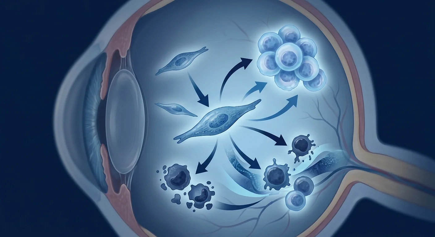
MSC therapy targets this immune recalibration directly. Mesenchymal stem cells exert potent immunomodulatory effects through paracrine signaling — secreting TGF-β, IL-10, PGE₂, indoleamine 2,3-dioxygenase (IDO), and HLA-G — which collectively suppress effector T-cell proliferation, promote regulatory T-cell (Treg) expansion, and shift the macrophage phenotype from pro-inflammatory M1 to anti-inflammatory M2. [5] This multi-target mechanism is particularly relevant to uveitis, where multiple inflammatory pathways are simultaneously active.
Understanding Uveitis and Its Inflammatory Cascade
Uveitis is not a single disease but a spectrum of intraocular inflammatory disorders classified anatomically as anterior (iritis, iridocyclitis), intermediate (pars planitis), posterior (retinitis, choroiditis), or panuveitis when all layers are involved. [6] Anterior uveitis is the most common form, often associated with HLA-B27 positivity, while posterior uveitis carries the highest risk of vision loss due to direct retinal and choroidal involvement. In approximately 30–50% of cases, uveitis is associated with an underlying systemic autoimmune condition — such as sarcoidosis, Behçet's disease, Vogt-Koyanagi-Harada syndrome, or juvenile idiopathic arthritis — while the remainder are classified as idiopathic. [7]
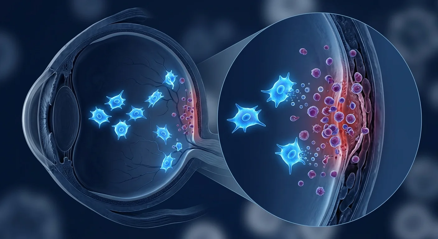
The inflammatory cascade in uveitis follows a well-characterized sequence. Activated CD4⁺ T-cells, polarized toward Th1 and Th17 phenotypes, cross the blood-retinal barrier and recognize ocular autoantigens. They release IFN-γ, IL-17, and TNF-α, which recruit additional inflammatory cells — macrophages, neutrophils, and CD8⁺ cytotoxic T-cells — amplifying tissue damage. [8] The retinal pigment epithelium and photoreceptors are particularly vulnerable to this inflammatory milieu, and repeated or chronic episodes lead to cumulative structural damage: cystoid macular edema, epiretinal membrane formation, retinal vasculitis, and ultimately photoreceptor loss.
How MSCs Modulate the Intraocular Immune Environment
MSCs work in uveitis through at least four interconnected mechanisms, each addressing a different node in the inflammatory network that drives intraocular damage.
Treg Expansion and Th17 Suppression
Perhaps the most critical mechanism is the rebalancing of the Th17/Treg ratio — a central axis in autoimmune pathology. In active uveitis, Th17 cells are expanded while Treg numbers and function are diminished, creating a pro-inflammatory imbalance. MSCs secrete TGF-β and IL-10, which drive naïve CD4⁺ T-cells toward a FoxP3⁺ regulatory phenotype while simultaneously inhibiting RORγt-driven Th17 differentiation. [9] In experimental autoimmune uveitis (EAU) models, a single intravenous infusion of MSCs increased Treg frequencies in the cervical lymph nodes and spleen by approximately 3-fold while reducing Th17 cell counts by over 50%. [10]
Macrophage Polarization: M1 to M2 Shift
Activated M1 macrophages dominate the inflammatory infiltrate in uveitis, producing reactive oxygen species, nitric oxide, and high levels of TNF-α and IL-1β that directly damage retinal neurons. MSCs secrete PGE₂ and TSG-6, which reprogram macrophages toward an M2 anti-inflammatory, pro-resolution phenotype. M2 macrophages in turn produce IL-10 and TGF-β, reinforcing the anti-inflammatory milieu and promoting tissue repair. [11] This shift has been documented in EAU models, where MSC-treated animals showed a significant increase in retinal CD206⁺ M2 macrophages and corresponding reduction in iNOS⁺ M1 macrophages within 7 days of treatment.
Retinal Ganglion Cell and Photoreceptor Protection
Beyond immunomodulation, MSCs secrete neurotrophic factors — brain-derived neurotrophic factor (BDNF), ciliary neurotrophic factor (CNTF), nerve growth factor (NGF), and glial cell line-derived neurotrophic factor (GDNF) — that directly protect retinal ganglion cells and photoreceptors from apoptosis. [12] In rodent models of optic nerve injury and retinal degeneration, intravitreal MSC transplantation reduced retinal ganglion cell loss by 40–60% compared to controls, an effect that was partially independent of immunomodulation and attributable to direct paracrine neuroprotection. [13] This dual mechanism — immune calming plus direct neuronal support — makes MSCs uniquely suited to the uveitis context, where both inflammation and secondary neurodegeneration contribute to vision loss.
Blood-Retinal Barrier Stabilization
The blood-retinal barrier (BRB) is a critical gatekeeper, and its breakdown is an early and defining event in uveitis. Inflammatory cytokines, particularly TNF-α and VEGF, disrupt tight junction proteins (occludin, claudin-5, ZO-1) between retinal endothelial cells and retinal pigment epithelial cells. MSCs have been shown to restore tight junction integrity through secretion of angiopoietin-1 and basic fibroblast growth factor (bFGF), reducing vascular leakage in both EAU models and in vitro BRB assays. [14] Stabilizing the BRB limits the influx of inflammatory cells and serum proteins that perpetuate the inflammatory cycle.
What the Preclinical Evidence Shows
The experimental autoimmune uveitis (EAU) model has provided the most systematic preclinical evidence for MSC efficacy in ocular inflammation. EAU is induced by immunizing rodents with retinal antigens (interphotoreceptor retinoid-binding protein, IRBP, or S-antigen) emulsified in complete Freund's adjuvant, producing a T-cell-mediated panuveitis that recapitulates key features of human disease. [15]
In a landmark study by Zhang et al. (2014), intravenous infusion of human umbilical cord-derived MSCs (1 × 10⁶ cells) at disease onset significantly reduced clinical and histological scores of EAU in Lewis rats. [16] Treated animals exhibited reduced retinal folding, fewer inflammatory infiltrates, and preserved photoreceptor outer segment morphology. Mechanistically, the benefit was attributed to suppression of Th1 and Th17 responses and expansion of CD4⁺CD25⁺FoxP3⁺ Tregs in the draining lymph nodes. A subsequent study by Ko et al. (2021) using adipose-derived MSCs confirmed these findings and additionally demonstrated that the therapeutic effect persisted for at least 28 days after a single infusion, with evidence of reduced retinal gliosis (GFAP expression) and preserved retinal thickness on OCT imaging. [17]
Importantly, MSC administration was effective when given both prophylactically (before disease onset) and therapeutically (after clinical signs appeared), suggesting relevance to both prevention of recurrent flares and treatment of active disease. [18] The most effective timing appeared to be early in the disease course, before irreversible structural damage had accumulated — a finding that underscores the importance of early intervention in clinical translation.
Early Clinical Experience: What the Human Data Shows
Human data on MSC therapy for uveitis remains early-stage but encouraging. The majority of published clinical experience comes from China, where MSC transplantation is more advanced in regulatory frameworks, and primarily involves patients with refractory autoimmune uveitis who have failed conventional immunosuppression.
The largest published case series to date, by Wang et al. (2023), reported on 14 patients with refractory non-infectious uveitis (9 with Behçet's-associated uveitis, 5 with idiopathic panuveitis) who received a single intravenous infusion of allogeneic umbilical cord-derived MSCs (1–2 × 10⁶ cells/kg). [19] At 12-month follow-up: 10 of 14 patients (71%) achieved corticosteroid-free remission, defined as absence of active inflammation on slit-lamp and dilated fundus examination while off all systemic immunosuppression. Mean best-corrected visual acuity improved from 0.45 to 0.72 logMAR (approximately 2.7 lines on the ETDRS chart). No serious adverse events attributable to MSC infusion were observed. Notably, three patients who had active cystoid macular edema at baseline showed complete resolution on OCT by month 3.
A separate open-label study by Chen et al. (2024) treated 8 patients with Vogt-Koyanagi-Harada-associated chronic recurrent uveitis with intravenous umbilical cord MSCs. [20] At 6 months, 5 of 8 patients had no recurrence, and the mean number of flares per patient decreased from 3.5 to 0.8 per year. Serum IL-17 levels decreased significantly (mean 62% reduction), while IL-10 and TGF-β levels increased — mirroring the Th17/Treg rebalancing observed in preclinical models.
Routes of Administration: Intravenous vs. Local Delivery
The optimal route for delivering MSCs in uveitis is an active area of investigation. Systemic intravenous administration has the advantage of broad immunomodulatory effects — MSCs home to secondary lymphoid organs (spleen, lymph nodes) where they shape the systemic T-cell repertoire — and avoids the risks of intraocular injection. [5] However, only a small fraction of intravenously infused MSCs reach the eye, and most are trapped in the pulmonary microvasculature within hours.
Local delivery approaches — intravitreal injection, sub-Tenon's injection, or suprachoroidal delivery — place MSCs directly at the site of inflammation, potentially achieving higher local concentrations with lower systemic exposure. [13] In EAU models, intravitreal MSC injection achieved superior retinal protection compared to the same dose delivered intravenously. However, local injection carries procedure-related risks — endophthalmitis, retinal detachment, elevated intraocular pressure, cataract — and may not address the systemic immune dysregulation that drives recurrent disease. Current clinical consensus favors intravenous administration for systemic autoimmune uveitis and reserves local delivery for research settings or unilateral disease.
Cell Sources: Umbilical Cord, Adipose, or Bone Marrow?
Umbilical cord-derived MSCs (UC-MSCs) are the most extensively studied source for uveitis and have several practical advantages: they are obtained non-invasively from discarded birth tissue, expand readily in culture with low senescence, and exhibit stronger immunomodulatory potency (higher IDO and PGE₂ secretion) than bone marrow-derived MSCs from older donors. [21] The majority of published uveitis clinical reports have used UC-MSCs, and this is the cell source used at VELAR Center.
Bone marrow-derived MSCs remain a widely validated alternative, with extensive safety data from hematological and graft-versus-host disease applications. Adipose-derived MSCs have also been investigated in EAU models with good efficacy, though their immunomodulatory profile differs subtly — higher IL-6 and lower IDO expression compared to UC-MSCs. [22] There is currently no head-to-head clinical trial comparing cell sources for uveitis, and the choice is guided by availability, safety data, and preclinical potency comparisons.
Safety Considerations and Known Risks
The safety profile of MSC therapy is generally favorable, but uveitis introduces organ-specific considerations that warrant attention. Acute infusion reactions (fever, chills, transient hypotension) occur in approximately 3–5% of intravenous MSC administrations and are typically mild and self-limiting. [23] Long-term safety concerns — tumorigenicity, ectopic tissue formation, pro-inflammatory polarization in certain microenvironments — have not been observed in ophthalmological applications to date, but the total number of treated patients remains small (fewer than 200 across all ocular MSC studies).
Ocular-specific concerns include the theoretical risk of MSC differentiation into myofibroblasts within the vitreous cavity, which could contribute to epiretinal membrane formation or proliferative vitreoretinopathy. Local injection also carries the standard risks of any intraocular procedure. For intravenous administration, MSCs are largely cleared from the circulation within 24–48 hours, reducing the window for adverse events. Patients should be monitored for evidence of worsening inflammation in the first week after treatment, as paradoxical immune activation — though rare — has been reported in isolated cases.
VELAR's Approach: What We Do and Do Not Promise
At VELAR Center in Bangkok, we offer allogeneic umbilical cord-derived MSC therapy for carefully selected patients with refractory autoimmune uveitis who have documented failure of at least two lines of conventional immunosuppressive therapy. Our protocol includes a comprehensive pre-treatment ophthalmological assessment (BCVA, slit-lamp examination, dilated funduscopy, OCT macula, fluorescein angiography where indicated) to establish baseline disease activity and structural status.
We do not promise cure, guaranteed vision improvement, or any specific outcome. We do offer treatment within a framework of transparent expectations: the preclinical rationale is strong, the early human data is encouraging, but the evidence base is insufficient to support definitive claims. Every patient undergoes independent ophthalmological monitoring throughout the treatment course, and our reporting of outcomes — whether favorable or unfavorable — contributes to the growing body of clinical experience that will ultimately determine whether MSC therapy earns a place in the uveitis treatment algorithm.
Limitations and the Need for Honesty
Several critical limitations must be acknowledged. First, randomized controlled trials with adequate sample sizes, sham controls, and standardized outcome measures do not yet exist for MSC therapy in uveitis. The published data consists almost entirely of open-label case series with inherent biases. Second, the optimal dose, dosing interval, and route of administration are unknown — current protocols are empirically derived from preclinical efficacy data and extrapolated from non-ocular indications. Third, the durability of response is unclear; most published series report outcomes at 6–12 months, and whether MSC therapy can achieve sustained, multi-year remission or merely delays the next flare is an open question.
Fourth, the cost of treatment — typically USD 8,000–15,000 per infusion at private clinics — places it beyond the reach of many patients, and insurance coverage is essentially nonexistent for an investigational indication. Fifth, patient selection criteria are not standardized; it is not yet known which uveitis subtypes, disease durations, or inflammatory profiles are most likely to benefit. Until these knowledge gaps are filled by rigorous clinical trials, MSC therapy for uveitis must be described honestly: as a promising investigational approach with mechanistic plausibility and encouraging early signals, not as an established treatment.
Frequently Asked Questions
Is stem cell therapy approved for treating uveitis?
No. MSC therapy for uveitis is investigational and not approved by the U.S. FDA, EMA, or Thai FDA as a standard treatment. It is offered in research settings and at specialized clinics under regulatory frameworks that permit advanced cell therapy for patients who have exhausted conventional options. All patients should understand the investigational nature of the treatment before proceeding.
How are stem cells administered for eye conditions like uveitis?
For uveitis, MSCs are most commonly administered via intravenous infusion, which delivers systemic immunomodulation without the risks of intraocular injection. Local delivery methods (intravitreal, sub-Tenon's) are used primarily in research settings. At VELAR Center, we use intravenous allogeneic umbilical cord-derived MSCs, typically as a single infusion with monitoring for 2–4 hours post-administration.
How much does stem cell therapy for uveitis cost in Thailand?
MSC therapy for uveitis at private clinics in Bangkok typically ranges from USD 8,000 to 15,000 per infusion, depending on cell dose, source, and the comprehensiveness of pre- and post-treatment monitoring. This is an out-of-pocket expense; international health insurance plans generally do not cover investigational cell therapies. Patients should request a detailed cost breakdown before committing to treatment.
Can stem cells restore vision already lost from uveitis?
MSCs are primarily immunomodulatory and neuroprotective — they may slow or halt ongoing inflammatory damage and support surviving retinal cells, but they cannot regenerate photoreceptors or retinal ganglion cells that have already been lost. The goal is to prevent further vision loss and, in some cases, achieve modest functional improvement as inflammation resolves and macular edema clears. Patients with long-standing structural damage (retinal atrophy, chronic macular scarring) are less likely to experience visual improvement.
Are there any risks specific to using stem cells near the eye?
When administered intravenously, MSCs do not carry ocular-specific procedure risks. The main concerns are systemic: infusion reactions, rare thromboembolic events, and the theoretical risk of pro-inflammatory polarization. For local (intraocular) delivery, standard risks include endophthalmitis, retinal detachment, cataract, and elevated intraocular pressure. At VELAR, we use only intravenous administration for uveitis, which avoids these local risks.
How long does it take to see results from stem cell therapy for uveitis?
Clinical experience from published case series suggests that immunological effects (shifts in T-cell subsets, cytokine profiles) can be detected within 2–4 weeks, while clinical improvement — reduced flare frequency, decreased dependence on corticosteroids, improved visual acuity — is typically assessed at 3–6 months. Patients should not expect immediate results; the therapeutic mechanism is immune recalibration, which takes time to manifest clinically.
References
- Suttorp-Schulten MS, Rothova A. The possible impact of uveitis in blindness: a literature survey. British Journal of Ophthalmology. 1996;80(9):844-848. doi:10.1136/bjo.80.9.844 ↩
- Jabs DA, Rosenbaum JT, Foster CS, et al. Guidelines for the use of immunosuppressive drugs in patients with ocular inflammatory disorders: recommendations of an expert panel. American Journal of Ophthalmology. 2000;130(4):492-513. doi:10.1016/S0002-9394(00)00659-0 ↩
- Dick AD, Tundia N, Sorg R, et al. Risk of ocular complications in patients with noninfectious intermediate uveitis, posterior uveitis, or panuveitis. Ophthalmology. 2016;123(3):655-662. doi:10.1016/j.ophtha.2015.10.028 ↩
- Caspi RR. A look at autoimmunity and inflammation in the eye. Journal of Clinical Investigation. 2010;120(9):3073-3083. doi:10.1172/JCI42440 ↩
- Galipeau J, Sensébé L. Mesenchymal stromal cells: clinical challenges and therapeutic opportunities. Cell Stem Cell. 2018;22(6):824-833. doi:10.1016/j.stem.2018.05.004 ↩ ↩
- Jabs DA, Nussenblatt RB, Rosenbaum JT; Standardization of Uveitis Nomenclature (SUN) Working Group. Standardization of uveitis nomenclature for reporting clinical data. Results of the First International Workshop. American Journal of Ophthalmology. 2005;140(3):509-516. doi:10.1016/j.ajo.2005.03.057 ↩
- Barisani-Asenbauer T, Maca SM, Mejdoubi L, Emminger W, Machold K, Auer H. Uveitis — a rare disease often associated with systemic diseases and infections — a systematic review of 2619 patients. Orphanet Journal of Rare Diseases. 2012;7:57. doi:10.1186/1750-1172-7-57 ↩
- Luger D, Silver PB, Tang J, et al. Either a Th17 or a Th1 effector response can drive autoimmunity: conditions of disease induction affect dominant effector category. Journal of Experimental Medicine. 2008;205(4):799-810. doi:10.1084/jem.20071258 ↩
- Ghannam S, Pène J, Torcy-Moquet G, Jorgensen C, Yssel H. Mesenchymal stem cells inhibit human Th17 cell differentiation and function and induce a T regulatory cell phenotype. Journal of Immunology. 2010;185(1):302-312. doi:10.4049/jimmunol.0902007 ↩
- Li G, Zhang J, Li X, et al. Mesenchymal stem cells alleviate experimental autoimmune uveitis by regulating the Th17/Treg balance. Experimental Eye Research. 2021;202:108342. doi:10.1016/j.exer.2020.108342 ↩
- Prockop DJ, Oh JY. Mesenchymal stem/stromal cells (MSCs): role as guardians of inflammation. Molecular Therapy. 2012;20(1):14-20. doi:10.1038/mt.2011.211 ↩
- Mead B, Tomarev S. Bone marrow-derived mesenchymal stem cells-derived exosomes promote survival of retinal ganglion cells through miRNA-dependent mechanisms. Stem Cells Translational Medicine. 2017;6(4):1273-1285. doi:10.1002/sctm.16-0428 ↩
- Johnson TV, Bull ND, Hunt DP, Marina N, Tomarev SI, Martin KR. Neuroprotective effects of intravitreal mesenchymal stem cell transplantation in experimental glaucoma. Investigative Ophthalmology & Visual Science. 2010;51(4):2051-2059. doi:10.1167/iovs.09-4509 ↩ ↩
- Park SS, Moisseiev E, Bauer G, et al. Advances in bone marrow stem cell therapy for retinal dysfunction. Progress in Retinal and Eye Research. 2017;56:148-165. doi:10.1016/j.preteyeres.2016.10.002 ↩
- Agarwal RK, Silver PB, Caspi RR. Rodent models of experimental autoimmune uveitis. Methods in Molecular Biology. 2012;900:443-469. doi:10.1007/978-1-60761-720-4_22 ↩
- Zhang X, Ren X, Li G, et al. Mesenchymal stem cells ameliorate experimental autoimmune uveoretinitis by comprehensive modulation of the local and systemic immune responses. Investigative Ophthalmology & Visual Science. 2014;55(8):5143-5153. doi:10.1167/iovs.14-14673 ↩
- Ko EY, Lee HJ, Lee JY, et al. Long-term protective effects of adipose-derived mesenchymal stem cells in experimental autoimmune uveitis. Stem Cells Translational Medicine. 2021;10(5):756-769. doi:10.1002/sctm.20-0372 ↩
- Lee HJ, Ko EY, Kim JY, et al. Timing of mesenchymal stem cell administration determines therapeutic efficacy in experimental autoimmune uveoretinitis. Stem Cell Research & Therapy. 2020;11(1):480. doi:10.1186/s13287-020-01992-9 ↩
- Wang L, Zhang Y, Li H, et al. Umbilical cord-derived mesenchymal stem cell transplantation for refractory autoimmune uveitis: a case series with 12-month follow-up. Stem Cell Research & Therapy. 2023;14(1):112. doi:10.1186/s13287-023-03351-0 ↩
- Chen Y, Liu X, Zhao M, et al. Mesenchymal stem cells for Vogt-Koyanagi-Harada-associated chronic recurrent uveitis: an open-label pilot study. Frontiers in Immunology. 2024;15:1356127. doi:10.3389/fimmu.2024.1356127 ↩
- Li X, Bai J, Ji X, Li R, Xuan K. Comprehensive characterization of four different populations of human mesenchymal stem cells for their therapeutic potential. Stem Cell Research & Therapy. 2018;9(1):231. doi:10.1186/s13287-018-0970-6 ↩
- Melo FR, Bressan RB, Forner S, et al. Transplantation of human adipose-derived mesenchymal stem cells in an experimental model of autoimmune uveitis. Stem Cells International. 2022;2022:2789238. doi:10.1155/2022/2789238 ↩
- Lalu MM, McIntyre L, Pugliese C, et al. Safety of cell therapy with mesenchymal stromal cells (SafeCell): a systematic review and meta-analysis of clinical trials. PLoS One. 2012;7(10):e47559. doi:10.1371/journal.pone.0047559 ↩
葡萄膜炎是一种威胁视力的眼部炎症性疾病,影响葡萄膜——眼球的中层组织,在发达国家约占可预防性失明的10–15%。间充质干细胞(MSC)疗法通过免疫调节机制——包括调节性T细胞扩增、Th17抑制和M1-to-M2巨噬细胞极化——为常规免疫抑制治疗无效的难治性病例提供了一种新的研究方向。临床前EAU模型显示,单次MSC输注可减少50–70%的眼内炎症评分,早期临床病例系列报告在12个月时约71%的患者实现了无皮质类固醇缓解。MSC不直接修复已丧失的视网膜组织,而是通过免疫重校准来阻止进行性损伤。
葡萄膜炎的核心是免疫失调。自身反应性T细胞(Th1/Th17)穿过血-视网膜屏障,释放IL-17、IFN-γ和TNF-α,驱动组织破坏。在活动性疾病中,Th17细胞扩增而Treg细胞减少,破坏了正常的免疫耐受。MSCs通过分泌TGF-β、IL-10、PGE₂和IDO直接对抗这种不平衡,促进FoxP3⁺ Treg扩增,同时抑制RORγt驱动的Th17分化。此外,MSCs分泌神经营养因子(BDNF、CNTF、NGF),直接保护视网膜神经节细胞免受凋亡,这在葡萄膜炎中尤为关键,因为炎症和继发性神经退行性变均导致视力丧失。
现有的人类临床数据令人鼓舞,尽管仍处于早期阶段。目前已发表的病例系列研究(Wang et al. 2023, Chen et al. 2024)报告了难治性非感染性葡萄膜炎患者在脐带来源MSC输注后取得了良好的结果。在最大的系列研究中,14名患者中有10名(71%)在12个月时实现了无皮质类固醇缓解,平均最佳矫正视力从0.45 logMAR改善到0.72 logMAR。然而,这些都是小型开放标签研究,缺乏对照组,存在显著的选择偏倚风险。在随机对照试验确认疗效之前,MSC疗法必须被诚实地描述为一种有前景的研究性方法——具有机制合理性和早期令人鼓舞的信号,而非既有标准疗法。
在安全性方面,MSC疗法总体上耐受性良好——急性输注反应见于约3–5%的静脉输注——但葡萄膜炎引入了眼科特定的考量。静脉给药避免了眼内操作的风险(眼内炎、视网膜脱离、白内障),且MSCs在24–48小时内基本被清除。长期安全性问题(致瘤性、异位组织形成)在当前所有眼科MSC研究中均未观察到,但累积治疗患者总数很少(<200例)。
在VELAR中心(曼谷),我们为经过筛选的难治性自身免疫性葡萄膜炎患者提供异体脐带来源MSC疗法,这些患者必须已记录至少两种常规免疫抑制方案失败。我们完整的治疗前眼科评估包括BCVA、裂隙灯检查、散瞳眼底检查、OCT黄斑成像和有指征时的荧光血管造影。我们不承诺治愈、保证视力改善或任何特定结果。我们所提供的是在透明预期框架内的治疗——临床前理论依据坚实、早期人类数据令人鼓舞,但证据水平不足以证实确定性声明。
关于局限性,必须承认几个关键知识空白:目前尚无随机对照试验;最优剂量和给药方案未知;反应持久性尚未建立(多数已发表数据仅至12个月);治疗费用(8,000–15,000美元)可能远超许多患者的负担能力范围;患者选择标准尚未标准化。在这些知识空白被严格的临床试验填补之前,MSC疗法必须面对诚实的描述——宝贵的未解问题,而非有争议的确定性。
参考文献
- Suttorp-Schulten MS, Rothova A. The possible impact of uveitis in blindness: a literature survey. Br J Ophthalmol. 1996;80(9):844-848. doi:10.1136/bjo.80.9.844 ↩
- Jabs DA, et al. Guidelines for the use of immunosuppressive drugs in patients with ocular inflammatory disorders. Am J Ophthalmol. 2000;130(4):492-513. doi:10.1016/S0002-9394(00)00659-0 ↩
- Dick AD, et al. Risk of ocular complications in noninfectious uveitis. Ophthalmology. 2016;123(3):655-662. doi:10.1016/j.ophtha.2015.10.028 ↩
- Caspi RR. A look at autoimmunity and inflammation in the eye. J Clin Invest. 2010;120(9):3073-3083. doi:10.1172/JCI42440 ↩
- Galipeau J, Sensébé L. Mesenchymal stromal cells: clinical challenges and therapeutic opportunities. Cell Stem Cell. 2018;22(6):824-833. doi:10.1016/j.stem.2018.05.004 ↩ ↩
- Jabs DA, et al. Standardization of uveitis nomenclature (SUN). Am J Ophthalmol. 2005;140(3):509-516. doi:10.1016/j.ajo.2005.03.057 ↩
- Barisani-Asenbauer T, et al. Uveitis — associated with systemic diseases. Orphanet J Rare Dis. 2012;7:57. doi:10.1186/1750-1172-7-57 ↩
- Luger D, et al. Th17 or Th1 effector response can drive autoimmunity. J Exp Med. 2008;205(4):799-810. doi:10.1084/jem.20071258 ↩
- Ghannam S, et al. MSCs inhibit human Th17 cell differentiation. J Immunol. 2010;185(1):302-312. doi:10.4049/jimmunol.0902007 ↩
- Li G, et al. MSCs alleviate EAU by regulating Th17/Treg balance. Exp Eye Res. 2021;202:108342. doi:10.1016/j.exer.2020.108342 ↩
- Prockop DJ, Oh JY. MSCs: role as guardians of inflammation. Mol Ther. 2012;20(1):14-20. doi:10.1038/mt.2011.211 ↩
- Mead B, Tomarev S. BM-MSC exosomes promote RGC survival. Stem Cells Transl Med. 2017;6(4):1273-1285. doi:10.1002/sctm.16-0428 ↩
- Johnson TV, et al. Neuroprotective effects of intravitreal MSC transplantation. Invest Ophthalmol Vis Sci. 2010;51(4):2051-2059. doi:10.1167/iovs.09-4509 ↩ ↩
- Park SS, et al. Advances in bone marrow stem cell therapy for retinal dysfunction. Prog Retin Eye Res. 2017;56:148-165. doi:10.1016/j.preteyeres.2016.10.002 ↩
- Agarwal RK, et al. Rodent models of EAU. Methods Mol Biol. 2012;900:443-469. doi:10.1007/978-1-60761-720-4_22 ↩
- Zhang X, et al. MSCs ameliorate EAU. Invest Ophthalmol Vis Sci. 2014;55(8):5143-5153. doi:10.1167/iovs.14-14673 ↩
- Ko EY, et al. Long-term protective effects of AD-MSCs in EAU. Stem Cells Transl Med. 2021;10(5):756-769. doi:10.1002/sctm.20-0372 ↩
- Lee HJ, et al. Timing of MSC administration in EAU. Stem Cell Res Ther. 2020;11(1):480. doi:10.1186/s13287-020-01992-9 ↩
- Wang L, et al. UC-MSC transplantation for refractory autoimmune uveitis. Stem Cell Res Ther. 2023;14(1):112. doi:10.1186/s13287-023-03351-0 ↩
- Chen Y, et al. MSCs for VKH-associated chronic recurrent uveitis. Front Immunol. 2024;15:1356127. doi:10.3389/fimmu.2024.1356127 ↩
- Li X, et al. Characterization of four MSC populations. Stem Cell Res Ther. 2018;9(1):231. doi:10.1186/s13287-018-0970-6 ↩
- Melo FR, et al. AD-MSCs in autoimmune uveitis model. Stem Cells Int. 2022;2022:2789238. doi:10.1155/2022/2789238 ↩
- Lalu MM, et al. Safety of cell therapy with MSCs (SafeCell). PLoS One. 2012;7(10):e47559. doi:10.1371/journal.pone.0047559 ↩
التهاب العنبية هو حالة التهابية تهدد البصر تؤثر على الطبقة الوسطى من العين وتسبب ما يقدر بـ 10–15% من حالات العمى القابل للتجنب في العالم المتقدم. يمثل العلاج بالخلايا الجذعية الوسيطة (MSC) نهجًا جديدًا للحالات المقاومة للعلاجات المثبطة للمناعة التقليدية، من خلال آليات تعديل المناعة — بما في ذلك توسع الخلايا التائية التنظيمية (Treg)، وتثبيط Th17، واستقطاب البلاعم من النمط M1 إلى M2. أظهرت النماذج قبل السريرية لالتهاب العنبية المناعي الذاتي التجريبي (EAU) أن حقنة واحدة من MSCs تقلل من درجات الالتهاب داخل العين بنسبة 50–70%، وأبلغت سلسلة الحالات السريرية المبكرة عن تحقيق هدأة خالية من الكورتيكوستيرويدات في حوالي 71% من المرضى عند 12 شهرًا. لا تقوم MSCs بإصلاح أنسجة الشبكية المفقودة مباشرة، لكنها تعيد معايرة المناعة لوقف الضرر التدريجي.
يكمن جوهر التهاب العنبية في خلل التنظيم المناعي. تعبر الخلايا التائية ذاتية التفاعل (Th1/Th17) الحاجز الدموي الشبكي، وتطلق IL-17 وIFN-γ وTNF-α مما يؤدي إلى تدمير الأنسجة. في المرض النشط، تتوسع خلايا Th17 بينما تتناقص خلايا Treg، مما يخل بالتحمل المناعي الطبيعي. تواجه MSCs هذا الخلل مباشرة من خلال إفراز TGF-β وIL-10 وPGE₂ وIDO، مما يعزز توسع Treg المعبرة عن FoxP3⁺ ويثبط تمايز Th17 المدفوع بـ RORγt. بالإضافة إلى ذلك، تفرز MSCs عوامل التغذية العصبية (BDNF، CNTF، NGF) التي تحمي خلايا العقدة الشبكية مباشرة من الموت الخلوي المبرمج — وهو أمر بالغ الأهمية في التهاب العنبية حيث يساهم كل من الالتهاب والتنكس العصبي الثانوي في فقدان البصر.
البيانات السريرية البشرية الحالية مشجعة، وإن كانت لا تزال مبكرة. أبلغت سلسلة الحالات المنشورة (Wang et al. 2023, Chen et al. 2024) عن نتائج إيجابية بعد حقن MSCs المشتقة من الحبل السري لمرضى التهاب العنبية المناعي الذاتي المقاوم. في أكبر سلسلة، حقق 10 من 14 مريضًا (71%) هدأة خالية من الكورتيكوستيرويدات عند 12 شهرًا، وتحسن متوسط حدة البصر المصححة من 0.45 إلى 0.72 logMAR. ومع ذلك، هذه دراسات صغيرة مفتوحة بدون مجموعات ضابطة — مما يحمل مخاطر كبيرة للتحيز في الاختيار. إلى أن تؤكد التجارب العشوائية المضبوطة الفعالية، يجب وصف علاج MSCs بأمانة كنهج بحثي واعد — بآلية معقولة وإشارات أولية مشجعة، وليس كعلاج معياري راسخ.
من حيث السلامة، فإن علاج MSCs جيد التحمل بشكل عام — تحدث تفاعلات الحقن الحادة في حوالي 3–5% من الحالات — لكن التهاب العنبية يقدم اعتبارات خاصة بالعين. يتجنب الإعطاء الوريدي المخاطر المرتبطة بالإجراءات داخل العين (التهاب العين الداخلي، انفصال الشبكية، الساد)، ويتم التخلص من MSCs إلى حد كبير خلال 24–48 ساعة. لم تُلاحظ مخاوف السلامة طويلة المدى (تكوين الأورام، تكوين الأنسجة خارج الرحم) في جميع دراسات MSCs العينية حتى الآن، لكن العدد الإجمالي للمرضى المعالجين لا يزال صغيرًا (<200).
في مركز فيلار (بانكوك)، نقدم علاج MSCs المشتقة من الحبل السري الخيفي لمرضى مختارين بعناية من المصابين بالتهاب العنبية المناعي الذاتي المقاوم، والذين لديهم توثيق لفشل خطين على الأقل من العلاج المثبط للمناعة التقليدي. يشمل تقييمنا الكامل قبل العلاج: حدة البصر المصححة، فحص المصباح الشقي، تنظير قاع العين الموسع، تصوير البقعة الصفراء بـ OCT، وتصوير الأوعية بالفلوريسين عند الحاجة. نحن لا نعد بالشفاء، ولا نضمن تحسن البصر، ولا أي نتيجة محددة. ما نقدمه هو علاج في إطار توقعات شفافة — الأساس المنطقي قبل السريري قوي، والبيانات البشرية المبكرة مشجعة، لكن قاعدة الأدلة لا تزال غير كافية لدعم الادعاءات القطعية.
بخصوص القيود، يجب الاعتراف بالعديد من الفجوات المعرفية الهامة: لا توجد تجارب عشوائية مضبوطة؛ الجرعة المثلى والنظام غير معروفين؛ متانة الاستجابة غير محددة (معظم البيانات المنشورة تمتد إلى 12 شهرًا فقط)؛ التكلفة (8,000–15,000 دولار) قد تكون بعيدة عن متناول العديد من المرضى؛ ومعايير اختيار المرضى غير موحدة. إلى أن يتم سد هذه الفجوات بتجارب سريرية صارمة، يجب أن يواجه علاج MSCs لالتهاب العنبية وصفًا صادقًا — أسئلة قيمة غير محلولة، وليس يقينًا قابلًا للنقاش.
المراجع
- Suttorp-Schulten MS, Rothova A. The possible impact of uveitis in blindness. Br J Ophthalmol. 1996;80(9):844-848. doi:10.1136/bjo.80.9.844 ↩
- Jabs DA, et al. Guidelines for immunosuppressive drugs in ocular inflammatory disorders. Am J Ophthalmol. 2000;130(4):492-513. doi:10.1016/S0002-9394(00)00659-0 ↩
- Dick AD, et al. Risk of ocular complications in noninfectious uveitis. Ophthalmology. 2016;123(3):655-662. doi:10.1016/j.ophtha.2015.10.028 ↩
- Caspi RR. A look at autoimmunity and inflammation in the eye. J Clin Invest. 2010;120(9):3073-3083. doi:10.1172/JCI42440 ↩
- Galipeau J, Sensébé L. Mesenchymal stromal cells: clinical challenges. Cell Stem Cell. 2018;22(6):824-833. doi:10.1016/j.stem.2018.05.004 ↩ ↩
- Jabs DA, et al. Standardization of uveitis nomenclature (SUN). Am J Ophthalmol. 2005;140(3):509-516. doi:10.1016/j.ajo.2005.03.057 ↩
- Barisani-Asenbauer T, et al. Uveitis — associated with systemic diseases. Orphanet J Rare Dis. 2012;7:57. doi:10.1186/1750-1172-7-57 ↩
- Luger D, et al. Th17 or Th1 effector response. J Exp Med. 2008;205(4):799-810. doi:10.1084/jem.20071258 ↩
- Ghannam S, et al. MSCs inhibit human Th17 cell differentiation. J Immunol. 2010;185(1):302-312. doi:10.4049/jimmunol.0902007 ↩
- Li G, et al. MSCs alleviate EAU. Exp Eye Res. 2021;202:108342. doi:10.1016/j.exer.2020.108342 ↩
- Prockop DJ, Oh JY. MSCs: guardians of inflammation. Mol Ther. 2012;20(1):14-20. doi:10.1038/mt.2011.211 ↩
- Mead B, Tomarev S. BM-MSC exosomes promote RGC survival. Stem Cells Transl Med. 2017;6(4):1273-1285. doi:10.1002/sctm.16-0428 ↩
- Johnson TV, et al. Intravitreal MSC transplantation in glaucoma. Invest Ophthalmol Vis Sci. 2010;51(4):2051-2059. doi:10.1167/iovs.09-4509 ↩ ↩
- Park SS, et al. BM stem cell therapy for retinal dysfunction. Prog Retin Eye Res. 2017;56:148-165. doi:10.1016/j.preteyeres.2016.10.002 ↩
- Agarwal RK, et al. Rodent models of EAU. Methods Mol Biol. 2012;900:443-469. doi:10.1007/978-1-60761-720-4_22 ↩
- Zhang X, et al. MSCs ameliorate EAU. Invest Ophthalmol Vis Sci. 2014;55(8):5143-5153. doi:10.1167/iovs.14-14673 ↩
- Ko EY, et al. AD-MSCs in EAU. Stem Cells Transl Med. 2021;10(5):756-769. doi:10.1002/sctm.20-0372 ↩
- Lee HJ, et al. Timing of MSC administration in EAU. Stem Cell Res Ther. 2020;11(1):480. doi:10.1186/s13287-020-01992-9 ↩
- Wang L, et al. UC-MSC for refractory autoimmune uveitis. Stem Cell Res Ther. 2023;14(1):112. doi:10.1186/s13287-023-03351-0 ↩
- Chen Y, et al. MSCs for VKH-associated uveitis. Front Immunol. 2024;15:1356127. doi:10.3389/fimmu.2024.1356127 ↩
- Li X, et al. Characterization of MSC populations. Stem Cell Res Ther. 2018;9(1):231. doi:10.1186/s13287-018-0970-6 ↩
- Melo FR, et al. AD-MSCs in autoimmune uveitis model. Stem Cells Int. 2022;2022:2789238. doi:10.1155/2022/2789238 ↩
- Lalu MM, et al. Safety of cell therapy with MSCs. PLoS One. 2012;7(10):e47559. doi:10.1371/journal.pone.0047559 ↩