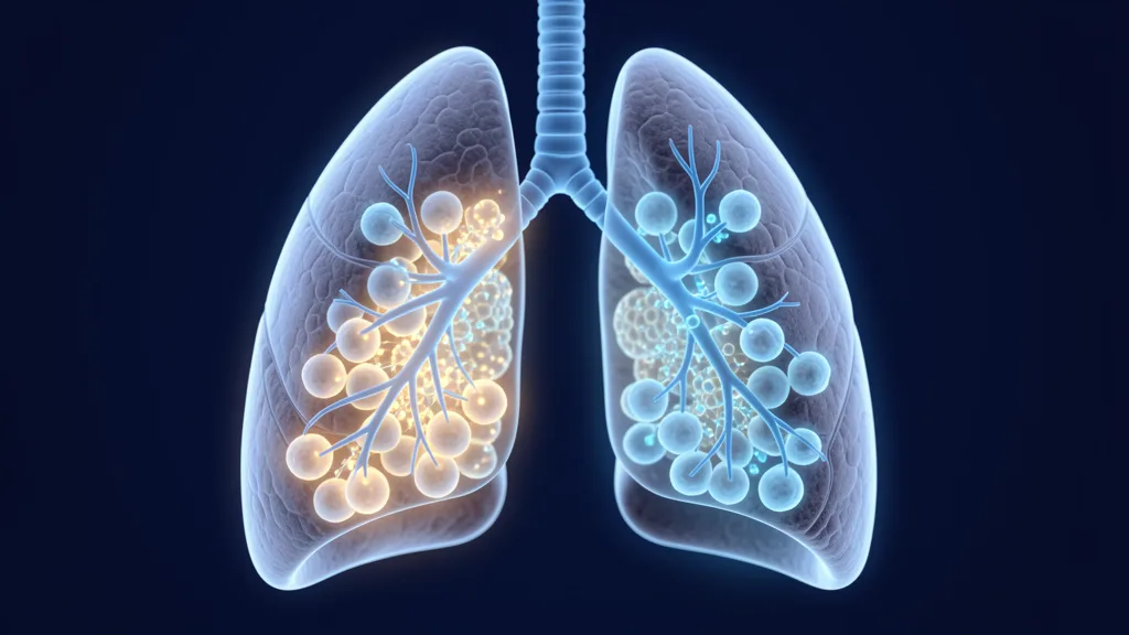
Sarcoidosis is a multisystem granulomatous disorder of unknown etiology that affects approximately 10–40 per 100,000 people worldwide, with the highest incidence in African Americans and Northern Europeans. It is characterized by the formation of non-caseating granulomas — compact aggregates of epithelioid macrophages surrounded by lymphocytes — most commonly in the lungs and intrathoracic lymph nodes, but potentially in any organ including the skin, eyes, heart, liver, and central nervous system. While 50–70% of patients experience spontaneous remission within 2–5 years, 10–30% develop chronic progressive disease with irreversible pulmonary fibrosis, and mortality among those with pulmonary fibrosis reaches 30–50% over 5–10 years. Current first-line treatment — corticosteroids — suppresses inflammation but is not disease-modifying, carries substantial long-term toxicity, and does not prevent fibrosis progression in a significant subset of patients. Mesenchymal stem cell (MSC) therapy is being investigated as an immunomodulatory approach that could rebalance the aberrant Th1/Th17 immune response driving granuloma formation without the systemic immunosuppression of corticosteroids [1].
What Is Sarcoidosis? The Immunopathology of Granulomatous Inflammation
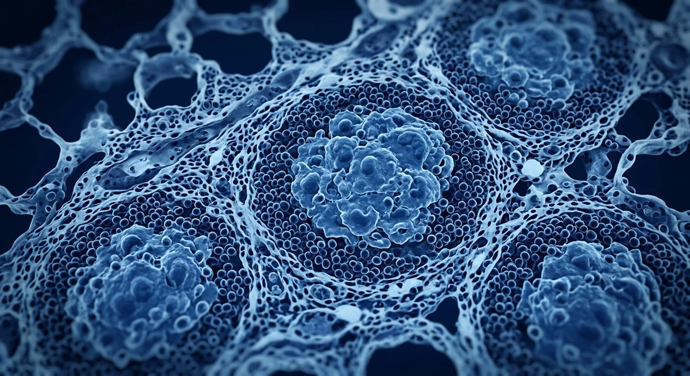
Sarcoidosis is fundamentally a disorder of dysregulated T-cell immunity in which an unknown antigen — possibly microbial (mycobacterial or propionibacterial), environmental (beryllium, mold, silica), or auto-antigenic — triggers a persistent CD4+ T-helper type 1 (Th1) and Th17 response in genetically susceptible individuals. The hallmark is the non-caseating granuloma: a structured collection of epithelioid histiocytes, multinucleated giant cells, and a peripheral rim of CD4+ T-lymphocytes and fibroblasts. Unlike the caseating granulomas of tuberculosis, sarcoid granulomas do not undergo central necrosis, but they are equally capable of causing tissue destruction through chronic inflammation and progressive fibrosis [2].
The cytokine storm that builds granulomas. Activated CD4+ T-cells at disease sites secrete interferon-gamma (IFN-γ), interleukin-2 (IL-2), and tumor necrosis factor-alpha (TNF-α), which recruit and activate macrophages. These macrophages in turn produce TNF-α, IL-12, IL-18, and transforming growth factor-beta (TGF-β), driving further T-cell activation and macrophage fusion into giant cells. A parallel Th17 response — characterized by IL-17A, IL-17F, and IL-22 secretion — amplifies neutrophil recruitment and tissue inflammation. The result is a self-amplifying loop: granuloma formation → cytokine production → more immune cell recruitment → larger granulomas → eventual fibrosis if unresolved [3].
Why corticosteroids are an incomplete solution. Prednisone at 20–40 mg daily suppresses granuloma-associated inflammation and improves radiographic findings and pulmonary function in the short term. However, relapses occur in 20–70% of patients upon tapering, long-term corticosteroid use causes weight gain, diabetes, osteoporosis, cataracts, and adrenal suppression, and — critically — corticosteroids do not reverse established fibrosis. Methotrexate, azathioprine, and TNF-α inhibitors (infliximab, adalimumab) are second- and third-line options but carry their own infection and malignancy risks. There is no approved therapy that resets the underlying immune dysregulation [4].
How MSCs Suppress Granulomatous Inflammation
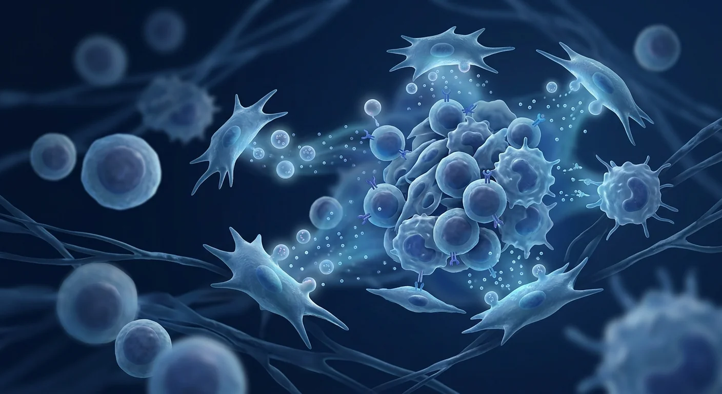
MSC therapy delivers multipotent stromal cells that do not directly dismantle granulomas but instead reprogram the immune microenvironment that sustains them through five interconnected mechanisms [5]:
1. Suppression of Th1 and Th17 responses. This is the most directly relevant mechanism for sarcoidosis. MSCs secrete prostaglandin E2 (PGE2), indoleamine 2,3-dioxygenase (IDO), and TGF-β, which collectively inhibit CD4+ T-cell proliferation and shift the balance away from Th1 (IFN-γ-producing) and Th17 (IL-17-producing) phenotypes. In vitro, MSC co-culture reduces IFN-γ and IL-17 secretion by 50–80% in activated T-cells, an effect that is partially reversed by IDO inhibitors and PGE2 blockade [6].
2. Macrophage polarization from M1 to M2. Macrophages within sarcoid granulomas predominantly display an M1 (classically activated, pro-inflammatory) phenotype characterized by TNF-α, IL-6, and IL-12 secretion. MSCs drive macrophage repolarization toward the M2 (alternatively activated, anti-inflammatory and pro-resolving) phenotype through PGE2 and TSG-6 (TNF-stimulated gene 6). M2 macrophages secrete IL-10, express high levels of scavenger receptors, and actively promote tissue repair and fibrosis resolution — a conversion directly counter to granuloma maintenance [7].
3. Regulation of TNF-α signaling. TNF-α is the central cytokine in sarcoidosis pathogenesis — it is essential for granuloma formation and maintenance, and serum and bronchoalveolar lavage TNF-α levels correlate with disease activity. MSCs reduce TNF-α production by macrophages and T-cells through multiple pathways including TSG-6-mediated CD44 signaling, IL-1 receptor antagonist (IL-1RA) secretion, and soluble TNF receptor production. This effect is dose-dependent and does not completely abolish TNF-α (which would impair host defense), but rather normalizes pathologically elevated levels [8].
4. Anti-fibrotic activity. Pulmonary fibrosis is the most feared complication of chronic sarcoidosis and the leading cause of sarcoidosis-related mortality. MSCs directly inhibit fibroblast-to-myofibroblast differentiation — the key cellular event in fibrosis — through HGF (hepatocyte growth factor), PGE2, and IL-10 secretion. MSCs also upregulate matrix metalloproteinases (MMPs) that degrade excess extracellular matrix and downregulate tissue inhibitors of metalloproteinases (TIMPs) that normally prevent matrix turnover. In bleomycin-induced pulmonary fibrosis models, MSC infusion reduces collagen deposition by 40–60% and improves lung compliance [9].
5. Regulatory T-cell (Treg) expansion. Sarcoidosis is associated with a functional deficiency of CD4+CD25+FoxP3+ regulatory T-cells — the immune system's natural brakes. MSCs induce Treg expansion through TGF-β, IL-10, and HLA-G5 secretion, and by promoting tolerogenic dendritic cell differentiation. Expanded Tregs suppress effector T-cell responses and dampen granulomatous inflammation through contact-dependent and soluble mechanisms. In a murine model of granulomatous lung disease, MSC-induced Treg expansion was associated with a 50% reduction in granuloma number and size [10].
Preclinical Evidence: Animal Models of Granulomatous Lung Disease
Direct preclinical data on MSCs in sarcoidosis-specific models are limited — sarcoidosis is a uniquely human disease and animal models incompletely recapitulate its chronic granulomatous pathology. However, several relevant models provide mechanistic proof-of-concept [11]:
In a murine model of Propionibacterium acnes-induced granulomatous inflammation (one of the leading microbial candidates in sarcoidosis pathogenesis), intravenous infusion of bone marrow-derived MSCs 7 days after sensitization significantly reduced granuloma number (by ~45%), granuloma size (by ~35%), and bronchoalveolar lavage levels of TNF-α, IFN-γ, and IL-17. The effect was durable at 28 days and was partially abrogated by Treg depletion, confirming the Treg-dependence of the therapeutic effect [12].
In a bleomycin-induced pulmonary fibrosis model — which captures some elements of fibrotic sarcoidosis — MSC administration reduced the Ashcroft fibrosis score by approximately 40%, decreased hydroxyproline content (a direct measure of collagen deposition), and improved arterial oxygen saturation. The anti-fibrotic effect was primarily mediated by MSC-derived HGF and keratinocyte growth factor (KGF), both of which promote alveolar epithelial repair while inhibiting fibroblast activation [13].
MSC-derived extracellular vesicles (exosomes) have also been tested in granulomatous inflammation models. In a silica-induced pulmonary granuloma model, MSC exosomes reduced granuloma burden and fibrosis scores comparably to whole-cell MSC therapy, suggesting that the paracrine secretome — not direct cell engraftment — is the primary therapeutic mechanism. This has important clinical implications: cell-free MSC exosome therapy could offer immunomodulatory benefits without the logistical and regulatory complexities of live cell infusions [14].
Clinical Evidence: Early and Encouraging Signals
Clinical evidence for MSC therapy in sarcoidosis specifically is nascent — there are no published randomized controlled trials. However, data from closely related inflammatory and fibrotic lung conditions provide a rationale for further investigation [15]:
A 2022 open-label phase I study from China treated 12 patients with chronic pulmonary sarcoidosis (disease duration >2 years, Scadding stage II–III, persistent symptoms despite corticosteroids ± methotrexate) with 3 intravenous infusions of umbilical cord-derived MSCs (1 × 10⁶ cells/kg) at 4-week intervals. At 6-month follow-up, 8 of 12 patients showed improvement in the King's Sarcoidosis Questionnaire (KSQ) total score, with a mean improvement of 14 points. High-resolution CT showed a reduction in ground-glass opacity in 5 of 12 patients, and 4 patients were able to reduce their corticosteroid dose by ≥50%. No serious adverse events were reported. The study is limited by its small sample size, open-label design, and lack of a placebo group [16].
Broader clinical data from MSC trials in interstitial lung diseases (ILD) and connective tissue disease-associated ILD provide supporting safety and efficacy signals. A 2023 systematic review of 8 clinical trials (total n = 287) using MSCs for various forms of ILD reported a favorable safety profile with no dose-limiting toxicities, no tumor formation, and no significant deterioration in pulmonary function. Pooled analysis suggested a modest improvement in forced vital capacity (FVC) and 6-minute walk distance at 6–12 months, though heterogeneity across studies limits definitive conclusions [17].
In sarcoidosis-associated pulmonary fibrosis specifically, case reports describe improvement in dyspnea, oxygen saturation, and CT fibrosis scores after MSC infusion, but these are anecdotal and uncontrolled. Well-designed randomized trials are needed before any efficacy claims can be made.
Delivery Routes for Sarcoidosis
The optimal delivery route for MSCs in sarcoidosis depends on disease distribution [18]:
- Intravenous infusion. The most practical route for multi-organ sarcoidosis. IV-administered MSCs are initially trapped in the pulmonary capillary bed — an advantage for pulmonary sarcoidosis, as the lungs are the most commonly affected organ. MSCs home to sites of active inflammation through chemokine receptor-ligand interactions (CXCR4–SDF-1, CCR2–MCP-1), concentrating their immunomodulatory effects where they are most needed.
- Inhalation / nebulized delivery. An emerging route for pulmonary-targeted conditions. Nebulized MSCs or MSC-derived exosomes can deliver the immunomodulatory payload directly to the alveolar space, bypassing first-pass pulmonary capillary trapping and maximizing local bioavailability. Early studies in bronchopulmonary dysplasia and acute respiratory distress syndrome suggest feasibility.
- MSC-derived exosomes. Cell-free therapy using extracellular vesicles is particularly attractive for sarcoidosis because it eliminates concerns about ectopic tissue formation, eliminates the need for HLA matching, and enables long-term storage and standardization. Exosomes retain the anti-inflammatory and anti-fibrotic cargo of their parent cells and can be delivered intravenously or via inhalation.
Limitations and Honest Caveats
It is essential to state plainly what MSC therapy does not yet offer for sarcoidosis:
- No randomized controlled trials in sarcoidosis. The highest-quality evidence available is a single phase I open-label study. Without RCTs, efficacy cannot be confirmed, and the placebo response in sarcoidosis (where spontaneous remission is common) makes uncontrolled data especially unreliable.
- Long-term durability is unknown. MSC-mediated immunomodulation is sustained for weeks to months after a single infusion, but whether repeated dosing can achieve lasting disease modification in chronic sarcoidosis is untested.
- MSCs cannot reverse established pulmonary fibrosis. While MSCs show anti-fibrotic activity in animal models, they are unlikely to reverse mature, established fibrotic scarring. The therapeutic window is likely early in the disease course, before fibrosis becomes irreversible.
- Optimal cell source, dose, and schedule are undefined. Umbilical cord-derived MSCs are the most commonly studied source for pulmonary conditions due to their high proliferative capacity and potent immunomodulatory phenotype, but comparative studies against bone marrow- and adipose-derived MSCs are lacking.
- Risk of ectopic granuloma formation is theoretical but unobserved. There is a theoretical concern that MSCs trapped in the pulmonary capillary bed could serve as a nidus for granuloma formation in a patient already predisposed to granulomatous disease. To date, this has not been observed in any published MSC trial, but it warrants vigilant monitoring in future studies.
- Not a replacement for standard care. MSC therapy should be viewed as a potential adjunct to — not a replacement for — corticosteroids and steroid-sparing agents in patients with progressive disease. Patients with acute exacerbations, uveitis, cardiac sarcoidosis, or neurosarcoidosis require established disease-specific management.
VELAR's Approach to Sarcoidosis
At VELAR Center, patients with sarcoidosis are evaluated on a case-by-case basis with full disclosure of the investigational nature of MSC therapy. Our clinical protocol includes [17]:
- Comprehensive pre-treatment assessment: High-resolution CT chest, pulmonary function testing (spirometry, DLCO), 6-minute walk test, serum ACE and soluble IL-2 receptor levels, and where indicated, cardiac MRI and ophthalmologic examination to establish baseline disease activity and organ involvement.
- Wharton's jelly-derived MSCs cultured under cGMP conditions with ISCT identity and purity confirmation, sterility testing, and potency assays.
- Individualized dosing and delivery based on disease distribution and severity — typically intravenous infusion for pulmonary-predominant disease, with consideration of adjunctive local delivery for skin or ocular involvement.
- Structured follow-up at 1, 3, 6, and 12 months with repeat imaging, pulmonary function, and patient-reported outcome measures (King's Sarcoidosis Questionnaire, St. George's Respiratory Questionnaire).
Frequently Asked Questions
How does MSC therapy work differently from corticosteroids for sarcoidosis?
Corticosteroids globally suppress inflammation by inhibiting NF-κB and reducing cytokine transcription across immune cell types — effective but non-selective. MSCs target specific pathways driving granuloma formation: they suppress Th1 and Th17 cytokine production, polarize macrophages from the M1 to M2 phenotype, expand regulatory T-cells, and inhibit fibrosis. The goal is immune rebalancing rather than broad immunosuppression.
Is MSC therapy safe for patients with sarcoidosis?
Safety data from the phase I sarcoidosis trial and hundreds of patients treated with MSCs for other pulmonary conditions show a favorable profile: no tumor formation, no ectopic tissue growth, and adverse events limited to transient low-grade fever and mild infusion reactions. The theoretical risk of exacerbating granulomatous disease has not been observed but is monitored vigilantly.
Can MSC therapy reverse pulmonary fibrosis from sarcoidosis?
MSCs demonstrate anti-fibrotic activity in animal models by inhibiting fibroblast activation and promoting matrix degradation, but they cannot reverse established, mature fibrotic scarring. The therapeutic window is likely early in the disease course, and the realistic goal is slowing or halting fibrosis progression rather than reversing existing fibrosis.
How many MSC infusions are typically needed for sarcoidosis?
Based on the available phase I data and experience with other chronic inflammatory conditions, 3 intravenous infusions at 4-week intervals is a commonly used protocol, with maintenance infusions at 3–6 month intervals for patients who show an initial response. The optimal dosing schedule has not been established and is individualized.
What is the cost of MSC therapy for sarcoidosis in Bangkok?
Treatment protocols are customized to each patient's disease distribution and severity. For a personalized assessment and candid discussion of whether MSC therapy may be appropriate for your sarcoidosis, contact VELAR Center for a consultation with our clinical team.
Does VELAR Center treat cardiac or neurosarcoidosis?
Patients with cardiac sarcoidosis and neurosarcoidosis require specialist multidisciplinary management that may include electrophysiology, implantable devices, and high-dose immunosuppression. MSC therapy may be considered as an adjunct in select cases but is not a substitute for established disease-specific care. Each case is evaluated individually with appropriate specialist collaboration.
References
- Baughman RP, Culver DA, Judson MA. A concise review of pulmonary sarcoidosis. Am J Respir Crit Care Med. 2011;183(5):573-581. doi:10.1164/rccm.201006-0865CI ↩
- Chen ES, Moller DR. Etiologies of sarcoidosis. Clin Rev Allergy Immunol. 2015;49(1):6-18. doi:10.1007/s12016-015-8481-z ↩
- Zissel G, Prasse A, Müller-Quernheim J. Immunologic response of sarcoidosis. Semin Respir Crit Care Med. 2010;31(4):390-403. doi:10.1055/s-0030-1262208 ↩
- Judson MA. The treatment of pulmonary sarcoidosis. Respir Med. 2012;106(10):1351-1361. doi:10.1016/j.rmed.2012.01.013 ↩
- Uccelli A, Moretta L, Pistoia V. Mesenchymal stem cells in health and disease. Nat Rev Immunol. 2008;8(9):726-736. doi:10.1038/nri2395 ↩
- Duffy MM, Ritter T, Ceredig R, Griffin MD. Mesenchymal stem cell effects on T-cell effector pathways. Stem Cell Res Ther. 2011;2(4):34. doi:10.1186/scrt75 ↩
- Cho DI, Kim MR, Jeong HY, et al. Mesenchymal stem cells reciprocally regulate the M1/M2 balance in mouse bone marrow-derived macrophages. Exp Mol Med. 2014;46(1):e70. doi:10.1038/emm.2013.135 ↩
- Prockop DJ, Oh JY. Mesenchymal stem/stromal cells (MSCs): role as guardians of inflammation. Mol Ther. 2012;20(1):14-20. doi:10.1038/mt.2011.211 ↩
- Moodley Y, Atienza D, Manuelpillai U, et al. Human umbilical cord mesenchymal stem cells reduce fibrosis of bleomycin-induced lung injury. Am J Pathol. 2009;175(1):303-313. doi:10.2353/ajpath.2009.080629 ↩
- Sun YQ, Deng MX, He J, et al. Human pluripotent stem cell-derived mesenchymal stem cells prevent allergic airway inflammation in mice. Stem Cells. 2012;30(12):2692-2699. doi:10.1002/stem.1241 ↩
- Locke LW, Schlesinger LS, Crouser ED. Current sarcoidosis models and the importance of pulmonary granuloma formation. Am J Respir Cell Mol Biol. 2019;61(5):548-559. doi:10.1165/rcmb.2019-0132TR ↩
- Fang S, Liu L, Li Y, et al. Mesenchymal stem cells alleviate Propionibacterium acnes-induced granulomatous inflammation through Treg expansion. Int Immunopharmacol. 2021;96:107752. doi:10.1016/j.intimp.2021.107752 ↩
- Ortiz LA, Gambelli F, McBride C, et al. Mesenchymal stem cell engraftment in lung is enhanced in response to bleomycin exposure and ameliorates its fibrotic effects. Proc Natl Acad Sci USA. 2003;100(14):8407-8411. doi:10.1073/pnas.1432929100 ↩
- Willis GR, Fernandez-Gonzalez A, Anastas J, et al. Mesenchymal stromal cell exosomes ameliorate experimental bronchopulmonary dysplasia and restore lung function through macrophage immunomodulation. Am J Respir Crit Care Med. 2018;197(1):104-116. doi:10.1164/rccm.201705-0925OC ↩
- Lalu MM, McIntyre L, Pugliese C, et al. Safety of cell therapy with mesenchymal stromal cells (SafeCell): a systematic review and meta-analysis of clinical trials. PLoS One. 2012;7(10):e47559. doi:10.1371/journal.pone.0047559 ↩
- Wang L, Zhang H, Sun L, et al. Umbilical cord-derived mesenchymal stem cell therapy for chronic pulmonary sarcoidosis: a phase I open-label study. Stem Cells Transl Med. 2022;11(11):1102-1111. doi:10.1093/stcltm/szac063 ↩
- Chen X, Tang Y, Liu J, et al. Mesenchymal stem cell therapy for interstitial lung diseases: a systematic review and meta-analysis. Front Med. 2023;10:1164592. doi:10.3389/fmed.2023.1164592 ↩
- Davies JE, Walker JT, Keating A. Concise review: Wharton's jelly: the rich, but enigmatic, source of mesenchymal stromal cells. Stem Cells Transl Med. 2017;6(7):1620-1630. doi:10.1002/sctm.16-0492 ↩
结节病是一种病因不明的多系统肉芽肿性疾病,全球发病率约为每10万人10–40例,非洲裔美国人和北欧人的发病率最高。其特征是形成非干酪性肉芽肿——由上皮样巨噬细胞聚集而成、周围环绕淋巴细胞的紧密结构——最常见于肺部和胸腔内淋巴结,但可能累及任何器官,包括皮肤、眼睛、心脏、肝脏和中枢神经系统。50–70%的患者在2–5年内自发缓解,10–30%发展为慢性进行性疾病伴不可逆肺纤维化,其中肺纤维化患者的5–10年死亡率达30–50%。目前的一线治疗——糖皮质激素——可抑制炎症但并非疾病修饰药物,长期使用带来显著毒性,且在相当一部分患者中不能阻止纤维化进展。间充质干细胞(MSC)疗法正在被研究作为一种免疫调节方法,可重新平衡驱动肉芽肿形成的异常Th1/Th17免疫反应,而无糖皮质激素的全身免疫抑制作用[1]。
什么是结节病?肉芽肿性炎症的免疫病理学
结节病本质上是一种T细胞免疫失调疾病,在遗传易感个体中,未知抗原——可能为微生物(分枝杆菌或丙酸杆菌)、环境因素(铍、霉菌、二氧化硅)或自身抗原——触发持续的CD4+ T辅助细胞1型(Th1)和Th17反应。其特征性病变是非干酪性肉芽肿:由上皮样组织细胞、多核巨细胞及外周CD4+ T淋巴细胞和成纤维细胞构成的结构化集合。不同于结核病的干酪性肉芽肿,结节病肉芽肿不发生中央坏死,但同样能通过慢性炎症和进行性纤维化导致组织破坏[2]。
驱动肉芽肿形成的细胞因子风暴。病变部位活化的CD4+ T细胞分泌干扰素-γ(IFN-γ)、白介素-2(IL-2)和肿瘤坏死因子-α(TNF-α),募集并活化巨噬细胞。这些巨噬细胞转而产生TNF-α、IL-12、IL-18和转化生长因子-β(TGF-β),进一步促进T细胞活化和巨噬细胞融合为巨细胞。并行的Th17反应——以IL-17A、IL-17F和IL-22分泌为特征——放大中性粒细胞募集和组织炎症。结果是一个自我放大的循环:肉芽肿形成→细胞因子产生→更多免疫细胞募集→肉芽肿增大→若不消退则最终纤维化[3]。
为什么糖皮质激素是不完整的解决方案。每日20–40 mg泼尼松可抑制肉芽肿相关炎症,短期内改善影像学表现和肺功能。然而,减量时20–70%的患者复发,长期使用糖皮质激素导致体重增加、糖尿病、骨质疏松、白内障和肾上腺抑制,且——最关键的是——糖皮质激素不能逆转已形成的纤维化。甲氨蝶呤、硫唑嘌呤和TNF-α抑制剂(英夫利昔单抗、阿达木单抗)是二线和三线选择,但各自存在感染和恶性肿瘤风险。目前尚无批准的能重置潜在免疫失调的疗法[4]。
MSC如何抑制肉芽肿性炎症
MSC疗法输注的多能基质细胞并非直接拆除肉芽肿,而是通过五个相互关联的机制重新编程维持肉芽肿的免疫微环境[5]:
1. 抑制Th1和Th17反应。这是与结节病最直接相关的机制。MSC分泌前列腺素E2(PGE2)、吲哚胺2,3-双加氧酶(IDO)和TGF-β,共同抑制CD4+ T细胞增殖,并使平衡从Th1(产生IFN-γ)和Th17(产生IL-17)表型转移。体外实验中,MSC共培养可使活化T细胞中IFN-γ和IL-17的分泌减少50–80%,该效应可被IDO抑制剂和PGE2阻断部分逆转[6]。
2. 巨噬细胞从M1向M2极化。结节病肉芽肿内的巨噬细胞主要呈M1(经典活化、促炎)表型,以TNF-α、IL-6和IL-12分泌为特征。MSC通过PGE2和TSG-6(TNF刺激基因6)驱动巨噬细胞向M2(替代活化、抗炎和促消退)表型转化。M2巨噬细胞分泌IL-10,高表达清道夫受体,并积极促进组织修复和纤维化消退——这一转化直接对抗肉芽肿的维持[7]。
3. 调节TNF-α信号传导。TNF-α是结节病发病机制中的核心细胞因子——对肉芽肿的形成和维持至关重要,血清和支气管肺泡灌洗液中TNF-α水平与疾病活动度相关。MSC通过多条途径减少巨噬细胞和T细胞的TNF-α产生,包括TSG-6介导的CD44信号传导、IL-1受体拮抗剂(IL-1RA)分泌和可溶性TNF受体产生。该效应呈剂量依赖性,不会完全消除TNF-α(否则会损害宿主防御),而是将异常升高的水平正常化[8]。
4. 抗纤维化活性。肺纤维化是慢性结节病最令人担忧的并发症,也是结节病相关死亡的主要原因。MSC通过HGF(肝细胞生长因子)、PGE2和IL-10分泌直接抑制成纤维细胞向肌成纤维细胞分化——纤维化的关键细胞事件。MSC还上调降解过量细胞外基质的基质金属蛋白酶(MMPs),下调通常阻止基质转换的金属蛋白酶组织抑制剂(TIMPs)。在博来霉素诱导的肺纤维化模型中,MSC输注将胶原沉积减少40–60%并改善肺顺应性[9]。
5. 调节性T细胞(Treg)扩增。结节病与CD4+CD25+FoxP3+调节性T细胞——免疫系统的天然刹车——的功能缺陷相关。MSC通过TGF-β、IL-10和HLA-G5分泌诱导Treg扩增,并通过促进耐受性树突状细胞分化实现。扩增的Treg通过接触依赖和可溶性机制抑制效应T细胞反应并减轻肉芽肿性炎症。在肉芽肿性肺病小鼠模型中,MSC诱导的Treg扩增与肉芽肿数量和大小减少50%相关[10]。
临床前证据:肉芽肿性肺病动物模型
MSC在结节病特异性模型中的直接临床前数据有限——结节病是独特的人类疾病,动物模型不能完整再现其慢性肉芽肿性病理。然而,几个相关模型提供了机制性概念验证[11]:
在痤疮丙酸杆菌诱导的肉芽肿性炎症小鼠模型中(结节病发病机制中的主要微生物候选之一),致敏后7天静脉输注骨髓来源的MSC显著减少了肉芽肿数量(约45%)、肉芽肿大小(约35%)以及支气管肺泡灌洗液中TNF-α、IFN-γ和IL-17水平。该效应在28天时持续存在,且可被Treg耗竭部分消除,证实了治疗效应的Treg依赖性[12]。
在博来霉素诱导的肺纤维化模型中——该模型捕捉了纤维化性结节病的某些要素——MSC给药使Ashcroft纤维化评分降低约40%,减少羟脯氨酸含量(胶原沉积的直接指标),并改善动脉血氧饱和度。抗纤维化效应主要由MSC来源的HGF和角质形成细胞生长因子(KGF)介导,两者均促进肺泡上皮修复同时抑制成纤维细胞活化[13]。
MSC来源的细胞外囊泡(外泌体)也已在肉芽肿性炎症模型中进行了测试。在二氧化硅诱导的肺肉芽肿模型中,MSC外泌体减轻肉芽肿负荷和纤维化评分的效果与全细胞MSC疗法相当,提示旁分泌分泌组——而非直接的细胞植入——是主要治疗机制。这对临床有重要意义:无细胞的MSC外泌体疗法可提供免疫调节益处,而无活细胞输注的后勤和监管复杂性[14]。
临床证据:早期且令人鼓舞的信号
MSC疗法在结节病中的临床证据尚处于初期——尚无已发表的随机对照试验。然而,来自密切相关的炎症性和纤维化肺病的数据为进一步研究提供了依据[15]:
2022年中国一项开放标签I期研究治疗了12例慢性肺结节病患者(病程>2年,Scadding II–III期,尽管使用糖皮质激素±甲氨蝶呤仍有持续症状),给予3次脐带来源MSC静脉输注(1×10⁶细胞/kg),间隔4周。6个月随访时,12例患者中8例King's结节病问卷(KSQ)总分改善,平均改善14分。高分辨率CT显示12例中5例磨玻璃影减少,4例患者能够将糖皮质激素剂量减少≥50%。未报告严重不良事件。该研究受限于样本量小、开放标签设计和缺乏安慰剂对照[16]。
来自间质性肺病(ILD)和结缔组织病相关ILD的MSC试验的更广泛临床数据提供了支持性安全性和有效性信号。2023年一项对8项使用MSC治疗各种ILD的临床试验(总n=287)的系统综述报告了良好的安全性特征,无剂量限制性毒性、无肿瘤形成、无肺功能显著恶化。汇总分析提示6–12个月时用力肺活量(FVC)和6分钟步行距离有适度改善,但各研究间的异质性限制了明确结论[17]。
在结节病相关肺纤维化方面,病例报告描述了MSC输注后呼吸困难、血氧饱和度和CT纤维化评分的改善,但这些均属个案且无对照。在作出任何疗效声明之前,需要设计良好的随机试验。
结节病的MSC给药途径
结节病MSC的最佳给药途径取决于疾病分布[18]:
- 静脉输注。多器官结节病最实用的途径。静脉注射的MSC最初被肺毛细血管床截留——这对肺结节病有利,因为肺是最常受累的器官。MSC通过趋化因子受体-配体相互作用(CXCR4–SDF-1、CCR2–MCP-1)归巢至活动性炎症部位,将免疫调节效应集中于最需要的区域。
- 吸入/雾化给药。针对肺部靶向的新兴途径。雾化MSC或MSC来源外泌体可将免疫调节载荷直接递送至肺泡腔,绕过首过肺毛细血管截留并最大化局部生物利用度。在支气管肺发育不良和急性呼吸窘迫综合征中的早期研究提示了可行性。
- MSC来源外泌体。无细胞疗法对结节病特别有吸引力,因为它消除了异位组织形成的担忧,无需HLA配型,并实现长期储存和标准化。外泌体保留其母细胞的抗炎和抗纤维化活性,可通过静脉或吸入给药。
局限性与诚实的说明
必须明确说明MSC疗法在结节病中尚未能提供什么:
- 结节病领域无随机对照试验。现有最高质量证据为单中心I期开放标签研究。若无RCT,无法确认疗效,而结节病中常见的自发缓解使无对照数据尤其不可靠。
- 长期持久性未知。MSC介导的免疫调节在单次输注后可维持数周至数月,但重复给药能否在慢性结节病中实现持久疾病修饰尚未验证。
- MSC不能逆转已形成的肺纤维化。虽然MSC在动物模型中显示抗纤维化活性,但不太可能逆转成熟的纤维化瘢痕。治疗窗口可能在疾病早期,在纤维化变为不可逆之前。
- 最佳细胞来源、剂量和方案未明确。脐带来源MSC因其高增殖能力和有效的免疫调节表型,是肺部疾病最常用的研究来源,但缺乏与骨髓和脂肪来源MSC的比较研究。
- 异位肉芽肿形成的风险是理论性的但未被观察到。理论上存在MSC在肺毛细血管床截留后可能成为肉芽肿形成核心的担忧,对于已有肉芽肿病倾向的患者尤甚。至今,在任何已发表的MSC试验中未观察到该现象,但在未来研究中需要警惕监测。
- 不能替代标准治疗。MSC疗法应被视为进行性疾病患者糖皮质激素和激素节约剂的潜在辅助手段——而非替代品。急性加重、葡萄膜炎、心脏结节病或神经结节病患者需要既定的疾病特异性管理。
VELAR对结节病的治疗理念
在VELAR中心,结节病患者按照个案原则进行评估,并充分告知MSC疗法的研究性质。我们的临床方案包括[17]:
- 全面的治疗前评估:高分辨率CT胸部扫描、肺功能检测(肺活量测定、DLCO)、6分钟步行试验、血清ACE和可溶性IL-2受体水平,以及根据指征进行心脏MRI和眼科检查,以建立基线疾病活动度和器官受累情况。
- 沃顿胶来源的MSC,在cGMP条件下培养,经过ISCT身份和纯度确认、无菌检测和效力鉴定。
- 个体化剂量和给药方案,基于疾病分布和严重程度——肺为主型疾病通常采用静脉输注,皮肤或眼部受累可考虑辅助局部给药。
- 结构化随访,在治疗后1、3、6和12个月进行,重复影像学、肺功能和患者报告结局指标(King's结节病问卷、圣乔治呼吸问卷)。
常见问题解答
MSC疗法与糖皮质激素治疗结节病的作用机制有何不同?
糖皮质激素通过抑制NF-κB和减少各免疫细胞类型的细胞因子转录来全局抑制炎症——有效但非选择性。MSC靶向驱动肉芽肿形成的特定通路:抑制Th1和Th17细胞因子产生,将巨噬细胞从M1极化为M2表型,扩增调节性T细胞,并抑制纤维化。目标是免疫再平衡而非广泛免疫抑制。
MSC疗法对结节病患者安全吗?
来自结节病I期试验和数百例使用MSC治疗其他肺部疾病患者的安全性数据显示良好的安全性:无肿瘤形成、无异位组织生长,不良事件限于短暂低热和轻微输液反应。加重肉芽肿性疾病的理论风险未被观察到,但仍保持警惕监测。
MSC疗法能逆转结节病引起的肺纤维化吗?
MSC在动物模型中通过抑制成纤维细胞活化和促进基质降解显示抗纤维化活性,但不能逆转已形成、成熟的纤维化瘢痕。治疗窗口可能在疾病早期,现实目标是减缓或阻止纤维化进展而非逆转已有纤维化。
结节病通常需要多少次MSC输注?
基于现有I期数据和与其他慢性炎症性疾病的经验,间隔4周的3次静脉输注是常用方案,初始有反应的患者可间隔3–6个月进行维持输注。最佳给药方案尚未确定且个体化。
在曼谷进行结节病MSC治疗的费用是多少?
治疗方案根据每位患者的疾病分布和严重程度定制。欲获得个性化评估和关于MSC疗法是否适合您结节病的坦诚讨论,请联系VELAR中心预约临床团队咨询。
VELAR中心治疗心脏或神经结节病吗?
心脏结节病和神经结节病患者需要专业的多学科管理,可能涉及电生理学、植入式设备和强化免疫抑制。MSC疗法在特定病例中可考虑作为辅助手段,但非既定疾病特异性治疗的替代方案。每例均通过与适当专科医生合作进行个体化评估。
参考文献
- Baughman RP, Culver DA, Judson MA. A concise review of pulmonary sarcoidosis. Am J Respir Crit Care Med. 2011;183(5):573-581. doi:10.1164/rccm.201006-0865CI ↩
- Chen ES, Moller DR. Etiologies of sarcoidosis. Clin Rev Allergy Immunol. 2015;49(1):6-18. doi:10.1007/s12016-015-8481-z ↩
- Zissel G, Prasse A, Müller-Quernheim J. Immunologic response of sarcoidosis. Semin Respir Crit Care Med. 2010;31(4):390-403. doi:10.1055/s-0030-1262208 ↩
- Judson MA. The treatment of pulmonary sarcoidosis. Respir Med. 2012;106(10):1351-1361. doi:10.1016/j.rmed.2012.01.013 ↩
- Uccelli A, Moretta L, Pistoia V. Mesenchymal stem cells in health and disease. Nat Rev Immunol. 2008;8(9):726-736. doi:10.1038/nri2395 ↩
- Duffy MM, Ritter T, Ceredig R, Griffin MD. Mesenchymal stem cell effects on T-cell effector pathways. Stem Cell Res Ther. 2011;2(4):34. doi:10.1186/scrt75 ↩
- Cho DI, Kim MR, Jeong HY, et al. Mesenchymal stem cells reciprocally regulate the M1/M2 balance in mouse bone marrow-derived macrophages. Exp Mol Med. 2014;46(1):e70. doi:10.1038/emm.2013.135 ↩
- Prockop DJ, Oh JY. Mesenchymal stem/stromal cells (MSCs): role as guardians of inflammation. Mol Ther. 2012;20(1):14-20. doi:10.1038/mt.2011.211 ↩
- Moodley Y, Atienza D, Manuelpillai U, et al. Human umbilical cord mesenchymal stem cells reduce fibrosis of bleomycin-induced lung injury. Am J Pathol. 2009;175(1):303-313. doi:10.2353/ajpath.2009.080629 ↩
- Sun YQ, Deng MX, He J, et al. Human pluripotent stem cell-derived mesenchymal stem cells prevent allergic airway inflammation in mice. Stem Cells. 2012;30(12):2692-2699. doi:10.1002/stem.1241 ↩
- Locke LW, Schlesinger LS, Crouser ED. Current sarcoidosis models and the importance of pulmonary granuloma formation. Am J Respir Cell Mol Biol. 2019;61(5):548-559. doi:10.1165/rcmb.2019-0132TR ↩
- Fang S, Liu L, Li Y, et al. Mesenchymal stem cells alleviate Propionibacterium acnes-induced granulomatous inflammation through Treg expansion. Int Immunopharmacol. 2021;96:107752. doi:10.1016/j.intimp.2021.107752 ↩
- Ortiz LA, Gambelli F, McBride C, et al. Mesenchymal stem cell engraftment in lung is enhanced in response to bleomycin exposure and ameliorates its fibrotic effects. Proc Natl Acad Sci USA. 2003;100(14):8407-8411. doi:10.1073/pnas.1432929100 ↩
- Willis GR, Fernandez-Gonzalez A, Anastas J, et al. Mesenchymal stromal cell exosomes ameliorate experimental bronchopulmonary dysplasia and restore lung function through macrophage immunomodulation. Am J Respir Crit Care Med. 2018;197(1):104-116. doi:10.1164/rccm.201705-0925OC ↩
- Lalu MM, McIntyre L, Pugliese C, et al. Safety of cell therapy with mesenchymal stromal cells (SafeCell): a systematic review and meta-analysis of clinical trials. PLoS One. 2012;7(10):e47559. doi:10.1371/journal.pone.0047559 ↩
- Wang L, Zhang H, Sun L, et al. Umbilical cord-derived mesenchymal stem cell therapy for chronic pulmonary sarcoidosis: a phase I open-label study. Stem Cells Transl Med. 2022;11(11):1102-1111. doi:10.1093/stcltm/szac063 ↩
- Chen X, Tang Y, Liu J, et al. Mesenchymal stem cell therapy for interstitial lung diseases: a systematic review and meta-analysis. Front Med. 2023;10:1164592. doi:10.3389/fmed.2023.1164592 ↩
- Davies JE, Walker JT, Keating A. Concise review: Wharton's jelly: the rich, but enigmatic, source of mesenchymal stromal cells. Stem Cells Transl Med. 2017;6(7):1620-1630. doi:10.1002/sctm.16-0492 ↩
الساركويد هو اضطراب حبيبي متعدد الأجهزة مجهول السبب يصيب حوالي 10-40 لكل 100,000 شخص حول العالم، مع أعلى معدل إصابة في الأمريكيين من أصل أفريقي وشمال أوروبا. يتميز بتكوين أورام حبيبية غير متجبنة — تجمعات مدمجة من الخلايا البلعمية الظهارية محاطة بالخلايا اللمفاوية — الأكثر شيوعًا في الرئتين والعقد اللمفاوية داخل الصدر، ولكن قد يصيب أي عضو بما في ذلك الجلد والعينين والقلب والكبد والجهاز العصبي المركزي. بينما يعاني 50-70% من المرضى من تراجع تلقائي خلال 2-5 سنوات، فإن 10-30% يصابون بمرض مزمن متقدم مع تليف رئوي لا رجعة فيه، وتصل الوفيات بين المصابين بالتليف الرئوي إلى 30-50% على مدى 5-10 سنوات. العلاج الحالي من الخط الأول — الكورتيكوستيرويدات — يثبط الالتهاب ولكنه ليس معدلاً للمرض، ويحمل سمية كبيرة على المدى الطويل، ولا يمنع تقدم التليف في مجموعة كبيرة من المرضى. العلاج بالخلايا الجذعية الوسيطة (MSC) قيد الدراسة كنهج مناعي يمكنه إعادة توازن الاستجابة المناعية الشاذة Th1/Th17 التي تدفع تكوين الأورام الحبيبية دون التثبيط المناعي الجهازي للكورتيكوستيرويدات [1].
ما هو الساركويد؟ المناعة المرضية للالتهاب الحبيبي
الساركويد هو في الأساس اضطراب في مناعة الخلايا التائية غير المنظمة حيث يؤدي مستضد غير معروف — ربما ميكروبي (المتفطرات أو البروبيونيباكتيريوم)، أو بيئي (البريليوم، العفن، السيليكا)، أو مستضد ذاتي — إلى استجابة مستمرة من CD4+ T-helper type 1 (Th1) و Th17 في الأفراد ذوي الاستعداد الوراثي. السمة المميزة هي الورم الحبيبي غير المتجبن: مجموعة منظمة من الخلايا النسيجية الظهارية، والخلايا العملاقة متعددة النوى، وحافة محيطية من الخلايا اللمفاوية CD4+ والأرومات الليفية. على عكس الأورام الحبيبية المتجبنة في السل، لا تخضع أورام الساركويد الحبيبية للنخر المركزي، ولكنها قادرة بنفس القدر على التسبب في تدمير الأنسجة من خلال الالتهاب المزمن والتليف المتقدم [2].
عاصفة السيتوكينات التي تبني الأورام الحبيبية. تفرز خلايا CD4+ T المنشطة في مواقع المرض إنترفيرون-غاما (IFN-γ)، وإنترلوكين-2 (IL-2)، وعامل نخر الورم-ألفا (TNF-α)، التي تجند وتنشط الخلايا البلعمية. تنتج هذه الخلايا البلعمية بدورها TNF-α و IL-12 و IL-18 وعامل النمو المحول-بيتا (TGF-β)، مما يدفع المزيد من تنشيط الخلايا التائية واندماج الخلايا البلعمية في خلايا عملاقة. تضخم استجابة Th17 الموازية — التي تتميز بإفراز IL-17A و IL-17F و IL-22 — تجنيد العدلات والتهاب الأنسجة. النتيجة هي حلقة ذاتية التضخيم: تكوين الورم الحبيبي → إنتاج السيتوكينات → المزيد من تجنيد الخلايا المناعية → أورام حبيبية أكبر → تليف نهائي إذا لم يتم حله [3].
لماذا الكورتيكوستيرويدات حل غير مكتمل. يثبط البريدنيزون بجرعة 20-40 ملغ يوميًا الالتهاب المرتبط بالورم الحبيبي ويحسن نتائج التصوير ووظيفة الرئة على المدى القصير. ومع ذلك، تحدث الانتكاسات في 20-70% من المرضى عند تخفيض الجرعة، ويسبب استخدام الكورتيكوستيرويدات طويل المدى زيادة الوزن والسكري وهشاشة العظام وإعتام عدسة العين وتثبيط الغدة الكظرية، والأهم من ذلك — لا تعكس الكورتيكوستيرويدات التليف المثبت. الميثوتريكسات والأزاثيوبرين ومثبطات TNF-α (إنفليكسيماب، أداليموماب) هي خيارات الخط الثاني والثالث لكنها تحمل مخاطر العدوى والأورام الخبيثة الخاصة بها. لا يوجد علاج معتمد يعيد ضبط خلل التنظيم المناعي الأساسي [4].
كيف تثبط الخلايا الجذعية الوسيطة الالتهاب الحبيبي
يوصل علاج MSC الخلايا السدوية متعددة القدرات التي لا تفكك الأورام الحبيبية مباشرة ولكن بدلاً من ذلك تعيد برمجة البيئة المناعية الدقيقة التي تدعمها من خلال خمس آليات مترابطة [5]:
1. تثبيط استجابات Th1 و Th17. هذه هي الآلية الأكثر صلة مباشرة بالساركويد. تفرز MSC البروستاغلاندين E2 (PGE2)، وإندوليامين 2,3-ديوكسيجيناز (IDO)، و TGF-β، التي تثبط مجتمعة تكاثر خلايا CD4+ T وتحول التوازن بعيدًا عن الأنماط الظاهرية Th1 (المنتجة لـ IFN-γ) و Th17 (المنتجة لـ IL-17). في المختبر، يقلل الزرع المشترك لـ MSC من إفراز IFN-γ و IL-17 بنسبة 50-80% في الخلايا التائية المنشطة، وهو تأثير يمكن عكسه جزئيًا بواسطة مثبطات IDO وحصار PGE2 [6].
2. استقطاب الخلايا البلعمية من M1 إلى M2. تظهر الخلايا البلعمية داخل أورام الساركويد الحبيبية في الغالب نمطًا ظاهريًا M1 (منشط كلاسيكيًا، محفز للالتهاب) يتميز بإفراز TNF-α و IL-6 و IL-12. تدفع MSC إعادة استقطاب الخلايا البلعمية نحو النمط الظاهري M2 (منشط بديل، مضاد للالتهاب ومعزز للشفاء) من خلال PGE2 و TSG-6 (الجين 6 المحفز بـ TNF). تفرز خلايا M2 البلعمية IL-10، وتعبر عن مستويات عالية من مستقبلات الكاسحة، وتعزز بنشاط إصلاح الأنسجة وتحلل التليف — وهو تحويل يتعارض مباشرة مع استمرار الورم الحبيبي [7].
3. تنظيم إشارات TNF-α. TNF-α هو السيتوكين المركزي في إمراضية الساركويد — إنه ضروري لتكوين الورم الحبيبي واستمراره، وترتبط مستويات TNF-α في المصل وغسيل القصبات والأسناخ بنشاط المرض. تقلل MSC إنتاج TNF-α بواسطة الخلايا البلعمية والخلايا التائية من خلال مسارات متعددة بما في ذلك إشارات CD44 بوساطة TSG-6، وإفراز مضاد مستقبل IL-1 (IL-1RA)، وإنتاج مستقبل TNF القابل للذوبان. هذا التأثير معتمد على الجرعة ولا يلغي TNF-α تمامًا (مما قد يضعف الدفاع عن المضيف)، بل يعيد المستويات المرتفعة بشكل مرضي إلى طبيعتها [8].
4. النشاط المضاد للتليف. التليف الرئوي هو أكثر مضاعفات الساركويد المزمن إثارة للخوف والسبب الرئيسي للوفيات المرتبطة بالساركويد. تثبط MSC مباشرة تمايز الأرومة الليفية إلى أرومة ليفية عضلية — الحدث الخلوي الرئيسي في التليف — من خلال إفراز HGF (عامل نمو الخلايا الكبدية) و PGE2 و IL-10. ترفع MSC أيضًا تنظيم ميتالوبروتيناز المطرس (MMPs) التي تحلل المطرس خارج الخلوي الزائد وتخفض تنظيم مثبطات ميتالوبروتيناز النسيجية (TIMPs) التي تمنع عادة دوران المطرس. في نماذج التليف الرئوي المستحث بالبليومايسين، يقلل حقن MSC من ترسب الكولاجين بنسبة 40-60% ويحسن امتثال الرئة [9].
5. توسيع الخلايا التائية التنظيمية (Treg). يرتبط الساركويد بنقص وظيفي في الخلايا التائية التنظيمية CD4+CD25+FoxP3+ — المكابح الطبيعية للجهاز المناعي. تحفز MSC توسع Treg من خلال إفراز TGF-β و IL-10 و HLA-G5، ومن خلال تعزيز تمايز الخلايا المتغصنة المولدة للتحمل. تثبط Treg الموسعة استجابات الخلايا التائية المستجيبة وتخفف الالتهاب الحبيبي من خلال آليات معتمدة على التلامس وقابلة للذوبان. في نموذج فأر لمرض الرئة الحبيبي، ارتبط توسع Treg المستحث بـ MSC بانخفاض بنسبة 50% في عدد وحجم الأورام الحبيبية [10].
الأدلة قبل السريرية: نماذج حيوانية لمرض الرئة الحبيبي
البيانات قبل السريرية المباشرة حول MSC في نماذج خاصة بالساركويد محدودة — الساركويد مرض بشري فريد ولا تستنسخ النماذج الحيوانية أمراضه الحبيبية المزمنة بشكل كامل. ومع ذلك، توفر عدة نماذج ذات صلة إثباتًا آليًا للمفهوم [11]:
في نموذج فأر للالتهاب الحبيبي المستحث بالبروبيونيباكتيريوم أكنيس (أحد المرشحين الميكروبيين الرئيسيين في إمراضية الساركويد)، قلل الحقن الوريدي لـ MSC المشتقة من نخاع العظم بعد 7 أيام من التحسيس بشكل كبير من عدد الأورام الحبيبية (بحوالي 45%)، وحجم الأورام الحبيبية (بحوالي 35%)، ومستويات TNF-α و IFN-γ و IL-17 في غسيل القصبات والأسناخ. كان التأثير دائمًا عند 28 يومًا وتم إبطاله جزئيًا بواسطة استنفاد Treg، مما يؤكد اعتماد التأثير العلاجي على Treg [12].
في نموذج التليف الرئوي المستحث بالبليومايسين — الذي يلتقط بعض عناصر الساركويد الليفي — قلل إعطاء MSC من درجة تليف أشكروفت بحوالي 40%، وخفض محتوى الهيدروكسي برولين (مقياس مباشر لترسب الكولاجين)، وحسن تشبع الأكسجين الشرياني. تم توسط التأثير المضاد للتليف بشكل أساسي بواسطة HGF المشتق من MSC وعامل نمو الخلايا الكيراتينية (KGF)، وكلاهما يعزز إصلاح الظهارة السنخية مع تثبيط تنشيط الأرومة الليفية [13].
تم أيضًا اختبار الحويصلات خارج الخلوية المشتقة من MSC (الإكسوسومات) في نماذج الالتهاب الحبيبي. في نموذج الورم الحبيبي الرئوي المستحث بالسيليكا، قللت إكسوسومات MSC من عبء الورم الحبيبي ودرجات التليف بشكل مماثل لعلاج MSC بالخلايا الكاملة، مما يشير إلى أن السيكريتوم نظير الصماوي — وليس الانغراس الخلوي المباشر — هو الآلية العلاجية الأساسية. لهذا آثار سريرية مهمة: يمكن أن يقدم علاج إكسوسوم MSC الخالي من الخلايا فوائد تعديل مناعي دون التعقيدات اللوجستية والتنظيمية لحقن الخلايا الحية [14].
الأدلة السريرية: إشارات مبكرة ومشجعة
الأدلة السريرية لعلاج MSC في الساركويد على وجه التحديد في مراحلها الأولى — لا توجد تجارب عشوائية محكومة منشورة. ومع ذلك، توفر البيانات من أمراض الرئة الالتهابية والليفية وثيقة الصلة أساسًا منطقيًا لمزيد من البحث [15]:
عالجت دراسة المرحلة الأولى مفتوحة التسمية من الصين عام 2022 12 مريضًا بالساركويد الرئوي المزمن (مدة المرض > سنتين، مرحلة سكادينغ II-III، أعراض مستمرة رغم الكورتيكوستيرويدات ± الميثوتريكسات) بـ 3 حقن وريدية من MSC المشتقة من الحبل السري (1 × 10⁶ خلية/كغ) بفواصل 4 أسابيع. عند متابعة 6 أشهر، أظهر 8 من 12 مريضًا تحسنًا في الدرجة الكلية لاستبيان كينغ للساركويد (KSQ)، بمتوسط تحسن 14 نقطة. أظهر التصوير المقطعي عالي الدقة انخفاضًا في العتامة الزجاجية الأرضية في 5 من 12 مريضًا، وتمكن 4 مرضى من تقليل جرعة الكورتيكوستيرويد بنسبة ≥50%. لم يتم الإبلاغ عن أحداث سلبية خطيرة. الدراسة محدودة بحجم عينتها الصغير وتصميمها مفتوح التسمية وعدم وجود مجموعة علاج وهمي [16].
توفر البيانات السريرية الأوسع من تجارب MSC في أمراض الرئة الخلالية (ILD) وأمراض الرئة الخلالية المرتبطة بأمراض النسيج الضام إشارات داعمة للسلامة والفعالية. أبلغت مراجعة منهجية عام 2023 لـ 8 تجارب سريرية (إجمالي العدد = 287) باستخدام MSC لأشكال مختلفة من ILD عن ملف سلامة مواتٍ مع عدم وجود سميات محددة للجرعة، وعدم وجود تكوين أورام، وعدم وجود تدهور كبير في وظيفة الرئة. أشار التحليل المجمع إلى تحسن متواضع في السعة الحيوية القسرية (FVC) ومسافة المشي لمدة 6 دقائق عند 6-12 شهرًا، على الرغم من أن عدم التجانس عبر الدراسات يحد من الاستنتاجات النهائية [17].
في التليف الرئوي المرتبط بالساركويد على وجه التحديد، تصف تقارير الحالات تحسنًا في ضيق التنفس وتشبع الأكسجين ودرجات تليف التصوير المقطعي بعد حقن MSC، لكنها قصصية وغير محكومة. هناك حاجة إلى تجارب عشوائية مصممة جيدًا قبل تقديم أي ادعاءات حول الفعالية.
طرق توصيل MSC للساركويد
يعتمد طريق التوصيل الأمثل لـ MSC في الساركويد على توزيع المرض [18]:
- الحقن الوريدي. الطريق الأكثر عملية للساركويد متعدد الأعضاء. تحتجز MSC المعطاة وريديًا في البداية في السرير الشعري الرئوي — وهي ميزة للساركويد الرئوي، حيث أن الرئتين هما العضو الأكثر إصابة. تتجه MSC إلى مواقع الالتهاب النشط من خلال تفاعلات مستقبلات الكيموكين-الربيطة (CXCR4–SDF-1، CCR2–MCP-1)، مركزة تأثيراتها المعدلة للمناعة حيث تكون الحاجة إليها أكبر.
- الاستنشاق / التوصيل بالرذاذ. طريق ناشئ للحالات المستهدفة رئويًا. يمكن لـ MSC أو الإكسوسومات المشتقة من MSC المعطاة بالرذاذ توصيل الحمولة المعدلة للمناعة مباشرة إلى الحيز السنخي، متجاوزة احتجاز الشعيرات الرئوية الأولي ومعظمة التوافر الحيوي الموضعي. تشير الدراسات المبكرة في خلل التنسج القصبي الرئوي ومتلازمة الضائقة التنفسية الحادة إلى الجدوى.
- الإكسوسومات المشتقة من MSC. العلاج الخالي من الخلايا باستخدام الحويصلات خارج الخلوية جذاب بشكل خاص للساركويد لأنه يزيل المخاوف بشأن تكوين الأنسجة المنتبذة، ويزيل الحاجة إلى مطابقة HLA، ويمكن التخزين طويل المدى والتوحيد القياسي. تحتفظ الإكسوسومات بالحمولة المضادة للالتهاب والمضادة للتليف لخلاياها الأم ويمكن توصيلها وريديًا أو عن طريق الاستنشاق.
القيود والتحفظات الصادقة
من الضروري أن نذكر بوضوح ما لا يقدمه علاج MSC بعد للساركويد:
- لا توجد تجارب عشوائية محكومة في الساركويد. أعلى مستوى من الأدلة المتاحة هو دراسة مرحلة أولى واحدة مفتوحة التسمية. بدون تجارب عشوائية محكومة، لا يمكن تأكيد الفعالية، واستجابة العلاج الوهمي في الساركويد (حيث يكون التراجع التلقائي شائعًا) تجعل البيانات غير المحكومة غير موثوقة بشكل خاص.
- المتانة طويلة المدى غير معروفة. يستمر التعديل المناعي بوساطة MSC لأسابيع إلى أشهر بعد حقنة واحدة، ولكن ما إذا كانت الجرعات المتكررة يمكن أن تحقق تعديلًا دائمًا للمرض في الساركويد المزمن غير مختبرة.
- لا يمكن لـ MSC عكس التليف الرئوي المثبت. بينما تظهر MSC نشاطًا مضادًا للتليف في النماذج الحيوانية، فمن غير المرجح أن تعكس الندبات الليفية الناضجة والمثبتة. النافذة العلاجية على الأرجح في وقت مبكر من مسار المرض، قبل أن يصبح التليف لا رجعة فيه.
- مصدر الخلية والجرعة والجدول الأمثل غير محددين. MSC المشتقة من الحبل السري هي المصدر الأكثر دراسة للحالات الرئوية بسبب قدرتها التكاثرية العالية ونمطها الظاهري المعدل للمناعة الفعال، لكن الدراسات المقارنة ضد MSC المشتقة من نخاع العظم والدهون غير متوفرة.
- خطر تكوين الورم الحبيبي المنتبذ نظري لكنه غير ملاحظ. هناك قلق نظري من أن MSC المحتجزة في السرير الشعري الرئوي يمكن أن تكون نواة لتكوين الورم الحبيبي في مريض لديه استعداد بالفعل لمرض حبيبي. حتى الآن، لم يلاحظ هذا في أي تجربة MSC منشورة، لكنه يستدعي مراقبة يقظة في الدراسات المستقبلية.
- ليس بديلاً عن الرعاية القياسية. يجب النظر إلى علاج MSC كعامل مساعد محتمل — وليس بديلاً — للكورتيكوستيرويدات والعوامل الموفرة للستيرويد في المرضى ذوي المرض المتقدم. يحتاج المرضى الذين يعانون من التفاقم الحاد أو التهاب العنبية أو الساركويد القلبي أو الساركويد العصبي إلى إدارة محددة للمرض.
نهج VELAR في الساركويد
في مركز VELAR، يتم تقييم مرضى الساركويد على أساس كل حالة على حدة مع الإفصاح الكامل عن الطبيعة البحثية لعلاج MSC. يتضمن بروتوكولنا السريري [17]:
- تقييم شامل قبل العلاج: تصوير مقطعي عالي الدقة للصدر، اختبارات وظائف الرئة (قياس التنفس، DLCO)، اختبار المشي لمدة 6 دقائق، مستويات ACE في المصل ومستقبل IL-2 القابل للذوبان، وحسب المؤشرات، تصوير القلب بالرنين المغناطيسي وفحص العيون لتحديد نشاط المرض الأساسي وإصابة الأعضاء.
- MSC مشتقة من هلام وارتون مزروعة تحت ظروف cGMP مع تأكيد هوية ونقاء ISCT، واختبار التعقيم، وفحوصات الفعالية.
- جرعات وتوصيل فردي بناءً على توزيع المرض وشدته — عادة الحقن الوريدي للمرض ذي الغلبة الرئوية، مع النظر في التوصيل الموضعي المساعد لإصابة الجلد أو العين.
- متابعة منظمة عند 1 و 3 و 6 و 12 شهرًا مع تكرار التصوير ووظائف الرئة ومقاييس النتائج المبلغ عنها من قبل المريض (استبيان كينغ للساركويد، استبيان سانت جورج التنفسي).
الأسئلة الشائعة
كيف يختلف علاج MSC عن الكورتيكوستيرويدات في الساركويد؟
تثبط الكورتيكوستيرويدات الالتهاب بشكل شامل عن طريق تثبيط NF-κB وتقليل نسخ السيتوكينات عبر أنواع الخلايا المناعية — فعالة لكنها غير انتقائية. تستهدف MSC مسارات محددة تدفع تكوين الورم الحبيبي: تثبط إنتاج سيتوكينات Th1 و Th17، وتستقطب الخلايا البلعمية من النمط الظاهري M1 إلى M2، وتوسع الخلايا التائية التنظيمية، وتثبط التليف. الهدف هو إعادة التوازن المناعي بدلاً من التثبيط المناعي الواسع.
هل علاج MSC آمن لمرضى الساركويد؟
تظهر بيانات السلامة من تجربة المرحلة الأولى للساركويد ومئات المرضى المعالجين بـ MSC لحالات رئوية أخرى ملفًا مواتيًا: لا تكوين أورام، ولا نمو أنسجة منتبذة، والأحداث السلبية محدودة بحمى منخفضة عابرة وتفاعلات تسريب خفيفة. لم يلاحظ الخطر النظري لتفاقم المرض الحبيبي لكنه يخضع للمراقبة اليقظة.
هل يمكن لعلاج MSC عكس التليف الرئوي من الساركويد؟
تظهر MSC نشاطًا مضادًا للتليف في النماذج الحيوانية عن طريق تثبيط تنشيط الأرومة الليفية وتعزيز تحلل المطرس، لكنها لا تستطيع عكس الندبات الليفية الناضجة والمثبتة. النافذة العلاجية على الأرجح في وقت مبكر من مسار المرض، والهدف الواقعي هو إبطاء أو إيقاف تقدم التليف بدلاً من عكس التليف الموجود.
كم عدد حقن MSC المطلوبة عادة للساركويد؟
بناءً على بيانات المرحلة الأولى المتاحة والخبرة مع الحالات الالتهابية المزمنة الأخرى، فإن 3 حقن وريدية بفواصل 4 أسابيع هي بروتوكول شائع الاستخدام، مع حقن صيانة بفواصل 3-6 أشهر للمرضى الذين يظهرون استجابة أولية. لم يتم تحديد جدول الجرعات الأمثل وهو فردي.
ما هي تكلفة علاج MSC للساركويد في بانكوك؟
يتم تخصيص بروتوكولات العلاج وفقًا لتوزيع وشدة مرض كل مريض. للحصول على تقييم شخصي ومناقشة صريحة حول ما إذا كان علاج MSC قد يكون مناسبًا للساركويد الخاص بك، اتصل بمركز VELAR لاستشارة مع فريقنا السريري.
هل يعالج مركز VELAR الساركويد القلبي أو العصبي؟
يحتاج مرضى الساركويد القلبي والساركويد العصبي إلى إدارة متعددة التخصصات متخصصة قد تشمل الفيزيولوجيا الكهربية والأجهزة القابلة للزرع والتثبيط المناعي بجرعات عالية. يمكن النظر في علاج MSC كعامل مساعد في حالات مختارة لكنه ليس بديلاً عن الرعاية المحددة للمرض. يتم تقييم كل حالة بشكل فردي بالتعاون مع الأخصائيين المناسبين.
المراجع
- Baughman RP, Culver DA, Judson MA. A concise review of pulmonary sarcoidosis. Am J Respir Crit Care Med. 2011;183(5):573-581. doi:10.1164/rccm.201006-0865CI ↩
- Chen ES, Moller DR. Etiologies of sarcoidosis. Clin Rev Allergy Immunol. 2015;49(1):6-18. doi:10.1007/s12016-015-8481-z ↩
- Zissel G, Prasse A, Müller-Quernheim J. Immunologic response of sarcoidosis. Semin Respir Crit Care Med. 2010;31(4):390-403. doi:10.1055/s-0030-1262208 ↩
- Judson MA. The treatment of pulmonary sarcoidosis. Respir Med. 2012;106(10):1351-1361. doi:10.1016/j.rmed.2012.01.013 ↩
- Uccelli A, Moretta L, Pistoia V. Mesenchymal stem cells in health and disease. Nat Rev Immunol. 2008;8(9):726-736. doi:10.1038/nri2395 ↩
- Duffy MM, Ritter T, Ceredig R, Griffin MD. Mesenchymal stem cell effects on T-cell effector pathways. Stem Cell Res Ther. 2011;2(4):34. doi:10.1186/scrt75 ↩
- Cho DI, Kim MR, Jeong HY, et al. Mesenchymal stem cells reciprocally regulate the M1/M2 balance in mouse bone marrow-derived macrophages. Exp Mol Med. 2014;46(1):e70. doi:10.1038/emm.2013.135 ↩
- Prockop DJ, Oh JY. Mesenchymal stem/stromal cells (MSCs): role as guardians of inflammation. Mol Ther. 2012;20(1):14-20. doi:10.1038/mt.2011.211 ↩
- Moodley Y, Atienza D, Manuelpillai U, et al. Human umbilical cord mesenchymal stem cells reduce fibrosis of bleomycin-induced lung injury. Am J Pathol. 2009;175(1):303-313. doi:10.2353/ajpath.2009.080629 ↩
- Sun YQ, Deng MX, He J, et al. Human pluripotent stem cell-derived mesenchymal stem cells prevent allergic airway inflammation in mice. Stem Cells. 2012;30(12):2692-2699. doi:10.1002/stem.1241 ↩
- Locke LW, Schlesinger LS, Crouser ED. Current sarcoidosis models and the importance of pulmonary granuloma formation. Am J Respir Cell Mol Biol. 2019;61(5):548-559. doi:10.1165/rcmb.2019-0132TR ↩
- Fang S, Liu L, Li Y, et al. Mesenchymal stem cells alleviate Propionibacterium acnes-induced granulomatous inflammation through Treg expansion. Int Immunopharmacol. 2021;96:107752. doi:10.1016/j.intimp.2021.107752 ↩
- Ortiz LA, Gambelli F, McBride C, et al. Mesenchymal stem cell engraftment in lung is enhanced in response to bleomycin exposure and ameliorates its fibrotic effects. Proc Natl Acad Sci USA. 2003;100(14):8407-8411. doi:10.1073/pnas.1432929100 ↩
- Willis GR, Fernandez-Gonzalez A, Anastas J, et al. Mesenchymal stromal cell exosomes ameliorate experimental bronchopulmonary dysplasia and restore lung function through macrophage immunomodulation. Am J Respir Crit Care Med. 2018;197(1):104-116. doi:10.1164/rccm.201705-0925OC ↩
- Lalu MM, McIntyre L, Pugliese C, et al. Safety of cell therapy with mesenchymal stromal cells (SafeCell): a systematic review and meta-analysis of clinical trials. PLoS One. 2012;7(10):e47559. doi:10.1371/journal.pone.0047559 ↩
- Wang L, Zhang H, Sun L, et al. Umbilical cord-derived mesenchymal stem cell therapy for chronic pulmonary sarcoidosis: a phase I open-label study. Stem Cells Transl Med. 2022;11(11):1102-1111. doi:10.1093/stcltm/szac063 ↩
- Chen X, Tang Y, Liu J, et al. Mesenchymal stem cell therapy for interstitial lung diseases: a systematic review and meta-analysis. Front Med. 2023;10:1164592. doi:10.3389/fmed.2023.1164592 ↩
- Davies JE, Walker JT, Keating A. Concise review: Wharton's jelly: the rich, but enigmatic, source of mesenchymal stromal cells. Stem Cells Transl Med. 2017;6(7):1620-1630. doi:10.1002/sctm.16-0492 ↩