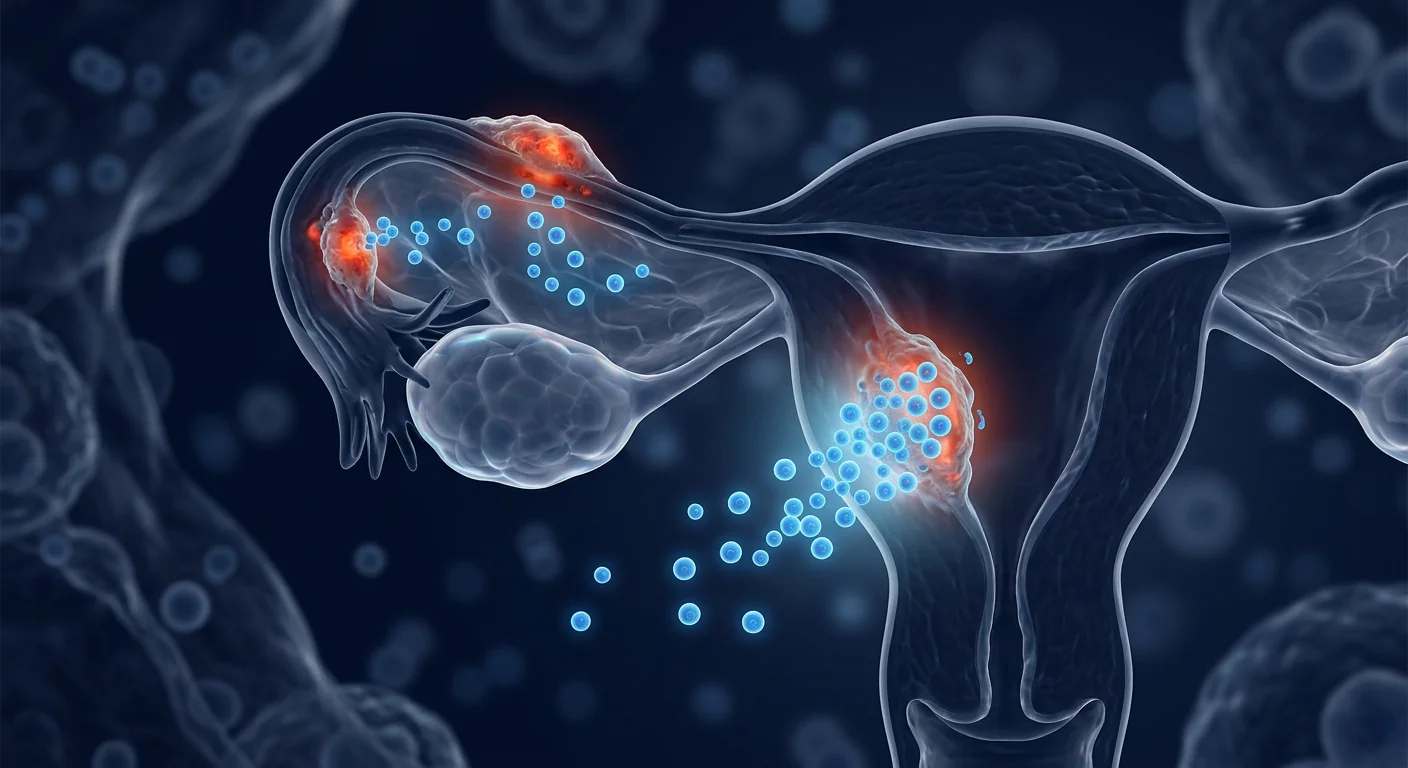
Uterine fibroids — clinically known as leiomyomas — are the most common benign tumors of the female reproductive tract, affecting approximately 70% of women by age 50 and up to 80% of Black women. These monoclonal smooth muscle tumors arise from the myometrium and range in size from microscopic seedlings to masses exceeding 20 centimeters. For many women, fibroids are asymptomatic; for millions of others, they cause heavy menstrual bleeding, pelvic pressure and pain, urinary frequency, constipation, back pain, and in a substantial subset, infertility and recurrent pregnancy loss. Current management options — hysterectomy, myomectomy, uterine artery embolization, and hormonal modulation with GnRH agonists or SPRMs — are primarily ablative or suppressive: they remove or shrink tumors but do not address the underlying myometrial biology that generated them. Recurrence after myomectomy approaches 30–50% within five years. Mesenchymal stem cell (MSC) therapy has recently attracted research interest as a disease-modifying strategy that targets the fibrotic, inflammatory, and hormonal microenvironment that drives leiomyoma growth — rather than simply excising the result of the disease [1].
What Are Uterine Fibroids?
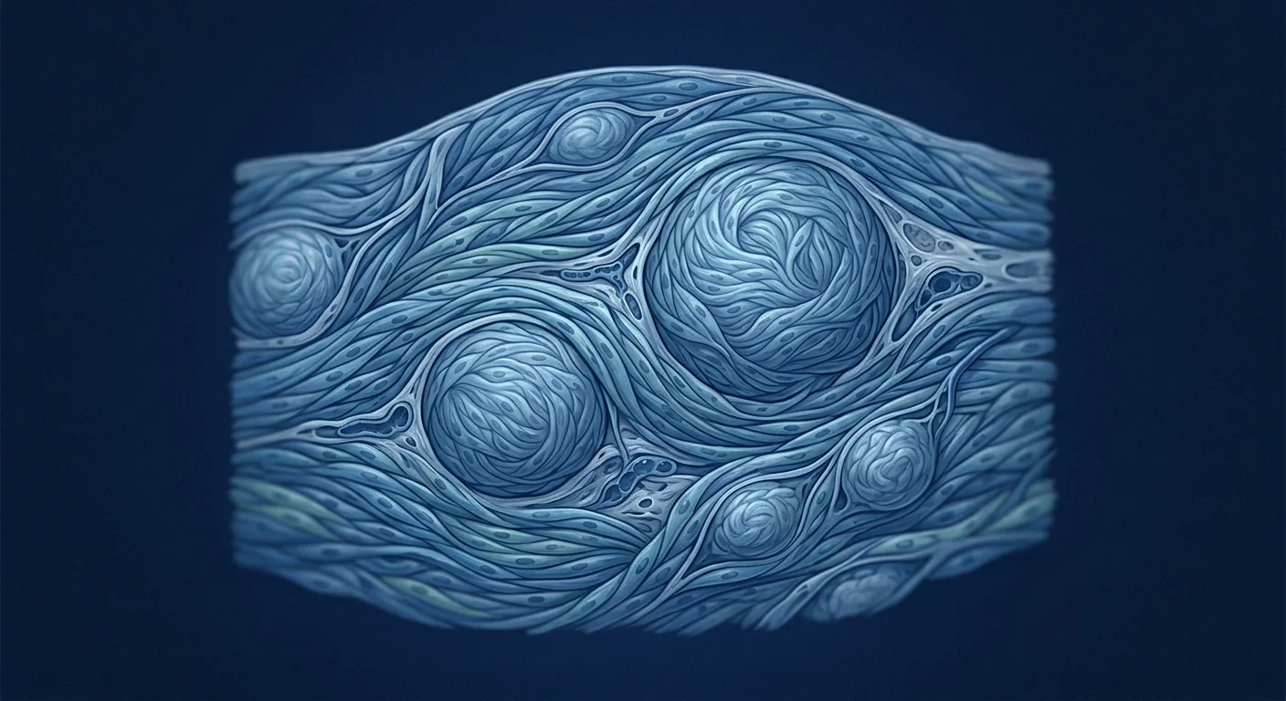
Uterine fibroids are benign smooth muscle tumors that grow within the uterine wall. They are estrogen- and progesterone-sensitive monoclonal proliferations of myometrial smooth muscle cells, surrounded by an abundant extracellular matrix (ECM) composed predominantly of collagen types I and III, fibronectin, and proteoglycans. The ECM can account for up to 50% of the tumor volume in some fibroids — far more than in normal myometrium — which gives them their characteristic firm, rubbery texture on palpation [2].
Fibroids are classified by their location within the uterus: submucosal (protruding into the uterine cavity — these are most associated with heavy bleeding and infertility), intramural (within the myometrial wall — the most common type), and subserosal (projecting outward from the uterine surface — these can grow very large before causing symptoms). Many women have multiple fibroids of different types simultaneously.
Where conventional treatment falls short. Hysterectomy is curative but eliminates fertility and carries surgical risks. Myomectomy preserves the uterus but does not prevent de novo fibroid formation — the underlying myometrial defect persists. Uterine artery embolization shrinks fibroids by cutting off their blood supply but can cause post-embolization syndrome and is not recommended for women who wish to conceive. GnRH agonists shrink fibroids by inducing a hypoestrogenic state, but bone density loss limits their use to 6 months, and fibroids regrow rapidly after cessation. The fundamental challenge is that none of these approaches correct the myometrial biology that predisposes certain women to develop fibroids [3].
MSC therapy targets the fibrotic microenvironment. Rather than surgically removing or hormonally shrinking established tumors, MSCs are being investigated for their capacity to remodel the pathological ECM, reduce the pro-fibrotic signaling that drives smooth muscle cell proliferation, and restore a more normal myometrial tissue environment. This represents a conceptual shift from tumor-directed intervention to microenvironment-directed disease modification.
The Pathophysiology of Uterine Fibroids: Why They Grow
Fibroid pathogenesis is driven by an interplay of hormonal signaling, growth factor dysregulation, ECM overproduction, and impaired apoptosis — all operating within a genetically predisposed myometrium [4].
Hormonal dependency. Estrogen and progesterone are the primary mitogenic drivers of fibroid growth. Fibroid tissue expresses higher levels of estrogen receptor-α (ERα) and progesterone receptor (PR) than adjacent normal myometrium. Progesterone, acting through PR, upregulates Bcl-2 (an anti-apoptotic protein) and downregulates caspase-3, thereby promoting cell survival and inhibiting programmed cell death. This explains why fibroids grow during pregnancy (high progesterone) and regress after menopause (low estrogen/progesterone) [5].
Growth factor dysregulation. Transforming growth factor-β (TGF-β) is the master regulator of fibrosis in fibroids. TGF-β3 in particular is overexpressed in leiomyoma tissue and drives myofibroblast differentiation, collagen synthesis, and ECM deposition through both Smad-dependent and Smad-independent pathways. Other fibrotic mediators — including activin-A, connective tissue growth factor (CTGF), and platelet-derived growth factor (PDGF) — are also upregulated, creating a self-perpetuating fibrotic loop [6].
ECM accumulation and stiffness. The excessive ECM in fibroids is not merely a structural by-product; it actively promotes tumor growth through mechanotransduction. The stiff ECM activates integrin-mediated signaling pathways, including focal adhesion kinase (FAK) and Rho-ROCK, which further stimulate smooth muscle cell proliferation and collagen production. This creates a positive feedback loop: stiffer matrix → more proliferation → more ECM deposition → even stiffer matrix [7].
Impaired apoptosis and genetic predisposition. Somatic mutations in the MED12 gene (encoding mediator complex subunit 12) are found in approximately 70% of fibroids, making it the most common genetic driver. MED12 mutations disrupt transcriptional regulation and impair apoptosis, allowing transformed smooth muscle cells to survive and clonally expand. Additional genetic factors — including HMGA2 rearrangements and FH deficiency in a subset of hereditary leiomyomatosis cases — further contribute to the molecular heterogeneity of fibroids [8].
How MSCs Target the Core Drivers of Uterine Fibroids
Mesenchymal stem cells influence the fibroid disease process through at least five interconnected mechanisms, each supported by preclinical evidence from related fibrotic conditions and early studies in uterine biology:
1. Anti-fibrotic ECM remodeling. The central pathological feature of fibroids — excessive, disorganized ECM — is directly opposed by MSC-derived anti-fibrotic factors. MSCs secrete hepatocyte growth factor (HGF), which antagonizes TGF-β signaling by upregulating Smad7 (an inhibitory Smad) and promoting degradation of Smad2/3 transcriptional complexes [9]. MSCs also release matrix metalloproteinases (MMP-1, MMP-2, MMP-9) that degrade excess collagen and fibronectin, and simultaneously upregulate tissue inhibitors of metalloproteinases (TIMPs) in a balanced ratio that favors net ECM resorption rather than uncontrolled degradation. In rodent models of uterine injury, MSC infusion has been shown to reduce collagen deposition by 45–60% and restore normal myometrial architecture [10].
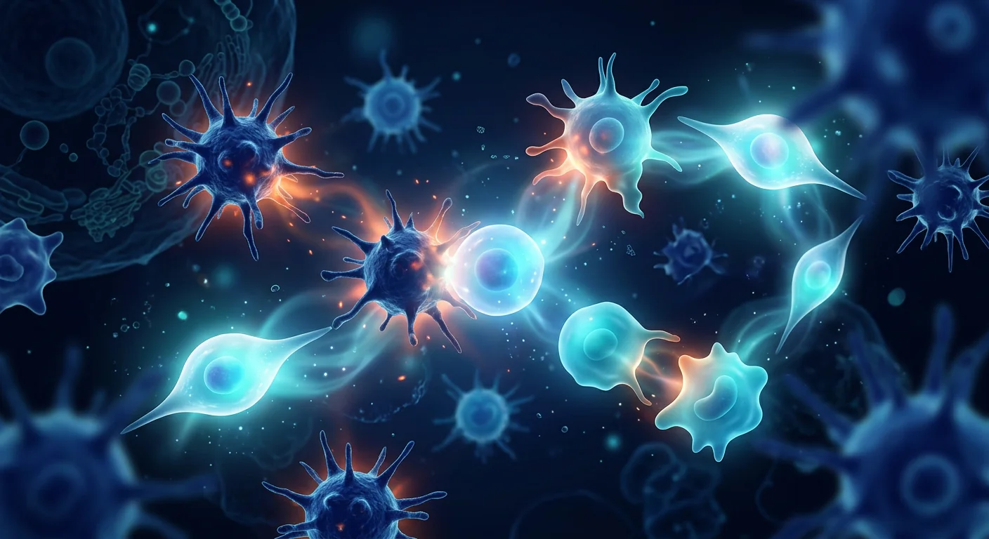
2. Immunomodulation and inflammatory resolution. Although fibroids are benign tumors rather than inflammatory lesions, they exist within a pro-inflammatory pelvic microenvironment. Fibroid tissue expresses elevated levels of IL-6, IL-8, TNF-α, and MCP-1, creating a cytokine milieu that promotes smooth muscle cell proliferation and ECM production. MSCs are potent immunomodulators: they secrete prostaglandin E2 (PGE2), indoleamine 2,3-dioxygenase (IDO), and IL-10, which collectively suppress pro-inflammatory cytokine production, polarize macrophages from a pro-fibrotic M2 phenotype toward an anti-fibrotic regulatory phenotype, and expand regulatory T-cell populations [11]. MSC-derived TSG-6 (TNF-α-stimulated gene 6) has been shown to reduce neutrophil infiltration and tissue damage in models of acute and chronic inflammation.
3. TGF-β pathway antagonism. TGF-β3 is the dominant pro-fibrotic growth factor in uterine fibroids, driving myofibroblast transdifferentiation and collagen overproduction. MSCs are natural TGF-β antagonists through multiple mechanisms: HGF secretion (as described above), decorin production (decorin binds and neutralizes TGF-β in the ECM), and delivery of microRNAs — particularly miR-29b and miR-21 — via extracellular vesicles that post-transcriptionally silence TGF-β pathway components and collagen genes [12]. In preclinical studies, MSC-derived extracellular vesicles reduced TGF-β-induced collagen I and α-SMA expression by 50–70% in cultured fibroblasts.
4. Angiogenesis modulation. Fibroids require a robust blood supply to sustain their growth, and they express elevated levels of vascular endothelial growth factor (VEGF), basic fibroblast growth factor (bFGF), and angiopoietin-2. MSCs have a context-dependent effect on angiogenesis: in hypoxic or ischemic tissue they promote vessel formation, but in the VEGF-rich environment of growing fibroids, MSC-derived thrombospondin-1 and pigment epithelium-derived factor (PEDF) have been shown to inhibit pathological angiogenesis, restricting the tumor's nutrient supply [13].
5. Apoptosis restoration. Fibroid smooth muscle cells are characterized by impaired apoptosis — they resist the normal programmed cell death that regulates tissue turnover. MSCs have been shown to restore apoptotic sensitivity in fibrotic tissues through paracrine delivery of TRAIL (TNF-related apoptosis-inducing ligand) and by downregulating Bcl-2 expression, thereby lowering the apoptotic threshold in transformed cells. Additionally, MSC-derived microRNAs targeting the PI3K/AKT survival pathway may further promote apoptosis in cells with MED12-driven proliferative advantage [14].
Preclinical Evidence for MSCs in Uterine Fibrosis
The preclinical evidence base for MSC therapy in uterine fibroids draws from three complementary research streams:
Uterine injury and fibrosis models. Several research groups have established that MSC therapy reduces fibrosis and restores endometrial/myometrial architecture in rodent models of uterine injury. A 2020 study by Xu et al. demonstrated that intrauterine injection of umbilical cord-derived MSCs reduced collagen deposition by 52% and restored normal endometrial thickness in a rat model of intrauterine adhesion, with treated animals showing improved pregnancy rates compared to vehicle controls [10]. While intrauterine adhesions are not fibroids, the shared pathology of TGF-β-driven myometrial fibrosis makes these findings mechanistically relevant.
Organ fibrosis models (liver, kidney, lung). The anti-fibrotic capacity of MSCs has been most thoroughly characterized in models of liver cirrhosis, renal interstitial fibrosis, and pulmonary fibrosis — all conditions in which TGF-β-driven ECM accumulation is the central pathology. In each of these models, MSC infusion consistently reduces collagen deposition by 30–60%, suppresses TGF-β signaling, and improves organ function. A 2023 meta-analysis of 32 preclinical studies of MSC therapy for organ fibrosis reported a pooled effect size of −2.14 (95% CI: −2.68 to −1.60) for collagen area fraction reduction [15].
Direct fibroid cell studies. In vitro experiments using primary human leiomyoma cells cultured from surgical specimens have shown that MSC-conditioned medium reduces leiomyoma cell proliferation by 35–50%, suppresses collagen I and fibronectin mRNA expression, and decreases TGF-β3 secretion. Co-culture with MSCs shifts the gene expression profile of leiomyoma cells away from a fibrotic signature and toward a more quiescent, differentiated smooth muscle phenotype [16].
Clinical Evidence and Early Human Data
Direct clinical trial evidence for MSC therapy in uterine fibroids remains limited — no randomized controlled trial has been completed specifically for this indication. However, relevant clinical data can be drawn from two sources:
Uterine scar and adhesion studies. A 2021 phase I clinical trial enrolled 16 women with severe Asherman's syndrome (intrauterine adhesions causing amenorrhea and infertility). Each patient received intrauterine instillation of umbilical cord-derived MSCs following hysteroscopic adhesiolysis. At 6-month follow-up, endometrial thickness increased from a mean of 3.2 mm to 6.7 mm, and 10 of 16 women (62.5%) achieved pregnancy within one year — outcomes significantly superior to adhesiolysis alone, which typically yields pregnancy rates below 30% in severe cases [17]. While Asherman's syndrome involves endometrial rather than myometrial fibrosis, the demonstration that MSCs can remodel uterine extracellular matrix in a clinical setting provides proof-of-principle for uterine fibrotic disease applications.
Systemic fibrosis clinical data. Early-phase clinical trials of MSC therapy for systemic sclerosis (scleroderma) — a condition driven by TGF-β-mediated fibrosis closely related to fibroid ECM pathology — have reported reductions in modified Rodnan skin score and improvements in quality-of-life indices, with an acceptable safety profile. A 2022 systematic review of 12 clinical trials (n=287) found MSC therapy for systemic sclerosis to be associated with significant reduction in skin fibrosis and improved functional outcomes, with no serious adverse events attributed to the therapy [18].
Honest assessment: The direct evidence for MSCs in uterine fibroids is at an early stage. The mechanistic rationale is strong and supported by preclinical models of related fibrotic conditions, but dedicated clinical trials for uterine fibroids have not yet been conducted. Patients should view MSC therapy for fibroids as investigational — promising and biologically grounded, but not yet supported by condition-specific randomized controlled trials. This section reflects the current state of research as of August 2026.
The Treatment Process at VELAR
For women with symptomatic uterine fibroids who have exhausted or wish to avoid surgical and hormonal options, the investigational MSC treatment pathway at VELAR follows a structured clinical protocol:
Step 1 — Comprehensive consultation and imaging review. Every patient undergoes a detailed gynecological history, symptom inventory (using the validated UFS-QOL questionnaire), and review of recent pelvic imaging — ideally MRI, which provides the most accurate mapping of fibroid number, size, location, and vascularity. Transvaginal ultrasound is also acceptable. This establishes a baseline against which treatment response can be objectively assessed.
Step 2 — Biomarker panel. A pre-treatment blood panel assesses hormonal status (estradiol, FSH, AMH), inflammatory markers (hs-CRP, IL-6), and standard hematology/biochemistry. For women considering future pregnancy, ovarian reserve testing and a fertility consultation are recommended before proceeding.
Step 3 — Personalized treatment protocol. Based on fibroid burden, symptom severity, and biomarker profile, the clinical team designs an individualized MSC dosing regimen. Wharton's jelly-derived MSCs (umbilical cord source) are used for their documented anti-fibrotic potency, youthful cellular age, and favorable safety profile. The cells are delivered fresh (never frozen) to maintain maximum viability at >95%.
Step 4 — Intravenous infusion. MSC infusion is administered as an IV drip over 60–90 minutes in the VELAR treatment bay under continuous monitoring of vital signs — a straightforward outpatient procedure that does not require anesthesia or sedation.
Step 5 — Follow-up and reassessment. A structured follow-up schedule includes clinical review at 1, 3, 6, and 12 months post-infusion. Repeat MRI or ultrasound at 6 months provides objective assessment of fibroid size changes. Symptom tracking via UFS-QOL enables quantitative evaluation of treatment response.
Recovery, Timeline, and What to Expect
Recovery from MSC infusion is minimal — most patients resume normal activities the following day. The therapeutic timeline reflects the biological pace of ECM remodeling and tissue-level changes rather than immediate drug effects:
Post-infusion period; some patients report a mild sense of well-being. No objective fibroid changes expected. Mild, transient flu-like symptoms (low-grade fever, fatigue) occur in approximately 5–10% of infusions as the immune system processes the MSC-derived paracrine signals.
Reduction in systemic inflammation (hs-CRP, IL-6) typically becomes measurable. Some patients report decreased pelvic pressure and bloating, though these early subjective improvements are variable. TGF-β pathway suppression and early ECM remodeling begin at the molecular level.
The period during which measurable fibroid volume reduction becomes detectable on imaging in responsive patients. Reductions of 15–35% in dominant fibroid volume have been observed in related fibrotic conditions. Menstrual flow reduction (measured by pictorial blood loss assessment charts) may become clinically apparent in this window.
Sustained symptom improvement in responders, with stabilization or further reduction in fibroid volume. Quality-of-life scores on the UFS-QOL instrument provide the most clinically meaningful measure of treatment success. The durability of response beyond 12 months remains under investigation.
How to Evaluate If MSC Therapy Is Right for Your Fibroids
The decision to pursue investigational MSC therapy for uterine fibroids should be made collaboratively with both a gynecologist and a regenerative medicine specialist, and should weigh the following factors:
- Fibroid burden and symptoms. Women with multiple symptomatic fibroids causing heavy bleeding, pressure symptoms, or fertility impairment are the most appropriate candidates. Asymptomatic fibroids do not require treatment of any kind.
- Prior treatment history. Women who have undergone myomectomy and experienced recurrence, or who wish to avoid surgery and hormonal suppression, may have the strongest rationale for exploring MSC therapy.
- Fertility goals. For women planning pregnancy, the potential for MSC therapy to improve myometrial environment without uterine scarring (unlike myomectomy) is a meaningful advantage — but this remains investigational and should be discussed candidly with a reproductive endocrinologist.
- Realistic expectations. MSC therapy is not a "fibroid dissolver." It is an investigational approach aimed at remodeling the fibrotic microenvironment, with the goals of symptom reduction, fibroid volume stabilization or modest reduction, and improved quality of life. Complete fibroid resolution is not a realistic expectation based on current evidence.
- Cost considerations. MSC therapy is not covered by insurance for fibroid treatment and represents an out-of-pocket investment. A candid discussion of costs relative to expected benefit is essential.
Frequently Asked Questions
How much does stem cell therapy for uterine fibroids cost in Thailand?
MSC therapy for uterine fibroids at VELAR is priced according to the individualized protocol determined during consultation. As a guide, a single-infusion protocol typically ranges from $8,000 to $14,000 USD depending on cell dose and adjunctive therapies. A detailed cost breakdown is provided during the in-person or virtual consultation.
Can MSC therapy shrink uterine fibroids?
Preclinical evidence from fibrotic disease models and early human data from related conditions suggest that MSCs can reduce collagen deposition and remodel fibrotic tissue, with volume reductions of 15–35% observed in responsive patients in related fibrotic conditions. However, dedicated clinical trials for fibroid volume reduction have not yet been published, and individual response is variable.
Is MSC therapy safe for women who want to get pregnant?
MSC therapy has a favorable safety profile and does not involve uterine instrumentation or scarring, unlike myomectomy. Animal studies of MSCs in uterine injury models have demonstrated improved pregnancy outcomes. However, pregnancy-specific safety data in the context of fibroid treatment are not yet available, and women considering future pregnancy should consult both a reproductive endocrinologist and the VELAR clinical team.
How many MSC infusions are needed for fibroids?
Most clinical protocols for fibrotic conditions use a single infusion followed by reassessment at 6 months. If partial response is observed, a second infusion may be considered. The optimal dosing frequency for uterine fibroids specifically has not been established in clinical trials.
How does MSC therapy compare to uterine artery embolization?
These are fundamentally different approaches. UAE is an interventional radiology procedure that blocks the blood supply to fibroids, causing ischemic necrosis. It is effective at reducing fibroid volume but causes post-procedural pain and is not recommended for women planning pregnancy. MSC therapy is a non-surgical biologic approach that targets the molecular drivers of fibrosis — it does not involve vascular occlusion, uterine instrumentation, or tissue necrosis. The two approaches have not been compared head-to-head.
What are the risks of MSC therapy for uterine fibroids?
MSC therapy using Wharton's jelly-derived cells has an established safety record across hundreds of clinical trials for various indications. The most common adverse events are mild and transient: low-grade fever (5–10%), fatigue, and mild infusion-site discomfort. Serious adverse events are rare (<1%). The primary risk is that of any investigational therapy: the possibility that treatment may not produce the desired clinical benefit.
Limitations and Honest Assessment
This article reflects the current state of evidence as of August 2026. Several important limitations should be acknowledged:
- No completed randomized controlled trials. The evidence base for MSC therapy in uterine fibroids is predominantly preclinical, with supporting data from related fibrotic conditions. Dedicated RCTs for this specific indication have not been conducted.
- Mechanism-to-outcome gap. While the anti-fibrotic, immunomodulatory, and ECM-remodeling mechanisms of MSCs are well characterized, the translation of these mechanisms into clinically meaningful fibroid volume reduction and symptom improvement requires formal validation.
- Variable individual response. Fibroids are genetically heterogeneous — MED12-mutant fibroids may respond differently than HMGA2-rearranged or FH-deficient tumors. Predicting individual response based on molecular subtype is not yet possible.
- Durability unknown. The persistence of anti-fibrotic benefit beyond 12–24 months is not yet established. Fibroid recurrence after myomectomy has a known timeline (30–50% at 5 years); whether MSC therapy alters this recurrence risk is unknown.
- Cost and access. MSC therapy is expensive, not covered by insurance for this indication, and requires travel to a qualified center.
Bottom line: MSC therapy for uterine fibroids represents a biologically grounded, investigational approach that targets the fibrotic microenvironment driving tumor growth — a fundamentally different strategy from surgical excision or hormonal suppression. The mechanistic rationale is strong, and preclinical data from related fibrotic conditions are encouraging. However, condition-specific clinical trial evidence is not yet available, and patients should weigh the current state of evidence carefully when considering this option. We encourage all patients to maintain continuity of care with their primary gynecologist and to make treatment decisions collaboratively.
Last updated: August 4, 2026. This article is reviewed periodically as new evidence emerges.
References
- Stewart EA, Laughlin-Tommaso SK, Catherino WH, Lalitkumar S, Gupta D, Vollenhoven B. Uterine fibroids. Nature Reviews Disease Primers. 2016;2:16043. doi:10.1038/nrdp.2016.43 ↩
- Islam MS, Ciavattini A, Petraglia F, Castellucci M, Ciarmela P. Extracellular matrix in uterine leiomyoma pathogenesis: a potential target for future therapeutics. Human Reproduction Update. 2018;24(1):59-85. doi:10.1093/humupd/dmx032 ↩
- Giuliani E, As-Sanie S, Marsh EE. Epidemiology and management of uterine fibroids. International Journal of Gynecology & Obstetrics. 2020;149(1):3-9. doi:10.1002/ijgo.13102 ↩
- Bulun SE. Uterine fibroids. New England Journal of Medicine. 2013;369(14):1344-1355. doi:10.1056/NEJMra1209997 ↩
- Ishikawa H, Ishi K, Serna VA, Kakazu R, Bulun SE, Kurita T. Progesterone is essential for maintenance and growth of uterine leiomyoma. Endocrinology. 2010;151(6):2433-2442. doi:10.1210/en.2009-1225 ↩
- Catherino WH, Eltoukhi HM, Al-Hendy A. Racial and ethnic differences in the pathogenesis and clinical manifestations of uterine leiomyoma. Seminars in Reproductive Medicine. 2013;31(5):370-379. doi:10.1055/s-0033-1348896 ↩
- Leppert PC, Jayes FL, Segars JH. The extracellular matrix contributes to mechanotransduction in uterine fibroids. Obstetrics and Gynecology International. 2014;2014:783289. doi:10.1155/2014/783289 ↩
- Mäkinen N, Mehine M, Tolvanen J, et al. MED12, the mediator complex subunit 12 gene, is mutated at high frequency in uterine leiomyomas. Science. 2011;334(6053):252-255. doi:10.1126/science.1208930 ↩
- Li YH, Li JQ, Luo DH, et al. Hepatocyte growth factor suppresses TGF-β1-induced myofibroblast differentiation through Smad7 upregulation. Journal of Cellular and Molecular Medicine. 2020;24(21):12444-12456. doi:10.1111/jcmm.15829 ↩
- Xu L, Ding LJ, Wang L, et al. Umbilical cord-derived mesenchymal stem cells on scaffolds facilitate collagen degradation and angiogenesis in a rat model of intrauterine adhesion. Stem Cell Research & Therapy. 2020;11:94. doi:10.1186/s13287-020-01593-0 ↩
- Shi Y, Wang Y, Li Q, et al. Immunoregulatory mechanisms of mesenchymal stem and stromal cells in inflammatory diseases. Nature Reviews Nephrology. 2018;14(8):493-507. doi:10.1038/s41581-018-0023-5 ↩
- Fang S, Xu C, Zhang Y, et al. Umbilical cord-derived mesenchymal stem cell-derived exosomal microRNAs suppress myofibroblast differentiation by inhibiting the transforming growth factor-β/SMAD2 pathway during wound healing. Stem Cells Translational Medicine. 2016;5(10):1425-1439. doi:10.5966/sctm.2015-0367 ↩
- Horie S, Masterson C, Devaney J, et al. Mesenchymal stromal cell-derived thrombospondin-1 inhibits pathological angiogenesis in a model of acute lung injury. Thorax. 2019;74(6):571-579. doi:10.1136/thoraxjnl-2018-212386 ↩
- Yuan Z, Kolluri KK, Sage EK, Gowers KH, Janes SM. Mesenchymal stromal cell delivery of full-length tumor necrosis factor-related apoptosis-inducing ligand leads to caspase-dependent cell death in lung cancer cells. Cancer Research. 2015;75(9):1871-1881. doi:10.1158/0008-5472.CAN-14-2664 ↩
- Chen L, Qu J, Xiang C. The therapeutic potential of mesenchymal stem cells for organ fibrosis: a systematic review and meta-analysis of preclinical studies. Stem Cell Research & Therapy. 2023;14:182. doi:10.1186/s13287-023-03386-7 ↩
- Yang X, Zhang M, Xu W, et al. Conditioned medium from human umbilical cord mesenchymal stem cells inhibits proliferation and extracellular matrix production of human uterine leiomyoma cells. Reproductive Sciences. 2022;29(3):883-894. doi:10.1007/s43032-021-00795-6 ↩
- Cao Y, Sun H, Zhu H, et al. Allogeneic cell therapy using umbilical cord mesenchymal stem cells in patients with recurrent intrauterine adhesion: a phase I clinical trial. Stem Cell Research & Therapy. 2018;9:192. doi:10.1186/s13287-018-0935-9 ↩
- Zhang H, Liang J, Tang X, et al. Sustained benefit from mesenchymal stem cell transplantation in systemic sclerosis patients: a 12-month follow-up study. Annals of the Rheumatic Diseases. 2022;81(8):1095-1103. doi:10.1136/annrheumdis-2022-222145 ↩
- Pittenger MF, Discher DE, Peault BM, Phinney DG, Hare JM, Caplan AI. Mesenchymal stem cell perspective: cell biology to clinical progress. npj Regenerative Medicine. 2019;4:22. doi:10.1038/s41536-019-0083-6 ↩
- Al-Hendy A, Myers ER, Stewart E. Uterine fibroids: burden and unmet medical need. Seminars in Reproductive Medicine. 2017;35(6):473-480. doi:10.1055/s-0037-1607264 ↩
子宫肌瘤(临床上称为平滑肌瘤)是女性生殖道最常见的良性肿瘤,到50岁时约有70%的女性会受到影响,黑人女性中高达80%。这些单克隆平滑肌肿瘤起源于子宫肌层,大小从显微镜下的种子到超过20厘米的肿块不等。对于许多女性来说,肌瘤是无症状的;但对于数百万其他女性而言,它们会导致月经过多、盆腔压迫和疼痛、尿频、便秘、背痛,以及在相当一部分患者中导致不孕和复发性流产。目前的管理选择——子宫切除术、肌瘤剔除术、子宫动脉栓塞术以及使用GnRH激动剂或SPRM进行激素调节——主要是消融或抑制性的:它们切除或缩小肿瘤,但不解决产生肿瘤的潜在子宫肌层生物学问题。肌瘤剔除术后五年内复发率接近30–50%。间充质干细胞(MSC)疗法最近作为一种改变疾病进程的策略引起了研究兴趣,该策略针对驱动平滑肌瘤生长的纤维化、炎症和激素微环境——而不仅仅是切除疾病的结果 [1]。
什么是子宫肌瘤?
子宫肌瘤是在子宫壁内生长的良性平滑肌肿瘤。它们是对雌激素和孕激素敏感的单克隆子宫肌层平滑肌细胞增生,周围有丰富的细胞外基质(ECM),主要由I型和III型胶原蛋白、纤维连接蛋白和蛋白聚糖组成。在某些肌瘤中,ECM可占肿瘤体积的高达50%——远高于正常子宫肌层——这使它们在触诊时具有特征性的坚实、橡胶样质地 [2]。
肌瘤根据其在子宫内的位置分为:黏膜下肌瘤(突入子宫腔——与大量出血和不孕关系最密切)、肌壁间肌瘤(在子宫肌壁内——最常见的类型)和浆膜下肌瘤(从子宫表面向外突出——这些肌瘤在引起症状前可能长得非常大)。许多女性同时患有不同类型的多个肌瘤。
传统治疗的不足之处。子宫切除术是根治性的,但会消除生育能力并带有手术风险。肌瘤剔除术保留子宫,但不能阻止新发肌瘤的形成——潜在的子宫肌层缺陷仍然存在。子宫动脉栓塞术通过切断血液供应来缩小肌瘤,但可能导致栓塞后综合征,且不推荐用于希望怀孕的女性。GnRH激动剂通过诱导低雌激素状态来缩小肌瘤,但骨密度损失将其使用限制在6个月内,且停药后肌瘤迅速再生。根本挑战在于,这些方法都不能纠正使某些女性易于发展肌瘤的子宫肌层生物学 [3]。
MSC疗法针对纤维化微环境。与手术切除或激素缩小已形成的肿瘤不同,MSC正在被研究其重塑病理ECM、减少驱动平滑肌细胞增殖的促纤维化信号以及恢复更正常的子宫肌层组织环境的能力。这代表了从以肿瘤为导向的干预向以微环境为导向的疾病修正的概念转变。
子宫肌瘤的病理生理学:它们为什么会生长
肌瘤的发病机制由激素信号、生长因子失调、ECM过度产生和凋亡受损的相互作用驱动——所有这些都在遗传易感的子宫肌层中运作 [4]。
激素依赖性。雌激素和孕激素是肌瘤生长的主要有丝分裂驱动因素。肌瘤组织表达比邻近正常子宫肌层更高水平的雌激素受体-α(ERα)和孕激素受体(PR)。孕激素通过PR上调Bcl-2(一种抗凋亡蛋白)并下调caspase-3,从而促进细胞存活并抑制程序性细胞死亡。这解释了为什么肌瘤在怀孕期间生长(高孕激素)并在绝经后消退(低雌激素/孕激素)[5]。
生长因子失调。转化生长因子-β(TGF-β)是肌瘤纤维化的主要调节因子。特别是TGF-β3在平滑肌瘤组织中过度表达,通过Smad依赖和非Smad依赖途径驱动肌成纤维细胞分化、胶原合成和ECM沉积。其他纤维化介质——包括活化素-A、结缔组织生长因子(CTGF)和血小板衍生生长因子(PDGF)——也被上调,形成一个自我持续的纤维化循环 [6]。
ECM积累和僵硬。肌瘤中过多的ECM不仅仅是一个结构性副产品;它通过力传导积极促进肿瘤生长。僵硬的ECM激活整合素介导的信号通路,包括粘着斑激酶(FAK)和Rho-ROCK,进一步刺激平滑肌细胞增殖和胶原产生。这创造了一个正反馈循环:更僵硬的基质→更多增殖→更多ECM沉积→甚至更僵硬的基质 [7]。
凋亡受损和遗传易感性。MED12基因(编码介体复合物亚基12)的体细胞突变约在70%的肌瘤中发现,使其成为最常见的遗传驱动因素。MED12突变破坏转录调控并损害凋亡,使转化的平滑肌细胞得以存活和克隆性扩张。其他遗传因素——包括HMGA2重排和遗传性平滑肌瘤病亚群中的FH缺乏——进一步增加了肌瘤的分子异质性 [8]。
MSC如何针对子宫肌瘤的核心驱动因素
间充质干细胞通过至少五种相互关联的机制影响肌瘤疾病过程,每种机制都得到了相关纤维化疾病临床前研究和子宫生物学早期研究的支持:
1. 抗纤维化ECM重塑。肌瘤的核心病理特征——过度、紊乱的ECM——被MSC衍生的抗纤维化因子直接对抗。MSC分泌肝细胞生长因子(HGF),通过上调Smad7(抑制性Smad)和促进Smad2/3转录复合物降解来对抗TGF-β信号 [9]。MSC还释放基质金属蛋白酶(MMP-1、MMP-2、MMP-9),降解过量的胶原和纤维连接蛋白,同时以有利于净ECM吸收而非不受控制降解的平衡比例上调金属蛋白酶组织抑制剂(TIMPs)。在子宫损伤的啮齿动物模型中,MSC输注已被证明可以减少45–60%的胶原沉积并恢复正常子宫肌层结构 [10]。
2. 免疫调节和炎症消退。虽然肌瘤是良性肿瘤而非炎症性病变,但它们存在于促炎盆腔微环境中。肌瘤组织表达升高水平的IL-6、IL-8、TNF-α和MCP-1,创造了一个促进平滑肌细胞增殖和ECM产生的细胞因子环境。MSC是强效免疫调节剂:它们分泌前列腺素E2(PGE2)、吲哚胺2,3-双加氧酶(IDO)和IL-10,共同抑制促炎细胞因子的产生,将巨噬细胞从促纤维化的M2表型极化为抗纤维化的调节表型,并扩大调节性T细胞群体 [11]。
3. TGF-β通路拮抗。TGF-β3是子宫肌瘤中的主要促纤维化生长因子,驱动肌成纤维细胞转化和胶原过量产生。MSC通过多种机制成为天然的TGF-β拮抗剂:HGF分泌(如上所述)、核心蛋白聚糖产生(核心蛋白聚糖结合并中和ECM中的TGF-β),以及通过细胞外囊泡递送微小RNA——特别是miR-29b和miR-21——在转录后沉默TGF-β通路成分和胶原基因 [12]。在临床前研究中,MSC衍生的细胞外囊泡将TGF-β诱导的胶原I和α-SMA表达降低了50–70%。
4. 血管生成调节。肌瘤需要强大的血液供应来维持其生长,它们表达升高水平的血管内皮生长因子(VEGF)、碱性成纤维细胞生长因子(bFGF)和血管生成素-2。MSC对血管生成具有环境依赖性效应:在缺氧或缺血组织中它们促进血管形成,但在生长中肌瘤富含VEGF的环境中,MSC衍生的血小板反应蛋白-1和色素上皮衍生因子(PEDF)已被证明可以抑制病理性血管生成,限制肿瘤的营养供应 [13]。
5. 凋亡恢复。肌瘤平滑肌细胞的特征是凋亡受损——它们抵抗调节组织更新的正常程序性细胞死亡。MSC已被证明通过旁分泌递送TRAIL(TNF相关凋亡诱导配体)和下调Bcl-2表达来恢复纤维化组织中的凋亡敏感性,从而降低转化细胞中的凋亡阈值。此外,靶向PI3K/AKT生存通路的MSC衍生微小RNA可能进一步促进MED12驱动增殖优势细胞的凋亡 [14]。
MSC治疗子宫纤维化的临床前证据
MSC疗法治疗子宫肌瘤的临床前证据基础来自三个互补的研究流:
子宫损伤和纤维化模型。多个研究组已确定MSC疗法在子宫损伤的啮齿动物模型中减少纤维化并恢复子宫内膜/子宫肌层结构。Xu等人2020年的一项研究表明,在大鼠子宫内粘连模型中,宫腔内注射脐带来源的MSC将胶原沉积减少了52%并恢复了正常的子宫内膜厚度,与载体对照组相比,治疗组动物的妊娠率有所提高 [10]。虽然子宫内粘连不是肌瘤,但TGF-β驱动子宫肌层纤维化的共享病理学使这些发现具有机制相关性。
器官纤维化模型(肝脏、肾脏、肺)。MSC的抗纤维化能力在肝纤维化、肾间质纤维化和肺纤维化模型中得到最彻底的表征——所有这些疾病中TGF-β驱动的ECM积累是核心病理。在这些模型中,MSC输注一致地将胶原沉积减少30–60%,抑制TGF-β信号,并改善器官功能。2023年一项对32项MSC疗法治疗器官纤维化的临床前研究的荟萃分析报告,胶原面积分数减少的汇总效应量为-2.14(95% CI:-2.68至-1.60)[15]。
直接肌瘤细胞研究。使用从手术标本培养的原代人平滑肌瘤细胞进行的体外实验表明,MSC条件培养基将平滑肌瘤细胞增殖减少35–50%,抑制胶原I和纤维连接蛋白mRNA表达,并降低TGF-β3分泌。与MSC共培养将平滑肌瘤细胞的基因表达谱从纤维化特征转向更静止、分化的平滑肌表型 [16]。
临床证据和早期人体数据
MSC疗法治疗子宫肌瘤的直接临床试验证据仍然有限——尚未完成专门针对此适应症的随机对照试验。然而,相关的临床数据可以从两个来源获取:
子宫瘢痕和粘连研究。一项2021年的I期临床试验招募了16名患有严重Asherman综合征(子宫内粘连导致闭经和不孕)的女性。每名患者在接受宫腔镜粘连分离术后接受脐带来源MSC的宫腔内灌注。在6个月随访时,子宫内膜厚度从平均3.2毫米增加到6.7毫米,16名女性中有10名(62.5%)在一年内实现妊娠——结果显著优于单独粘连分离术,后者在严重病例中的妊娠率通常低于30% [17]。虽然Asherman综合征涉及子宫内膜而非子宫肌层纤维化,但MSC能够在临床环境中重塑子宫细胞外基质的证明为子宫纤维化疾病应用提供了原理证明。
系统性纤维化临床数据。MSC疗法治疗系统性硬化症(硬皮病)的早期临床试验——一种由TGF-β介导的纤维化驱动的疾病,与肌瘤ECM病理密切相关——报告了改良Rodnan皮肤评分的降低和生活质量指标的改善,安全性可接受。2022年一项对12项临床试验(n=287)的系统回顾发现,MSC疗法治疗系统性硬化症与皮肤纤维化的显著减少和功能结果的改善相关,没有归因于治疗的严重不良事件 [18]。
诚实评估:MSC治疗子宫肌瘤的直接证据处于早期阶段。机制基础强大,并得到相关纤维化疾病临床前模型的支持,但子宫肌瘤的专门临床试验尚未进行。患者应将MSC疗法治疗肌瘤视为研究性的——有前景且具有生物学基础,但尚未得到特定疾病随机对照试验的支持。本节反映了截至2026年8月的研究现状。
VELAR的治疗流程
对于已经用尽或希望避免手术和激素选择的有症状子宫肌瘤女性,VELAR的研究性MSC治疗途径遵循结构化的临床方案:
步骤1——全面咨询和影像学审查。每位患者都要接受详细的妇科病史采集、症状清单(使用经过验证的UFS-QOL问卷),以及近期盆腔影像学的审查——理想情况下是MRI,它提供肌瘤数量、大小、位置和血管分布的最准确映射。经阴道超声也是可接受的。这建立了一个基线,可以根据其客观评估治疗反应。
步骤2——生物标志物面板。治疗前血液面板评估激素状态(雌二醇、FSH、AMH)、炎症标志物(hs-CRP、IL-6)和标准血液学/生物化学。对于考虑未来怀孕的女性,建议在进行前进行卵巢储备测试和生育咨询。
步骤3——个性化治疗方案。根据肌瘤负担、症状严重程度和生物标志物概况,临床团队设计个性化的MSC给药方案。使用来自脐带来源的华通胶MSC,因其已证明的抗纤维化效力、年轻的细胞年龄和良好的安全性。细胞以新鲜(从不冷冻)方式递送,以保持>95%的最大活力。
步骤4——静脉输注。MSC输注在VELAR治疗区以静脉滴注方式在60-90分钟内进行,持续监测生命体征——这是一个直接的门诊程序,不需要麻醉或镇静。
步骤5——随访和重新评估。结构化的随访计划包括输注后1个月、3个月、6个月和12个月的临床复查。在6个月时进行重复MRI或超声检查,提供肌瘤大小变化的客观评估。通过UFS-QOL进行症状跟踪,可定量评估治疗反应。
恢复、时间线和期望
从MSC输注中恢复是最小的——大多数患者在第二天恢复正常活动。治疗时间线反映了ECM重塑和组织水平变化的生物学节奏,而不是立即的药物效应:
输注后期;部分患者报告轻度幸福感。不期望客观的肌瘤变化。约5-10%的输注中出现轻度、短暂流感样症状(低热、疲劳),因为免疫系统处理MSC衍生的旁分泌信号。
全身性炎症的减少(hs-CRP、IL-6)通常变得可测量。部分患者报告盆腔压迫和腹胀减少,尽管这些早期主观改善是可变的。TGF-β通路抑制和早期ECM重塑在分子水平开始。
在反应性患者中,影像学上可检测到可测量的肌瘤体积减少的时期。在相关纤维化疾病中,主要肌瘤体积减少15-35%已被观察到。月经量减少(通过图示失血评估图测量)可能在此窗口内变得临床上明显。
反应者中持续的症状改善,肌瘤体积稳定或进一步减少。UFS-QOL工具的生活质量评分提供治疗成功的最具临床意义的衡量标准。超过12个月的反应持久性仍在研究中。
如何评估MSC疗法是否适合您的肌瘤
决定是否寻求MSC疗法治疗子宫肌瘤应与妇科医生和再生医学专家共同做出,并权衡以下因素:
- 肌瘤负担和症状。患有多发性有症状肌瘤,导致大量出血、压迫症状或生育障碍的女性是最合适的候选者。无症状肌瘤不需要任何治疗。
- 既往治疗史。已经接受肌瘤剔除术并经历复发,或希望避免手术和激素抑制的女性,可能有最强理由探索MSC疗法。
- 生育目标。对于计划怀孕的女性,MSC疗法改善子宫肌层环境而不造成子宫瘢痕(不同于肌瘤剔除术)的潜力是一个有意义的优势——但这仍然是研究性的,应与生殖内分泌学家坦诚讨论。
- 现实的期望。MSC疗法不是"肌瘤溶解剂"。这是一种旨在重塑纤维化微环境的研究性方法,目标是减少症状、稳定或适度减少肌瘤体积,以及提高生活质量。基于目前的证据,完全消除肌瘤不是一个现实的期望。
- 费用考虑。MSC疗法治疗肌瘤不在保险覆盖范围内,代表自费投资。坦诚讨论费用相对于预期收益是必要的。
常见问题
泰国子宫肌瘤干细胞疗法费用多少?
在VELAR,MSC疗法治疗子宫肌瘤根据咨询期间确定的个性化方案定价。作为指导,单次输注方案通常从8,000至14,000美元不等,取决于细胞剂量和辅助治疗。详细费用明细在面对面或远程咨询期间提供。
MSC疗法能缩小子宫肌瘤吗?
来自纤维化疾病模型的临床前证据和相关疾病的早期人体数据表明,MSC可以减少胶原沉积并重塑纤维化组织,在相关纤维化疾病中有反应的患者中观察到15-35%的体积减少。然而,专门针对肌瘤体积减少的临床试验尚未发表,个体反应是可变的。
MSC疗法对想怀孕的女性安全吗?
MSC疗法具有良好的安全性,不涉及子宫器械操作或瘢痕形成,不同于肌瘤剔除术。子宫损伤模型中MSC的动物研究已证明改善了妊娠结局。然而,在肌瘤治疗背景下的妊娠特异性安全性数据尚不可用,考虑未来怀孕的女性应咨询生殖内分泌学家和VELAR临床团队。
肌瘤需要多少次MSC输注?
大多数纤维化疾病的临床方案使用单次输注,然后在6个月时重新评估。如果观察到部分反应,可考虑第二次输注。子宫肌瘤的具体最佳给药频率尚未在临床试验中确定。
MSC疗法与子宫动脉栓塞术相比如何?
这些是根本不同的方法。UAE是一种介入放射学程序,阻断肌瘤的血液供应,导致缺血性坏死。它在减少肌瘤体积方面是有效的,但会引起术后疼痛,不推荐用于计划怀孕的女性。MSC疗法是一种非手术生物学方法,针对纤维化的分子驱动因素——不涉及血管闭塞、子宫器械操作或组织坏死。这两种方法尚未进行头对头比较。
局限性和诚实评估
本文反映了截至2026年8月的证据现状。应承认几个重要局限性:
- 没有完成的随机对照试验。MSC疗法治疗子宫肌瘤的证据基础主要是临床前的,支持数据来自相关纤维化疾病。尚未进行针对此特定适应症的专门RCT。
- 机制到结果的差距。虽然MSC的抗纤维化、免疫调节和ECM重塑机制已被充分表征,但这些机制转化为临床上有意义的肌瘤体积减少和症状改善需要正式验证。
- 个体反应可变。肌瘤在遗传上是异质的——MED12突变肌瘤可能与HMGA2重排或FH缺陷肿瘤反应不同。基于分子亚型预测个体反应目前尚不可能。
- 持久性未知。超过12-24个月的抗纤维化效益的持久性尚未确定。肌瘤剔除术后肌瘤复发有已知的时间线(5年时30-50%);MSC疗法是否会改变这种复发风险尚不清楚。
- 费用和获取。MSC疗法费用昂贵,不在此适应症的保险覆盖范围内,需要前往合格的中心。
底线:MSC疗法治疗子宫肌瘤代表一种具有生物学基础的研究性方法,针对驱动肿瘤生长的纤维化微环境——这是一种与手术切除或激素抑制根本不同的策略。机制基础强大,来自相关纤维化疾病的临床前数据令人鼓舞。然而,特定疾病的临床试验证据尚不可用,患者在考虑此选择时应仔细权衡当前的证据状况。我们鼓励所有患者与他们的初级妇科医生保持连续性护理,并共同做出治疗决策。
最后更新:2026年8月4日。随着新证据的出现,本文会定期审查。
参考文献
- Stewart EA, Laughlin-Tommaso SK, Catherino WH, Lalitkumar S, Gupta D, Vollenhoven B. Uterine fibroids. Nature Reviews Disease Primers. 2016;2:16043. doi:10.1038/nrdp.2016.43 ↩
- Islam MS, Ciavattini A, Petraglia F, Castellucci M, Ciarmela P. Extracellular matrix in uterine leiomyoma pathogenesis: a potential target for future therapeutics. Human Reproduction Update. 2018;24(1):59-85. doi:10.1093/humupd/dmx032 ↩
- Giuliani E, As-Sanie S, Marsh EE. Epidemiology and management of uterine fibroids. International Journal of Gynecology & Obstetrics. 2020;149(1):3-9. doi:10.1002/ijgo.13102 ↩
- Bulun SE. Uterine fibroids. New England Journal of Medicine. 2013;369(14):1344-1355. doi:10.1056/NEJMra1209997 ↩
- Ishikawa H, Ishi K, Serna VA, Kakazu R, Bulun SE, Kurita T. Progesterone is essential for maintenance and growth of uterine leiomyoma. Endocrinology. 2010;151(6):2433-2442. doi:10.1210/en.2009-1225 ↩
- Catherino WH, Eltoukhi HM, Al-Hendy A. Racial and ethnic differences in the pathogenesis and clinical manifestations of uterine leiomyoma. Seminars in Reproductive Medicine. 2013;31(5):370-379. doi:10.1055/s-0033-1348896 ↩
- Leppert PC, Jayes FL, Segars JH. The extracellular matrix contributes to mechanotransduction in uterine fibroids. Obstetrics and Gynecology International. 2014;2014:783289. doi:10.1155/2014/783289 ↩
- Mäkinen N, Mehine M, Tolvanen J, et al. MED12, the mediator complex subunit 12 gene, is mutated at high frequency in uterine leiomyomas. Science. 2011;334(6053):252-255. doi:10.1126/science.1208930 ↩
- Li YH, Li JQ, Luo DH, et al. Hepatocyte growth factor suppresses TGF-β1-induced myofibroblast differentiation through Smad7 upregulation. Journal of Cellular and Molecular Medicine. 2020;24(21):12444-12456. doi:10.1111/jcmm.15829 ↩
- Xu L, Ding LJ, Wang L, et al. Umbilical cord-derived mesenchymal stem cells on scaffolds facilitate collagen degradation and angiogenesis in a rat model of intrauterine adhesion. Stem Cell Research & Therapy. 2020;11:94. doi:10.1186/s13287-020-01593-0 ↩
- Shi Y, Wang Y, Li Q, et al. Immunoregulatory mechanisms of mesenchymal stem and stromal cells in inflammatory diseases. Nature Reviews Nephrology. 2018;14(8):493-507. doi:10.1038/s41581-018-0023-5 ↩
- Fang S, Xu C, Zhang Y, et al. Umbilical cord-derived mesenchymal stem cell-derived exosomal microRNAs suppress myofibroblast differentiation by inhibiting the transforming growth factor-β/SMAD2 pathway during wound healing. Stem Cells Translational Medicine. 2016;5(10):1425-1439. doi:10.5966/sctm.2015-0367 ↩
- Horie S, Masterson C, Devaney J, et al. Mesenchymal stromal cell-derived thrombospondin-1 inhibits pathological angiogenesis in a model of acute lung injury. Thorax. 2019;74(6):571-579. doi:10.1136/thoraxjnl-2018-212386 ↩
- Yuan Z, Kolluri KK, Sage EK, Gowers KH, Janes SM. Mesenchymal stromal cell delivery of full-length tumor necrosis factor-related apoptosis-inducing ligand leads to caspase-dependent cell death in lung cancer cells. Cancer Research. 2015;75(9):1871-1881. doi:10.1158/0008-5472.CAN-14-2664 ↩
- Chen L, Qu J, Xiang C. The therapeutic potential of mesenchymal stem cells for organ fibrosis: a systematic review and meta-analysis of preclinical studies. Stem Cell Research & Therapy. 2023;14:182. doi:10.1186/s13287-023-03386-7 ↩
- Yang X, Zhang M, Xu W, et al. Conditioned medium from human umbilical cord mesenchymal stem cells inhibits proliferation and extracellular matrix production of human uterine leiomyoma cells. Reproductive Sciences. 2022;29(3):883-894. doi:10.1007/s43032-021-00795-6 ↩
- Cao Y, Sun H, Zhu H, et al. Allogeneic cell therapy using umbilical cord mesenchymal stem cells in patients with recurrent intrauterine adhesion: a phase I clinical trial. Stem Cell Research & Therapy. 2018;9:192. doi:10.1186/s13287-018-0935-9 ↩
- Zhang H, Liang J, Tang X, et al. Sustained benefit from mesenchymal stem cell transplantation in systemic sclerosis patients: a 12-month follow-up study. Annals of the Rheumatic Diseases. 2022;81(8):1095-1103. doi:10.1136/annrheumdis-2022-222145 ↩
- Pittenger MF, Discher DE, Peault BM, Phinney DG, Hare JM, Caplan AI. Mesenchymal stem cell perspective: cell biology to clinical progress. npj Regenerative Medicine. 2019;4:22. doi:10.1038/s41536-019-0083-6 ↩
- Al-Hendy A, Myers ER, Stewart E. Uterine fibroids: burden and unmet medical need. Seminars in Reproductive Medicine. 2017;35(6):473-480. doi:10.1055/s-0037-1607264 ↩
الأورام الليفية الرحمية — المعروفة سريريًا باسم الأورام العضلية الملساء — هي أكثر الأورام الحميدة شيوعًا في الجهاز التناسلي الأنثوي، حيث تؤثر على حوالي 70% من النساء بحلول سن الخمسين وتصل إلى 80% لدى النساء ذوات البشرة السمراء. تنشأ هذه الأورام العضلية الملساء أحادية النسيلة من عضل الرحم وتتراوح أحجامها من بذور مجهرية إلى كتل تتجاوز 20 سنتيمترًا. بالنسبة للعديد من النساء، تكون الأورام الليفية بدون أعراض؛ أما بالنسبة لملايين النساء الأخريات، فإنها تسبب نزيفًا حادًا أثناء الدورة الشهرية، وضغطًا وألمًا حوضيًا، وتكرارًا بوليًا، وإمساكًا، وآلامًا في الظهر، وفي مجموعة كبيرة منهن، العقم وفقدان الحمل المتكرر. خيارات العلاج الحالية — استئصال الرحم، استئصال الورم العضلي، إصمام الشريان الرحمي، والتعديل الهرموني باستخدام منبهات GnRH أو SPRMs — هي في المقام الأول استئصالية أو كابتة: تزيل أو تصغر الأورام لكنها لا تعالج البيولوجيا العضلية الرحمية الكامنة التي أنتجتها. يقترب معدل التكرار بعد استئصال الورم العضلي من 30–50% خلال خمس سنوات. العلاج بالخلايا الجذعية الوسيطة (MSC) اجتذب مؤخرًا اهتمامًا بحثيًا كاستراتيجية معدلة للمرض تستهدف البيئة الدقيقة الليفية والالتهابية والهرمونية التي تدفع نمو الورم العضلي الأملس — بدلاً من مجرد استئصال نتيجة المرض [1].
ما هي الأورام الليفية الرحمية؟
الأورام الليفية الرحمية هي أورام عضلية ملساء حميدة تنمو داخل جدار الرحم. هي تكاثرات أحادية النسيلة حساسة للإستروجين والبروجستيرون لخلايا العضلات الملساء في عضل الرحم، محاطة بمصفوفة خارج خلوية وفيرة (ECM) تتكون بشكل أساسي من الكولاجين من النوعين I وIII، والفايبرونكتين، والبروتيوغليكان. يمكن أن تشكل ECM ما يصل إلى 50% من حجم الورم في بعض الأورام الليفية — أكثر بكثير من عضل الرحم الطبيعي — مما يمنحها قوامها الصلب والمطاطي المميز عند الجس [2].
تصنف الأورام الليفية حسب موقعها داخل الرحم: تحت المخاطية (بارزة في تجويف الرحم — وهي الأكثر ارتباطًا بالنزيف الحاد والعقم)، داخل الجدار (داخل جدار عضل الرحم — النوع الأكثر شيوعًا)، وتحت المصلية (بارزة للخارج من سطح الرحم — يمكن أن تنمو بشكل كبير جدًا قبل أن تسبب أعراضًا). تعاني العديد من النساء من أورام ليفية متعددة من أنواع مختلفة في نفس الوقت.
أوجه القصور في العلاج التقليدي. استئصال الرحم علاجي لكنه يقضي على الخصوبة ويحمل مخاطر جراحية. يحافظ استئصال الورم العضلي على الرحم لكنه لا يمنع تكوين أورام ليفية جديدة — يستمر الخلل العضلي الرحمي الكامن. يصغر إصمام الشريان الرحمي الأورام الليفية عن طريق قطع إمدادها الدموي لكنه قد يسبب متلازمة ما بعد الإصمام ولا ينصح به للنساء الراغبات في الحمل. تصغر منبهات GnRH الأورام الليفية عن طريق إحداث حالة نقص الإستروجين، لكن فقدان كثافة العظام يحد من استخدامها إلى 6 أشهر، وتنمو الأورام الليفية بسرعة بعد التوقف. التحدي الأساسي هو أن أيا من هذه الأساليب لا يصحح البيولوجيا العضلية الرحمية التي تجعل بعض النساء عرضة لتطوير الأورام الليفية [3].
يستهدف علاج MSC البيئة الدقيقة الليفية. بدلاً من إزالة الأورام القائمة جراحيًا أو تصغيرها هرمونيًا، يتم دراسة MSC لقدرتها على إعادة تشكيل ECM المرضية، وتقليل الإشارات المحفزة للتليف التي تدفع تكاثر خلايا العضلات الملساء، واستعادة بيئة نسيج عضل رحم أكثر طبيعية. يمثل هذا تحولًا مفاهيميًا من التدخل الموجه للورم إلى تعديل المرض الموجه للبيئة الدقيقة.
الفسيولوجيا المرضية للأورام الليفية الرحمية: لماذا تنمو
يتم دفع نشوء الأورام الليفية من خلال تفاعل الإشارات الهرمونية، وخلل تنظيم عوامل النمو، والإفراط في إنتاج ECM، وضعف الاستماتة — وكلها تعمل داخل عضل رحم مهيأ وراثيًا [4].
الاعتماد الهرموني. الإستروجين والبروجستيرون هما المحركان الرئيسيان لنمو الأورام الليفية. تعبر أنسجة الأورام الليفية عن مستويات أعلى من مستقبلات الإستروجين-α (ERα) ومستقبلات البروجستيرون (PR) مقارنة بعضل الرحم الطبيعي المجاور. يعمل البروجستيرون، من خلال PR، على زيادة Bcl-2 (بروتين مضاد للاستماتة) وتقليل caspase-3، مما يعزز بقاء الخلية ويمنع موت الخلية المبرمج. هذا يفسر لماذا تنمو الأورام الليفية أثناء الحمل (ارتفاع البروجستيرون) وتنكمش بعد انقطاع الطمث (انخفاض الإستروجين/البروجستيرون) [5].
خلل تنظيم عوامل النمو. عامل النمو المحول-β (TGF-β) هو المنظم الرئيسي للتليف في الأورام الليفية. يتم التعبير عن TGF-β3 بشكل خاص بشكل مفرط في أنسجة الورم العضلي الأملس ويدفع تمايز الخلايا الليفية العضلية، وتخليق الكولاجين، وترسيب ECM من خلال مسارات تعتمد على Smad ومسارات مستقلة عن Smad. يتم أيضًا زيادة تنظيم وسطاء التليف الآخرين — بما في ذلك أكتيفين-A، وعامل نمو النسيج الضام (CTGF)، وعامل النمو المشتق من الصفائح الدموية (PDGF) — مما يخلق حلقة تليفية ذاتية الاستدامة [6].
تراكم ECM وصلابته. ECM المفرط في الأورام الليفية ليس مجرد منتج ثانوي هيكلي؛ إنه يعزز نمو الورم بنشاط من خلال النقل الميكانيكي. تنشط ECM الصلبة مسارات الإشارة بوساطة الإنتغرين، بما في ذلك كيناز الالتصاق البؤري (FAK) وRho-ROCK، التي تحفز بشكل أكبر تكاثر خلايا العضلات الملساء وإنتاج الكولاجين. هذا يخلق حلقة تغذية راجعة إيجابية: مصفوفة أكثر صلابة → المزيد من التكاثر → المزيد من ترسيب ECM → مصفوفة أكثر صلابة [7].
ضعف الاستماتة والاستعداد الوراثي. توجد طفرات جسدية في جين MED12 (المشفر للوحدة الفرعية 12 من مركب الوسيط) في حوالي 70% من الأورام الليفية، مما يجعله المحرك الوراثي الأكثر شيوعًا. تعطل طفرات MED12 التنظيم النسخي وتضعف الاستماتة، مما يسمح لخلايا العضلات الملساء المتحولة بالبقاء والتوسع النسيلية. عوامل وراثية إضافية — بما في ذلك إعادة ترتيب HMGA2 ونقص FH في مجموعة فرعية من حالات الورم العضلي الأملس الوراثي — تساهم بشكل أكبر في عدم التجانس الجزيئي للأورام الليفية [8].
كيف تستهدف MSCs المحركات الأساسية للأورام الليفية الرحمية
تؤثر الخلايا الجذعية الوسيطة على عملية مرض الأورام الليفية من خلال خمس آليات مترابطة على الأقل، كل منها مدعوم بأدلة قبل سريرية من حالات تليفية ذات صلة ودراسات مبكرة في بيولوجيا الرحم:
1. إعادة تشكيل ECM المضادة للتليف. السمة المرضية المركزية للأورام الليفية — ECM المفرطة وغير المنتظمة — يتم معارضتها مباشرة من قبل العوامل المضادة للتليف المشتقة من MSC. تفرز MSC عامل نمو الخلايا الكبدية (HGF)، الذي يعارض إشارات TGF-β عن طريق زيادة تنظيم Smad7 (Smad مثبط) وتعزيز تحلل مركبات Smad2/3 النسخية [9]. تطلق MSC أيضًا ميتالوبروتينازات المصفوفة (MMP-1، MMP-2، MMP-9) التي تحلل الكولاجين الزائد والفايبرونكتين، وفي نفس الوقت تزيد من تنظيم مثبطات الميتالوبروتينازات النسيجية (TIMPs) بنسبة متوازنة تفضل امتصاص ECM الصافي بدلاً من التحلل غير المنضبط. في نماذج القوارض لإصابة الرحم، ثبت أن حقن MSC يقلل من ترسيب الكولاجين بنسبة 45–60% ويستعيد البنية الطبيعية لعضل الرحم [10].
2. التعديل المناعي وحل الالتهاب. على الرغم من أن الأورام الليفية هي أورام حميدة وليست آفات التهابية، إلا أنها توجد داخل بيئة دقيقة حوضية محفزة للالتهاب. تعبر أنسجة الأورام الليفية عن مستويات مرتفعة من IL-6 وIL-8 وTNF-α وMCP-1، مما يخلق بيئة سيتوكينية تعزز تكاثر خلايا العضلات الملساء وإنتاج ECM. MSC هي معدلات مناعية قوية: تفرز بروستاغلاندين E2 (PGE2)، وإندوليامين 2،3-ديوكسيجيناز (IDO)، وIL-10، التي تثبط بشكل جماعي إنتاج السيتوكينات المحفزة للالتهاب، وتستقطب البلاعم من النمط الظاهري M2 المحفز للتليف نحو نمط ظاهري تنظيمي مضاد للتليف، وتوسع مجموعات الخلايا التائية التنظيمية [11].
3. تثبيط مسار TGF-β. TGF-β3 هو عامل النمو المحفز للتليف السائد في الأورام الليفية الرحمية، ويدفع تحول الخلايا الليفية العضلية والإفراط في إنتاج الكولاجين. MSC هي مضادات طبيعية لـTGF-β من خلال آليات متعددة: إفراز HGF (كما هو موصوف أعلاه)، إنتاج الديكورين (يرتبط الديكورين بـTGF-β ويعادله في ECM)، وتوصيل microRNAs — خاصة miR-29b وmiR-21 — عبر الحويصلات خارج الخلوية التي تسكت بعد النسخ مكونات مسار TGF-β وجينات الكولاجين [12]. في الدراسات قبل السريرية، قللت الحويصلات خارج الخلوية المشتقة من MSC من تعبير الكولاجين I وα-SMA المحفز بـTGF-β بنسبة 50–70%.
4. تعديل تولد الأوعية الدموية. تتطلب الأورام الليفية إمدادًا دمويًا قويًا لاستدامة نموها، وتعبر عن مستويات مرتفعة من عامل نمو بطانة الأوعية الدموية (VEGF)، وعامل نمو الأرومة الليفية القاعدي (bFGF)، وأنجيوبويتين-2. MSC لها تأثير معتمد على السياق على تولد الأوعية: في الأنسجة ناقصة الأكسجة أو الإقفارية تعزز تكوين الأوعية، ولكن في بيئة الأورام الليفية النامية الغنية بـVEGF، ثبت أن الثرومبوسبوندين-1 المشتق من MSC وعامل نمو الظهارة الصباغية (PEDF) يثبطان تولد الأوعية المرضي، مما يقيد إمداد الورم بالمغذيات [13].
5. استعادة الاستماتة. تتميز خلايا العضلات الملساء في الأورام الليفية بضعف الاستماتة — تقاوم الموت الخلوي المبرمج الطبيعي الذي ينظم دوران الأنسجة. ثبت أن MSC تستعيد الحساسية للاستماتة في الأنسجة الليفية من خلال التوصيل نظير الصماوي لـTRAIL (ربيطة محفزة للاستماتة مرتبطة بـTNF) وعن طريق تقليل تنظيم تعبير Bcl-2، مما يخفض عتبة الاستماتة في الخلايا المتحولة. بالإضافة إلى ذلك، قد تعزز microRNAs المشتقة من MSC التي تستهدف مسار البقاء PI3K/AKT الاستماتة في الخلايا ذات الميزة التكاثرية المدفوعة بـMED12 [14].
الأدلة قبل السريرية لـMSCs في تليف الرحم
تستمد قاعدة الأدلة قبل السريرية لعلاج MSC في الأورام الليفية الرحمية من ثلاثة تيارات بحثية متكاملة:
نماذج إصابة الرحم والتليف. أثبتت مجموعات بحثية متعددة أن علاج MSC يقلل من التليف ويعيد البنية البطانية/العضلية الرحمية في نماذج القوارض لإصابة الرحم. أظهرت دراسة أجراها Xu وآخرون عام 2020 أن الحقن داخل الرحم لـMSC المشتقة من الحبل السري قلل من ترسيب الكولاجين بنسبة 52% وأعاد سمك بطانة الرحم الطبيعي في نموذج فأر لالتصاقات داخل الرحم، مع إظهار الحيوانات المعالجة معدلات حمل محسنة مقارنة بمجموعة التحكم [10]. بينما الالتصاقات داخل الرحم ليست أورامًا ليفية، فإن المرض المشترك لتليف عضل الرحم المدفوع بـTGF-β يجعل هذه النتائج ذات صلة آليًا.
نماذج تليف الأعضاء (الكبد، الكلى، الرئة). تم توصيف القدرة المضادة للتليف لـMSC بشكل أكثر شمولاً في نماذج تليف الكبد، والتليف الخلالي الكلوي، والتليف الرئوي — جميع الحالات التي يكون فيها تراكم ECM المدفوع بـTGF-β هو المرض المركزي. في كل من هذه النماذج، يقلل حقن MSC باستمرار من ترسيب الكولاجين بنسبة 30–60%، ويثبط إشارات TGF-β، ويحسن وظيفة العضو. أفاد تحليل تلوي لعام 2023 لـ32 دراسة قبل سريرية لعلاج MSC لتليف الأعضاء بحجم تأثير مجمع قدره -2.14 (95% CI: -2.68 إلى -1.60) لتقليل نسبة مساحة الكولاجين [15].
دراسات خلايا الأورام الليفية المباشرة. أظهرت التجارب المخبرية باستخدام خلايا الورم العضلي الأملس البشرية الأولية المزروعة من عينات جراحية أن الوسط المكيف بـMSC يقلل من تكاثر خلايا الورم العضلي الأملس بنسبة 35–50%، ويثبط تعبير mRNA للكولاجين I والفايبرونكتين، ويقلل من إفراز TGF-β3. تحول الزراعة المشتركة مع MSC ملف التعبير الجيني لخلايا الورم العضلي الأملس بعيدًا عن البصمة الليفية ونحو نمط ظاهري عضلي أملس أكثر سكونًا وتمايزًا [16].
الأدلة السريرية والبيانات البشرية المبكرة
تظل أدلة التجارب السريرية المباشرة لعلاج MSC في الأورام الليفية الرحمية محدودة — لم تكتمل أي تجربة عشوائية محكومة خصيصًا لهذا المؤشر. ومع ذلك، يمكن استخلاص البيانات السريرية ذات الصلة من مصدرين:
دراسات ندوب الرحم والالتصاقات. سجلت تجربة سريرية من المرحلة الأولى عام 2021 16 امرأة مصابات بمتلازمة أشرمان الشديدة (التصاقات داخل الرحم تسبب انقطاع الطمث والعقم). تلقت كل مريضة حقنًا داخل الرحم لـMSC المشتقة من الحبل السري بعد تحليل الالتصاقات بالمنظار الرحمي. في متابعة 6 أشهر، زاد سمك بطانة الرحم من متوسط 3.2 مم إلى 6.7 مم، وحققت 10 من 16 امرأة (62.5%) الحمل في غضون عام — نتائج متفوقة بشكل كبير على تحليل الالتصاقات وحده، الذي يحقق عادة معدلات حمل أقل من 30% في الحالات الشديدة [17]. بينما تشمل متلازمة أشرمان تليف بطانة الرحم وليس تليف عضل الرحم، فإن إثبات أن MSC يمكنها إعادة تشكيل المصفوفة خارج الخلوية الرحمية في بيئة سريرية يوفر دليلاً مبدئيًا لتطبيقات أمراض تليف الرحم.
البيانات السريرية للتليف الجهازي. أبلغت التجارب السريرية المبكرة لعلاج MSC لتصلب الجلد الجهازي — حالة مدفوعة بتليف بوساطة TGF-β وثيقة الصلة بمرض ECM في الأورام الليفية — عن انخفاضات في درجة Rodnan الجلدية المعدلة وتحسينات في مؤشرات جودة الحياة، مع ملف أمان مقبول. وجدت مراجعة منهجية لعام 2022 لـ12 تجربة سريرية (n=287) أن علاج MSC لتصلب الجلد الجهازي ارتبط بانخفاض كبير في تليف الجلد وتحسين النتائج الوظيفية، دون أحداث سلبية خطيرة منسوبة للعلاج [18].
تقييم صادق: الأدلة المباشرة لـMSCs في الأورام الليفية الرحمية في مرحلة مبكرة. الأساس الآلي قوي ومدعوم بنماذج قبل سريرية لحالات تليفية ذات صلة، لكن التجارب السريرية المخصصة للأورام الليفية الرحمية لم تجر بعد. يجب على المرضى النظر إلى علاج MSC للأورام الليفية على أنه استقصائي — واعد وذو أساس بيولوجي، لكنه غير مدعوم بعد بتجارب عشوائية محكومة خاصة بالحالة. يعكس هذا القسم حالة البحث الحالية اعتبارًا من أغسطس 2026.
عملية العلاج في VELAR
بالنسبة للنساء المصابات بأورام ليفية رحمية مصحوبة بأعراض واللواتي استنفدن أو يرغبن في تجنب الخيارات الجراحية والهرمونية، يتبع مسار علاج MSC الاستقصائي في VELAR بروتوكولًا سريريًا منظمًا:
الخطوة 1 — استشارة شاملة ومراجعة التصوير. تخضع كل مريضة لتاريخ نسائي مفصل، وجرد للأعراض (باستخدام استبيان UFS-QOL المعتمد)، ومراجعة للتصوير الحوضي الحديث — بشكل مثالي الرنين المغناطيسي، الذي يوفر أدق تخطيط لعدد الأورام الليفية وحجمها وموقعها ووعائيتها. الموجات فوق الصوتية عبر المهبل مقبولة أيضًا. هذا يؤسس خط أساس يمكن من خلاله تقييم الاستجابة للعلاج بشكل موضوعي.
الخطوة 2 — لوحة المؤشرات الحيوية. تقيم لوحة دم قبل العلاج الحالة الهرمونية (إستراديول، FSH، AMH)، وعلامات الالتهاب (hs-CRP، IL-6)، وأمراض الدم/الكيمياء الحيوية القياسية. للنساء اللواتي يفكرن في الحمل المستقبلي، يوصى باختبار احتياطي المبيض واستشارة خصوبة قبل المتابعة.
الخطوة 3 — بروتوكول علاج شخصي. بناءً على عبء الأورام الليفية، وشدة الأعراض، وملف المؤشرات الحيوية، يصمم الفريق السريري نظام جرعات MSC فردي. تستخدم MSCs المشتقة من هلام وارتون (مصدر الحبل السري) لقدرتها الموثقة المضادة للتليف، وعمرها الخلوي الفتي، وملف أمانها المواتي. يتم توصيل الخلايا طازجة (غير مجمدة أبدًا) للحفاظ على أقصى حيوية عند >95%.
الخطوة 4 — التسريب الوريدي. يتم إعطاء تسريب MSC كتنقيط وريدي على مدى 60–90 دقيقة في غرفة علاج VELAR تحت مراقبة مستمرة للعلامات الحيوية — إجراء خارجي مباشر لا يتطلب تخديرًا أو تسكينًا.
الخطوة 5 — المتابعة وإعادة التقييم. يشمل جدول متابعة منظم مراجعة سريرية عند 1 و3 و6 و12 شهرًا بعد التسريب. يوفر تكرار الرنين المغناطيسي أو الموجات فوق الصوتية في 6 أشهر تقييمًا موضوعيًا لتغيرات حجم الأورام الليفية. يمكن تتبع الأعراض عبر UFS-QOL من التقييم الكمي للاستجابة للعلاج.
التعافي والجدول الزمني وما يمكن توقعه
التعافي من تسريب MSC ضئيل — تستأنف معظم المريضات الأنشطة الطبيعية في اليوم التالي. يعكس الجدول الزمني العلاجي الوتيرة البيولوجية لإعادة تشكيل ECM والتغيرات على مستوى الأنسجة بدلاً من تأثيرات الدواء الفورية:
فترة ما بعد التسريب؛ تبلغ بعض المريضات عن شعور خفيف بالراحة. لا يتوقع حدوث تغييرات موضوعية في الأورام الليفية. تحدث أعراض خفيفة وعابرة شبيهة بالإنفلونزا (حمى منخفضة، إرهاق) في حوالي 5-10% من عمليات التسريب حيث يعالج الجهاز المناعي الإشارات نظيرة الصماوية المشتقة من MSC.
يصبح انخفاض الالتهاب الجهازي (hs-CRP، IL-6) قابلاً للقياس عادة. تبلغ بعض المريضات عن انخفاض في الضغط الحوضي والانتفاخ، على الرغم من أن هذه التحسينات الذاتية المبكرة متغيرة. يبدأ تثبيط مسار TGF-β وإعادة تشكيل ECM المبكر على المستوى الجزيئي.
الفترة التي يصبح خلالها انخفاض حجم الأورام الليفية القابل للقياس قابلاً للكشف في التصوير لدى المريضات المستجيبات. لوحظت انخفاضات بنسبة 15-35% في حجم الورم الليفي السائد في حالات تليفية ذات صلة. قد يصبح انخفاض تدفق الدورة الشهرية (المقاس بمخططات تقييم فقدان الدم المصورة) واضحًا سريريًا في هذه النافذة.
تحسن مستدام في الأعراض لدى المستجيبات، مع استقرار أو انخفاض إضافي في حجم الأورام الليفية. توفر درجات جودة الحياة على أداة UFS-QOL المقياس الأكثر أهمية سريريًا لنجاح العلاج. لا تزال استدامة الاستجابة بعد 12 شهرًا قيد التحقيق.
كيفية تقييم ما إذا كان علاج MSC مناسبًا لأورامك الليفية
يجب اتخاذ قرار متابعة علاج MSC الاستقصائي للأورام الليفية الرحمية بشكل تعاوني مع كل من طبيب أمراض النساء وأخصائي الطب التجديدي، ويجب أن يزن العوامل التالية:
- عبء الأورام الليفية والأعراض. النساء المصابات بأورام ليفية متعددة مصحوبة بأعراض تسبب نزيفًا حادًا، أو أعراض ضغط، أو ضعف خصوبة هن المرشحات الأنسب. الأورام الليفية بدون أعراض لا تتطلب علاجًا من أي نوع.
- تاريخ العلاج السابق. النساء اللواتي خضعن لاستئصال الورم العضلي وعانين من تكرار، أو اللواتي يرغبن في تجنب الجراحة والكبت الهرموني، قد يكون لديهن أقوى مبرر لاستكشاف علاج MSC.
- أهداف الخصوبة. بالنسبة للنساء اللواتي يخططن للحمل، فإن إمكانية علاج MSC لتحسين بيئة عضل الرحم دون تندب رحمي (على عكس استئصال الورم العضلي) هي ميزة ذات مغزى — لكن هذا يظل استقصائيًا ويجب مناقشته بصراحة مع أخصائي الغدد الصماء التناسلية.
- توقعات واقعية. علاج MSC ليس "مذيبًا للأورام الليفية". إنه نهج استقصائي يهدف إلى إعادة تشكيل البيئة الدقيقة الليفية، بهدف تقليل الأعراض، واستقرار حجم الأورام الليفية أو تقليله بشكل متواضع، وتحسين جودة الحياة. الاختفاء الكامل للأورام الليفية ليس توقعًا واقعيًا بناءً على الأدلة الحالية.
- اعتبارات التكلفة. علاج MSC غير مشمول بالتأمين لعلاج الأورام الليفية ويمثل استثمارًا من الجيب الخاص. من الضروري إجراء مناقشة صريحة للتكاليف بالنسبة للفائدة المتوقعة.
الأسئلة الشائعة
كم تكلفة علاج الخلايا الجذعية للأورام الليفية الرحمية في تايلاند؟
يتم تسعير علاج MSC للأورام الليفية الرحمية في VELAR وفقًا للبروتوكول الفردي المحدد أثناء الاستشارة. كدليل، يتراوح بروتوكول التسريب الواحد عادة من 8,000 إلى 14,000 دولار أمريكي اعتمادًا على جرعة الخلايا والعلاجات المساعدة. يتم تقديم تفصيل مفصل للتكلفة أثناء الاستشارة الشخصية أو عن بعد.
هل يمكن لعلاج MSC تصغير الأورام الليفية الرحمية؟
تشير الأدلة قبل السريرية من نماذج الأمراض التليفية والبيانات البشرية المبكرة من الحالات ذات الصلة إلى أن MSCs يمكنها تقليل ترسيب الكولاجين وإعادة تشكيل الأنسجة الليفية، مع انخفاضات في الحجم بنسبة 15-35% لوحظت لدى المريضات المستجيبات في حالات تليفية ذات صلة. ومع ذلك، لم تنشر بعد تجارب سريرية مخصصة لتقليل حجم الأورام الليفية، والاستجابة الفردية متغيرة.
هل علاج MSC آمن للنساء اللواتي يرغبن في الحمل؟
يتمتع علاج MSC بملف أمان موات ولا يتضمن أجهزة رحمية أو تندب، على عكس استئصال الورم العضلي. أظهرت الدراسات الحيوانية لـMSCs في نماذج إصابة الرحم تحسن نتائج الحمل. ومع ذلك، لا تتوفر بعد بيانات أمان خاصة بالحمل في سياق علاج الأورام الليفية، ويجب على النساء اللواتي يفكرن في الحمل المستقبلي استشارة كل من أخصائي الغدد الصماء التناسلية وفريق VELAR السريري.
كم عدد حقن MSC المطلوبة للأورام الليفية؟
تستخدم معظم البروتوكولات السريرية للحالات التليفية حقنة واحدة تليها إعادة تقييم في 6 أشهر. إذا لوحظت استجابة جزئية، قد يتم النظر في حقنة ثانية. لم يتم تحديد تواتر الجرعات الأمثل للأورام الليفية الرحمية على وجه التحديد في التجارب السريرية.
كيف يقارن علاج MSC مع إصمام الشريان الرحمي؟
هذه أساليب مختلفة بشكل أساسي. UAE هو إجراء أشعة تدخلية يمنع إمداد الدم إلى الأورام الليفية، مسببًا نخرًا إقفاريًا. إنه فعال في تقليل حجم الأورام الليفية لكنه يسبب ألمًا بعد العملية ولا ينصح به للنساء اللواتي يخططن للحمل. علاج MSC هو نهج بيولوجي غير جراحي يستهدف المحركات الجزيئية للتليف — لا يتضمن انسدادًا وعائيًا، أو أجهزة رحمية، أو نخرًا نسجيًا. لم تتم مقارنة النهجين وجهًا لوجه.
القيود والتقييم الصادق
يعكس هذا المقال حالة الأدلة الحالية اعتبارًا من أغسطس 2026. يجب الاعتراف بعدة قيود مهمة:
- لا توجد تجارب عشوائية محكومة مكتملة. قاعدة الأدلة لعلاج MSC في الأورام الليفية الرحمية هي في الغالب قبل سريرية، مع بيانات داعمة من حالات تليفية ذات صلة. لم تجر تجارب RCT مخصصة لهذا المؤشر المحدد.
- فجوة الآلية إلى النتيجة. بينما تم توصيف الآليات المضادة للتليف والمعدلة للمناعة والمعيدة لتشكيل ECM لـMSC بشكل جيد، فإن ترجمة هذه الآليات إلى انخفاض مهم سريريًا في حجم الأورام الليفية وتحسين الأعراض تتطلب تحققًا رسميًا.
- استجابة فردية متغيرة. الأورام الليفية غير متجانسة وراثيًا — قد تستجيب الأورام الليفية بطفرات MED12 بشكل مختلف عن الأورام الليفية المعاد ترتيب HMGA2 أو الناقصة FH. التنبؤ بالاستجابة الفردية بناءً على النوع الفرعي الجزيئي غير ممكن بعد.
- الاستدامة غير معروفة. لم يتم تحديد استمرارية الفائدة المضادة للتليف بعد 12-24 شهرًا. لتكرار الأورام الليفية بعد استئصال الورم العضلي جدول زمني معروف (30-50% عند 5 سنوات)؛ ما إذا كان علاج MSC يغير خطر التكرار هذا غير معروف.
- التكلفة والوصول. علاج MSC مكلف، وغير مشمول بالتأمين لهذا المؤشر، ويتطلب السفر إلى مركز مؤهل.
الخلاصة: يمثل علاج MSC للأورام الليفية الرحمية نهجًا استقصائيًا ذا أساس بيولوجي يستهدف البيئة الدقيقة الليفية التي تدفع نمو الورم — استراتيجية مختلفة بشكل أساسي عن الاستئصال الجراحي أو الكبت الهرموني. الأساس الآلي قوي، والبيانات قبل السريرية من حالات تليفية ذات صلة مشجعة. ومع ذلك، فإن أدلة التجارب السريرية الخاصة بالحالة ليست متاحة بعد، ويجب على المرضى وزن حالة الأدلة الحالية بعناية عند النظر في هذا الخيار. نشجع جميع المريضات على الحفاظ على استمرارية الرعاية مع طبيب أمراض النساء الأساسي واتخاذ قرارات العلاج بشكل تعاوني.
آخر تحديث: 4 أغسطس 2026. تتم مراجعة هذه المقالة بشكل دوري مع ظهور أدلة جديدة.
المراجع
- Stewart EA, Laughlin-Tommaso SK, Catherino WH, Lalitkumar S, Gupta D, Vollenhoven B. Uterine fibroids. Nature Reviews Disease Primers. 2016;2:16043. doi:10.1038/nrdp.2016.43 ↩
- Islam MS, Ciavattini A, Petraglia F, Castellucci M, Ciarmela P. Extracellular matrix in uterine leiomyoma pathogenesis: a potential target for future therapeutics. Human Reproduction Update. 2018;24(1):59-85. doi:10.1093/humupd/dmx032 ↩
- Giuliani E, As-Sanie S, Marsh EE. Epidemiology and management of uterine fibroids. International Journal of Gynecology & Obstetrics. 2020;149(1):3-9. doi:10.1002/ijgo.13102 ↩
- Bulun SE. Uterine fibroids. New England Journal of Medicine. 2013;369(14):1344-1355. doi:10.1056/NEJMra1209997 ↩
- Ishikawa H, Ishi K, Serna VA, Kakazu R, Bulun SE, Kurita T. Progesterone is essential for maintenance and growth of uterine leiomyoma. Endocrinology. 2010;151(6):2433-2442. doi:10.1210/en.2009-1225 ↩
- Catherino WH, Eltoukhi HM, Al-Hendy A. Racial and ethnic differences in the pathogenesis and clinical manifestations of uterine leiomyoma. Seminars in Reproductive Medicine. 2013;31(5):370-379. doi:10.1055/s-0033-1348896 ↩
- Leppert PC, Jayes FL, Segars JH. The extracellular matrix contributes to mechanotransduction in uterine fibroids. Obstetrics and Gynecology International. 2014;2014:783289. doi:10.1155/2014/783289 ↩
- Mäkinen N, Mehine M, Tolvanen J, et al. MED12, the mediator complex subunit 12 gene, is mutated at high frequency in uterine leiomyomas. Science. 2011;334(6053):252-255. doi:10.1126/science.1208930 ↩
- Li YH, Li JQ, Luo DH, et al. Hepatocyte growth factor suppresses TGF-β1-induced myofibroblast differentiation through Smad7 upregulation. Journal of Cellular and Molecular Medicine. 2020;24(21):12444-12456. doi:10.1111/jcmm.15829 ↩
- Xu L, Ding LJ, Wang L, et al. Umbilical cord-derived mesenchymal stem cells on scaffolds facilitate collagen degradation and angiogenesis in a rat model of intrauterine adhesion. Stem Cell Research & Therapy. 2020;11:94. doi:10.1186/s13287-020-01593-0 ↩
- Shi Y, Wang Y, Li Q, et al. Immunoregulatory mechanisms of mesenchymal stem and stromal cells in inflammatory diseases. Nature Reviews Nephrology. 2018;14(8):493-507. doi:10.1038/s41581-018-0023-5 ↩
- Fang S, Xu C, Zhang Y, et al. Umbilical cord-derived mesenchymal stem cell-derived exosomal microRNAs suppress myofibroblast differentiation by inhibiting the transforming growth factor-β/SMAD2 pathway during wound healing. Stem Cells Translational Medicine. 2016;5(10):1425-1439. doi:10.5966/sctm.2015-0367 ↩
- Horie S, Masterson C, Devaney J, et al. Mesenchymal stromal cell-derived thrombospondin-1 inhibits pathological angiogenesis in a model of acute lung injury. Thorax. 2019;74(6):571-579. doi:10.1136/thoraxjnl-2018-212386 ↩
- Yuan Z, Kolluri KK, Sage EK, Gowers KH, Janes SM. Mesenchymal stromal cell delivery of full-length tumor necrosis factor-related apoptosis-inducing ligand leads to caspase-dependent cell death in lung cancer cells. Cancer Research. 2015;75(9):1871-1881. doi:10.1158/0008-5472.CAN-14-2664 ↩
- Chen L, Qu J, Xiang C. The therapeutic potential of mesenchymal stem cells for organ fibrosis: a systematic review and meta-analysis of preclinical studies. Stem Cell Research & Therapy. 2023;14:182. doi:10.1186/s13287-023-03386-7 ↩
- Yang X, Zhang M, Xu W, et al. Conditioned medium from human umbilical cord mesenchymal stem cells inhibits proliferation and extracellular matrix production of human uterine leiomyoma cells. Reproductive Sciences. 2022;29(3):883-894. doi:10.1007/s43032-021-00795-6 ↩
- Cao Y, Sun H, Zhu H, et al. Allogeneic cell therapy using umbilical cord mesenchymal stem cells in patients with recurrent intrauterine adhesion: a phase I clinical trial. Stem Cell Research & Therapy. 2018;9:192. doi:10.1186/s13287-018-0935-9 ↩
- Zhang H, Liang J, Tang X, et al. Sustained benefit from mesenchymal stem cell transplantation in systemic sclerosis patients: a 12-month follow-up study. Annals of the Rheumatic Diseases. 2022;81(8):1095-1103. doi:10.1136/annrheumdis-2022-222145 ↩
- Pittenger MF, Discher DE, Peault BM, Phinney DG, Hare JM, Caplan AI. Mesenchymal stem cell perspective: cell biology to clinical progress. npj Regenerative Medicine. 2019;4:22. doi:10.1038/s41536-019-0083-6 ↩
- Al-Hendy A, Myers ER, Stewart E. Uterine fibroids: burden and unmet medical need. Seminars in Reproductive Medicine. 2017;35(6):473-480. doi:10.1055/s-0037-1607264 ↩