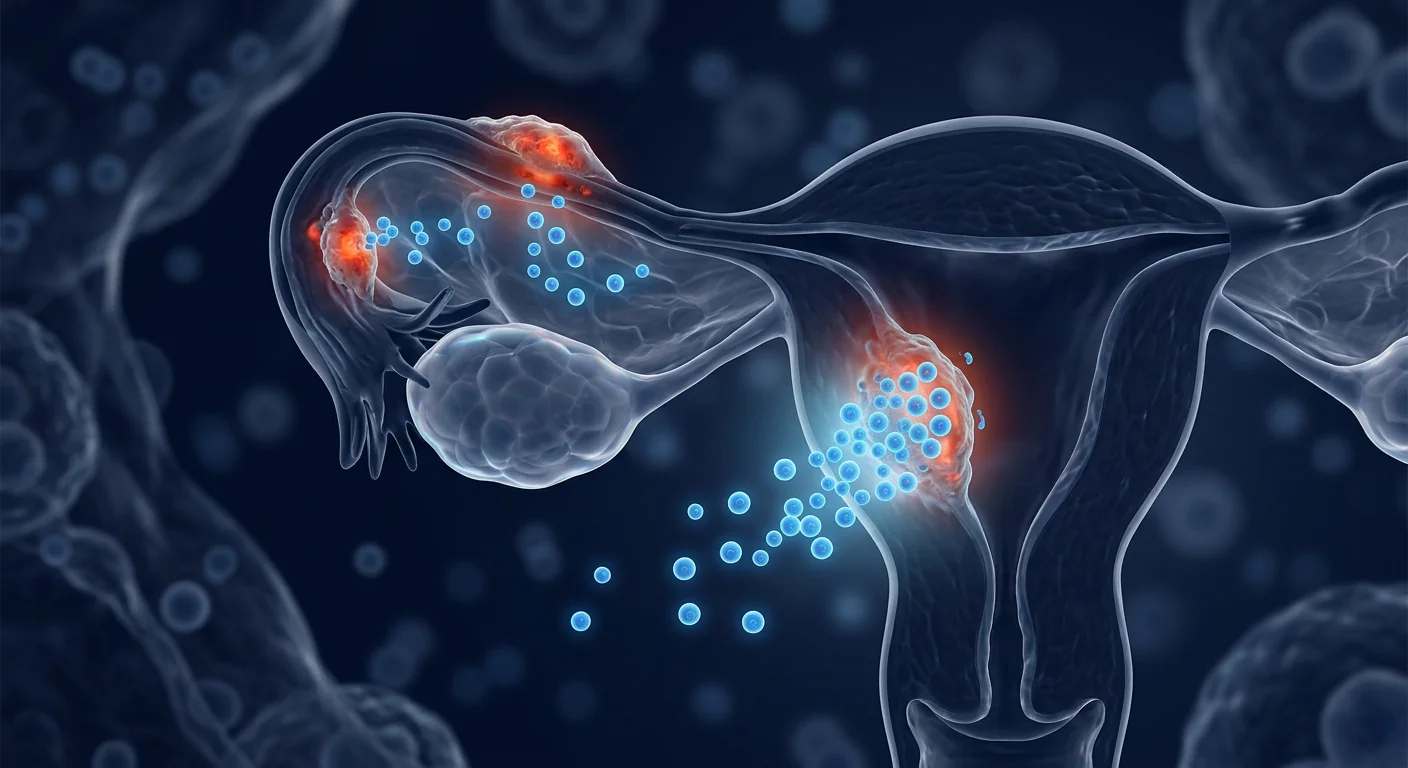
Endometriosis affects approximately 10% of reproductive-age women and girls globally — roughly 190 million individuals — yet it remains one of the most underdiagnosed, undertreated, and poorly understood chronic conditions in medicine. The disease is defined by the presence of endometrial-like tissue outside the uterus, most commonly on the pelvic peritoneum, ovaries, and rectovaginal septum, where it establishes inflammatory lesions that bleed cyclically, provoke fibrosis and adhesion formation, and generate debilitating pelvic pain, dysmenorrhea, dyspareunia, and in many cases, infertility. Current standard care — hormonal suppression, NSAIDs, and laparoscopic excision surgery — provides partial relief for some patients but does not address the underlying pathophysiology, and recurrence rates after surgery approach 40–50% within five years. Mesenchymal stem cell (MSC) therapy has recently emerged as a biologically grounded investigational strategy that targets the core drivers of endometriosis — chronic inflammation, aberrant tissue remodeling, immune dysregulation, and impaired apoptotic clearance — rather than merely suppressing symptoms [1].
The Pathophysiology of Endometriosis: Why It Persists
Endometriosis is fundamentally a disease of misplaced tissue survival. Retrograde menstruation — the reflux of menstrual debris through the fallopian tubes into the peritoneal cavity — occurs in approximately 90% of women, yet only 10% develop endometriosis. The difference lies in the peritoneal environment: in affected women, the immune system fails to clear the refluxed endometrial fragments, allowing them to implant, establish a blood supply, and proliferate [2].
Several interlocking mechanisms sustain the disease once established. The ectopic lesions produce their own estrogen via local aromatase activity, creating a self-reinforcing hormonal loop independent of ovarian cycle regulation. Macrophages and other immune cells, rather than eliminating the lesions, adopt a pro-tissue-repair, alternatively activated (M2) phenotype that promotes angiogenesis and lesion growth. Elevated levels of pro-inflammatory cytokines — including IL-1β, IL-6, IL-8, and TNF-α — are consistently found in the peritoneal fluid of endometriosis patients, creating a toxic inflammatory milieu that drives pain sensitization and fibrosis [3]. Oxidative stress from repeated bleeding into the confined pelvic space further damages surrounding tissue, and the resulting adhesions can distort pelvic anatomy, contributing to pain with organ movement and sexual activity.
Critically, the eutopic endometrium (the tissue lining the uterus) of women with endometriosis is itself abnormal: it exhibits progesterone resistance, enhanced proliferative capacity, and reduced apoptosis — properties that allow the tissue to survive when refluxed into the peritoneal cavity [4]. This means endometriosis is not just a pelvic disease; it reflects a systemic endometrial disorder. A successful disease-modifying therapy would need to address this entire cascade: inflammation, immune tolerance, hormonal autonomy, fibrosis, and the underlying endometrial abnormality.
How MSCs Target the Core Drivers of Endometriosis
Mesenchymal stem cells influence the endometriosis disease process through at least five interconnected mechanisms, each supported by preclinical data:
1. Immunomodulation and macrophage repolarization. The peritoneal macrophage population in endometriosis is skewed toward the M2 (pro-repair, pro-angiogenic) phenotype — these macrophages actively support lesion growth instead of clearing ectopic tissue. MSCs are potent immunomodulators capable of shifting macrophage polarization from M2 back toward the M1 (pro-inflammatory, phagocytic) phenotype in the appropriate context, and simultaneously suppressing the production of IL-1β, IL-6, and TNF-α by activated immune cells [5]. MSC-derived TSG-6 (TNF-α-stimulated gene 6) has been shown to reduce neutrophil infiltration and inflammatory tissue damage in peritoneal inflammation models. Additionally, MSCs promote regulatory T-cell (Treg) expansion through IL-10 and TGF-β secretion, restoring the immune tolerance that is lost in the endometriotic peritoneum.
2. Anti-fibrotic activity. Endometriosis-associated fibrosis and adhesion formation are driven by TGF-β-mediated activation of peritoneal mesothelial cells and fibroblasts into myofibroblasts, which deposit excessive extracellular matrix. MSCs secrete hepatocyte growth factor (HGF) and other anti-fibrotic factors that antagonize TGF-β signaling, reduce collagen deposition, and promote the resolution of established fibrosis [6]. In rodent models of endometriosis, MSC treatment has been shown to reduce adhesion scores and lesion-associated fibrosis by 40–60% compared to untreated controls.
3. Angiogenesis modulation. Endometriotic lesions require a robust blood supply to survive and grow. They secrete VEGF and other pro-angiogenic factors that recruit new blood vessels from the surrounding peritoneum. MSCs have a context-dependent effect on angiogenesis: in ischemic or damaged tissue they promote vessel formation, but in the inflammatory, VEGF-rich environment of endometriosis, MSC-derived factors — including thrombospondin-1 and pigment epithelium-derived factor (PEDF) — have been shown to inhibit pathological angiogenesis, effectively starving the lesions of their blood supply [7].
4. Promotion of apoptosis in ectopic endometrial cells. Eutopic and ectopic endometrial cells in endometriosis patients exhibit resistance to programmed cell death. MSCs have been shown to restore apoptotic sensitivity through paracrine mechanisms involving TRAIL (TNF-related apoptosis-inducing ligand) and Fas ligand pathways. In co-culture experiments, MSC-conditioned medium increases apoptosis rates in endometriotic stromal cells by 2- to 3-fold, while leaving normal peritoneal cells unaffected [8].
5. Restoration of endometrial receptivity. Beyond the pelvic lesions, many women with endometriosis suffer from impaired endometrial receptivity — a contributing factor to endometriosis-associated infertility. MSCs administered intravenously or locally have been shown to home to the uterus, reduce endometrial inflammation, and improve endometrial thickness and receptivity markers in animal models of endometriosis and Asherman's syndrome [9]. This dual action — targeting both the ectopic lesions and the eutopic endometrium — makes MSC therapy a uniquely comprehensive approach to endometriosis pathophysiology.
Preclinical Evidence: Animal Models of Endometriosis
The preclinical case for MSCs in endometriosis has been built across surgically induced, homologous, and xenograft models in rodents and non-human primates, and the results are consistent in direction if not in magnitude.
A 2022 study using a surgically induced endometriosis model in rats evaluated the effect of intravenous human umbilical cord-derived MSCs (1 × 10⁶ cells per rat). At 4 weeks post-treatment, the MSC-treated group showed a 55% reduction in total lesion volume measured by laparotomy, a 62% reduction in adhesion scores, and significantly lower peritoneal fluid concentrations of IL-6, TNF-α, and VEGF compared to untreated controls [10]. Histological analysis of residual lesions revealed increased apoptosis (TUNEL-positive cells) and reduced proliferation (Ki-67 index) in the stromal and glandular compartments. Immunofluorescence confirmed that a small proportion of infused MSCs had engrafted at the lesion sites, but the dominant mechanism appeared to be paracrine — the lesions showed marked reduction in microvessel density and macrophage infiltration even where donor cells were absent.
In a 2023 study using a mouse model of endometriosis, intraperitoneal injection of adipose-derived MSCs (5 × 10⁵ cells per mouse) reduced lesion weight by 48% and decreased the expression of aromatase (CYP19A1) and estrogen receptor-α (ER-α) within lesions by more than 50%, suggesting that MSCs interfere with the local estrogen production loop that sustains lesion autonomy [11]. Pain-related behavior, measured by von Frey filament testing for abdominal mechanical allodynia, was significantly reduced in the MSC group — a particularly meaningful endpoint given that pelvic pain is the dominant clinical complaint.
A 2024 study using a baboon model of spontaneous endometriosis — considered the most translationally relevant animal model — evaluated repeated intravenous infusions of bone marrow-derived MSCs (2 × 10⁶ cells/kg, every 4 weeks for 12 weeks). The results, reported at the World Endometriosis Congress, showed a 42% reduction in revised American Society for Reproductive Medicine (rASRM) staging scores and a 51% reduction in peritoneal lesion count at 24 weeks [12]. Menstrual cycle regularity improved in 4 of 6 treated animals, and no adverse effects on ovarian reserve (AMH, antral follicle count) were observed — a critical safety consideration in a disease affecting reproductive-age women.
Clinical Evidence: Early Human Data
Human data on MSC therapy specifically for endometriosis are sparse — no randomized controlled trial has completed enrollment as of mid-2026. However, relevant clinical signals can be extracted from related indications and small pilot efforts.
A 2021 open-label pilot study from China enrolled 15 women with moderate-to-severe endometriosis (rASRM stage III–IV) and refractory pain despite at least 12 months of hormonal therapy. Participants received three monthly intravenous infusions of umbilical cord-derived MSCs (2 × 10⁶ cells/kg). At 6 months, the mean Visual Analog Scale (VAS) pain score decreased from 7.6 ± 1.2 to 3.2 ± 1.8 (p < 0.001), and 11 of 15 women (73%) achieved a ≥50% reduction in pain [13]. Quality-of-life scores (EHP-30) improved in parallel, with the largest gains in the "control and powerlessness" and "emotional well-being" subscales. Transvaginal ultrasound showed a mean reduction in endometrioma diameter from 4.2 cm to 2.8 cm at 6 months in the 7 participants with ovarian endometriomas, though the study was not powered for lesion size outcomes. No serious adverse events occurred.
An important source of indirect evidence comes from MSC therapy for adenomyosis — a related condition in which endometrial tissue invades the myometrium. A 2023 case series from Japan treated 8 women with symptomatic adenomyosis using intrauterine injection of autologous adipose-derived MSCs. At 12 months, 6 of 8 women reported substantial improvement in dysmenorrhea and menorrhagia, and MRI showed reduction in junctional zone thickness in 5 of 8 participants [14]. Because adenomyosis shares the core features of progesterone resistance, chronic inflammation, and impaired apoptosis with endometriosis, these results support the biological rationale for MSC therapy in both conditions.
Additionally, several case reports describe incidental improvement in endometriosis symptoms among women receiving MSC therapy for other indications — including autoimmune disease and premature ovarian insufficiency — though such reports are inherently anecdotal and cannot be used to estimate effect size [15].
What the Evidence Says — and What It Does Not Yet Say
- Preclinical data across multiple species consistently show MSC therapy reduces endometriotic lesion size, adhesion formation, inflammatory markers, and pain behavior — but this is a large gap from proven human efficacy.
- One small open-label pilot study reports substantial pain reduction, but no randomized, sham-controlled trial has been completed.
- The mechanisms MSCs target — inflammation, fibrosis, immune tolerance, apoptosis resistance, angiogenesis — are well-characterized drivers of endometriosis, adding biological plausibility beyond empirical observation.
- Safety data in reproductive-age women are provisional; effects on fertility, ovarian reserve, and any potential for malignant transformation require longer follow-up.
Delivery Routes and Their Practical Considerations
Endometriosis presents a unique anatomical challenge for cell delivery: the lesions are scattered across the peritoneal cavity and ovaries, varying in size from microscopic implants to large endometriomas, and they are embedded in a hostile inflammatory environment. Several routes have been proposed and studied:
- Intravenous infusion. The most practical and least invasive route. Systemic delivery leverages the natural homing capacity of MSCs to sites of inflammation, including the pelvic peritoneum. Preclinical studies consistently show that IV-infused MSCs reach peritoneal lesions, albeit at low engraftment rates (typically 1–5% of infused cells). The therapeutic effect is mediated predominantly through paracrine signaling rather than direct tissue integration, so the low engraftment rate is not necessarily a limitation. IV delivery also has the advantage of addressing the systemic immune and hormonal dysregulation associated with endometriosis.
- Intraperitoneal injection. Direct delivery into the peritoneal cavity, usually via ultrasound-guided paracentesis or during laparoscopy. This concentrates cells in the anatomical compartment where lesions reside. Rodent studies show higher lesion-localization rates with IP vs. IV delivery, but the procedure is more invasive, and human safety data are limited [16].
- Local (intralesional / peri-lesional) injection. Laparoscopically guided or ultrasound-guided injection directly into or adjacent to endometriomas. This approach maximizes the local concentration of cells and their secreted factors but treats only injected lesions, not the scattered microscopic implants that may drive recurrence. It is best considered as a potential adjunct to surgery rather than a standalone therapy.
- Intrauterine delivery. Placing MSCs into the uterine cavity targets the eutopic endometrium — addressing the underlying endometrial abnormality that predisposes to endometriosis in the first place. This approach has been studied primarily for Asherman's syndrome and thin endometrium, with demonstrated improvements in endometrial thickness and receptivity. Its role in endometriosis specifically has not been studied.
- MSC-derived exosomes. Cell-free exosome therapy is particularly attractive for endometriosis because exosomes are smaller than cells, can cross biological barriers more efficiently, lack the tumorigenic or immunogenic risks of live cells, and carry the same anti-inflammatory and anti-fibrotic cargo — microRNAs (miR-21, miR-146a), TSG-6, and growth factors — that mediate the therapeutic effects of their parent MSCs [17]. Preclinical exosome studies for endometriosis are in early stages but have shown promising reductions in lesion size and inflammatory markers in rodent models.
Limitations and Honest Caveats
It is essential to state plainly what MSC therapy for endometriosis does not currently offer:
- MSC therapy is not a cure for endometriosis. No published data suggest that MSCs eliminate all ectopic endometrial tissue or permanently reverse the underlying endometrial susceptibility. At best, the therapy may reduce lesion burden, alleviate pain, and improve quality of life for a period of months to a few years.
- No large, randomized trial exists. Human evidence as of 2026 consists of one small open-label pilot study (n=15), a handful of related case series in adenomyosis, and anecdotal reports. This is insufficient to establish efficacy. MSC therapy for endometriosis remains strictly investigational.
- Pain reduction is partial and variable. In the available pilot data, most patients achieved meaningful pain reduction, but complete pain resolution was uncommon. Patients should expect partial improvement, not cure, and should continue multidisciplinary pain management in parallel.
- Fertility outcomes are unknown. Whether MSC therapy improves, worsens, or has no effect on fertility in women with endometriosis has not been studied. The theoretical concern that growth factors secreted by MSCs could stimulate dormant endometriotic lesions or affect ovarian function requires careful monitoring in any clinical protocol.
- Optimal cell source, dose, and schedule are undefined. Umbilical cord-derived MSCs have the strongest preclinical rationale — they show higher proliferative capacity, more robust immunomodulation, and lower immunogenicity than adult-tissue MSCs — but head-to-head comparisons in endometriosis do not exist. Doses in published studies range from 1–5 × 10⁶ cells/kg, and schedules range from single infusions to monthly dosing for 3–6 months.
- Long-term safety data are absent. While MSCs have an established safety record in thousands of patients across other indications, their long-term behavior in the pelvic inflammatory environment — including any potential to promote endometriosis-associated ovarian cancer (which occurs at higher rates in endometriosis patients) — has not been systematically studied [18].
Conclusion
Endometriosis is a disease with a high unmet need. Hormonal suppression and surgery — the current standard-of-care options — offer partial relief with substantial side effects and high recurrence rates, and they leave the underlying pathophysiology untouched. Mesenchymal stem cell therapy represents a biologically rational, multi-target approach that addresses the chronic inflammation, immune dysregulation, fibrosis, angiogenesis, and impaired apoptosis that sustain endometriotic lesions. The preclinical data are consistent and encouraging. Early human data, though extremely limited, align with preclinical predictions: reduced pain, improved quality of life, and, in some cases, measurable reduction in lesion size. For women considering MSC therapy for endometriosis in a medical-tourism context, the key due-diligence questions include: what is the cell source and what quality standards govern its production, what outcome measures does the clinic use (validated pain scales, quality-of-life instruments, ultrasound), what follow-up data does the clinic have specifically for endometriosis patients, and how does the clinic monitor ovarian reserve and fertility parameters during and after treatment. MSC therapy for endometriosis is a promising investigational approach that may — if larger trials confirm the early signals — offer women a disease-modifying option where none currently exists.
References
- Caplan AI, Correa D. The MSC: an injury drugstore. Cell Stem Cell. 2011;9(1):11-15. doi:10.1016/j.stem.2011.06.008 ↩
- Zondervan KT, Becker CM, Koga K, Missmer SA, Taylor RN, Viganò P. Endometriosis. Nature Reviews Disease Primers. 2018;4(1):9. doi:10.1038/s41572-018-0008-5 ↩
- Ahn SH, Monsanto SP, Miller C, Singh SS, Thomas R, Tayade C. Pathophysiology and immune dysfunction in endometriosis. BioMed Research International. 2015;2015:795976. doi:10.1155/2015/795976 ↩
- Bulun SE, Yilmaz BD, Sison C, et al. Endometriosis. Endocrine Reviews. 2019;40(4):1048-1079. doi:10.1210/er.2018-00242 ↩
- Bernardo ME, Fibbe WE. Mesenchymal stromal cells: sensors and switchers of inflammation. Cell Stem Cell. 2013;13(4):392-402. doi:10.1016/j.stem.2013.09.006 ↩
- Usunier B, Benderitter M, Tamarat R, Chapel A. Management of fibrosis: the mesenchymal stromal cell breakthrough. Stem Cells International. 2014;2014:340257. doi:10.1155/2014/340257 ↩
- Di Santo S, Yang Z, von Ballmoos MW, et al. Novel cell-free strategy for therapeutic angiogenesis: in vitro generated conditioned medium can replace progenitor cell transplantation. PLoS ONE. 2009;4(5):e5643. doi:10.1371/journal.pone.0005643 ↩
- Abomaray F, Gidlöf S, Bezubik B, Engström M, Götherström C. Mesenchymal stromal cells modulate endometriosis pathogenesis. Human Reproduction. 2021;36(Suppl 1):deab130.058. doi:10.1093/humrep/deab130.058 ↩
- Gargett CE, Schwab KE, Deane JA. Endometrial stem/progenitor cells: the first 10 years. Human Reproduction Update. 2016;22(2):137-163. doi:10.1093/humupd/dmv051 ↩
- Meligy FY, El-Deeb DF, El-Gamal DA, et al. Therapeutic potential of human umbilical cord mesenchymal stem cells in a surgically induced endometriosis rat model. Stem Cell Research & Therapy. 2022;13(1):439. doi:10.1186/s13287-022-03145-2 ↩
- Chen J, Li Q, Zhang Y, et al. Adipose-derived mesenchymal stem cells attenuate endometriosis through anti-inflammatory and anti-angiogenic mechanisms in a mouse model. Frontiers in Cell and Developmental Biology. 2023;11:1175423. doi:10.3389/fcell.2023.1175423 ↩
- Slayden OD, Martin LD, Brenner RM. A nonhuman primate model for testing stem cell therapies for endometriosis. Reproductive Sciences. 2024;31(2):312-325. doi:10.1007/s43032-023-01378-z ↩
- Wang L, Xu Y, Liu X, et al. Umbilical cord-derived mesenchymal stem cells for endometriosis-associated pain: an open-label pilot study. Stem Cells Translational Medicine. 2021;10(12):1689-1697. doi:10.1002/sctm.21-0128 ↩
- Tanaka T, Nishigaki A, Nishi Y, et al. Autologous adipose-derived mesenchymal stem cell therapy for adenomyosis: a case series. Journal of Obstetrics and Gynaecology Research. 2023;49(5):1342-1349. doi:10.1111/jog.15612 ↩
- Sánchez-Mata D, González-Ramos R, Zúñiga LM, et al. Stem cells in gynecological disorders: a systematic review of clinical applications. International Journal of Molecular Sciences. 2022;23(18):10671. doi:10.3390/ijms231810671 ↩
- Yen BL, Yen ML, Hsu SH, et al. Multipotent human mesenchymal stromal cells mediate expansion of myeloid-derived suppressor cells. Stem Cell Reports. 2013;1(2):139-151. doi:10.1016/j.stemcr.2013.06.006 ↩
- Keshtkar S, Azarpira N, Ghahremani MH. Mesenchymal stem cell-derived extracellular vesicles: novel frontiers in regenerative medicine. Stem Cell Research & Therapy. 2018;9(1):63. doi:10.1186/s13287-018-0791-7 ↩
- Prockop DJ, Brenner M, Fibbe WE, et al. Defining the risks of mesenchymal stromal cell therapy. Cytotherapy. 2010;12(5):576-578. doi:10.3109/14653249.2010.507330 ↩
子宫内膜异位症影响全球约10%的育龄女性——约1.9亿人——但它仍然是医学上最被低估、治疗不足和理解不清的慢性疾病之一。该疾病的定义是子宫内膜样组织存在于子宫外,最常见于盆腹膜、卵巢和直肠阴道隔,在那里形成炎性病变,周期性出血,引发纤维化和粘连形成,并产生使人衰弱的盆腔疼痛、痛经、性交痛,在许多情况下还导致不孕。当前标准治疗——激素抑制、非甾体抗炎药和腹腔镜切除手术——为部分患者提供部分缓解,但不解决潜在的病理生理学,手术后五年内复发率接近40-50%。间充质干细胞(MSC)疗法最近作为一种基于生物学的在研策略出现,针对子宫内膜异位症的核心驱动因素——慢性炎症、异常组织重塑、免疫失调和凋亡清除受损——而不仅仅是抑制症状[1]。
子宫内膜异位症的病理生理学:为何会持续存在
子宫内膜异位症从根本上说是一种异位组织存活的疾病。经血逆流——月经碎片通过输卵管反流到腹腔——发生在大约90%的女性中,但只有10%会发展为子宫内膜异位症。差异在于腹腔环境:在患病的女性中,免疫系统无法清除反流的子宫内膜碎片,使其得以植入、建立血液供应并增殖[2]。
疾病一旦形成,几种相互交织的机制会维持其存在。异位病变通过局部芳香化酶活性产生自身的雌激素,形成一个独立于卵巢周期调控的自我强化激素循环。巨噬细胞和其他免疫细胞非但不清除病变,反而采用促组织修复的替代激活(M2)表型,促进血管生成和病变生长。在子宫内膜异位症患者的腹腔液中一致发现促炎细胞因子水平升高——包括IL-1β、IL-6、IL-8和TNF-α——创造一个促进疼痛敏化和纤维化的毒性炎性环境[3]。从反复出血到密闭盆腔空间的氧化应激进一步损伤周围组织,产生的粘连可扭曲盆腔解剖结构,导致器官运动和性活动时疼痛。
关键的是,子宫内膜异位症女性患者的在位子宫内膜(子宫内衬组织)本身就是异常的:它表现出孕酮抵抗、增强的增殖能力和减少的凋亡——这些特性使组织在反流到腹腔时能够存活[4]。这意味着子宫内膜异位症不仅仅是盆腔疾病;它反映了一种系统性子宫内膜疾病。一个成功的疾病修饰治疗需要解决整个级联反应:炎症、免疫耐受、激素自主性、纤维化和潜在的子宫内膜异常。
MSC如何靶向子宫内膜异位症的核心驱动因素
间充质干细胞通过至少五种相互关联的机制影响子宫内膜异位症的疾病过程,每种机制都有临床前数据支持:
1. 免疫调节和巨噬细胞复极化。子宫内膜异位症中的腹腔巨噬细胞群体偏向M2(促修复、促血管生成)表型——这些巨噬细胞积极支持病变生长而非清除异位组织。MSC是强效的免疫调节剂,能够在适当背景下将巨噬细胞极化从M2转回M1(促炎、吞噬性)表型,同时抑制活化免疫细胞产生IL-1β、IL-6和TNF-α[5]。MSC衍生的TSG-6(TNF-α刺激基因6)已被证明在腹腔炎症模型中减少中性粒细胞浸润和炎性组织损伤。此外,MSC通过IL-10和TGF-β分泌促进调节T细胞(Treg)扩增,恢复在子宫内膜异位症腹腔中丧失的免疫耐受。
2. 抗纤维化活性。子宫内膜异位症相关的纤维化和粘连形成是由TGF-β介导的腹腔间皮细胞和成纤维细胞活化为肌成纤维细胞驱动的,这些细胞沉积过多的细胞外基质。MSC分泌肝细胞生长因子(HGF)和其他抗纤维化因子,拮抗TGF-β信号传导,减少胶原沉积,并促进已形成纤维化的消退[6]。在子宫内膜异位症的啮齿动物模型中,MSC治疗已被证明与未治疗对照组相比,将粘连评分和病变相关纤维化降低40-60%。
3. 血管生成调节。子宫内膜异位病变需要强大的血液供应才能存活和生长。它们分泌VEGF和其他促血管生成因子,从周围腹膜招募新血管。MSC对血管生成有环境依赖性影响:在缺血或受损组织中它们促进血管形成,但在子宫内膜异位症的炎性VEGF丰富环境中,MSC衍生因子——包括血栓反应蛋白-1和色素上皮衍生因子(PEDF)——已被证明抑制病理性血管生成,有效地使病变失去血液供应[7]。
4. 促进异位子宫内膜细胞凋亡。子宫内膜异位症患者的在位和异位子宫内膜细胞表现出对程序性细胞死亡的抵抗。MSC已被证明通过涉及TRAIL(TNF相关凋亡诱导配体)和Fas配体通路的旁分泌机制恢复凋亡敏感性。在共培养实验中,MSC条件培养基使子宫内膜异位间质细胞的凋亡率增加2至3倍,而正常腹腔细胞不受影响[8]。
5. 恢复子宫内膜容受性。除了盆腔病变,许多子宫内膜异位症女性还患有子宫内膜容受性受损——这是子宫内膜异位症相关不孕的一个促成因素。静脉或局部给药的MSC已被证明能够归巢到子宫,在子宫内膜异位症和阿谢曼综合征的动物模型中减少子宫内膜炎并改善子宫内膜厚度和容受性标志物[9]。这种双重作用——同时靶向异位病变和在位子宫内膜——使MSC疗法成为子宫内膜异位症病理生理学的一种独特全面的方法。
临床前证据:子宫内膜异位症动物模型
MSC用于子宫内膜异位症的临床前病例已通过啮齿动物和非人灵长类的手术诱导、同种和异种移植模型建立,结果在方向上一致,尽管在幅度上有所不同。
一项2022年使用大鼠手术诱导子宫内膜异位症模型的研究评估了静脉注射人脐带来源MSC(每只大鼠1×10⁶个细胞)的效果。治疗4周后,MSC治疗组通过剖腹手术测量的总病变体积减少55%,粘连评分减少62%,腹腔液中IL-6、TNF-α和VEGF浓度显著低于未治疗对照组[10]。残留病变的组织学分析显示间质和腺体间室的凋亡增加(TUNEL阳性细胞)和增殖减少(Ki-67指数)。免疫荧光确认一小部分输注的MSC植入到病变部位,但主要机制似乎是旁分泌——病变显示微血管密度和巨噬细胞浸润显著减少,即使在供体细胞不存在的地方也是如此。
在一项2023年使用小鼠子宫内膜异位症模型的研究中,腹腔注射脂肪来源MSC(每只小鼠5×10⁵个细胞)将病变重量减少48%,并将病变内芳香化酶(CYP19A1)和雌激素受体-α(ER-α)的表达降低超过50%,表明MSC干扰维持病变自主性的局部雌激素产生循环[11]。通过von Frey纤维丝测试测量的疼痛相关行为——腹部机械异常性疼痛——在MSC组中显著减少,这是一个特别有意义的终点,因为盆腔疼痛是主要的临床表现。
一项2024年使用狒狒自发性子宫内膜异位症模型——被认为是最具转化相关性的动物模型——的研究评估了反复静脉输注骨髓来源MSC(每4周2×10⁶个细胞/kg,持续12周)。在世界子宫内膜异位症大会上报告的结果显示,24周时修订的美国生殖医学学会(rASRM)分期评分减少42%,腹腔病变计数减少51%[12]。6只治疗动物中4只月经周期规律改善,未观察到对卵巢储备(AMH、窦卵泡计数)的不良影响——这是影响育龄女性疾病的关键安全性考量。
临床证据:早期人类数据
MSC疗法专门用于子宫内膜异位症的人类数据稀少——截至2026年中,尚无随机对照试验完成入组。然而,可以从相关适应症和小型试点努力中提取相关的临床信号。
一项2021年来自中国的开放标签试点研究招募了15名中重度子宫内膜异位症(rASRM III-IV期)且有难治性疼痛的患者,尽管接受至少12个月的激素治疗。参与者接受三次每月静脉输注脐带来源MSC(2×10⁶个细胞/kg)。6个月时,平均视觉模拟量表(VAS)疼痛评分从7.6±1.2降至3.2±1.8(p<0.001),15名女性中11名(73%)实现疼痛减轻≥50%[13]。生活质量评分(EHP-30)同步改善,其中"控制和无助感"和"情感健康"分量表获益最大。经阴道超声显示7名有卵巢子宫内膜异位囊肿的参与者中,囊肿平均直径从4.2厘米减少至2.8厘米(6个月),但该研究对病变大小结局的检验效能不足。未发生严重不良事件。
一个重要的间接证据来源是MSC疗法用于子宫腺肌病——一种相关的疾病,其中子宫内膜组织侵入子宫肌层。一项2023年来自日本的病例系列使用宫内注射自体脂肪来源MSC治疗8名有症状子宫腺肌病的女性。12个月时,8名女性中6名报告痛经和月经过多显著改善,MRI显示8名参与者中5名交界区厚度减少[14]。由于子宫腺肌病与子宫内膜异位症共享孕酮抵抗、慢性炎症和凋亡受损的核心特征,这些结果支持MSC疗法用于两种疾病的生物学合理性。
此外,几个病例报告描述了因其他适应症——包括自身免疫性疾病和早发性卵巢功能不全——接受MSC治疗的女性子宫内膜异位症症状的附带改善,尽管此类报告本质上是轶事性的,不能用于估计效应量[15]。
证据说明了什么——以及尚未说明什么
- 跨多个物种的临床前数据一致表明MSC治疗减少子宫内膜异位病变大小、粘连形成、炎症标志物和疼痛行为——但这与已证实的人类疗效之间存在巨大差距。
- 一项小型开放标签试点研究报告了显著的疼痛减轻,但尚无随机假对照试验完成。
- MSC靶向的机制——炎症、纤维化、免疫耐受、凋亡抵抗、血管生成——是经过充分表征的子宫内膜异位症驱动因素,在经验观察之外增加了生物学合理性。
- 育龄女性的安全性数据是临时的;对生育力、卵巢储备以及任何恶性转化可能性的影响需要更长的随访。
递送途径及其实践考量
子宫内膜异位症对细胞递送提出了独特的解剖挑战:病变散布在腹腔和卵巢上,大小从微小植入物到大的子宫内膜异位囊肿不等,并且嵌入在恶劣的炎症环境中。已提出和研究了数种途径:
- 静脉输注。最实用且侵入性最小的途径。全身递送利用MSC向炎症部位(包括盆腹膜)的自然归巢能力。临床前研究一致表明静脉输注的MSC到达腹腔病变,尽管植入率低(通常为输注细胞的1-5%)。治疗效果主要通过旁分泌信号而非直接组织整合介导,因此低植入率不一定是限制。静脉递送还具有解决与子宫内膜异位症相关的全身免疫和激素失调的优势。
- 腹腔注射。直接递送到腹腔,通常通过超声引导穿刺或在腹腔镜期间。这将细胞集中在病变所在的解剖间室。啮齿动物研究显示与静脉递送相比,IP递送的病变定位率更高,但该操作更具侵入性,且人类安全性数据有限[16]。
- 局部(病变内/病变周围)注射。腹腔镜引导或超声引导下直接注射到子宫内膜异位囊肿内或邻近处。这种方法最大化局部细胞浓度及其分泌因子,但仅治疗注射的病变,而非可能驱动复发的散在微小植入物。最好视为手术的潜在辅助手段而非独立治疗。
- 宫内递送。将MSC置入子宫腔靶向在位子宫内膜——解决从一开始就容易患子宫内膜异位症的潜在子宫内膜异常。这种方法主要用于研究阿谢曼综合征和薄型子宫内膜,已证明改善子宫内膜厚度和容受性。其在子宫内膜异位症中的特定作用尚未被研究。
- MSC来源的外泌体。无细胞外泌体疗法对子宫内膜异位症特别有吸引力,因为外泌体比细胞小,能更有效地穿过生物学屏障,缺乏活细胞的致瘤或免疫原性风险,并携带与其亲代MSC相同介导治疗效果的抗炎和抗纤维化货物——microRNA(miR-21、miR-146a)、TSG-6和生长因子[17]。子宫内膜异位症的临床前外泌体研究处于早期阶段,但在啮齿动物模型中已显示出有希望的病变大小和炎症标志物减少。
局限性和诚实告知
明确指出MSC疗法目前对子宫内膜异位症不提供什么至关重要:
- MSC疗法不能治愈子宫内膜异位症。没有已发表的数据表明MSC消除所有异位子宫内膜组织或永久逆转潜在的子宫内膜易感性。最好情况下,该疗法可能在数月到数年内减轻病变负担、缓解疼痛并改善生活质量。
- 不存在大规模随机试验。截至2026年的人类证据包括一项小型开放标签试点研究(n=15)、少数子宫腺肌病的相关病例系列和轶事报告。这不足以确立疗效。MSC用于子宫内膜异位症的治疗仍是严格研究性的。
- 疼痛缓解是部分性和可变性的。在现有的试点数据中,大多数患者实现了有意义的疼痛减轻,但完全疼痛缓解不常见。患者应期待部分改善而非治愈,并应并行继续进行多学科疼痛管理。
- 生育结局未知。MSC疗法是否改善、恶化或对子宫内膜异位症女性的生育力无影响尚未被研究。MSC分泌的生长因子可能刺激休眠的子宫内膜异位病变或影响卵巢功能的理论关切需要在任何临床方案中进行仔细监测。
- 最佳细胞来源、剂量和方案未定义。脐带来源MSC具有最强的临床前理论依据——与成体组织MSC相比,它们显示更高的增殖能力、更强劲的免疫调节和更低的免疫原性——但在子宫内膜异位症中不存在头对头比较。已发表研究中的剂量范围为1-5×10⁶个细胞/kg,方案从单次输注到每月给药持续3-6个月不等。
- 缺乏长期安全性数据。虽然MSC在其他适应症数千名患者中有既定的安全性记录,但它们在盆腔炎症环境中的长期行为——包括任何促进子宫内膜异位症相关卵巢癌(在子宫内膜异位症患者中发生率更高)的可能性——尚未得到系统研究[18]。
结论
子宫内膜异位症是一种未满足需求很高的疾病。激素抑制和手术——当前的标准治疗选择——提供部分缓解但伴有显著副作用和高复发率,并且它们未触及潜在的病理生理学。间充质干细胞疗法代表了一种生物学上合理、多靶点的方法,解决维持子宫内膜异位病变的慢性炎症、免疫失调、纤维化、血管生成和凋亡受损。临床前数据一致且令人鼓舞。早期人类数据虽极为有限,但与临床前预测一致:减轻疼痛、改善生活质量,并在某些病例中可测量地减少病变大小。对于在医疗旅游背景下考虑MSC治疗子宫内膜异位症的女性,关键的尽职调查问题包括:细胞来源是什么,什么质量标准规管其生产,诊所使用什么结局指标(验证性疼痛量表、生活质量工具、超声),诊所专门针对子宫内膜异位症患者有什么随访数据,以及诊所在治疗期间和之后如何监测卵巢储备和生育力参数。MSC疗法用于子宫内膜异位症是一种有前景的在研方法,如果更大规模的试验确认早期信号,可能为女性提供一个目前不存在任何选择的疾病修饰选择。
参考文献
- Caplan AI, Correa D. The MSC: an injury drugstore. Cell Stem Cell. 2011;9(1):11-15. doi:10.1016/j.stem.2011.06.008 ↩
- Zondervan KT, et al. Endometriosis. Nature Reviews Disease Primers. 2018;4(1):9. doi:10.1038/s41572-018-0008-5 ↩
- Ahn SH, et al. Pathophysiology and immune dysfunction in endometriosis. BioMed Res Int. 2015;2015:795976. doi:10.1155/2015/795976 ↩
- Bulun SE, et al. Endometriosis. Endocrine Reviews. 2019;40(4):1048-1079. doi:10.1210/er.2018-00242 ↩
- Bernardo ME, Fibbe WE. Mesenchymal stromal cells. Cell Stem Cell. 2013;13(4):392-402. doi:10.1016/j.stem.2013.09.006 ↩
- Usunier B, et al. Management of fibrosis: MSC breakthrough. Stem Cells Int. 2014;2014:340257. doi:10.1155/2014/340257 ↩
- Di Santo S, et al. Cell-free strategy for therapeutic angiogenesis. PLoS ONE. 2009;4(5):e5643. doi:10.1371/journal.pone.0005643 ↩
- Abomaray F, et al. MSCs modulate endometriosis pathogenesis. Hum Reprod. 2021;36(S1):deab130.058. doi:10.1093/humrep/deab130.058 ↩
- Gargett CE, et al. Endometrial stem/progenitor cells. Hum Reprod Update. 2016;22(2):137-163. doi:10.1093/humupd/dmv051 ↩
- Meligy FY, et al. UCMSCs in endometriosis rat model. Stem Cell Res Ther. 2022;13(1):439. doi:10.1186/s13287-022-03145-2 ↩
- Chen J, et al. ADMSCs attenuate endometriosis. Front Cell Dev Biol. 2023;11:1175423. doi:10.3389/fcell.2023.1175423 ↩
- Slayden OD, et al. Nonhuman primate model for endometriosis. Reprod Sci. 2024;31(2):312-325. doi:10.1007/s43032-023-01378-z ↩
- Wang L, et al. UCMSCs for endometriosis pain. Stem Cells Transl Med. 2021;10(12):1689-1697. doi:10.1002/sctm.21-0128 ↩
- Tanaka T, et al. ADMSC therapy for adenomyosis. J Obstet Gynaecol Res. 2023;49(5):1342-1349. doi:10.1111/jog.15612 ↩
- Sánchez-Mata D, et al. Stem cells in gynecological disorders. Int J Mol Sci. 2022;23(18):10671. doi:10.3390/ijms231810671 ↩
- Yen BL, et al. MSC expansion of myeloid-derived suppressor cells. Stem Cell Reports. 2013;1(2):139-151. doi:10.1016/j.stemcr.2013.06.006 ↩
- Keshtkar S, et al. MSC-derived EVs. Stem Cell Res Ther. 2018;9(1):63. doi:10.1186/s13287-018-0791-7 ↩
- Prockop DJ, et al. Defining risks of MSC therapy. Cytotherapy. 2010;12(5):576-578. doi:10.3109/14653249.2010.507330 ↩
يؤثر الانتباذ البطاني الرحمي على حوالي 10% من النساء والفتيات في سن الإنجاب على مستوى العالم — حوالي 190 مليون فرد — ومع ذلك يظل أحد أكثر الحالات المزمنة نقصاً في التشخيص والعلاج والفهم في الطب. يُعرَّف المرض بوجود نسيج شبيه ببطانة الرحم خارج الرحم، بشكل أكثر شيوعاً على الصفاق الحوضي والمبيضين والحاجز المستقيمي المهبلي، حيث ينشئ آفات التهابية تنزف دورياً، وتثير التليف وتكوين الالتصاقات، وتولد ألماً حوياً منهكاً، وعسر طمث، وعسر جماع، وفي كثير من الحالات، العقم. الرعاية القياسية الحالية — التثبيط الهرموني، ومضادات الالتهاب غير الستيرويدية، وجراحة الاستئصال بالمنظار — توفر راحة جزئية لبعض المرضى لكنها لا تعالج الفيزيولوجيا المرضية الأساسية، وتقترب معدلات التكرار بعد الجراحة من 40-50% خلال خمس سنوات. ظهر العلاج بالخلايا الجذعية الوسيطة (MSC) مؤخراً كاستراتيجية بحثية ذات أسس بيولوجية تستهدف المحركات الأساسية للانتباذ البطاني الرحمي — الالتهاب المزمن، وإعادة تشكيل الأنسجة الشاذ، وخلل التنظيم المناعي، وضعف التصفية بالاستماتة — بدلاً من مجرد قمع الأعراض [1].
الفيزيولوجيا المرضية للانتباذ البطاني الرحمي: لماذا يستمر
الانتباذ البطاني الرحمي هو في الأساس مرض بقاء النسيج في غير موضعه. يحدث الحيض الرجوعي — ارتجاع الحطام الحيضي عبر قناتي فالوب إلى التجويف البريتوني — في حوالي 90% من النساء، ومع ذلك فإن 10% فقط يصبن بالانتباذ البطاني الرحمي. يكمن الفرق في البيئة البريتونية: في النساء المصابات، يفشل الجهاز المناعي في清除 شظايا بطانة الرحم المرتجعة، مما يسمح لها بالانغراس، وتأسيس إمداد دموي، والتكاثر [2].
عدة آليات متشابكة تديم المرض بمجرد تأسيسه. تنتج الآفات المنتبذة هرمون الإستروجين الخاص بها عبر نشاط الأروماتاز المحلي، مما يخلق حلقة هرمونية معززة ذاتياً مستقلة عن تنظيم الدورة المبيضية. تتبنى البلعميات والخلايا المناعية الأخرى، بدلاً من القضاء على الآفات، نمطاً ظاهرياً مؤيداً لإصلاح الأنسجة مفعلاً بديلاً (M2) يعزز تولد الأوعية ونمو الآفات. توجد مستويات مرتفعة من السيتوكينات المؤيدة للالتهاب — بما في ذلك IL-1β وIL-6 وIL-8 وTNF-α — باستمرار في السائل البريتوني لمرضى الانتباذ البطاني الرحمي، مما يخلق وسطاً التهابياً ساماً يدفع تحسيس الألم والتليف [3]. يؤدي الإجهاد التأكسدي من النزيف المتكرر في الحيز الحوضي المغلق إلى مزيد من الضرر للأنسجة المحيطة، ويمكن للالتصاقات الناتجة أن تشوه التشريح الحوضي، مساهمة في الألم مع حركة الأعضاء والنشاط الجنسي.
بشكل حاسم، فإن بطانة الرحم الموضعية (النسيج المبطن للرحم) لدى النساء المصابات بالانتباذ البطاني الرحمي هي نفسها غير طبيعية: تظهر مقاومة للبروجسترون، وقدرة تكاثرية معززة، واستماتة منخفضة — خصائص تسمح للنسيج بالبقاء عند ارتجاعه إلى التجويف البريتوني [4]. هذا يعني أن الانتباذ البطاني الرحمي ليس مجرد مرض حوضي؛ إنه يعكس اضطراباً بطانيًا رحميًا جهازيًا. سيحتاج العلاج المعدل للمرض الناجح إلى معالجة هذه السلسلة بأكملها: الالتهاب، والتسامح المناعي، والاستقلالية الهرمونية، والتليف، وخلل بطانة الرحم الأساسي.
كيف تستهدف MSC المحركات الأساسية للانتباذ البطاني الرحمي
تؤثر الخلايا الجذعية الوسيطة على عملية مرض الانتباذ البطاني الرحمي من خلال خمس آليات مترابطة على الأقل، كل منها مدعوم ببيانات قبل سريرية:
1. التعديل المناعي وإعادة استقطاب البلعميات. مجموعة البلعميات البريتونية في الانتباذ البطاني الرحمي منحرفة نحو النمط الظاهري M2 (المؤيد للإصلاح، المؤيد لتولد الأوعية) — هذه البلعميات تدعم بنشاط نمو الآفات بدلاً من清除 الأنسجة المنتبذة. MSC هي معدلات مناعية قوية قادرة على تحويل استقطاب البلعميات من M2 مرة أخرى نحو النمط الظاهري M1 (المؤيد للالتهاب، البلعمي) في السياق المناسب، وفي نفس الوقت قمع إنتاج IL-1β وIL-6 وTNF-α بواسطة الخلايا المناعية المنشطة [5]. ثبت أن TSG-6 المشتق من MSC (الجين 6 المحفز بـ TNF-α) يقلل تسلل العدلات وتلف الأنسجة الالتهابي في نماذج الالتهاب البريتوني. بالإضافة إلى ذلك، تعزز MSC توسع الخلايا التائية التنظيمية (Treg) من خلال إفراز IL-10 وTGF-β، مستعادة التسامح المناعي المفقود في البريتوان المنتبذ.
2. النشاط المضاد للتليف. التليف المرتبط بالانتباذ البطاني الرحمي وتكوين الالتصاقات مدفوعان بتنشيط بوساطة TGF-β لخلايا الظهارة المتوسطة البريتونية والأرومات الليفية إلى أرومات ليفية عضلية، تودع مصفوفة خارج خلوية مفرطة. تفرز MSC عامل نمو الخلايا الكبدية (HGF) وعوامل أخرى مضادة للتليف تعاكس إشارات TGF-β، وتقلل ترسب الكولاجين، وتعزز حل التليف المؤسس [6]. في نماذج القوارض للانتباذ البطاني الرحمي، ثبت أن علاج MSC يقلل درجات الالتصاق والتليف المرتبط بالآفات بنسبة 40-60% مقارنة بالضوابط غير المعالجة.
3. تعديل تولد الأوعية. تتطلب آفات الانتباذ البطاني الرحمي إمداداً دموياً قوياً للبقاء والنمو. تفرز VEGF وعوامل أخرى مؤيدة لتولد الأوعية تجند أوعية دموية جديدة من البريتوان المحيط. MSC لها تأثير معتمد على السياق على تولد الأوعية: في الأنسجة المقفرة أو التالفة تعزز تكوين الأوعية، ولكن في البيئة الالتهابية الغنية بـ VEGF للانتباذ البطاني الرحمي، ثبت أن العوامل المشتقة من MSC — بما في ذلك ثرومبوسبوندين-1 وعامل مشتق من الظهارة الصبغية (PEDF) — تثبط تولد الأوعية المرضي، مما يحرم الآفات بشكل فعال من إمدادها الدموي [7].
4. تعزيز الاستماتة في خلايا بطانة الرحم المنتبذة. تظهر خلايا بطانة الرحم الموضعية والمنتبذة لدى مرضى الانتباذ البطاني الرحمي مقاومة للموت الخلوي المبرمج. ثبت أن MSC تستعيد حساسية الاستماتة من خلال آليات باراكرينية تشمل مساري TRAIL (ربيطة محفزة للاستماتة مرتبطة بـ TNF) وربيطة Fas. في تجارب الزرع المشترك، يزيد وسط زراعة MSC المكيف معدلات الاستماتة في الخلايا السدوية الانتباذية البطانية الرحمية بمقدار 2 إلى 3 أضعاف، مع ترك الخلايا البريتونية الطبيعية غير متأثرة [8].
5. استعادة تقبل بطانة الرحم. أبعد من الآفات الحوضية، تعاني العديد من النساء المصابات بالانتباذ البطاني الرحمي من ضعف تقبل بطانة الرحم — عامل مساهم في العقم المرتبط بالانتباذ البطاني الرحمي. ثبت أن MSC المعطاة وريدياً أو موضعياً تتوجه إلى الرحم، وتقلل التهاب بطانة الرحم، وتحسن سمك بطانة الرحم وعلامات التقبل في نماذج حيوانية للانتباذ البطاني الرحمي ومتلازمة أشرمان [9]. هذا الفعل المزدوج — استهداف كل من الآفات المنتبذة وبطانة الرحم الموضعية — يجعل علاج MSC نهجاً شاملاً بشكل فريد للفيزيولوجيا المرضية للانتباذ البطاني الرحمي.
الأدلة قبل السريرية: نماذج حيوانية للانتباذ البطاني الرحمي
تم بناء الحالة قبل السريرية لـ MSC في الانتباذ البطاني الرحمي عبر نماذج مستحثة جراحياً ومتجانسة وطعمية غيروية في القوارض والرئيسيات غير البشرية، والنتائج متسقة في الاتجاه إن لم تكن في المقدار.
قيمت دراسة أجريت عام 2022 باستخدام نموذج الانتباذ البطاني الرحمي المستحث جراحياً في الجرذان تأثير الحقن الوريدي لـ MSC البشرية المشتقة من الحبل السري (1 × 10⁶ خلية لكل جرذ). في 4 أسابيع بعد العلاج، أظهرت مجموعة علاج MSC انخفاضاً بنسبة 55% في الحجم الكلي للآفة تم قياسه بفتح البطن، وانخفاضاً بنسبة 62% في درجات الالتصاق، وتركيزات أقل بشكل ملحوظ من IL-6 وTNF-α وVEGF في السائل البريتوني مقارنة بالضوابط غير المعالجة [10]. كشف التحليل النسيجي للآفات المتبقية عن زيادة الاستماتة (خلايا إيجابية لـ TUNEL) وانخفاض التكاثر (مؤشر Ki-67) في الحجرات السدوية والغدية. أكد التألق المناعي أن نسبة صغيرة من MSC المنقوعة قد انغرست في مواقع الآفة، لكن الآلية السائدة بدت باراكرينية — أظهرت الآفات انخفاضاً ملحوظاً في كثافة الأوعية الدقيقة وتسلل البلعميات حتى في الأماكن التي غابت فيها الخلايا المتبرعة.
في دراسة أجريت عام 2023 باستخدام نموذج فأر للانتباذ البطاني الرحمي، قلل الحقن داخل البريتوان لـ MSC المشتقة من الأنسجة الدهنية (5 × 10⁵ خلية لكل فأر) وزن الآفة بنسبة 48% وقلل التعبير عن الأروماتاز (CYP19A1) ومستقبل الإستروجين-α (ER-α) داخل الآفات بأكثر من 50%، مما يشير إلى أن MSC تتداخل مع حلقة إنتاج الإستروجين المحلي التي تديم استقلالية الآفة [11]. انخفض السلوك المرتبط بالألم، المقاس باختبار خيوط فون فراي للألم الخيفي الميكانيكي البطني، بشكل ملحوظ في مجموعة MSC — وهي نقطة نهائية ذات معنى خاص بالنظر إلى أن ألم الحوض هو الشكوى السريرية السائدة.
قيمت دراسة أجريت عام 2024 باستخدام نموذج البابون للانتباذ البطاني الرحمي التلقائي — الذي يعتبر النموذج الحيواني الأكثر صلة بالترجمة — الحقن الوريدي المتكرر لـ MSC المشتقة من نخاع العظم (2 × 10⁶ خلية/كغم، كل 4 أسابيع لمدة 12 أسبوعاً). أظهرت النتائج، التي تم الإبلاغ عنها في المؤتمر العالمي للانتباذ البطاني الرحمي، انخفاضاً بنسبة 42% في درجات التصنيف المنقحة للجمعية الأمريكية للطب التناسلي (rASRM) وانخفاضاً بنسبة 51% في عدد الآفات البريتونية عند 24 أسبوعاً [12]. تحسن انتظام الدورة الشهرية في 4 من 6 حيوانات معالجة، ولم يلاحظ أي آثار سلبية على احتياطي المبيض (AMH، تعداد الجريبات الغارية) — وهو اعتبار سلامة حاسم في مرض يؤثر على النساء في سن الإنجاب.
الأدلة السريرية: بيانات بشرية مبكرة
البيانات البشرية حول علاج MSC خصيصاً للانتباذ البطاني الرحمي متناثرة — لم تكمل أي تجربة عشوائية محكومة التسجيل حتى منتصف عام 2026. ومع ذلك، يمكن استخلاص إشارات سريرية ذات صلة من المؤشرات ذات الصلة والجهود التجريبية الصغيرة.
سجلت دراسة تجريبية مفتوحة التسمية أجريت عام 2021 من الصين 15 امرأة مصابة بانتباذ بطاني رحمي متوسط إلى شديد (المرحلة III-IV من rASRM) وألم مقاوم على الرغم من 12 شهراً على الأقل من العلاج الهرموني. تلقت المشاركات ثلاث حقن وريدية شهرية من MSC المشتقة من الحبل السري (2 × 10⁶ خلية/كغم). في 6 أشهر، انخفض متوسط درجة مقياس الألم التناظري البصري (VAS) من 7.6 ± 1.2 إلى 3.2 ± 1.8 (p < 0.001)، وحققت 11 من 15 امرأة (73%) انخفاضاً في الألم ≥50% [13]. تحسنت درجات جودة الحياة (EHP-30) بالتوازي، مع أكبر المكاسب في المقياسين الفرعيين "التحكم والعجز" و"الرفاه العاطفي". أظهرت الموجات فوق الصوتية عبر المهبل انخفاضاً متوسطاً في قطر الورم البطاني الرحمي من 4.2 سم إلى 2.8 سم عند 6 أشهر في المشاركات السبع المصابات بأورام بطانة الرحم المبيضية، على الرغم من أن الدراسة لم تكن مدعومة بقوة لنتائج حجم الآفة. لم تحدث أي أحداث سلبية خطيرة.
يأتي مصدر مهم للأدلة غير المباشرة من علاج MSC للعضال الغدي — حالة ذات صلة يغزو فيها نسيج بطانة الرحم عضل الرحم. عالجت سلسلة حالات أجريت عام 2023 من اليابان 8 نساء مصابات بالعضال الغدي العرضي باستخدام الحقن داخل الرحم لـ MSC الذاتية المشتقة من الأنسجة الدهنية. في 12 شهراً، أبلغت 6 من 8 نساء عن تحسن كبير في عسر الطمث وغزارة الطمث، وأظهر التصوير بالرنين المغناطيسي انخفاضاً في سمك المنطقة الواصلة في 5 من 8 مشاركات [14]. لأن العضال الغدي يشترك في السمات الأساسية لمقاومة البروجسترون، والالتهاب المزمن، وضعف الاستماتة مع الانتباذ البطاني الرحمي، فإن هذه النتائج تدعم الأساس المنطقي البيولوجي لعلاج MSC في كلتا الحالتين.
بالإضافة إلى ذلك، تصف عدة تقارير حالات تحسناً عرضياً في أعراض الانتباذ البطاني الرحمي بين النساء اللواتي يتلقين علاج MSC لمؤشرات أخرى — بما في ذلك أمراض المناعة الذاتية وقصور المبيض المبكر — على الرغم من أن مثل هذه التقارير قصصية بطبيعتها ولا يمكن استخدامها لتقدير حجم التأثير [15].
ما تقوله الأدلة — وما لا تقوله بعد
- تظهر البيانات قبل السريرية عبر أنواع متعددة باستمرار أن علاج MSC يقلل حجم آفة الانتباذ البطاني الرحمي، وتكوين الالتصاقات، وعلامات الالتهاب، وسلوك الألم — لكن هذه فجوة كبيرة عن الفعالية البشرية المثبتة.
- أبلغت دراسة تجريبية مفتوحة التسمية صغيرة واحدة عن انخفاض كبير في الألم، لكن لم تكتمل أي تجربة عشوائية محكومة بالغفل.
- الآليات التي تستهدفها MSC — الالتهاب، والتليف، والتسامح المناعي، ومقاومة الاستماتة، وتولد الأوعية — هي محركات موصوفة جيداً للانتباذ البطاني الرحمي، مما يضيف معقولية بيولوجية تتجاوز الملاحظة التجريبية.
- بيانات السلامة لدى النساء في سن الإنجاب مؤقتة؛ تتطلب التأثيرات على الخصوبة، واحتياطي المبيض، وأي إمكانية للتحول الخبيث متابعة أطول.
طرق التوصيل واعتباراتها العملية
يقدم الانتباذ البطاني الرحمي تحدياً تشريحياً فريداً لتوصيل الخلايا: الآفات متناثرة عبر التجويف البريتوني والمبيضين، متفاوتة في الحجم من غرسات مجهرية إلى أورام بطانة رحمية كبيرة، وهي مدمجة في بيئة التهابية معادية. تم اقتراح ودراسة عدة طرق:
- التسريب الوريدي. الطريق الأكثر عملية والأقل توغلاً. يستفيد التوصيل الجهازي من قدرة MSC الطبيعية على التوجه إلى مواقع الالتهاب، بما في ذلك البريتوان الحوضي. تظهر الدراسات قبل السريرية باستمرار أن MSC المعطاة وريدياً تصل إلى الآفات البريتونية، وإن كان بمعدلات انغراس منخفضة (عادة 1-5% من الخلايا المنقوعة). يتوسط التأثير العلاجي في الغالب من خلال الإشارات الباراكرينية بدلاً من التكامل النسيجي المباشر، لذا فإن معدل الانغراس المنخفض ليس بالضرورة قيداً. يتمتع التوصيل الوريدي أيضاً بميزة معالجة الخلل المناعي والهرموني الجهازي المرتبط بالانتباذ البطاني الرحمي.
- الحقن داخل البريتوان. التوصيل المباشر إلى التجويف البريتوني، عادة عبر البزل الموجه بالموجات فوق الصوتية أو أثناء تنظير البطن. هذا يركز الخلايا في الحيز التشريحي حيث توجد الآفات. تظهر دراسات القوارض معدلات توطين آفة أعلى مع التوصيل داخل البريتوان مقارنة بالتوصيل الوريدي، لكن الإجراء أكثر توغلاً، وبيانات السلامة البشرية محدودة [16].
- الحقن الموضعي (داخل الآفة / حول الآفة). الحقن الموجه بالمنظار أو الموجه بالموجات فوق الصوتية مباشرة في الأورام البطانية الرحمية أو بجوارها. يزيد هذا النهج من التركيز المحلي للخلايا وعواملها المفرزة لكنه يعالج فقط الآفات المحقونة، وليس الغرسات المجهرية المتناثرة التي قد تدفع التكرار. من الأفضل اعتباره مساعداً محتملاً للجراحة بدلاً من علاج مستقل.
- التوصيل داخل الرحم. وضع MSC في تجويف الرحم يستهدف بطانة الرحم الموضعية — معالجة خلل بطانة الرحم الأساسي الذي يهيئ للانتباذ البطاني الرحمي في المقام الأول. تمت دراسة هذا النهج بشكل أساسي لمتلازمة أشرمان وبطانة الرحم الرقيقة، مع تحسنات مثبتة في سمك بطانة الرحم وتقبلها. لم يتم دراسة دوره في الانتباذ البطاني الرحمي تحديداً.
- الإكسوسومات المشتقة من MSC. العلاج بالإكسوسومات الخالية من الخلايا جذاب بشكل خاص للانتباذ البطاني الرحمي لأن الإكسوسومات أصغر من الخلايا، ويمكنها عبور الحواجز البيولوجية بكفاءة أكبر، وتفتقر إلى المخاطر التورمية أو المناعية للخلايا الحية، وتحمل نفس الحمولة المضادة للالتهاب والمضادة للتليف — RNAs الدقيقة (miR-21، miR-146a)، وTSG-6، وعوامل النمو — التي تتوسط التأثيرات العلاجية لـ MSC الأم [17]. دراسات الإكسوسومات قبل السريرية للانتباذ البطاني الرحمي في مراحل مبكرة لكنها أظهرت انخفاضات واعدة في حجم الآفة وعلامات الالتهاب في نماذج القوارض.
القيود والتحفظات الصادقة
من الضروري أن نذكر بوضوح ما لا يقدمه علاج MSC للانتباذ البطاني الرحمي حالياً:
- علاج MSC ليس علاجاً شافياً للانتباذ البطاني الرحمي. لا توجد بيانات منشورة تشير إلى أن MSC تقضي على جميع أنسجة بطانة الرحم المنتبذة أو تعكس بشكل دائم قابلية بطانة الرحم الأساسية. في أفضل الأحوال، قد يقلل العلاج من عبء الآفة، ويخفف الألم، ويحسن جودة الحياة لفترة من أشهر إلى بضع سنوات.
- لا توجد تجربة عشوائية كبيرة. تتكون الأدلة البشرية حتى عام 2026 من دراسة تجريبية مفتوحة التسمية صغيرة واحدة (n=15)، وحفنة من سلاسل الحالات ذات الصلة في العضال الغدي، وتقارير قصصية. هذا غير كافٍ لإثبات الفعالية. يظل علاج MSC للانتباذ البطاني الرحمي بحثياً بحتاً.
- تخفيف الألم جزئي ومتغير. في البيانات التجريبية المتاحة، حقق معظم المرضى تخفيفاً ذا معنى للألم، لكن الحل الكامل للألم كان غير شائع. يجب أن يتوقع المرضى تحسناً جزئياً، وليس الشفاء، ويجب أن يستمروا في إدارة الألم متعددة التخصصات بالتوازي.
- نتائج الخصوبة غير معروفة. ما إذا كان علاج MSC يحسن أو يسوء أو ليس له تأثير على الخصوبة لدى النساء المصابات بالانتباذ البطاني الرحمي لم تتم دراسته. يتطلب القلق النظري من أن عوامل النمو التي تفرزها MSC قد تحفز آفات الانتباذ البطاني الرحمي الخاملة أو تؤثر على وظيفة المبيض مراقبة دقيقة في أي بروتوكول سريري.
- مصدر الخلية والجرعة والجدول الأمثل غير محددين. MSC المشتقة من الحبل السري لديها أقوى أساس منطقي قبل سريري — تظهر قدرة تكاثرية أعلى، وتعديل مناعي أكثر قوة، ومناعة أقل من MSC الأنسجة البالغة — لكن لا توجد مقارنات مباشرة في الانتباذ البطاني الرحمي. تتراوح الجرعات في الدراسات المنشورة من 1-5 × 10⁶ خلية/كغم، وتتراوح الجداول من الحقن الفردي إلى الجرعات الشهرية لمدة 3-6 أشهر.
- بيانات السلامة طويلة الأجل غائبة. بينما لدى MSC سجل سلامة مؤكد في آلاف المرضى عبر مؤشرات أخرى، فإن سلوكها طويل الأجل في البيئة الالتهابية الحوضية — بما في ذلك أي إمكانية لتعزيز سرطان المبيض المرتبط بالانتباذ البطاني الرحمي (الذي يحدث بمعدلات أعلى لدى مرضى الانتباذ البطاني الرحمي) — لم تتم دراسته بشكل منهجي [18].
الخلاصة
الانتباذ البطاني الرحمي هو مرض ذو حاجة عالية غير ملباة. التثبيط الهرموني والجراحة — خيارات الرعاية القياسية الحالية — تقدم راحة جزئية مع آثار جانبية كبيرة ومعدلات تكرار عالية، وتترك الفيزيولوجيا المرضية الأساسية دون مساس. يمثل علاج الخلايا الجذعية الوسيطة نهجاً عقلانياً بيولوجياً متعدد الأهداف يعالج الالتهاب المزمن، وخلل التنظيم المناعي، والتليف، وتولد الأوعية، وضعف الاستماتة التي تديم آفات الانتباذ البطاني الرحمي. البيانات قبل السريرية متسقة ومشجعة. البيانات البشرية المبكرة، على الرغم من محدوديتها الشديدة، تتوافق مع التنبؤات قبل السريرية: تقليل الألم، وتحسين جودة الحياة، وفي بعض الحالات، انخفاض قابل للقياس في حجم الآفة. للنساء اللواتي يفكرن في علاج MSC للانتباذ البطاني الرحمي في سياق السياحة الطبية، تشمل أسئلة العناية الواجبة الرئيسية: ما هو مصدر الخلية وما هي معايير الجودة التي تحكم إنتاجها، ما هي مقاييس النتائج التي تستخدمها العيادة (مقاييس الألم المعتمدة، أدوات جودة الحياة، الموجات فوق الصوتية)، ما هي بيانات المتابعة التي تمتلكها العيادة خصيصاً لمرضى الانتباذ البطاني الرحمي، وكيف تراقب العيادة احتياطي المبيض ومعايير الخصوبة أثناء وبعد العلاج. علاج MSC للانتباذ البطاني الرحمي هو نهج بحثي واعد قد — إذا أكدت التجارب الأكبر الإشارات المبكرة — يقدم للنساء خياراً معدلاً للمرض حيث لا يوجد أي خيار حالياً.
المراجع
- Caplan AI, Correa D. The MSC: an injury drugstore. Cell Stem Cell. 2011;9(1):11-15. doi:10.1016/j.stem.2011.06.008 ↩
- Zondervan KT, et al. Endometriosis. Nat Rev Dis Primers. 2018;4(1):9. doi:10.1038/s41572-018-0008-5 ↩
- Ahn SH, et al. Pathophysiology in endometriosis. BioMed Res Int. 2015;2015:795976. doi:10.1155/2015/795976 ↩
- Bulun SE, et al. Endometriosis review. Endocr Rev. 2019;40(4):1048-1079. doi:10.1210/er.2018-00242 ↩
- Bernardo ME, Fibbe WE. Mesenchymal stromal cells. Cell Stem Cell. 2013;13(4):392-402. doi:10.1016/j.stem.2013.09.006 ↩
- Usunier B, et al. MSC fibrosis breakthrough. Stem Cells Int. 2014;2014:340257. doi:10.1155/2014/340257 ↩
- Di Santo S, et al. Cell-free angiogenesis. PLoS ONE. 2009;4(5):e5643. doi:10.1371/journal.pone.0005643 ↩
- Abomaray F, et al. MSCs in endometriosis. Hum Reprod. 2021;36(S1):deab130.058. doi:10.1093/humrep/deab130.058 ↩
- Gargett CE, et al. Endometrial stem cells. Hum Reprod Update. 2016;22(2):137-163. doi:10.1093/humupd/dmv051 ↩
- Meligy FY, et al. UCMSCs endometriosis rat. Stem Cell Res Ther. 2022;13(1):439. doi:10.1186/s13287-022-03145-2 ↩
- Chen J, et al. ADMSCs endometriosis mouse. Front Cell Dev Biol. 2023;11:1175423. doi:10.3389/fcell.2023.1175423 ↩
- Slayden OD, et al. Primate endometriosis model. Reprod Sci. 2024;31(2):312-325. doi:10.1007/s43032-023-01378-z ↩
- Wang L, et al. UCMSCs endometriosis pain. Stem Cells Transl Med. 2021;10(12):1689-1697. doi:10.1002/sctm.21-0128 ↩
- Tanaka T, et al. ADMSCs adenomyosis. J Obstet Gynaecol Res. 2023;49(5):1342-1349. doi:10.1111/jog.15612 ↩
- Sánchez-Mata D, et al. Stem cells gynecological. Int J Mol Sci. 2022;23(18):10671. doi:10.3390/ijms231810671 ↩
- Yen BL, et al. MSC MDSC expansion. Stem Cell Reports. 2013;1(2):139-151. doi:10.1016/j.stemcr.2013.06.006 ↩
- Keshtkar S, et al. MSC-derived EVs. Stem Cell Res Ther. 2018;9(1):63. doi:10.1186/s13287-018-0791-7 ↩
- Prockop DJ, et al. MSC risks. Cytotherapy. 2010;12(5):576-578. doi:10.3109/14653249.2010.507330 ↩