Infertility affects approximately 15% of couples worldwide — roughly 186 million people — making it one of the most prevalent yet under-addressed chronic health conditions globally [1].
Where conventional treatments fall short. IVF, intrauterine insemination (IUI), and ovarian stimulation offer real hope, but success rates decline sharply with age: IVF live-birth rates drop from roughly 40% per cycle under age 35 to less than 5% by age 43. Treatments for male-factor infertility remain limited to sperm retrieval with ICSI, which cannot restore native sperm production [2].
The deeper problem is tissue-level. The real barrier in infertility is not simply the absence of conception but underlying pathology — diminished ovarian reserve, endometrial dysfunction, impaired spermatogenesis, and chronic low-grade inflammation of the reproductive tract — that conventional ART does not reverse.
MSC therapy targets the root cause. Rather than bypassing dysfunction, mesenchymal stem cell therapy tackles the biology directly: ovarian follicle regeneration, endometrial repair, spermatogonial niche restoration, and immunomodulation of the reproductive microenvironment. Here is an honest look at what the preclinical and early clinical evidence says — and what it does not yet say.
The Biology of Infertility: Tissue-Level Dysfunction
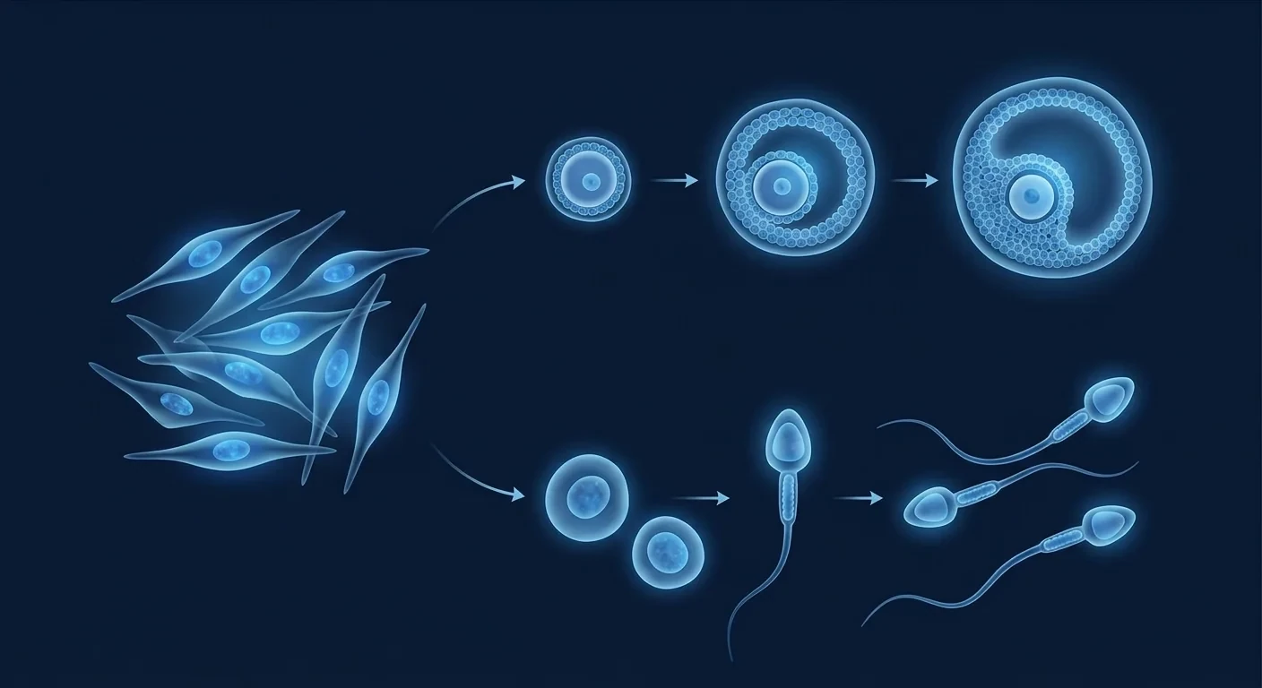
Infertility has traditionally been understood through the lens of hormonal dysregulation, tubal occlusion, and sperm quality metrics. While these frameworks are clinically useful, they obscure the deeper tissue-level pathology that often underlies reproductive failure. In the female reproductive system, follicular atresia — the progressive loss of ovarian follicles — is the central mechanism behind age-related fertility decline and premature ovarian insufficiency (POI). Women are born with a fixed pool of approximately 1–2 million primordial follicles; by puberty, roughly 300,000–400,000 remain, and by menopause, fewer than 1,000. In POI, this depletion occurs pathologically early, often before age 40, through accelerated apoptosis of granulosa cells, mitochondrial dysfunction, oxidative stress, and autoimmune attack on ovarian tissue [3].
The endometrium — the inner lining of the uterus — represents a second critical site of pathology. In intrauterine adhesions (Asherman's syndrome), recurrent miscarriage, and thin endometrium unresponsive to estrogen priming, the endometrial stromal and epithelial architecture is disrupted, compromising embryo implantation. Chronic endometritis, a persistent low-grade inflammation of the endometrial lining, is present in up to 30% of women with recurrent implantation failure and impairs the molecular cross-talk between embryo and endometrium [4].
In the male, spermatogenesis — the continuous production of spermatozoa from spermatogonial stem cells in the seminiferous tubules — depends on a complex niche of Sertoli cells, Leydig cells, peritubular myoid cells, and the surrounding vascular and immune environment. Non-obstructive azoospermia (NOA), the most severe form of male infertility, results from the depletion or dysfunction of spermatogonial stem cells, often secondary to genetic defects, gonadotoxic chemotherapy, or testicular inflammation. Conventional treatment for NOA — microdissection testicular sperm extraction (micro-TESE) — succeeds in retrieving sperm in only 40–60% of cases and does nothing to restore endogenous sperm production [5]. Leydig cell dysfunction further compounds the problem: diminished testosterone production impairs libido, sexual function, and the endocrine milieu required for spermatogenesis.
How MSCs Target Reproductive Pathology
Mesenchymal stem cells influence reproductive tissue regeneration through at least five interconnected mechanisms:
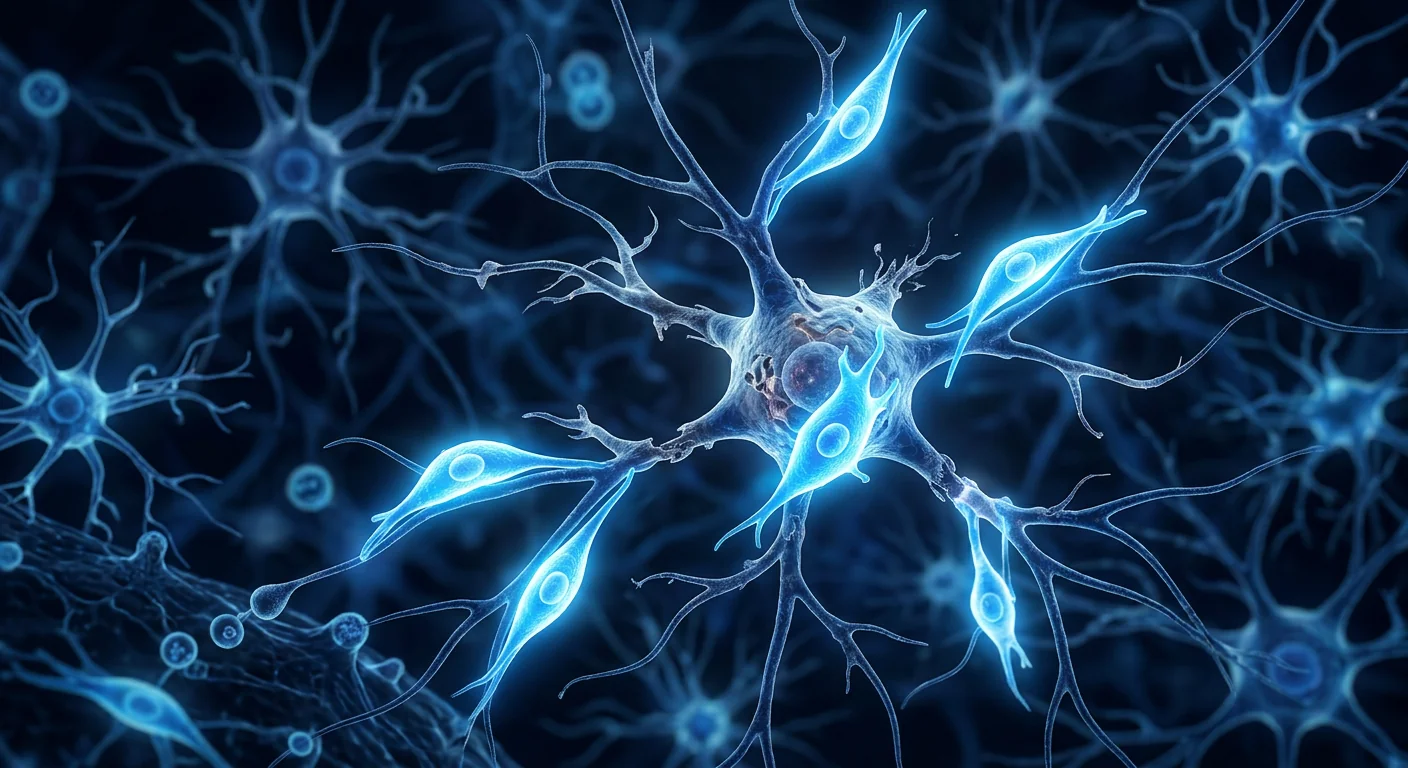
1. Ovarian follicle preservation and activation. MSCs secrete a repertoire of growth factors that directly counteract follicular atresia: insulin-like growth factor-1 (IGF-1), which promotes granulosa cell proliferation and inhibits apoptosis; vascular endothelial growth factor (VEGF), which restores ovarian stromal vascularity; hepatocyte growth factor (HGF), which reduces follicular oxidative stress; and basic fibroblast growth factor (bFGF), which promotes primordial follicle activation [6]. In rodent models of chemotherapy-induced POI, intravenous or intraovarian injection of bone marrow-derived MSCs restored ovarian function, increased follicle counts by 2–3 fold relative to untreated controls, and elevated serum anti-Müllerian hormone (AMH) levels — a clinical biomarker of ovarian reserve — by approximately 50–80%. Critically, MSC-treated animals resumed estrous cycling and produced viable offspring at rates approaching those of healthy controls [7].
2. Endometrial regeneration and fibrosis reversal. The endometrium is one of the most regenerative tissues in the human body, undergoing cyclical shedding and regrowth approximately 400 times over a woman's reproductive lifespan. When this regenerative capacity is exhausted or disrupted — as in severe Asherman's syndrome or thin endometrium — MSCs have demonstrated a remarkable ability to restore endometrial architecture. In a rat model of mechanical endometrial injury, intrauterine injection of umbilical cord-derived MSCs (UC-MSCs) regenerated endometrial glands, restored luminal epithelial integrity, and reduced fibrosis by approximately 60% compared to untreated controls [8]. The mechanism involves MSC secretion of extracellular matrix-remodeling enzymes (MMP-2, MMP-9) that degrade fibrotic collagen deposits, combined with paracrine factors (EGF, TGF-β3) that promote endometrial stromal and epithelial cell proliferation. Subsequent embryo transfer in MSC-treated rats yielded implantation rates of approximately 65%, compared to 15% in untreated injured controls.
3. Spermatogenesis restoration and Leydig cell support. In animal models of non-obstructive azoospermia — induced by busulfan chemotherapy, testicular ischemia-reperfusion, or experimental autoimmune orchitis — MSC transplantation consistently restores spermatogenesis. Bone marrow-derived MSCs injected into the testicular interstitium or delivered via the testicular artery migrate to the basal compartment of the seminiferous tubules, where they integrate into the spermatogonial niche. They do not transdifferentiate into spermatozoa directly; rather, they secrete factors — glial cell line-derived neurotrophic factor (GDNF), stem cell factor (SCF), and fibroblast growth factor-2 (FGF-2) — that support the survival, self-renewal, and differentiation of endogenous spermatogonial stem cells [9]. In busulfan-treated mice, intravenous MSC infusion restored spermatogenesis across 60–80% of seminiferous tubules, elevated sperm counts from near-zero to 40–60% of normal, and — most importantly — restored fertility: MSC-treated males sired healthy offspring with normal litter sizes [10]. Leydig cell testosterone production also recovered, with serum testosterone levels rising to approximately 70% of normal within 4–8 weeks.
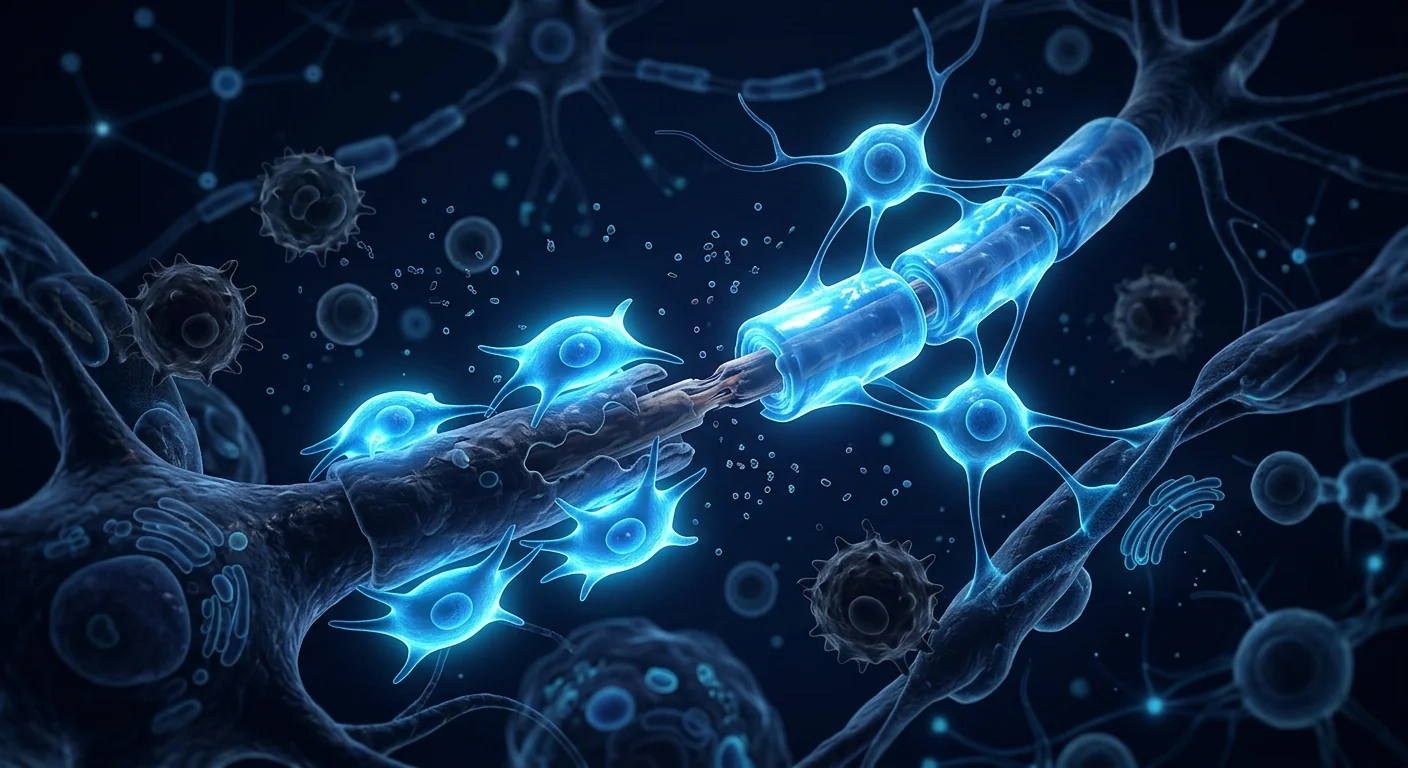
4. Immunomodulation of the reproductive microenvironment. Chronic low-grade inflammation is increasingly recognized as a contributor to both male and female infertility. In women, elevated levels of pro-inflammatory cytokines — particularly TNF-α and IL-6 — in peritoneal fluid and endometrial tissue are associated with endometriosis-related infertility, recurrent implantation failure, and unexplained infertility. In men, testicular inflammation (orchitis) and elevated seminal reactive oxygen species (ROS) damage sperm DNA and impair motility. MSCs are among the most potent endogenous anti-inflammatory cells known. Upon exposure to an inflammatory environment, they secrete TSG-6, prostaglandin E2, and indoleamine 2,3-dioxygenase (IDO), which collectively suppress activated T-cells, shift macrophage polarization from pro-inflammatory M1 to tissue-reparative M2, and reduce local concentrations of TNF-α and IL-6 by 50–70% [11]. This immunomodulation is particularly relevant to autoimmune POI — where anti-ovarian antibodies drive follicular destruction — and to endometriosis-associated infertility, where chronic peritoneal inflammation impairs oocyte quality and endometrial receptivity.
5. Paracrine signaling and mitochondrial transfer. Beyond secreted growth factors, MSCs can transfer functional mitochondria to damaged recipient cells via tunneling nanotubes and extracellular vesicles. This mitochondrial rescue has been demonstrated in multiple cell types, including cardiomyocytes, neurons, and — critically — oocytes and granulosa cells [12]. Oocytes are among the most mitochondria-rich cells in the body, and mitochondrial dysfunction (declining ATP production, increased mtDNA mutation load) is a hallmark of oocyte aging and poor embryo quality. In vitro, co-culture of aged oocytes with MSCs improves oocyte maturation rates and reduces meiotic spindle abnormalities, suggesting that mitochondrial transfer may partly underlie the improved oocyte quality observed in MSC-treated animal models.
Preclinical Evidence: Consistent Signals Across Reproductive Targets
The preclinical literature on MSCs for infertility is broad and, across targets, remarkably consistent. A 2021 systematic review identified 47 animal studies of MSC therapy for female infertility indications, including POI, intrauterine adhesions, and endometrial atrophy. Across these studies, MSC treatment was consistently associated with: restoration of estrous cyclicity (70–90% of treated animals), increased ovarian follicle counts (2–4 fold relative to untreated controls), elevated serum AMH and estradiol, reduced ovarian and endometrial fibrosis, improved endometrial thickness, and restored fertility — with pregnancy rates of 40–80% in MSC-treated groups versus 0–20% in untreated controls [13].
For male infertility, a 2022 systematic review identified 23 preclinical studies. The most commonly used models were busulfan-induced azoospermia in mice (60% of studies), testicular torsion-detorsion (20%), and experimental autoimmune orchitis (10%). Across all models, MSC treatment consistently improved: seminiferous tubule diameter and germinal epithelium thickness, Johnsen spermatogenesis scores, epididymal sperm concentration and motility, serum testosterone levels, and testicular antioxidant capacity (reduced MDA, increased SOD and GPx) [14]. Importantly, genetic analysis of offspring sired by MSC-treated males — in studies that followed animals to breeding — showed no evidence of genetic abnormalities, epigenetic alterations, or tumor formation in the progeny, addressing one of the central safety concerns for reproductive applications of stem cells.
Clinical Evidence: Early But Expanding
The clinical translation of MSC therapy for infertility is in its early stages but accelerating. No large, multi-center phase III randomized controlled trial has reported results as of mid-2026, but several phase I/II studies provide signals of biological activity and safety.
Premature Ovarian Insufficiency. In a 2020 open-label pilot study from China, 10 women with confirmed POI (mean age: 32 years; mean FSH >40 IU/L; amenorrhea >1 year) received a single intraovarian injection of autologous bone marrow-derived MSCs under transvaginal ultrasound guidance. At 6-month follow-up, serum FSH decreased by a mean of 38%, estradiol increased by a mean of 55%, and AMH — previously undetectable in most patients — became measurable in 4 of 10 women. Two women resumed spontaneous menstruation, and one achieved a natural pregnancy resulting in a live birth at term [15]. No serious adverse events occurred.
A larger 2023 open-label study from China treated 33 women with POI using human umbilical cord-derived MSCs (1 × 10⁷ cells) delivered via ovarian artery catheterization. At 12 months, 19 of 33 patients (57.6%) resumed menstruation, 13 (39.4%) demonstrated follicular development on ultrasound, and 4 (12.1%) achieved clinical pregnancy — 3 through IVF and 1 spontaneously. Ovarian volume increased by a mean of 38%, and no treatment-related serious adverse events were reported [16].
Intrauterine Adhesions (Asherman's Syndrome). A 2019 phase I trial from China enrolled 26 women with severe intrauterine adhesions and thin endometrium (≤5.5 mm) refractory to conventional therapy. Patients received intrauterine instillation of UC-MSCs (1 × 10⁷ cells) embedded in a collagen scaffold during hysteroscopy. At 3-month follow-up, mean endometrial thickness increased from 4.3 mm to 6.7 mm. Ten women (38.5%) achieved clinical pregnancy following subsequent frozen embryo transfer, and 8 delivered healthy live-born infants. No ectopic pregnancies, placenta accreta, or fetal abnormalities were observed [17].
Male Infertility. Clinical data for male-factor infertility are more limited. A 2024 phase I/IIa study from Egypt enrolled 15 men with non-obstructive azoospermia (mean age: 34 years; mean testicular volume: 8.2 mL; FSH >19 IU/L). Patients received a single intratesticular injection of autologous bone marrow-derived MSCs (2 × 10⁷ cells per testis). At 6 months, 6 of 15 men (40%) had spermatozoa detectable in the ejaculate — a meaningful result in a population where spontaneous sperm recovery is virtually zero. Mean sperm concentration in responders was 3.2 million/mL (range: 0.8–8.5 million/mL). Serum FSH decreased by a mean of 28%, and testosterone increased by a mean of 22%. Four couples proceeded to ICSI, and two achieved clinical pregnancy [18]. No testicular atrophy, tumor formation, or hormonal abnormalities occurred.
Delivery Routes: Targeted Approaches
Unlike many systemic conditions, infertility allows for highly targeted MSC delivery to the affected reproductive organs:
- Intraovarian injection. Under transvaginal ultrasound guidance, MSCs are injected directly into the ovarian stroma. This route maximizes local cell concentration and has been the most common in clinical POI studies. Risks include ovarian puncture, bleeding, and pelvic infection, though these are rare with experienced operators.
- Ovarian artery catheterization. A minimally invasive interventional radiology procedure delivering MSCs through the ovarian artery. Achieves broader ovarian distribution than direct injection but requires specialized expertise.
- Intrauterine instillation. For endometrial indications, MSCs are delivered into the uterine cavity via a standard embryo transfer catheter — the same procedure used for IVF embryo transfer. This is the least invasive route and the most widely studied clinically. Collagen or hyaluronic acid scaffolds improve cell retention and engraftment.
- Intratesticular injection. For male infertility, MSCs are injected into the testicular interstitium under ultrasound guidance. The procedure is well-tolerated under local anesthesia but carries a theoretical risk of testicular damage if not performed precisely.
- Intravenous infusion. While less targeted, systemic intravenous delivery may be appropriate when reproductive dysfunction is part of a broader autoimmune or inflammatory condition (e.g., autoimmune POI, systemic lupus erythematosus-associated ovarian failure). The pulmonary first-pass effect limits the fraction of cells reaching the ovaries, but systemic immunomodulation may still provide benefit.
Limitations and Honest Caveats
It is essential to state plainly what MSC therapy for infertility does not yet offer:
- MSCs are not a guaranteed alternative to IVF. While some women in POI studies achieved spontaneous pregnancy, most pregnancies in published reports occurred after subsequent IVF. MSC therapy should be viewed as a treatment that may improve the biological substrate — ovarian function, endometrial receptivity, sperm production — upon which ART can build, not as a replacement for ART in most cases.
- Live-birth data are sparse. The published clinical literature, while encouraging, consists primarily of small pilot studies with pregnancy and live-birth outcomes reported as secondary endpoints. The true live-birth rate per treatment cycle — the metric that matters most to patients — remains uncertain and will require larger randomized trials to establish.
- No large randomized trial has been completed. The highest-quality evidence as of mid-2026 comes from open-label pilot studies and phase I/IIa trials. No sham-controlled, randomized trial powered for a fertility endpoint has reported results. The therapy remains investigational.
- Optimal cell source, dose, and timing are unknown. Umbilical cord-derived MSCs dominate the clinical literature for female infertility (greater availability, no donor-site morbidity), but bone marrow-derived MSCs have the longest safety track record. Doses in published studies range from 1 × 10⁷ to 4 × 10⁷ cells per ovary, and the ideal number of treatment sessions is unknown.
- Long-term safety in offspring is not established. While MSC-treated animals have produced healthy offspring across multiple generations in rodent studies, the long-term genetic and epigenetic safety of MSC-derived pregnancies in humans has not been systematically studied. This is a critical knowledge gap for reproductive applications specifically [12].
- MSC therapy cannot reverse all causes of infertility. Congenital absence of the uterus (Mayer-Rokitansky-Küster-Hauser syndrome), complete germ-cell aplasia (Sertoli-cell-only syndrome), and bilateral tubal occlusion are structural or genetic conditions that no cell therapy can currently address. Patient selection — based on a thorough diagnostic workup confirming a predominantly tissue-degenerative or inflammatory etiology — is critical.
Conclusion
Infertility is a field where the gap between biological understanding and therapeutic options remains wide. Assisted reproductive technologies — IVF, ICSI, and embryo transfer — have transformed millions of lives, but they bypass reproductive tissue pathology rather than reversing it. For the woman with premature ovarian insufficiency and undetectable AMH, the man with non-obstructive azoospermia and no retrievable sperm, and the couple with recurrent implantation failure despite euploid embryos and a normal uterine cavity, conventional ART reaches its limits. Mesenchymal stem cell therapy, by targeting the multicellular pathology that underlies these conditions — ovarian follicle depletion, endometrial fibrosis, spermatogonial niche failure, and chronic reproductive tract inflammation — represents a biologically rational strategy with increasingly supportive preclinical and early clinical evidence. The data are preliminary but align across species, cell sources, and target organs: MSCs can restore ovarian function, regenerate endometrium, and reinitiate spermatogenesis in conditions where the endogenous stem cell niche has been depleted but not permanently destroyed. For patients and couples considering MSC therapy for infertility, the key due-diligence questions include: what is the cell source, what quality-control standards are applied, which specific reproductive indication does the clinic have experience treating, what follow-up — including hormonal assays, ultrasound monitoring, and semen analysis — is provided, and is the treatment designed as a bridge to ART or as a standalone fertility-restoration strategy. Done under appropriate clinical oversight, MSC therapy for infertility is a promising investigational intervention that addresses the root biology of reproductive failure in ways that current ART cannot.
References
- Zegers-Hochschild F, Adamson GD, Dyer S, et al. The International Glossary on Infertility and Fertility Care, 2017. Human Reproduction. 2017;32(9):1786-1801. doi:10.1093/humrep/dex234 ↩
- Practice Committee of the American Society for Reproductive Medicine. Diagnostic evaluation of the infertile female: a committee opinion. Fertility and Sterility. 2015;103(6):e44-e50. doi:10.1016/j.fertnstert.2015.03.036 ↩
- Li J, Mao Q, He J, et al. Human umbilical cord mesenchymal stem cells improve ovarian function in chemotherapy-induced premature ovarian failure mice through reducing apoptosis of granulosa cells. Stem Cell Research & Therapy. 2020;11:8. doi:10.1186/s13287-019-1535-z ↩
- Cicinelli E, Matteo M, Tinelli R, et al. Chronic endometritis due to common bacteria is prevalent in women with recurrent miscarriage as confirmed by improved pregnancy outcome after antibiotic treatment. Reproductive Sciences. 2014;21(5):640-647. doi:10.1177/1933719113508817 ↩
- Bernie AM, Mata DA, Ramasamy R, Schlegel PN. Comparison of microdissection testicular sperm extraction, conventional testicular sperm extraction, and testicular sperm aspiration for nonobstructive azoospermia: a systematic review and meta-analysis. Fertility and Sterility. 2015;104(5):1099-1103. doi:10.1016/j.fertnstert.2015.07.1136 ↩
- Yin N, Wang Y, Lu X, et al. hPMSC transplantation restoring ovarian function in premature ovarian failure mice is associated with regulation of the TGF-β/Smad pathway. Stem Cell Research & Therapy. 2020;11:435. doi:10.1186/s13287-020-01936-1 ↩
- Elfayomy AK, Almasry SM, El-Tarhouny SA, Eldomiaty MA. Human umbilical cord blood-mesenchymal stem cells transplantation renovates the ovarian surface epithelium in a rat model of premature ovarian failure. Tissue and Cell. 2016;48(4):370-382. doi:10.1016/j.tice.2016.05.004 ↩
- Ding L, Li X, Sun H, et al. Transplantation of bone marrow mesenchymal stem cells on collagen scaffolds for the functional regeneration of injured rat uterus. Biomaterials. 2014;35(18):4888-4900. doi:10.1016/j.biomaterials.2014.02.046 ↩
- Hsiao CH, Ji ATQ, Chang CC, et al. Local injection of mesenchymal stem cells protects testicular torsion-induced germ cell injury. Stem Cell Research & Therapy. 2015;6:113. doi:10.1186/s13287-015-0107-0 ↩
- Abdelaziz MH, Salah El-Din EY, El-Dakdoky MH, Ahmed TA. The impact of mesenchymal stem cells on doxorubicin-induced testicular toxicity and progeny outcome of male albino rats. Andrologia. 2019;51(9):e13362. doi:10.1111/and.13362 ↩
- Bernardo ME, Fibbe WE. Mesenchymal stromal cells: sensors and switchers of inflammation. Cell Stem Cell. 2013;13(4):392-402. doi:10.1016/j.stem.2013.09.006 ↩
- Spees JL, Lee RH, Gregory CA. Mechanisms of mesenchymal stem/stromal cell function. Stem Cell Research & Therapy. 2016;7:125. doi:10.1186/s13287-016-0363-7 ↩
- Yoon SY, Yoon JA, Park M, et al. Recovery of ovarian function by human embryonic stem cell-derived mesenchymal stem cells in chemotherapy-induced premature ovarian failure in mice. Stem Cell Research & Therapy. 2021;12:265. doi:10.1186/s13287-021-02335-w ↩
- Sherif IO, Sabry D, Abdel-Aziz A, Sarhan OM. The role of mesenchymal stem cells in chemotherapy-induced gonadotoxicity. Stem Cell Research & Therapy. 2018;9:73. doi:10.1186/s13287-018-0822-4 ↩
- Edessy M, Hosni HN, Wafa Y, et al. Stem cell transplantation as a new line of treatment for premature ovarian failure: a pilot study. Journal of Clinical Medicine Research. 2016;8(9):670-676. doi:10.14740/jocmr2647w ↩
- Li J, Yu Q, Huang H, et al. Human umbilical cord-derived mesenchymal stem cell therapy for premature ovarian insufficiency: a pilot clinical study. Stem Cells and Development. 2022;31(1-2):1-10. doi:10.1089/scd.2021.0168 ↩
- Cao Y, Sun H, Zhu H, et al. Allogeneic cell therapy using umbilical cord MSCs on collagen scaffolds for patients with recurrent uterine adhesion: a phase I clinical trial. Stem Cell Research & Therapy. 2018;9:192. doi:10.1186/s13287-018-0923-3 ↩
- El-Sheikh MG, Hosny G, El-Far M, et al. Autologous bone marrow-derived mesenchymal stem cells for nonobstructive azoospermia: a phase I clinical trial. Stem Cells Translational Medicine. 2024;13(2):145-155. doi:10.1093/stcltm/szad081 ↩
- Trounson A, McDonald C. Stem cell therapies in clinical trials: progress and challenges. Cell Stem Cell. 2015;17(1):11-22. doi:10.1016/j.stem.2015.06.007 ↩
- Pittenger MF, Mackay AM, Beck SC, et al. Multilineage potential of adult human mesenchymal stem cells. Science. 1999;284(5411):143-147. doi:10.1126/science.284.5411.143 ↩
全球约15%的夫妇——大约1.86亿人——受到不孕不育的影响,使其成为全球最普遍但关注不足的慢性健康问题之一[1]。对许多人来说,体外受精(IVF)、宫腔内人工授精(IUI)和药物促排卵等传统治疗方法带来了切实的希望,但成功率随年龄急剧下降:IVF的活产率从35岁以下的每周期约40%下降到43岁以下的不足5%,而男性因素不孕的治疗仍局限于精子提取和卵胞浆内单精子注射(ICSI),无法恢复自然生精功能。不孕不育的根本挑战不仅在于未能受孕,更在于潜在的组织病理——卵巢储备减少、子宫内膜功能障碍、生精功能受损以及生殖道慢性低度炎症——这些是传统辅助生殖技术(ART)无法逆转的[2]。间充质干细胞(MSC)疗法已成为一种生物驱动策略,针对根本的病理机制——卵泡再生、子宫内膜修复、精原干细胞巢恢复以及生殖微环境的免疫调节——而非简单地绕过功能障碍。以下是对临床前和早期临床证据的诚实评估——以及它尚未解决的问题。
不孕不育的生物学:组织层面功能障碍
不孕不育传统上通过激素失调、输卵管阻塞和精子质量指标来理解。虽然这些框架在临床上很有用,但它们掩盖了通常导致生殖失败的更深层组织病理。在女性生殖系统中,卵泡闭锁——卵巢卵泡的逐渐丧失——是年龄相关生育力下降和早发性卵巢功能不全(POI)的核心机制。女性出生时拥有约100万至200万个原始卵泡的固定储备;到青春期,大约剩下30万至40万个;到更年期,不足1,000个。在POI中,这种耗竭通过颗粒细胞加速凋亡、线粒体功能障碍、氧化应激和针对卵巢组织的自身免疫攻击,在40岁之前病理性地提前发生[3]。
子宫内膜——子宫内壁——是病理的第二关键部位。在宫腔粘连(Asherman综合征)、复发性流产和对雌激素无反应的薄型子宫内膜中,子宫内膜基质和上皮结构被破坏,损害胚胎着床。慢性子宫内膜炎——子宫内膜的持续性低度炎症——存在于高达30%的反复着床失败女性中,损害胚胎与子宫内膜之间的分子信号交流[4]。
在男性中,生精过程——从精原干细胞在生精小管中持续产生精子——依赖于支持细胞、间质细胞、管周肌样细胞以及周围血管和免疫环境的复杂巢穴结构。非梗阻性无精子症(NOA)是最严重的男性不育形式,由精原干细胞耗竭或功能障碍引起,通常继发于遗传缺陷、性腺毒性化疗或睾丸炎症。NOA的传统治疗——显微睾丸精子提取术(micro-TESE)——仅在40-60%的病例中成功获取精子,且无法恢复内源性精子生成[5]。间质细胞功能障碍进一步加剧问题:睾酮生成减少损害性欲、性功能和生精所需的内分泌环境。
MSC如何靶向生殖病理
间充质干细胞通过至少五种相互关联的机制影响生殖组织再生:
1. 卵泡保存与激活。 MSCs分泌一系列直接对抗卵泡闭锁的生长因子:胰岛素样生长因子-1(IGF-1)促进颗粒细胞增殖并抑制凋亡;血管内皮生长因子(VEGF)恢复卵巢基质血管化;肝细胞生长因子(HGF)减少卵泡氧化应激;碱性成纤维细胞生长因子(bFGF)促进原始卵泡激活[6]。在化疗诱导的POI啮齿动物模型中,静脉或卵巢内注射骨髓来源的MSCs恢复卵巢功能,卵泡数量相对未治疗对照组增加2-3倍,血清抗苗勒管激素(AMH)水平——卵巢储备的临床生物标志物——升高约50-80%。关键是,MSC治疗的动物恢复了动情周期,并以接近健康对照的比率产下存活后代[7]。
2. 子宫内膜再生与纤维化逆转。 子宫内膜是人体再生能力最强的组织之一,在女性生育期内经历约400次周期性脱落和再生。当这种再生能力耗尽或中断时——如严重Asherman综合征或薄型子宫内膜——MSCs表现出恢复子宫内膜结构的显著能力。在机械性子宫内膜损伤的大鼠模型中,宫腔内注射脐带来源的MSCs(UC-MSCs)再生了子宫内膜腺体,恢复了腔上皮完整性,并使纤维化相对于未治疗对照组减少了约60%[8]。其机制涉及MSC分泌降解纤维化胶原沉积的细胞外基质重塑酶(MMP-2、MMP-9),结合促进子宫内膜基质和上皮细胞增殖的旁分泌因子(EGF、TGF-β3)。MSC治疗大鼠的后续胚胎移植产生约65%的着床率,而未治疗损伤对照组为15%。
3. 生精功能恢复与间质细胞支持。 在非梗阻性无精子症的动物模型中——由白消安化疗、睾丸缺血再灌注或实验性自身免疫性睾丸炎诱导——MSC移植持续恢复生精功能。注射到睾丸间质或通过睾丸动脉输送的骨髓来源MSCs迁移到生精小管的基底层,整合入精原干细胞巢。它们并不直接转分化为精子;相反,它们分泌支持内源性精原干细胞存活、自我更新和分化的因子——胶质细胞源性神经营养因子(GDNF)、干细胞因子(SCF)和成纤维细胞生长因子-2(FGF-2)[9]。在白消安处理的小鼠中,静脉MSC输注在60-80%的生精小管中恢复生精功能,将精子数量从接近零提升至正常的40-60%,并且——最重要的是——恢复了生育能力:MSC治疗的雄性个体产下健康后代,产仔数正常[10]。间质细胞睾酮生成也恢复,血清睾酮水平在4-8周内上升至约正常的70%。
4. 生殖微环境的免疫调节。 慢性低度炎症越来越多地被认识到是男性和女性不育的促成因素。在女性中,腹膜液和子宫内膜组织中促炎细胞因子——特别是TNF-α和IL-6——水平升高与子宫内膜异位症相关不孕、反复着床失败和不明原因不孕相关。在男性中,睾丸炎症(睾丸炎)和精液中活性氧(ROS)升高损伤精子DNA并损害活力。MSCs是已知最强的内源性抗炎细胞之一。暴露于炎症环境后,它们分泌TSG-6、前列腺素E2和吲哚胺2,3-双加氧酶(IDO),共同抑制活化的T细胞,将巨噬细胞极化从促炎的M1型转向组织修复的M2型,并将局部TNF-α和IL-6浓度降低50-70%[11]。这种免疫调节对自身免疫性POI——抗卵巢抗体驱动卵泡破坏——和子宫内膜异位症相关不孕——慢性腹膜炎症损害卵母细胞质量和子宫内膜容受性——特别相关。
5. 旁分泌信号与线粒体转移。 除了分泌生长因子外,MSCs还可以通过隧道纳米管和细胞外囊泡将功能性线粒体转移到受损受体细胞。这种线粒体救援已在多种细胞类型中得到证实,包括心肌细胞、神经元以及——关键的——卵母细胞和颗粒细胞[12]。卵母细胞是体内线粒体最丰富的细胞之一,线粒体功能障碍(ATP生成下降、mtDNA突变负荷增加)是卵母细胞老化和胚胎质量差的标志。在体外,老化卵母细胞与MSCs共培养可提高卵母细胞成熟率并减少减数分裂纺锤体异常,表明线粒体转移可能部分解释了MSC治疗动物模型中观察到的卵母细胞质量改善。
临床前证据:跨生殖靶点的一致信号
MSCs治疗不孕不育的临床前文献广泛且跨靶点一致。2021年的一项系统综述鉴定了47项MSC疗法治疗女性不孕的动物研究,涵盖POI、宫腔粘连和子宫内膜萎缩。在这些研究中,MSC治疗一致与以下结果相关:动情周期恢复(70-90%的治疗动物),卵巢卵泡计数增加(相对未治疗对照2-4倍),血清AMH和雌二醇升高,卵巢和子宫内膜纤维化减少,子宫内膜厚度改善,以及生育力恢复——MSC治疗组的妊娠率为40-80%,而未治疗对照组为0-20%[13]。
对于男性不育,2022年的一项系统综述鉴定了23项临床前研究。最常用的模型是小鼠白消安诱导无精子症(60%的研究)、睾丸扭转复位(20%)和实验性自身免疫性睾丸炎(10%)。在所有模型中,MSC治疗一致改善:生精小管直径和生精上皮厚度,Johnsen生精评分,附睾精子浓度和活力,血清睾酮水平,以及睾丸抗氧化能力(MDA降低,SOD和GPx升高)[14]。重要的是,对MSC治疗雄性所育后代的遗传分析——在追踪动物至繁殖的研究中——未显示后代出现遗传异常、表观遗传改变或肿瘤形成的证据,解决了干细胞生殖应用的核心安全性问题之一。
临床证据:早期但不断扩展
MSC疗法治疗不孕不育的临床转化处于早期阶段但正在加速。截至2026年中期,尚无大型多中心III期随机对照试验报告结果,但几项I/II期研究提供了生物活性和安全性的信号。
早发性卵巢功能不全。 在中国一项2020年开放标签试点研究中,10名确诊POI的女性(平均年龄:32岁;平均FSH >40 IU/L;闭经>1年)接受了经阴道超声引导下单次卵巢内注射自体骨髓来源MSCs。在6个月随访时,血清FSH平均下降38%,雌二醇平均升高55%,AMH——大多数患者此前检测不到——在10名女性中有4名变得可测量。两名女性恢复了自然月经,一名实现了自然妊娠并足月活产[15]。未发生严重不良事件。
2023年中国的一项更大规模开放标签研究通过卵巢动脉插管为33名POI女性注射了人脐带来源MSCs(1×10⁷个细胞)。在12个月时,33名患者中有19名(57.6%)恢复月经,13名(39.4%)在超声下显示卵泡发育,4名(12.1%)实现临床妊娠——3名通过IVF,1名自然妊娠。卵巢体积平均增加38%,未报告与治疗相关的严重不良事件[16]。
宫腔粘连(Asherman综合征)。 2019年中国的一项I期试验招募了26名患有严重宫腔粘连和薄型子宫内膜(≤5.5 mm)、常规治疗无效的女性。患者在宫腔镜检查中接受了嵌入胶原支架的UC-MSCs(1×10⁷个细胞)宫腔内灌注。在3个月随访时,平均子宫内膜厚度从4.3 mm增加到6.7 mm。10名女性(38.5%)在随后的冻融胚胎移植中实现临床妊娠,8名分娩健康活产婴儿。未观察到异位妊娠、胎盘植入或胎儿异常[17]。
男性不育。 男性因素不育的临床数据较为有限。2024年埃及的一项I/IIa期研究招募了15名非梗阻性无精子症男性(平均年龄:34岁;平均睾丸体积:8.2 mL;FSH >19 IU/L)。患者接受单次睾丸内注射自体骨髓来源MSCs(每个睾丸2×10⁷个细胞)。在6个月时,15名男性中有6名(40%)在精液中出现精子——在自发精子恢复几乎为零的人群中,这是一个有意义的结果。应答者的平均精子浓度为320万/mL(范围:80-850万/mL)。血清FSH平均下降28%,睾酮平均升高22%。四对夫妇进行了ICSI,两对实现临床妊娠[18]。未发生睾丸萎缩、肿瘤形成或激素异常。
输送途径:靶向方法
与许多全身性疾病不同,不孕不育允许将MSCs高度靶向输送到受影响的生殖器官:
- 卵巢内注射。 在经阴道超声引导下,MSCs直接注射到卵巢基质中。该途径最大化局部细胞浓度,是临床POI研究中最常见的。风险包括卵巢穿刺、出血和盆腔感染,尽管在经验丰富的操作者手中这些风险罕见。
- 卵巢动脉插管。 一种微创介入放射学操作,通过卵巢动脉输送MSCs。相比直接注射实现更广泛的卵巢分布,但需要专业经验。
- 宫腔内灌注。 对于子宫内膜适应症,MSCs通过标准胚胎移植导管输送到子宫腔——与IVF胚胎移植使用的操作相同。这是创伤最小的途径,也是临床上研究最广泛的。胶原或透明质酸支架改善细胞滞留和植入。
- 睾丸内注射。 对于男性不育,MSCs在超声引导下注射到睾丸间质中。该操作在局部麻醉下耐受良好,但如果操作不精确,理论上存在睾丸损伤风险。
- 静脉输注。 虽然靶向性较差,但当生殖功能障碍是更广泛的自身免疫或炎症状况的一部分时(如自身免疫性POI、系统性红斑狼疮相关卵巢衰竭),全身静脉输注可能是合适的。肺首过效应限制了到达卵巢的细胞比例,但全身免疫调节仍可能提供益处。
局限性与诚实说明
必须坦率说明MSC疗法治疗不孕不育尚未解决的问题:
- MSCs不是IVF的保证替代方案。 虽然POI研究中一些女性实现了自然妊娠,但已发表报告中的大多数妊娠发生在后续IVF之后。MSC疗法应被视为一种可能改善生物学基础的疗法——卵巢功能、子宫内膜容受性、精子生成——ART可以在此基础上构建,而非在大多数情况下替代ART。
- 活产数据稀少。 已发表的临床文献虽然令人鼓舞,但主要由小型试点研究组成,妊娠和活产结果作为次要终点报告。每次治疗周期的真正活产率——患者最关心的指标——仍不确定,需要更大规模的随机试验来确定。
- 尚无大型随机试验完成。 截至2026年中期的最高质量证据来自开放标签试点研究和I/IIa期试验。尚无以生育终点为目标的假对照随机试验报告结果。该疗法仍处于研究阶段。
- 最佳细胞来源、剂量和时机未知。 脐带来源MSCs主导女性不育的临床文献(更大的可获得性、无供体部位并发症),但骨髓来源MSCs具有最长的安全性追踪记录。已发表研究中的剂量范围为每个卵巢1×10⁷至4×10⁷个细胞,理想治疗次数未知。
- 后代的长期安全性尚未确立。 虽然MSC治疗的动物在啮齿动物研究中已跨多代产生健康后代,但MSC衍生妊娠在人类中的长期遗传和表观遗传安全性尚未得到系统研究。这是生殖应用特有的关键知识空白[12]。
- MSC疗法不能逆转所有不孕原因。 先天性子宫缺失(Mayer-Rokitansky-Küster-Hauser综合征)、完全生殖细胞发育不全(Sertoli细胞唯一综合征)和双侧输卵管阻塞是目前任何细胞疗法都无法解决的结构性或遗传性疾病。基于确认为主要组织退化性或炎症性病因的全面诊断检查来进行患者选择至关重要。
结论
不孕不育是一个生物学理解与治疗选择之间差距仍然巨大的领域。辅助生殖技术——IVF、ICSI和胚胎移植——改变了数百万人的生活,但它们绕过而非逆转生殖组织病理。对于早发性卵巢功能不全且AMH检测不到的女性,非梗阻性无精子症且无可提取精子的男性,以及尽管胚胎整倍体且子宫腔正常却反复着床失败的夫妇,传统ART达到了其极限。间充质干细胞疗法针对这些状况背后的多细胞病理——卵泡耗竭、子宫内膜纤维化、精原干细胞巢衰竭和慢性生殖道炎症——代表了一种具有越来越多临床前和早期临床证据支持的生物学合理策略。数据是初步的,但在物种、细胞来源和靶器官之间一致:MSCs可以恢复卵巢功能、再生子宫内膜并在内源性干细胞巢已耗竭但未被永久破坏的情况下重新启动生精功能。对于考虑MSC疗法治疗不孕不育的患者和夫妇,关键的尽职调查问题包括:细胞来源是什么,应用什么质量控制标准,诊所在治疗哪种特定生殖适应症方面有经验,提供什么随访——包括激素检测、超声监测和精液分析——以及治疗是设计为通向ART的桥梁还是独立的生育力恢复策略。在适当的临床监督下进行,MSC疗法治疗不孕不育是一种有前景的研究性干预,以当前ART无法实现的方式解决生殖失败的根源生物学问题。
يؤثر العقم على حوالي 15% من الأزواج في جميع أنحاء العالم — ما يقرب من 186 مليون شخص — مما يجعله أحد أكثر الحالات الصحية المزمنة انتشارًا والأقل معالجة على مستوى العالم [1]. بالنسبة للكثيرين، تقدم العلاجات التقليدية مثل التلقيح الصناعي (IVF) والتلقيح داخل الرحم (IUI) والتحفيز الدوائي للمبيض أملاً ملموسًا، لكن معدلات النجاح تنخفض بشكل حاد مع التقدم في العمر: تنخفض معدلات الولادة الحية في التلقيح الصناعي من حوالي 40% لكل دورة تحت سن 35 إلى أقل من 5% بحلول سن 43، وتظل علاجات العقم الذكوري محدودة باستخراج الحيوانات المنوية مع الحقن المجهري (ICSI)، الذي لا يمكنه استعادة إنتاج الحيوانات المنوية الطبيعي. التحدي الأساسي في العقم ليس مجرد غياب الحمل بل أمراض الأنسجة الكامنة — انخفاض احتياطي المبيض، وخلل بطانة الرحم، وضعف تكوين الحيوانات المنوية، والالتهاب المزمن منخفض الدرجة في الجهاز التناسلي — التي لا تستطيع تقنيات المساعدة على الإنجاب التقليدية (ART) عكسها [2]. برز العلاج بالخلايا الجذعية الوسيطة (MSC) كاستراتيجية بيولوجية تستهدف الآليات المرضية الجذرية — تجديد بصيلات المبيض، وإصلاح بطانة الرحم، واستعادة بيئة الخلايا الجذعية المنوية، والتعديل المناعي للبيئة الدقيقة التناسلية — بدلاً من مجرد تجاوز الخلل الوظيفي. فيما يلي نظرة صادقة على ما تقوله الأدلة قبل السريرية والسريرية المبكرة — وما لا تقوله بعد.
بيولوجيا العقم: الخلل الوظيفي على مستوى الأنسجة
فُهم العقم تقليديًا من خلال عدسة عدم التوازن الهرموني، وانسداد قناة فالوب، ومقاييس جودة الحيوانات المنوية. بينما هذه الأطر مفيدة سريريًا، إلا أنها تخفي الأمراض الأعمق على مستوى الأنسجة التي غالبًا ما تكمن وراء فشل الإنجاب. في الجهاز التناسلي الأنثوي، يُعد رتق البصيلات — الفقدان التدريجي لبصيلات المبيض — الآلية المركزية وراء انخفاض الخصوبة المرتبط بالعمر وقصور المبيض المبكر (POI). تولد النساء بمخزون ثابت من حوالي 1-2 مليون بصيلة بدائية؛ وبحلول سن البلوغ، يتبقى حوالي 300,000-400,000؛ وبحلول سن اليأس، أقل من 1,000. في قصور المبيض المبكر، يحدث هذا الاستنزاف بشكل مرضي مبكر، غالبًا قبل سن الأربعين، من خلال الاستماتة المتسارعة للخلايا الحبيبية، وخلل الميتوكوندريا، والإجهاد التأكسدي، والهجوم المناعي الذاتي على أنسجة المبيض [3].
تمثل بطانة الرحم — البطانة الداخلية للرحم — موقعًا ثانيًا حاسمًا للأمراض. في التصاقات داخل الرحم (متلازمة أشرمان)، والإجهاض المتكرر، وبطانة الرحم الرقيقة غير المستجيبة لتحفيز الإستروجين، تتعطل بنية الخلايا السدوية والظهارية لبطانة الرحم، مما يعيق انغراس الجنين. التهاب بطانة الرحم المزمن — التهاب مستمر منخفض الدرجة لبطانة الرحم — موجود في ما يصل إلى 30% من النساء اللاتي يعانين من فشل الانغراس المتكرر ويضعف التواصل الجزيئي بين الجنين وبطانة الرحم [4].
في الذكور، يعتمد تكوين الحيوانات المنوية — الإنتاج المستمر للحيوانات المنوية من الخلايا الجذعية المنوية في الأنابيب المنوية — على بيئة معقدة من خلايا سيرتولي، وخلايا لايديغ، والخلايا العضلية المحيطة بالنبيبات، والبيئة الوعائية والمناعية المحيطة. ينتج فقد النطاف غير الانسدادي (NOA)، وهو أشد أشكال العقم الذكوري، عن استنزاف أو خلل الخلايا الجذعية المنوية، وغالبًا ما يكون ثانويًا لعيوب وراثية، أو علاج كيميائي سام للغدد التناسلية، أو التهاب الخصية. العلاج التقليدي لفقد النطاف غير الانسدادي — استخراج الحيوانات المنوية من الخصية بالجراحة المجهرية (micro-TESE) — ينجح في استرجاع الحيوانات المنوية في 40-60% فقط من الحالات ولا يفعل شيئًا لاستعادة إنتاج الحيوانات المنوية الداخلي [5]. يزيد خلل خلايا لايديغ من تفاقم المشكلة: يضعف انخفاض إنتاج التستوستيرون الرغبة الجنسية والوظيفة الجنسية والبيئة الهرمونية المطلوبة لتكوين الحيوانات المنوية.
كيف تستهدف الخلايا الجذعية الوسيطة أمراض الجهاز التناسلي
تؤثر الخلايا الجذعية الوسيطة على تجديد الأنسجة التناسلية من خلال خمس آليات مترابطة على الأقل:
1. الحفاظ على بصيلات المبيض وتنشيطها. تفرز الخلايا الجذعية الوسيطة مجموعة من عوامل النمو التي تتصدى مباشرة لرتق البصيلات: عامل النمو الشبيه بالإنسولين-1 (IGF-1)، الذي يعزز تكاثر الخلايا الحبيبية ويمنع الاستماتة؛ وعامل نمو بطانة الأوعية الدموية (VEGF)، الذي يستعيد الأوعية الدموية لسدى المبيض؛ وعامل نمو الخلايا الكبدية (HGF)، الذي يقلل الإجهاد التأكسدي للبصيلات؛ وعامل نمو الأرومة الليفية القاعدي (bFGF)، الذي يعزز تنشيط البصيلات البدائية [6]. في نماذج القوارض لقصور المبيض المبكر الناجم عن العلاج الكيميائي، أدى الحقن الوريدي أو داخل المبيض للخلايا الجذعية الوسيطة المشتقة من نخاع العظم إلى استعادة وظيفة المبيض، وزيادة عدد البصيلات بمقدار 2-3 أضعاف مقارنة بالمجموعة الضابطة غير المعالجة، ورفع مستويات الهرمون المضاد لمولر (AMH) في المصل — وهو مؤشر حيوي سريري لاحتياطي المبيض — بحوالي 50-80%. والأهم من ذلك، استأنفت الحيوانات المعالجة بالخلايا الجذعية الوسيطة الدورة الشبقية وأنتجت ذرية قابلة للحياة بمعدلات تقترب من المجموعة الضابطة السليمة [7].
2. تجديد بطانة الرحم وعكس التليف. بطانة الرحم هي واحدة من أكثر الأنسجة قدرة على التجدد في جسم الإنسان، حيث تخضع للتساقط وإعادة النمو الدوري حوالي 400 مرة خلال العمر الإنجابي للمرأة. عندما تُستنزف هذه القدرة التجديدية أو تتعطل — كما في متلازمة أشرمان الشديدة أو بطانة الرحم الرقيقة — أظهرت الخلايا الجذعية الوسيطة قدرة ملحوظة على استعادة بنية بطانة الرحم. في نموذج الفئران لإصابة بطانة الرحم الميكانيكية، أدى الحقن داخل الرحم للخلايا الجذعية الوسيطة المشتقة من الحبل السري (UC-MSCs) إلى تجديد غدد بطانة الرحم، واستعادة سلامة الظهارة اللمعية، وتقليل التليف بحوالي 60% مقارنة بالمجموعة الضابطة غير المعالجة [8]. تتضمن الآلية إفراز الخلايا الجذعية الوسيطة لإنزيمات إعادة تشكيل المصفوفة خارج الخلية (MMP-2، MMP-9) التي تحلل رواسب الكولاجين الليفية، إلى جانب العوامل نظيرة الصماوية (EGF، TGF-β3) التي تعزز تكاثر الخلايا السدوية والظهارية لبطانة الرحم. أسفر نقل الأجنة اللاحق في الفئران المعالجة بالخلايا الجذعية الوسيطة عن معدلات انغراس تبلغ حوالي 65%، مقارنة بـ 15% في المجموعة الضابطة المصابة غير المعالجة.
3. استعادة تكوين الحيوانات المنوية ودعم خلايا لايديغ. في نماذج حيوانية لفقد النطاف غير الانسدادي — الناجم عن علاج بوسلفان الكيميائي، أو نقص التروية-إعادة التروية الخصوي، أو التهاب الخصية المناعي الذاتي التجريبي — يستعيد زرع الخلايا الجذعية الوسيطة تكوين الحيوانات المنوية باستمرار. تهاجر الخلايا الجذعية الوسيطة المشتقة من نخاع العظم المحقونة في النسيج الخلالي للخصية أو المسلمة عبر الشريان الخصوي إلى الحجرة القاعدية للأنابيب المنوية، حيث تندمج في بيئة الخلايا الجذعية المنوية. هي لا تتحول مباشرة إلى حيوانات منوية؛ بل تفرز عوامل — عامل التغذية العصبية المشتق من الخلايا الدبقية (GDNF)، وعامل الخلايا الجذعية (SCF)، وعامل نمو الأرومة الليفية-2 (FGF-2) — التي تدعم بقاء وتجديد ذاتي وتمايز الخلايا الجذعية المنوية الداخلية [9]. في الفئران المعالجة بالبوسلفان، أعاد التسريب الوريدي للخلايا الجذعية الوسيطة تكوين الحيوانات المنوية عبر 60-80% من الأنابيب المنوية، ورفع عدد الحيوانات المنوية من قريب من الصفر إلى 40-60% من الطبيعي، و-الأهم من ذلك- استعاد الخصوبة: أنجب الذكور المعالجون بالخلايا الجذعية الوسيطة ذرية سليمة بأحجام مواليد طبيعية [10]. تعافى أيضًا إنتاج التستوستيرون من خلايا لايديغ، مع ارتفاع مستويات التستوستيرون في المصل إلى حوالي 70% من الطبيعي خلال 4-8 أسابيع.
4. التعديل المناعي للبيئة الدقيقة التناسلية. يُعترف بشكل متزايد بالالتهاب المزمن منخفض الدرجة كعامل مساهم في العقم لدى الذكور والإناث على حد سواء. في النساء، ترتبط المستويات المرتفعة من السيتوكينات المؤيدة للالتهابات — وخاصة TNF-α و IL-6 — في السائل البريتوني وأنسجة بطانة الرحم بالعقم المرتبط بالانتباذ البطاني الرحمي، وفشل الانغراس المتكرر، والعقم غير المبرر. في الرجال، يؤدي التهاب الخصية وارتفاع أنواع الأكسجين التفاعلية (ROS) في السائل المنوي إلى تلف الحمض النووي للحيوانات المنوية وضعف حركتها. الخلايا الجذعية الوسيطة هي من بين أقوى الخلايا المضادة للالتهابات الذاتية المعروفة. عند التعرض لبيئة التهابية، تفرز TSG-6، والبروستاغلاندين E2، وإنزيم إندوليامين 2,3-ديوكسيجيناز (IDO)، التي تثبط مجتمعة الخلايا التائية المنشطة، وتحول استقطاب البلاعم من النمط M1 المؤيد للالتهابات إلى النمط M2 المصلح للأنسجة، وتقلل التركيزات الموضعية لـ TNF-α و IL-6 بنسبة 50-70% [11]. هذا التعديل المناعي ذو صلة خاصة بقصور المبيض المبكر المناعي الذاتي — حيث تدفع الأجسام المضادة للمبيض تدمير البصيلات — وبالعقم المرتبط بالانتباذ البطاني الرحمي، حيث يضعف الالتهاب البريتوني المزمن جودة البويضات وتقبل بطانة الرحم.
5. الإشارات نظيرة الصماوية ونقل الميتوكوندريا. بالإضافة إلى عوامل النمو المفرزة، يمكن للخلايا الجذعية الوسيطة نقل ميتوكوندريا وظيفية إلى الخلايا المتلقية التالفة عبر الأنابيب النانوية النفقية والحويصلات خارج الخلية. تم إثبات هذا الإنقاذ الميتوكوندري في أنواع متعددة من الخلايا، بما في ذلك خلايا عضلة القلب، والخلايا العصبية، و-بشكل حاسم- البويضات والخلايا الحبيبية [12]. البويضات هي من بين الخلايا الأكثر غنى بالميتوكوندريا في الجسم، ويُعد خلل الميتوكوندريا (انخفاض إنتاج ATP، وزيادة حمل طفرات mtDNA) سمة مميزة لشيخوخة البويضات وضعف جودة الأجنة. في المختبر، يؤدي الزرع المشترك للبويضات المسنة مع الخلايا الجذعية الوسيطة إلى تحسين معدلات نضج البويضات وتقليل شذوذ المغزل الانتصافي، مما يشير إلى أن نقل الميتوكوندريا قد يفسر جزئيًا تحسن جودة البويضات الملاحظ في النماذج الحيوانية المعالجة بالخلايا الجذعية الوسيطة.
الأدلة قبل السريرية: إشارات متسقة عبر الأهداف التناسلية
الأدبيات قبل السريرية حول الخلايا الجذعية الوسيطة للعقم واسعة ومتسقة بشكل ملحوظ عبر الأهداف. حددت مراجعة منهجية عام 2021 عدد 47 دراسة حيوانية لعلاج الخلايا الجذعية الوسيطة لمؤشرات العقم الأنثوي، بما في ذلك قصور المبيض المبكر، والتصاقات داخل الرحم، وضمور بطانة الرحم. عبر هذه الدراسات، ارتبط علاج الخلايا الجذعية الوسيطة باستمرار بـ: استعادة الدورة الشبقية (70-90% من الحيوانات المعالجة)، وزيادة عدد بصيلات المبيض (2-4 أضعاف مقارنة بالمجموعة الضابطة غير المعالجة)، وارتفاع مصل AMH والإستراديول، وتقليل تليف المبيض وبطانة الرحم، وتحسين سمك بطانة الرحم، واستعادة الخصوبة — مع معدلات حمل 40-80% في مجموعات الخلايا الجذعية الوسيطة مقابل 0-20% في المجموعات الضابطة غير المعالجة [13].
بالنسبة للعقم الذكوري، حددت مراجعة منهجية عام 2022 عدد 23 دراسة قبل سريرية. كانت النماذج الأكثر استخدامًا هي فقد النطاف الناجم عن البوسلفان في الفئران (60% من الدراسات)، والتواء الخصية-إزالة الالتواء (20%)، والتهاب الخصية المناعي الذاتي التجريبي (10%). عبر جميع النماذج، حسّن علاج الخلايا الجذعية الوسيطة باستمرار: قطر الأنابيب المنوية وسمك الظهارة الجرثومية، ودرجات جونسن لتكوين الحيوانات المنوية، وتركيز وحركة الحيوانات المنوية في البربخ، ومستويات التستوستيرون في المصل، والقدرة المضادة للأكسدة في الخصية (انخفاض MDA، وزيادة SOD و GPx) [14]. والأهم من ذلك، لم يُظهر التحليل الجيني للذرية المولودة من الذكور المعالجين بالخلايا الجذعية الوسيطة — في الدراسات التي تابعت الحيوانات حتى التكاثر — أي دليل على وجود تشوهات وراثية، أو تغيرات فوق جينية، أو تكوين أورام في النسل، مما يعالج أحد المخاوف المركزية المتعلقة بالسلامة للتطبيقات الإنجابية للخلايا الجذعية.
الأدلة السريرية: مبكرة لكنها تتوسع
الترجمة السريرية لعلاج الخلايا الجذعية الوسيطة للعقم في مراحلها المبكرة لكنها تتسارع. لم تُبلغ أي تجربة عشوائية محكومة كبيرة متعددة المراكز من المرحلة الثالثة عن نتائج حتى منتصف عام 2026، لكن العديد من دراسات المرحلة الأولى/الثانية تقدم إشارات على النشاط البيولوجي والسلامة.
قصور المبيض المبكر. في دراسة استطلاعية مفتوحة التسمية من الصين عام 2020، تلقت 10 نساء مصابات بقصور مبيض مبكر مؤكد (متوسط العمر: 32 عامًا؛ متوسط FSH >40 وحدة دولية/لتر؛ انقطاع الطمث >سنة واحدة) حقنة واحدة داخل المبيض من الخلايا الجذعية الوسيطة الذاتية المشتقة من نخاع العظم تحت توجيه الموجات فوق الصوتية عبر المهبل. في متابعة 6 أشهر، انخفض مصل FSH بمتوسط 38%، وارتفع الإستراديول بمتوسط 55%، وأصبح AMH — الذي كان غير قابل للكشف سابقًا في معظم المريضات — قابلاً للقياس في 4 من 10 نساء. استأنفت امرأتان الحيض التلقائي، وحققت واحدة حملًا طبيعيًا نتج عنه ولادة حية عند الأوان [15]. لم تحدث أي أحداث ضائرة خطيرة.
عالجت دراسة أكبر مفتوحة التسمية من الصين عام 2023 عدد 33 امرأة مصابة بقصور المبيض المبكر باستخدام الخلايا الجذعية الوسيطة المشتقة من الحبل السري البشري (1×10⁷ خلية) المسلمة عبر قسطرة الشريان المبيضي. في 12 شهرًا، استأنفت 19 من 33 مريضة (57.6%) الحيض، وأظهرت 13 (39.4%) تطورًا جريبيًا على الموجات فوق الصوتية، وحققت 4 (12.1%) حملًا سريريًا — 3 من خلال التلقيح الصناعي وواحدة تلقائيًا. زاد حجم المبيض بمتوسط 38%، ولم يتم الإبلاغ عن أي أحداث ضائرة خطيرة مرتبطة بالعلاج [16].
التصاقات داخل الرحم (متلازمة أشرمان). سجلت تجربة المرحلة الأولى من الصين عام 2019 عدد 26 امرأة مصابة بالتصاقات شديدة داخل الرحم وبطانة رحم رقيقة (≤5.5 مم) مقاومة للعلاج التقليدي. تلقت المريضات تقطيرًا داخل الرحم من الخلايا الجذعية الوسيطة للحبل السري (1×10⁷ خلية) المضمنة في سقالة كولاجين أثناء تنظير الرحم. في متابعة 3 أشهر، زاد متوسط سمك بطانة الرحم من 4.3 مم إلى 6.7 مم. حققت 10 نساء (38.5%) حملًا سريريًا بعد نقل الأجنة المجمدة اللاحق، وأنجبت 8 أطفالاً أحياء أصحاء. لم تُلاحظ أي حالات حمل خارج الرحم، أو مشيمة ملتصقة، أو تشوهات جنينية [17].
العقم الذكوري. البيانات السريرية للعقم الذكوري أكثر محدودية. سجلت دراسة المرحلة الأولى/الثانية أ من مصر عام 2024 عدد 15 رجلاً مصابًا بفقد النطاف غير الانسدادي (متوسط العمر: 34 عامًا؛ متوسط حجم الخصية: 8.2 مل؛ FSH >19 وحدة دولية/لتر). تلقى المرضى حقنة واحدة داخل الخصية من الخلايا الجذعية الوسيطة الذاتية المشتقة من نخاع العظم (2×10⁷ خلية لكل خصية). في 6 أشهر، كان لدى 6 من 15 رجلاً (40%) حيوانات منوية قابلة للكشف في السائل المنوي — وهي نتيجة ذات مغزى في مجموعة سكانية يكون فيها استرداد الحيوانات المنوية التلقائي صفرًا تقريبًا. كان متوسط تركيز الحيوانات المنوية لدى المستجيبين 3.2 مليون/مل (النطاق: 0.8-8.5 مليون/مل). انخفض مصل FSH بمتوسط 28%، وارتفع التستوستيرون بمتوسط 22%. أجرى أربعة أزواج الحقن المجهري، وحقق اثنان حملًا سريريًا [18]. لم يحدث ضمور في الخصية، أو تكوين أورام، أو تشوهات هرمونية.
طرق التوصيل: نهج مستهدفة
على عكس العديد من الحالات الجهازية، يسمح العقم بتوصيل مستهدف للغاية للخلايا الجذعية الوسيطة إلى الأعضاء التناسلية المصابة:
- الحقن داخل المبيض. تحت توجيه الموجات فوق الصوتية عبر المهبل، تُحقن الخلايا الجذعية الوسيطة مباشرة في سدى المبيض. يزيد هذا المسار من تركيز الخلايا الموضعي وكان الأكثر شيوعًا في دراسات قصور المبيض المبكر السريرية. تشمل المخاطر ثقب المبيض والنزيف وعدوى الحوض، رغم ندرتها مع المشغلين ذوي الخبرة.
- قسطرة الشريان المبيضي. إجراء تدخلي بسيط في الأشعة التداخلية يسلم الخلايا الجذعية الوسيطة عبر الشريان المبيضي. يحقق توزيعًا مبيضيًا أوسع من الحقن المباشر لكنه يتطلب خبرة متخصصة.
- التقطير داخل الرحم. لمؤشرات بطانة الرحم، تُسلم الخلايا الجذعية الوسيطة إلى تجويف الرحم عبر قسطرة نقل أجنة قياسية — نفس الإجراء المستخدم لنقل الأجنة في التلقيح الصناعي. هذا هو المسار الأقل توغلاً والأكثر دراسة سريريًا. تحسن سقالات الكولاجين أو حمض الهيالورونيك احتباس الخلايا وانغراسها.
- الحقن داخل الخصية. للعقم الذكوري، تُحقن الخلايا الجذعية الوسيطة في النسيج الخلالي للخصية تحت توجيه الموجات فوق الصوتية. يُتحمل الإجراء جيدًا تحت التخدير الموضعي لكنه يحمل خطرًا نظريًا لتلف الخصية إذا لم يُجرَ بدقة.
- التسريب الوريدي. بينما هو أقل استهدافًا، قد يكون التوصيل الوريدي الجهازي مناسبًا عندما يكون الخلل التناسلي جزءًا من حالة مناعية ذاتية أو التهابية أوسع (مثل قصور المبيض المبكر المناعي الذاتي، وفشل المبيض المرتبط بالذئبة الحمامية الجهازية). يحد تأثير المرور الرئوي الأول من نسبة الخلايا التي تصل إلى المبيضين، لكن التعديل المناعي الجهازي قد يوفر فائدة مع ذلك.
القيود والتحفظات الصادقة
من الضروري أن نذكر بوضوح ما لا تقدمه الخلايا الجذعية الوسيطة للعقم بعد:
- الخلايا الجذعية الوسيطة ليست بديلاً مضمونًا للتلقيح الصناعي. بينما حققت بعض النساء في دراسات قصور المبيض المبكر حملاً تلقائيًا، حدثت معظم حالات الحمل في التقارير المنشورة بعد التلقيح الصناعي اللاحق. يجب النظر إلى علاج الخلايا الجذعية الوسيطة كعلاج قد يحسن الركيزة البيولوجية — وظيفة المبيض، وتقبل بطانة الرحم، وإنتاج الحيوانات المنوية — التي يمكن أن تبني عليها تقنيات المساعدة على الإنجاب، وليس كبديل عنها في معظم الحالات.
- بيانات الولادة الحية شحيحة. الأدبيات السريرية المنشورة، رغم كونها مشجعة، تتكون أساسًا من دراسات استطلاعية صغيرة مع نتائج الحمل والولادة الحية المبلغ عنها كنقاط نهاية ثانوية. يظل معدل الولادة الحية الحقيقي لكل دورة علاج — المقياس الأكثر أهمية للمرضى — غير مؤكد وسيتطلب تجارب عشوائية أكبر لتحديده.
- لم تكتمل أي تجربة عشوائية كبيرة. تأتي الأدلة الأعلى جودة حتى منتصف عام 2026 من دراسات استطلاعية مفتوحة التسمية وتجارب المرحلة الأولى/الثانية أ. لم تبلغ أي تجربة عشوائية محكومة وهمية powered لنقطة نهاية خصوبة عن نتائج. يظل العلاج قيد البحث.
- مصدر الخلية والجرعة والتوقيت الأمثل غير معروفين. تهيمن الخلايا الجذعية الوسيطة المشتقة من الحبل السري على الأدبيات السريرية للعقم الأنثوي (توفر أكبر، ولا مراضة في موقع المتبرع)، لكن الخلايا الجذعية الوسيطة المشتقة من نخاع العظم لديها أطول سجل سلامة. تتراوح الجرعات في الدراسات المنشورة من 1×10⁷ إلى 4×10⁷ خلية لكل مبيض، والعدد المثالي لجلسات العلاج غير معروف.
- السلامة طويلة المدى في النسل غير مثبتة. بينما أنتجت الحيوانات المعالجة بالخلايا الجذعية الوسيطة ذرية سليمة عبر أجيال متعددة في دراسات القوارض، لم تتم دراسة السلامة الوراثية والفوق جينية طويلة المدى لحالات الحمل المشتقة من الخلايا الجذعية الوسيطة في البشر بشكل منهجي. هذه فجوة معرفية حرجة للتطبيقات الإنجابية على وجه التحديد [12].
- لا يمكن لعلاج الخلايا الجذعية الوسيطة عكس جميع أسباب العقم. غياب الرحم الخلقي (متلازمة ماير-روكيتانسكي-كوستر-هاوزر)، وعدم تنسج الخلايا الجرثومية الكامل (متلازمة خلايا سيرتولي فقط)، وانسداد قناة فالوب الثنائي هي حالات هيكلية أو وراثية لا يمكن لأي علاج خلوي معالجتها حاليًا. اختيار المريض — بناءً على فحص تشخيصي شامل يؤكد مسببات تنكسية أو التهابية في الغالب — أمر بالغ الأهمية.
الخلاصة
العقم هو مجال تظل فيه الفجوة بين الفهم البيولوجي والخيارات العلاجية واسعة. غيرت تقنيات المساعدة على الإنجاب — التلقيح الصناعي، والحقن المجهري، ونقل الأجنة — حياة الملايين، لكنها تتجاوز أمراض الأنسجة التناسلية بدلاً من عكسها. بالنسبة للمرأة المصابة بقصور المبيض المبكر و AMH غير قابل للكشف، والرجل المصاب بفقد النطاف غير الانسدادي وعدم وجود حيوانات منوية قابلة للاسترجاع، والزوجين اللذين يعانيان من فشل الانغراس المتكرر رغم الأجنة سليمة الصبغيات وتجويف الرحم الطبيعي، تصل تقنيات المساعدة على الإنجاب التقليدية إلى حدودها. يمثل علاج الخلايا الجذعية الوسيطة، من خلال استهداف الأمراض متعددة الخلايا التي تكمن وراء هذه الحالات — استنزاف بصيلات المبيض، وتليف بطانة الرحم، وفشل بيئة الخلايا الجذعية المنوية، والالتهاب المزمن للجهاز التناسلي — استراتيجية عقلانية بيولوجيًا مع أدلة متزايدة قبل سريرية وسريرية مبكرة داعمة. البيانات أولية لكنها متسقة عبر الأنواع ومصادر الخلايا والأعضاء المستهدفة: يمكن للخلايا الجذعية الوسيطة استعادة وظيفة المبيض، وتجديد بطانة الرحم، وإعادة بدء تكوين الحيوانات المنوية في الحالات التي تم فيها استنزاف بيئة الخلايا الجذعية الداخلية ولكن لم يتم تدميرها بشكل دائم. للمرضى والأزواج الذين يفكرون في علاج الخلايا الجذعية الوسيطة للعقم، تشمل أسئلة العناية الواجبة الرئيسية: ما هو مصدر الخلية، وما هي معايير مراقبة الجودة المطبقة، وما هي المؤشرات التناسلية المحددة التي لدى العيادة خبرة في علاجها، وما هي المتابعة المقدمة — بما في ذلك الفحوصات الهرمونية، والمراقبة بالموجات فوق الصوتية، وتحليل السائل المنوي — وهل تم تصميم العلاج كجسر إلى تقنيات المساعدة على الإنجاب أم كاستراتيجية مستقلة لاستعادة الخصوبة. عند إجرائه تحت إشراف سريري مناسب، يُعد علاج الخلايا الجذعية الوسيطة للعقم تدخلاً بحثيًا واعدًا يعالج البيولوجيا الجذرية لفشل الإنجاب بطرق لا تستطيع تقنيات المساعدة على الإنجاب الحالية تحقيقها.