Type 1 diabetes is usually introduced as a disease of missing insulin: the immune system attacks and destroys the pancreatic beta-cells, so the body can no longer make the hormone it needs, and lifelong insulin therapy becomes the only way to keep glucose in a survivable range. That framing is true, but it describes the outcome, not the mechanism. The mechanism is an autoimmune process, and it is the mechanism that decides everything else — how fast the disease progresses, how much beta-cell function survives in early disease, how well insulin sensitivity is preserved, and how much damage the immune system causes beyond the pancreas.
Where standard care stops. Insulin therapy is remarkably effective at managing the output — glucose numbers — and modern delivery (insulin pumps, CGM, closed-loop systems) has transformed daily life for people with type 1 diabetes. But insulin does not stop the immune attack. It does not protect the beta-cells that are still alive, and it does not address the systemic low-grade inflammation that runs alongside many long-standing type 1 diabetes patients. That is the space where Mesenchymal Stem Cell therapy is being studied.
The tissue-level problem. In type 1 diabetes, autoimmune cells — primarily autoreactive T-lymphocytes with help from dysregulated macrophages and dendritic cells — infiltrate the pancreatic islets and kill beta-cells through inflammatory cytokines and direct cytotoxicity. In early disease, a meaningful fraction of beta-cells may still survive in a state of stress rather than death, and their continued function can blunt glucose variability for years. Any therapy that slows that process has real clinical value, even if it never restores full insulin independence.
How MSCs target it. Mesenchymal stem cells are professional modulators of immune environments. They sense the inflammatory milieu of damaged tissue and release factors that re-educate local immune cells away from a destructive phenotype. In a disease defined by exactly that kind of destructive local immunity, that property is the most biologically plausible reason to study them. What follows is an honest map of where the evidence stands.
What is type 1 diabetes, biologically
Type 1 diabetes is an autoimmune destruction of pancreatic beta-cells driven by a loss of immune tolerance to insulin-producing tissue — a process that unfolds over months to years before the first hyperglycaemic symptoms appear[1][2][3]. The key biological players are:
- Autoreactive T-cells — CD8+ cytotoxic and CD4+ helper T-cells that have lost tolerance to beta-cell antigens (insulin, GAD65, IA-2, ZnT8) and infiltrate the islets
- Dysregulated innate immunity — macrophages and dendritic cells in the islet environment skew toward a pro-inflammatory (M1-like) state, amplifying the T-cell attack and sustaining local cytokine production (IFN-γ, IL-1β, TNF-α)
- Islet inflammation — the local inflammatory cascade causes beta-cell stress, apoptosis, and eventually islet destruction; the rate of this destruction determines how much function is lost at clinical diagnosis
- Systemic immune dysregulation — many patients with long-standing type 1 diabetes carry a broader autoimmune tendency, and the islet-specific inflammation is part of a wider pattern of immune dysregulation that contributes to complications over time
An important nuance for anyone considering regenerative therapy: not all type 1 diabetes progresses at the same speed. A subset of newly diagnosed patients retains measurable C-peptide (residual insulin production) for years, and their beta-cells are stressed rather than fully destroyed. That subgroup — early disease, partial beta-cell survival — is where an immunomodulatory therapy has the most to gain. In established long-standing disease, the target tissue is largely gone, and the realistic goal shifts to supporting the metabolic and immune environment around it.
What MSCs may biologically contribute
MSCs are not a "beta-cell replacement" therapy in type 1 diabetes, and any clinic implying otherwise is overselling. The published research suggests several mechanisms that are plausible and partially validated in preclinical and early clinical work:
Anti-inflammatory recalibration of the islet environment
This is the best-supported mechanism. MSCs release anti-inflammatory mediators — TSG-6, PGE2, IDO, IL-10, and others — that suppress the pro-inflammatory cytokine cascade driving islet destruction. In animal models of autoimmune diabetes, MSC administration has been shown to reduce islet inflammatory cell infiltration and slow the rate of beta-cell loss. The clinical implication: in early disease, MSC therapy may preserve the beta-cells that standard insulin therapy cannot protect. The magnitude and durability of that effect in humans remain under active study[4][5][6].
Macrophage and dendritic-cell re-education
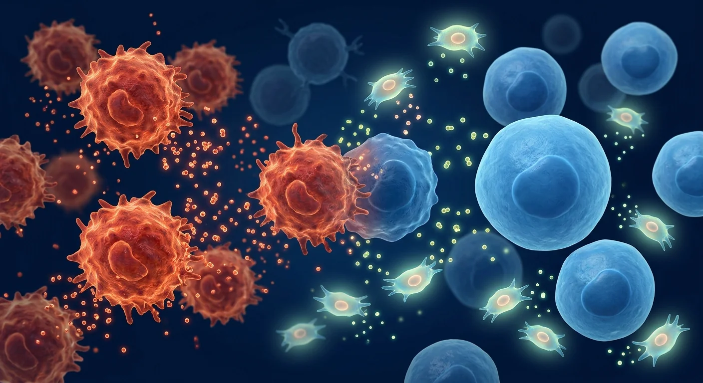
MSCs shift local macrophages from a destructive M1-like phenotype toward a restorative M2-like phenotype, and they modulate dendritic-cell maturation so that they present beta-cell antigens in a tolerogenic rather than inflammatory way. In a disease where the innate immune system is the engine of the autoimmune attack, re-educating the engine — rather than just suppressing it with broad immunosuppression — is an attractive theoretical advantage of MSCs over conventional immunosuppressants.
T-regulatory cell expansion
Autoimmune tolerance is maintained, in part, by regulatory T-cells (Tregs) that keep autoreactive T-cells in check. Type 1 diabetes is associated with reduced Treg function. Preclinical work shows that MSCs can expand and potentiate Tregs in vivo, which if it translates to humans would mean a more durable immune reset rather than a temporary suppression. This is one of the most actively studied and most promising directions in the field[7].
Beta-cell protection (not replacement)
MSC paracrine signalling — antioxidant, anti-apoptotic, and anti-inflammatory factors — can reduce beta-cell stress in early disease, where cells are surviving but under immune attack. The clinical goal is slowing further decline, not regenerating cells already destroyed. No published human trial demonstrates restoration of full insulin independence from MSC therapy, and the honest framing of current evidence is "protection and preservation," not "cure."
Microvascular and metabolic environment support
MSCs release angiogenic factors (VEGF, HGF) that may support microvascular health — relevant for patients developing early diabetic complications. There is also emerging preclinical evidence that MSCs can transfer healthy mitochondria to stressed cells via tunneling nanotubes, a mechanism with particular relevance to beta-cells whose metabolic workload is enormous. Both mechanisms are promising but still investigational in humans[8].
What the evidence supports — and what it doesn't
The published human evidence for MSC therapy in type 1 diabetes is still early. Most studies are small, single-arm, or uncontrolled case series, typically conducted in early or newly diagnosed disease. An honest summary:
What is plausible, and partially supported: Reduction in inflammatory markers (hsCRP, IL-6, TNF-α), modest improvement in beta-cell function markers (C-peptide, HOMA indices) in early disease, reduced glucose variability in some patients, and reduced insulin requirements in a minority of early-stage patients. Several small trials have reported safety and biologic plausibility, which is exactly the stage this research is at. A 2019 systematic review of MSC therapy in type 1 diabetes concluded that the data support continued investigation, not clinical adoption as a standard of care[9][10][11].
What is not supported by current human evidence: Cure of type 1 diabetes, freedom from insulin therapy, restoration of beta-cell mass in established long-standing disease, or guaranteed improvement in HbA1c. Any clinic implying these outcomes is overstating what the evidence supports. MSC therapy for type 1 diabetes is, as of 2026, an investigational adjunct — not a replacement for insulin, CGM, or the standard of care.
What remains under active investigation: Optimal cell source (Wharton's jelly vs bone marrow vs adipose), dose and dosing schedule, ideal timing in the disease course (newly diagnosed vs established), combination with other immunomodulatory strategies, and long-term durability of any immune effect. Larger, controlled, multi-centre trials are the critical next step, and their results will determine whether MSC therapy moves from "biologically plausible" to "clinically established."
The Honest Frame
MSC therapy for type 1 diabetes is, at present, an investigational adjunct — a possible addition to standard diabetes care, not a replacement for insulin. The most realistic near-term goals are: slowing immune-mediated beta-cell decline in early disease, reducing systemic low-grade inflammation, and improving the metabolic environment around the remaining beta-cells. Anyone selling a different promise is selling a different product than the science supports.
Who is the strongest candidate?
Within the honest boundaries of current evidence, the patients most likely to see meaningful response are those who:
- Are in the first 1–3 years of diagnosis, when a meaningful fraction of beta-cells may still be alive and the autoimmune process is active — the window where immunomodulation has the most to gain
- Retain measurable C-peptide — evidence that residual beta-cell function is still present and worth protecting
- Have suboptimal glucose variability (time-in-range below target, frequent lows or highs) despite good insulin therapy and CGM use — a marker that the metabolic environment is strained
- Have early signs of systemic inflammation or complications (elevated hsCRP, early microalbuminuria, early retinopathy) that may benefit from microvascular and anti-inflammatory support
- Are coordinating with their diabetes specialist — insulin dosing may need adjustment as the metabolic environment changes, and that decision belongs to the prescribing physician
- Have realistic expectations — understanding that the goal is protection and reduction of strain, not insulin independence
Patients with very long-standing type 1 diabetes (more than 10–15 years), no measurable C-peptide, or advanced complications are less likely to see a substantial change from MSC therapy alone. The immune damage to the islets is largely complete in that population, and the realistic role of MSC therapy shifts to supporting the broader metabolic and vascular environment — still potentially valuable, but with a different and more modest expectation.
What treatment looks like
A type 1 diabetes-focused MSC programme at a regulated clinical centre typically follows this structure:
- Comprehensive metabolic and immune assessment — HbA1c, C-peptide (fasting and stimulated), fasting glucose, time-in-range data from CGM, antibody panel (GAD65, IA-2, ZnT8), inflammatory markers (hsCRP, IL-6), kidney function, complication screening, current medications and insulin regimen
- Immunomodulation baseline — inflammatory cytokine profile and, where available, immune-cell subset analysis to establish a baseline for tracking MSC effects
- Coordination with the patient's diabetes specialist — MSC therapy supplements rather than replaces insulin therapy; any insulin dose adjustment is the prerogative of the prescribing physician and is done with awareness of the MSC cycle
- Personalised MSC protocol — typically intravenous infusion across a structured 8–12 week cycle, dose calibrated to body weight and indication, using clinical-grade Wharton's jelly–derived MSCs
- Lifestyle and metabolic protocol — nutrition, physical activity, and sleep guidance designed to reduce systemic inflammatory load and support the metabolic environment during the MSC cycle
- Outcome tracking — repeat C-peptide, HbA1c, inflammatory markers, and CGM-derived glucose variability metrics at 3, 6, and 12-month milestones
Realistic expectations
Outcomes vary substantially by disease duration, baseline beta-cell function, immune status, lifestyle adherence, MSC quality, and dosing protocol. Some patients in early disease see measurable improvement in C-peptide and inflammatory markers within 3–6 months; others build benefit gradually; a meaningful fraction may not respond at all. A reputable clinic discusses this variability honestly during the initial consultation, not after the patient has already paid.
Safety considerations specific to type 1 diabetes
Several considerations deserve attention in type 1 diabetes patients:
- Insulin dosing around the MSC cycle — if MSC therapy reduces the systemic inflammatory load and improves insulin sensitivity, insulin requirements may drop; this requires close coordination with the diabetes specialist and CGM monitoring to avoid hypoglycaemia
- Immune status — patients with concurrent autoimmune conditions or a history of immune dysregulation require careful protocol design; the goal is immunomodulation, not broad immunosuppression, and that distinction matters clinically
- Cardiovascular status — long-standing type 1 diabetes patients often have concurrent cardiovascular risk; medical clearance is part of every consultation
- Cell quality — clinical-grade, ISCT-verified MSCs with documented viability (>95% at delivery) and batch traceability, delivered under qualified medical supervision, is a non-negotiable foundation for any cell therapy in a chronic autoimmune disease
The right way to think about MSC therapy in type 1 diabetes is not "will this let me stop insulin?" — the biology does not support that today, and any clinic promising it is not being honest. The better question is: "Is there a measurable immune and metabolic improvement available to me, alongside the insulin and CGM that already form the core of my care?" For patients in early disease, the honest answer is increasingly yes — with appropriate expectations about what that improvement looks like.
— VELAR Clinical Team
The VELAR approach to type 1 diabetes
Type 1 diabetes protocols at VELAR Center are designed in coordination with each patient's existing diabetes team. Each programme uses clinical-grade Wharton's jelly–derived MSCs (≥95% identity verification, >95% viability at delivery, fresh and never-frozen) delivered via personalised intravenous infusion, paired with a structured lifestyle protocol and metabolic and inflammatory biomarker tracking at the 3, 6, and 12-month milestones.
If you are considering regenerative therapy as part of your type 1 diabetes care, the most useful first conversation is not about cells. It is about how long you have been on insulin, whether you retain measurable C-peptide, how your glucose variability looks over the last few months, and whether MSC therapy is a sensible addition to the care framework you already have. That conversation — honest, data-driven, in coordination with your existing team — is what we offer at VELAR.
Frequently Asked Questions
Can stem cell therapy cure type 1 diabetes?
No. As of 2026, no published human trial has demonstrated that MSC therapy can restore full insulin independence in type 1 diabetes. The realistic near-term goal is slowing immune-mediated beta-cell decline in early disease and reducing the systemic inflammatory load. Cures remain the domain of ongoing islet cell and immune-tolerance research, not current MSC therapy.
How much does stem cell therapy for type 1 diabetes cost in Bangkok?
Programme costs at a regulated Bangkok clinic vary by protocol length and personalisation, and are quoted during the initial consultation after a full metabolic and immune assessment. Any clinic that quotes a firm price before evaluating your specific case — disease duration, C-peptide status, complication profile — is prioritising sales over clinical judgement. The full cost context, including what a quality programme includes, is covered in our Thailand cost guide.
Is MSC therapy safe for people with type 1 diabetes?
Published MSC safety data across many indications — including SafeCell, a large meta-analysis — show a favourable safety profile when clinical-grade cells are used under medical supervision. The specific consideration in type 1 diabetes is insulin dosing: if MSC therapy improves insulin sensitivity, your insulin requirements may drop and need adjustment with your diabetes specialist. Cell quality and medical oversight are the two safety variables that matter most.
What cell source is best for type 1 diabetes?
Most published work in type 1 diabetes has used bone marrow–derived or Wharton's jelly–derived MSCs. Wharton's jelly MSCs are particularly attractive for this indication because they are allogeneic (available immediately without waiting for autologous processing), carry a low immunogenic profile, and can be delivered fresh. The optimal source for MSC therapy in type 1 diabetes is still an open research question; cell quality and processing standards matter more than the source label.
When should I consider MSC therapy relative to my diagnosis?
The evidence base is strongest for early disease — typically the first 1–3 years after diagnosis, when beta-cells are still present and the autoimmune process is active. The longer the disease has been established, the less islet tissue remains to protect, and the more the realistic goal shifts from beta-cell preservation to supporting the broader metabolic and vascular environment. Your diabetes specialist and C-peptide status are the best guides for timing.
Limitations and what we do not know
Several honest limitations should shape any decision about MSC therapy for type 1 diabetes:
- The human evidence base is still early. Most published data come from small, uncontrolled studies. The direction of the signal is consistent with the biology, but the magnitude and durability of clinical benefit in well-characterised populations is not yet established by large controlled trials.
- Response is not guaranteed. A meaningful fraction of patients may not respond, particularly in later-stage disease. No reliable biomarker currently predicts who will benefit before treatment begins.
- Optimal protocol parameters are not settled. Dose, dosing frequency, cell source, and timing are all still being optimised. Different clinics use meaningfully different protocols, and the differences are not yet validated against each other.
- Long-term immune effects are unknown. Whether the immune modulation MSCs produce is transient or durable is one of the most important open questions in the field, and the answer will only come from longer follow-up data.
- Cost and access. MSC therapy is not currently covered by standard insurance in most countries, and programme costs in Bangkok vary. Patients should approach pricing with the same scrutiny they apply to clinical credentials.
The responsible framing is this: MSC therapy for type 1 diabetes is a real, biologically grounded, and increasingly well-studied area of regenerative medicine — and it is not yet a standard of care. The patients who will get the most from it are those who approach it with that exact framing, coordinate it with their existing diabetes team, and hold their clinic to the same honesty they would hold a research trial to.
References
- Atkinson MA, Eisenbarth G, Michels AW. Type 1 diabetes. New England Journal of Medicine. 2014;371(24):2321-2332. doi:10.1056/NEJMra1314785 ↩
- Herold KC, Rothenberg SD, Faustner-Pirie V, et al. Beta-cell destruction in type 1 diabetes: the roles of autoimmunity, inflammation, and other pathogenic mechanisms. Endocrine Reviews. 2019;40(2):672-698. doi:10.1210/er.2018-00235 ↩
- Barker JM, Roep BO. Type 1 diabetes: a disease of T cell autoimmunity. Immunity. 2017;47(4):703-715. doi:10.1016/j.immuni.2017.10.006 ↩
- Gonzalez J, Linares ME, Gonzalez A, et al. Mesenchymal stem cell therapy in type 1 diabetes: a systematic review. Stem Cells Translational Medicine. 2018;7(8):621-632. doi:10.1002/sctm.17-0647 ↩
- Naji A, Eitoku M, Favier B, et al. Biological functions of mesenchymal stem cells and clinical implications. Cellular and Molecular Life Sciences. 2019;76(17):3323-3348. doi:10.1007/s00018-019-03125-1 ↩
- Liu G, Zhang Y, Zhang S, et al. Mesenchymal stem cell therapy for type 1 diabetes: a narrative review. Journal of Diabetes. 2021;13(4):315-326. doi:10.1111/1753-0407.12935 ↩
- Rabani MP, Ben-David U, Amit M. The effect of human mesenchymal stem cells on homeostatic and pathogenic T cell-mediated responses. Stem Cells. 2013;31(6):1253-1265. doi:10.1002/stem.1376 ↩
- Nainan A, Zhang C, Li W, et al. Mitochondrial transfer from mesenchymal stromal cells protects beta cells from glucose-induced apoptosis. Cellular Physiology and Biochemistry. 2017;44(3):919-930. doi:10.1159/000481709 ↩
- Ruan C, Zhao L, Yang H, et al. Mesenchymal stem cell therapy for type 1 diabetes: a systematic review and meta-analysis of clinical trials. Diabetes Therapy. 2019;10(5):1419-1435. doi:10.1007/s41541-019-0173-2 ↩
- Shimizu T, Tsuchida T, Nakano T, et al. Mesenchymal stem cell-based therapy for type 1 diabetes: current status and future directions. Islets. 2020;12(2):158-168. doi:10.1080/19382206.2020.1727716 ↩
- Cheng Y, Wang X, Li Y, et al. Wharton's jelly-derived mesenchymal stem cells in autoimmune diabetes: a preclinical and clinical perspective. Stem Cell Research & Therapy. 2021;12:545. doi:10.1186/s13287-021-02243-4 ↩
- Spees JL, Lee RH, Gregory CA. Mechanisms of mesenchymal stem/stromal cell function. Stem Cell Research & Therapy. 2016;7(1):125. doi:10.1186/s13287-016-0363-7 ↩
- Dominici M, Le Blanc K, Mueller I, et al. Minimal criteria for defining multipotent mesenchymal stromal cells. Cytotherapy. 2006;8(4):315-317. doi:10.1080/14653240600855905 ↩
- Lalu MM, McIntyre L, Pugliese C, et al. Safety of cell therapy with mesenchymal stromal cells (SafeCell): a systematic review and meta-analysis. PLoS One. 2012;7(10):e47559. doi:10.1371/journal.pone.0047559 ↩
- Caplan AI. Mesenchymal stem cells: time to change the name! Stem Cells Translational Medicine. 2017;6(6):1445-1451. doi:10.1002/sctm.17-0051 ↩
- Pittenger MF, Discher DE, Péault BM, et al. Mesenchymal stem cell perspective: cell biology to clinical progress. NPJ Regenerative Medicine. 2019;4:22. doi:10.1038/s41536-019-0083-6 ↩
参考文献
- Atkinson MA, Eisenbarth G, Michels AW. Type 1 diabetes. New England Journal of Medicine. 2014;371(24):2321-2332. doi:10.1056/NEJMra1314785 ↩
- Herold KC, Rothenberg SD, Faustner-Pirie V, et al. Beta-cell destruction in type 1 diabetes: the roles of autoimmunity, inflammation, and other pathogenic mechanisms. Endocrine Reviews. 2019;40(2):672-698. doi:10.1210/er.2018-00235 ↩
- Barker JM, Roep BO. Type 1 diabetes: a disease of T cell autoimmunity. Immunity. 2017;47(4):703-715. doi:10.1016/j.immuni.2017.10.006 ↩
- Gonzalez J, Linares ME, Gonzalez A, et al. Mesenchymal stem cell therapy in type 1 diabetes: a systematic review. Stem Cells Translational Medicine. 2018;7(8):621-632. doi:10.1002/sctm.17-0647 ↩
- Naji A, Eitoku M, Favier B, et al. Biological functions of mesenchymal stem cells and clinical implications. Cellular and Molecular Life Sciences. 2019;76(17):3323-3348. doi:10.1007/s00018-019-03125-1 ↩
- Liu G, Zhang Y, Zhang S, et al. Mesenchymal stem cell therapy for type 1 diabetes: a narrative review. Journal of Diabetes. 2021;13(4):315-326. doi:10.1111/1753-0407.12935 ↩
- Rabani MP, Ben-David U, Amit M. The effect of human mesenchymal stem cells on homeostatic and pathogenic T cell-mediated responses. Stem Cells. 2013;31(6):1253-1265. doi:10.1002/stem.1376 ↩
- Nainan A, Zhang C, Li W, et al. Mitochondrial transfer from mesenchymal stromal cells protects beta cells from glucose-induced apoptosis. Cellular Physiology and Biochemistry. 2017;44(3):919-930. doi:10.1159/000481709 ↩
- Ruan C, Zhao L, Yang H, et al. Mesenchymal stem cell therapy for type 1 diabetes: a systematic review and meta-analysis of clinical trials. Diabetes Therapy. 2019;10(5):1419-1435. doi:10.1007/s41541-019-0173-2 ↩
- Shimizu T, Tsuchida T, Nakano T, et al. Mesenchymal stem cell-based therapy for type 1 diabetes: current status and future directions. Islets. 2020;12(2):158-168. doi:10.1080/19382206.2020.1727716 ↩
- Cheng Y, Wang X, Li Y, et al. Wharton's jelly-derived mesenchymal stem cells in autoimmune diabetes: a preclinical and clinical perspective. Stem Cell Research & Therapy. 2021;12:545. doi:10.1186/s13287-021-02243-4 ↩
- Spees JL, Lee RH, Gregory CA. Mechanisms of mesenchymal stem/stromal cell function. Stem Cell Research & Therapy. 2016;7(1):125. doi:10.1186/s13287-016-0363-7 ↩
- Dominici M, Le Blanc K, Mueller I, et al. Minimal criteria for defining multipotent mesenchymal stromal cells. Cytotherapy. 2006;8(4):315-317. doi:10.1080/14653240600855905 ↩
- Lalu MM, McIntyre L, Pugliese C, et al. Safety of cell therapy with mesenchymal stromal cells (SafeCell): a systematic review and meta-analysis. PLoS One. 2012;7(10):e47559. doi:10.1371/journal.pone.0047559 ↩
- Caplan AI. Mesenchymal stem cells: time to change the name! Stem Cells Translational Medicine. 2017;6(6):1445-1451. doi:10.1002/sctm.17-0051 ↩
- Pittenger MF, Discher DE, Péault BM, et al. Mesenchymal stem cell perspective: cell biology to clinical progress. NPJ Regenerative Medicine. 2019;4:22. doi:10.1038/s41536-019-0083-6 ↩
1 型糖尿病通常被描述为缺乏胰岛素的疾病:免疫系统攻击并破坏胰腺 β 细胞,使身体无法再制造维持葡萄糖处于可生存范围所需的激素,终生胰岛素治疗成为唯一途径。这一描述是对的,但它描述的是结果,而非机制。机制是一种自身免疫过程,而正是机制决定了一切——疾病进展的速度、早期疾病中有多少 β 细胞功能得以保留、胰岛素敏感性保持得如何,以及免疫系统在胰腺之外造成多少损伤。
标准治疗的边界。胰岛素治疗在管理血糖这一"输出"方面非常有效,现代递送方式(胰岛素泵、连续葡萄糖监测、闭环系统)已彻底改变了 1 型糖尿病患者的日常生活。但胰岛素不能阻止免疫攻击。它不能保护仍存活的 β 细胞,也不能处理伴随许多长期 1 型糖尿病患者的全身性低度炎症。这正是间充质干细胞(MSC)疗法正在被研究的空间。
组织层面的问题。在 1 型糖尿病中,自身免疫细胞——主要是失去对胰岛素产生组织耐受性的自反应 T 淋巴细胞,由失调的巨噬细胞与树突细胞协助——渗入胰岛,通过炎症细胞因子与直接细胞毒性作用杀死 β 细胞。在疾病早期,仍有相当比例的 β 细胞可能处于应激而非死亡状态,它们的持续功能可在数年内减轻血糖波动。任何能减缓这一过程的疗法都具有真正的临床价值——即使它永远无法恢复完全的胰岛素独立。
MSC 如何针对它。间充质干细胞是免疫环境的"专业调节者"。它们感知受损组织中的炎症微环境,并释放能重新教育局部免疫细胞、使其脱离破坏性表型的因子。在一种恰恰由这种破坏性局部免疫所定义的疾病中,这一特性是研究它们的最具生物学合理性的理由。以下是证据现状的诚实图谱。
1 型糖尿病在生物学上究竟是什么
1 型糖尿病是一种自身免疫对胰腺 β 细胞的破坏,由对胰岛素产生组织的免疫耐受丧失所驱动——这一过程在出现首个高血糖症状前的数月乃至数年间缓慢展开[1][2][3]。关键生物学参与者包括:
- 自反应 T 细胞——CD8+ 细胞毒性与 CD4+ 辅助 T 细胞失去对 β 细胞抗原(胰岛素、GAD65、IA-2、ZnT8)的耐受,并渗入胰岛
- 先天免疫失调——胰岛环境中的巨噬细胞与树突细胞偏向促炎(M1 样)状态,放大 T 细胞攻击并维持局部细胞因子产生(IFN-γ、IL-1β、TNF-α)
- 胰岛炎症——局部炎症级联导致 β 细胞应激、凋亡,最终胰岛被破坏;该破坏速度决定了临床诊断时丧失多少功能
- 全身免疫失调——许多长期 1 型糖尿病患者带有更广泛的自身免疫倾向,胰岛特异性炎症是随时间推移促成并发症的更广泛免疫失调模式的一部分
对任何考虑再生疗法的人而言,一个重要的细节:并非所有 1 型糖尿病都以相同速度进展。新诊断患者中有相当一部分在数年内仍保留可测量的 C 肽(残余胰岛素产生),其 β 细胞处于应激而非完全被破坏的状态。这一亚群——早期疾病、部分 β 细胞存活——正是免疫调节疗法获益最多的地方。在已建立的长期疾病中,靶组织大多已消失,现实目标转向支持其周围的代谢与免疫环境。
MSC 可能在生物学上贡献什么
在 1 型糖尿病中,MSC 并非"β 细胞替代"疗法——任何暗示如此的诊所都在过度销售。已发表研究提示数个在临床前与早期临床工作中部分验证的合理机制:
胰岛环境的抗炎再校准
这是支持最充分的机制。MSC 释放抗炎介质——TSG-6、PGE2、IDO、IL-10 等——抑制驱动胰岛破坏的促炎细胞因子级联。在自身免疫糖尿病的动物模型中,MSC 输注已被证明可减少胰岛炎症细胞浸润并减缓 β 细胞丢失速度。临床含义:在疾病早期,MSC 疗法可能保护标准胰岛素治疗无法保护的 β 细胞。该效应在人体中的幅度与持久性仍在积极研究中[4][5][6]。
巨噬细胞与树突细胞再教育
MSC 将局部巨噬细胞从破坏性 M1 样表型转向修复性 M2 样表型,并调节树突细胞成熟,使其以耐受性而非炎症性的方式呈现 β 细胞抗原。在先天免疫系统是自身免疫攻击"引擎"的疾病中,"再教育引擎"而非用广谱免疫抑制"压制引擎",是 MSC 相比传统免疫抑制剂的一个吸引人的理论优势。
调节性 T 细胞扩增
自身免疫耐受在部分程度上由调节性 T 细胞(Treg)维持,它们抑制自反应 T 细胞。1 型糖尿病与 Treg 功能降低相关。临床前研究表明 MSC 可在体内扩增并增强 Treg——若能在人体中实现,这意味着更持久的免疫重置,而非暂时性抑制。这是该领域研究最活跃、也最有希望的方向之一[7]。
β 细胞保护(非替代)
MSC 的旁分泌信号——抗氧化、抗凋亡与抗炎因子——可减少早期疾病中 β 细胞的应激,此时细胞在免疫攻击下仍在存活。临床目标是减缓进一步衰退,而非再生已被破坏的细胞。没有任何已发表的人体试验证明 MSC 疗法能恢复完全的胰岛素独立,当前证据的诚实框架是"保护与保留",而非"治愈"。
微血管与代谢环境支持
MSC 释放促血管生成因子(VEGF、HGF),可能支持微血管健康——对开始发展早期糖尿病并发症的患者尤为重要。还有新兴临床前证据显示 MSC 可通过隧道纳米管向应激细胞转移健康线粒体——这一机制对代谢负荷巨大的 β 细胞具有特殊相关性。两种机制在人体中仍有前景但均属研究性阶段[8]。
证据支持什么——以及不支持什么
关于 MSC 疗法在 1 型糖尿病中的人体证据仍处于早期。多数研究规模小、单臂或非对照病例系列,通常在早期或新诊断疾病中进行。诚实的总结:
有合理性且部分得到支持的:炎症标志物(hsCRP、IL-6、TNF-α)下降、早期疾病中 β 细胞功能标志物(C 肽、HOMA 指数)的适度改善、部分患者血糖波动减少,以及少数早期阶段患者胰岛素需求降低。多项小型试验已报告了安全性与生物学合理性——这正是该研究目前所处的阶段。一项 2019 年关于 1 型糖尿病 MSC 疗法的系统综述得出结论:数据支持继续研究,而非作为标准护理被临床采纳[9][10][11]。
当前人体证据不支持的:1 型糖尿病的治愈、摆脱胰岛素治疗、在已建立的长期疾病中恢复 β 细胞质量,或保证 HbA1c 改善。任何暗示这些结果的诊所都过度声明了证据所支持的。截至 2026 年,MSC 疗法对 1 型糖尿病仍是一项探索性辅助治疗——不是胰岛素、CGM 或标准护理的替代。
仍处于积极研究中的:最佳细胞来源(华通氏胶 vs 骨髓 vs 脂肪)、剂量与给药方案、疾病进程中的理想时机(新诊断 vs 已建立)、与其他免疫调节策略的联合,以及任何免疫效应的长期持久性。更大规模的、有对照的、多中心试验是关键下一步,其结果将决定 MSC 疗法能否从"生物学合理"走向"临床确立"。
诚实的框架
目前 1 型糖尿病的 MSC 疗法是一项探索性辅助治疗——可能加入标准糖尿病护理的补充,而非胰岛素的替代。近期最现实的目标是:减缓早期疾病中免疫介导的 β 细胞衰退、降低全身低度炎症,并改善剩余 β 细胞周围的代谢环境。任何销售不同承诺的机构,销售的都不是科学所支持的产品。
谁是最强的候选人?
在当前证据的诚实边界内,最有可能看到有意义反应的患者是:
- 处于诊断后 1–3 年内——此时仍有相当比例的 β 细胞存活且自身免疫过程活跃,是免疫调节获益最大的窗口
- 保留可测量的 C 肽——残余 β 细胞功能仍存在、值得保护的证据
- 血糖波动欠佳(目标范围内时间低于靶值、频繁低血糖或高血糖)尽管胰岛素治疗与 CGM 使用良好——代谢环境紧张的标志
- 有全身炎症或早期并发症迹象(hsCRP 升高、早期微量白蛋白尿、早期视网膜病变)可能受益于微血管与抗炎支持
- 正与其糖尿病专科医师协调——胰岛素剂量可能需要随代谢环境变化而调整,这一决定属于处方医师
- 期望现实——理解目标是保护与减轻负担,而非胰岛素独立
1 型糖尿病病史很长(超过 10–15 年)、无可测量 C 肽或并发症已进展的患者,较少可能仅从 MSC 疗法看到实质性变化。该人群的胰岛免疫损伤大体已完成,MSC 疗法的现实角色转向支持更广泛的代谢与血管环境——仍可能有价值,但期望更为有限。
治疗的样子
受监管临床中心针对 1 型糖尿病的 MSC 项目通常遵循该结构:
- 全面代谢与免疫评估——HbA1c、C 肽(空腹与刺激后)、空腹血糖、CGM 目标范围内时间数据、抗体谱(GAD65、IA-2、ZnT8)、炎症标志物(hsCRP、IL-6)、肾功能、并发症筛查、当前用药与胰岛素方案
- 免疫调节基线——炎症细胞因子谱以及(如可获得)免疫细胞亚群分析,为追踪 MSC 效应建立基线
- 与患者的糖尿病专科医师协调——MSC 疗法补充而非替代胰岛素治疗;任何胰岛素剂量调整由处方医师决定,并知悉 MSC 周期
- 个性化 MSC 方案——通常在结构化的 8–12 周周期内静脉输注,剂量按体重与适应症校准,使用临床级华通氏胶来源 MSC
- 生活方式与代谢方案——营养、体力活动与睡眠指导,旨在降低全身炎症负荷并在 MSC 周期期间支持代谢环境
- 疗效追踪——重复 C 肽、HbA1c、炎症标志物及 CGM 导出的血糖波动指标,于 3、6 与 12 个月里程碑
实际预期
疗效因疾病持续时间、基线 β 细胞功能、免疫状态、生活方式依从性、MSC 质量与给药方案而显著不同。早期疾病的部分患者在 3–6 个月内看到 C 肽与炎症标志物的可测量改善;其他人逐渐建立受益;相当比例可能完全无反应。信誉良好的诊所在初次咨询期间诚实地讨论这一可变性,而不是在患者付费之后。
1 型糖尿病特有的安全性考量
1 型糖尿病患者中有几项考量值得关注:
- MSC 周期前后的胰岛素剂量——若 MSC 疗法降低全身炎症负荷并改善胰岛素敏感性,胰岛素需求可能下降;这需要与糖尿病专科医师密切协调并进行 CGM 监测以避免低血糖
- 免疫状态——合并自身免疫疾病或有免疫失调病史的患者需要仔细的方案设计;目标是免疫调节而非广谱免疫抑制,这一区别在临床上很重要
- 心血管状态——长期 1 型糖尿病患者常合并心血管风险;医疗评估是每次咨询的一部分
- 细胞质量——在合格医疗监督下递送的临床级、ISCT 验证 MSC(交付时活性 >95%、批次可追溯),是慢性自身免疫疾病中任何细胞治疗不可妥协的基础
思考 1 型糖尿病中 MSC 疗法的正确方式不是"这能让我停胰岛素吗?"——生物学今天不支持这一点,任何承诺如此的诊所都不诚实。更好的问题是:"在已构成我护理核心的胰岛素与 CGM 之外,是否有可测量的免疫与代谢改善可获得?"对早期疾病患者,诚实的答案越来越是肯定的——同时要对这种改善的样子持有适当的期望。
——VELAR 临床团队
VELAR 的 1 型糖尿病方法
VELAR Center 的 1 型糖尿病方案与每位患者现有的糖尿病团队协调设计。每个项目使用临床级华通氏胶来源 MSC(身份验证 ≥95%、交付时活性 >95%、新鲜从未冷冻)通过个性化静脉输注递送,搭配结构化的生活方式方案与在 3、6 与 12 个月里程碑的代谢与炎症生物标志物追踪。
如果您正在考虑作为 1 型糖尿病护理一部分的再生疗法,最有用的第一次对话不是关于细胞。是关于您使用胰岛素多久了、是否保留可测量的 C 肽、过去几个月的血糖波动情况,以及 MSC 疗法是否是您已有护理框架的合理补充。那种对话——诚实的、数据驱动的、与您现有团队协调的——就是我们在 VELAR 提供的。
常见问题
干细胞疗法能治愈 1 型糖尿病吗?
不能。截至 2026 年,没有任何已发表的人体试验证明 MSC 疗法能在 1 型糖尿病中恢复完全的胰岛素独立。近期最现实的目标是减缓早期疾病中免疫介导的 β 细胞衰退,并降低全身炎症负荷。治愈仍属于正在进行的胰岛细胞与免疫耐受研究领域,而非当前 MSC 疗法。
在曼谷,1 型糖尿病干细胞疗法费用是多少?
曼谷受监管诊所的项目费用因方案长度与个性化程度而异,并在完整的代谢与免疫评估后的初次咨询中报价。任何在评估您的具体情况——疾病持续时间、C 肽状态、并发症谱——之前就报出固定价格的诊所,都是在把销售置于临床判断之上。完整费用背景(包括高质量项目包含的内容)见我们的泰国费用指南。
MSC 疗法对 1 型糖尿病患者安全吗?
跨多种适应症发表的 MSC 安全性数据——包括 SafeCell 大型荟萃分析——显示,在医疗监督下使用临床级细胞时安全性良好。1 型糖尿病中的具体考量是胰岛素剂量:若 MSC 疗法改善胰岛素敏感性,您的胰岛素需求可能下降并需要与糖尿病专科医师共同调整。细胞质量与医疗监督是两个最重要的安全变量。
哪种细胞来源最适合 1 型糖尿病?
1 型糖尿病中已发表的大多数研究使用骨髓来源或华通氏胶来源 MSC。华通氏胶 MSC 对该适应症特别有吸引力,因为它们是同种异体的(可立即使用,无需等待自体处理)、免疫原性低,且可以新鲜状态递送。1 型糖尿病 MSC 疗法的最优来源仍是一个开放的研究问题;细胞质量与处理标准比来源标签更重要。
相对诊断时间,我什么时候应考虑 MSC 疗法?
证据基础在早期疾病最强——通常是诊断后 1–3 年,此时 β 细胞仍存在且自身免疫过程活跃。疾病建立得越久,可保护的胰岛组织越少,现实目标越从 β 细胞保护转向支持更广泛的代谢与血管环境。您的糖尿病专科医师与 C 肽状态是判断时机最好的依据。
局限性与我们尚不知道什么
若干诚实的局限应塑造任何关于 1 型糖尿病 MSC 疗法的决定:
- 人体证据基础仍处于早期。多数已发表数据来自小型、非对照研究。信号方向与生物学一致,但在特征明确的群体中临床获益的幅度与持久性尚未由大型对照试验确立。
- 反应并非有保证。相当比例的患者可能无反应,尤其在疾病后期。目前没有可靠的生物标志物能在治疗前预测谁将受益。
- 最优方案参数尚未确定。剂量、给药频率、细胞来源与时机都在优化中。不同诊所使用显著不同的方案,且这些差异尚未经过相互验证。
- 长期免疫效应未知。MSC 产生的免疫调节是短暂还是持久,是该领域最重要的未解问题之一,答案只能来自更长的随访数据。
- 费用与可及性。MSC 疗法目前在多数国家未获标准保险覆盖,曼谷项目费用不一。患者应对价格的审查力度,与对临床资质的审查力度相同。
负责任的框架是:1 型糖尿病的 MSC 疗法是一个真实的、有生物学基础的、研究日益深入的再生医学领域——但它尚未成为标准护理。从中获益最多的患者,正是以这一框架接近它、与现有糖尿病团队协调、并要求其诊所达到与要求研究试验相同诚实程度的人。
يُعرَض داء السكري من النوع 1 عادةً كمرض نقص في الأنسولين: يهاجم الجهاز المناعي خلايا بيتا في البنكرياس ويدمرها، فلا يستطيع الجسم إنتاج الهرمون الذي يحتاجه، ويصبح العلاج بالأنسولين مدى الحياة الطريقة الوحيدة للحفاظ على الجلوكوز ضمن النطاق الحيوي. هذا الوصف صحيح، لكنه يصف النتيجة لا الآلية. الآلية هي عملية مناعية ذاتية، وهي الآلية التي تحدد كل ما يتبعها — سرعة تقدم المرض، مقدار وظيفة خلايا بيتا الذي يبقى محفوظا في المرض المبكر، كيف يُحافَظ على حساسية الأنسولين، وكم من ضرر يسببه الجهاز المناعي خارج البنكرياس.
أين تتوقف الرعاية القياسية. علاج الأنسولين فعّال جداً في إدارة الناتج — أرقام الجلوكوز — وقد غيّرت أنظمة التوصيل الحديثة (مضخات الأنسولين، مراقبة الجلوكوز المستمرة، الأنظمة المغلقة) الحياة اليومية لمرضى النوع 1 تغييراً جذرياً. لكن الأنسولين لا يوقف الهجوم المناعي. ولا يحمي خلايا بيتا ما زالت حية، ولا يعالج الالتهاب منخفض الدرجة الجهازية الذي يسير بجانب كثير من مرضى النوع 1 طويل الأمد. هذا هو المجال الذي يُدرَس فيه الآن علاج الخلايا الجذعية الوسيطة.
المشكلة على مستوى النسيج. في النوع 1، تغزو خلايا مناعية ذاتية — وعلى رأسها خلايا T الذاتية التي فقدت تسامحها تجاه نسيج إنتاج الأنسولين بمساعدة خلايا بلعمية وخلايا شجرية متفلتة — جزر لانغرهانس وتقتل خلايا بيتا عبر سيتوكينات التهابية وسمية خلوية مباشرة. في المرض المبكر، قد تبقى نسبة معنوية من خلايا بيتا حية تحت الإجهاد بدل أن تموت، ويمكن لوظيفتها المتواصلة أن تخفف تقلبات الجلوكوز لسنوات. أي علاج يبطئ هذه العملية له قيمة سريرية حقيقية، حتى لو لم يستعِد استقلال الأنسولين كاملاً.
كيف تستهدف MSC الآلية. الخلايا الجذعية الوسيطة منظمات محترفة للبيئة المناعية. تستشعر وسط الالتهاب في النسيج المصاب وتطلق عوامل تعيد تعليم الخلايا المناعية المحلية بعيداً عن النمط المدمر. في مرض تعينه بالضبط هذه المناعة المحلية المدمرة، هذا هو السبب الأكثر قابلية بيولوجياً لدراسة الخلايا. ما يلي خريطة صادقة لموقع الأدلة حالياً.
ما هو داء السكري من النوع 1 بيولوجياً
النوع 1 هو تدمير مناعي ذاتي لخلايا بيتا في البنكرياس تدفعه فقدان التسامح المناعي تجاه نسيج إنتاج الأنسولين — عملية تتدرج خلال شهور إلى سنوات قبل ظهور أول أعراض ارتفاع الجلوكوز[1][2][3]. اللاعبون البيولوجيون الرئيسيون هم:
- خلايا T ذاتية — خلايا T سامة CD8+ ومساعدة CD4+ فقدت تسامحها تجاه مستضدات خلايا بيتا (الأنسولين، GAD65، IA-2، ZnT8) وتغزو الجزر
- خلل المناعة الفطرية — الخلايا البلعمية والخلايا الشجرية في بيئة الجزر تميل إلى حالة التهابية (M1-like)، فتضخم هجوم خلايا T وتحافظ على إنتاج السيتوكينات المحلية (IFN-γ، IL-1β، TNF-α)
- الالتهاب الجزيري — الشلال الالتهابي المحلي يسبب إجهاد خلايا بيتا واستماتتها ثم تدمير الجزر؛ سرعة هذا التدمير تحدد مقدار الوظيفة المفقودة عند التشخيص
- خلل مناعي جهازية — كثير من مرضى النوع 1 طويل الأمد يحملون ميلاً مناعياً ذاتياً أوسع، والالتهاب المحدد بالجزر جزء من نمط خلل مناعي أوسع يساهم في المضاعفات مع الوقت
تفصيل مهم لكل من يدرس العلاج التجديدي: ليس كل النوع 1 يتقدم بنفس السرعة. مجموعة من المرضى الجدد التشخيص تحتفظ ببيتا-سماك (C-peptide) قابل للقياس — إنتاج متبقٍ للأنسولين — لسنوات، وخلايا بيتا لديهم متوترة لا مدمرة كلياً. هذه المجموعة — مرض مبكر، بقاء جزئي لخلايا بيتا — هي التي تحقق أكبر فائدة من العلاج المناعي المعدل. في المرض المستقر طويل الأمد، النسيج المستهدف اختفى في جله، ويتحول الهدف الواقعي إلى دعم البيئة الأيضية والمناعية حوله.
ما يمكن أن تساهم به الخلايا الجذعية وسيطياً
MSC ليست علاج "استبدال خلايا بيتا" في النوع 1، وأي عيادة تلمح إلى خلاف ذلك تبالغ في البيع. تقيّم الأبحاث المنشورة عدة آليات معقولة ومحققة جزئياً في العمل قبل السريري والمبكر السريري:
إعادة معايرة مضادة للالتهاب في بيئة الجزر
هذه هي الآلية المدعومة بأفضل أساس. تطلق MSC وسطاء مضادين للالتهاب — TSG-6، PGE2، IDO، IL-10 وغيرها — تكبح الشلال السيتوكيني الالتهابي الذي يقود تدمير الجزر. في نماذج حيوانية للسكري المناعي الذاتي، ثبت أن إعطاء MSC يقلل من تغلغل الخلايا الالتهابية في الجزر ويبطئ معدل فقدان خلايا بيتا. الدلالة السريرية: في المرض المبكر، قد يحمي علاج MSC خلايا بيتا لا يحميها علاج الأنسولين القياسي. مدى هذا التأثير في الإنسان ومتانته ما زالا قيد الدراسة النشطة[4][5][6].
إعادة تعليم الخلايا البلعمية والخلايا الشجرية
تحول MSC الخلايا البلعمية المحلية من نمط M1 المدمر إلى نمط M2 المصلح، وتنضج الخلايا الشجرية بشكل يعرض مستضدات خلايا بيتا بطريقة تسامحية لا التهابية. في مرض تكون فيه المناعة الفطرية محرك الهجوم المناعي الذاتي، "إعادة تعليم المحرك" بدل كبحه بكبت مناعي واسع هي ميزة نظرية جذابة لـ MSC مقارنة بمثبطات المناعة التقليدية.
توسيع خلايا T التنظيمية
يُحافَظ على التسامح المناعي الذاتي جزئياً عبر خلايا T التنظيمية (Tregs) التي تكبح الخلايا الذاتية. يرتبط النوع 1 بانخفاض وظيفة Tregs. أظهر العمل قبل السريري أن MSC يمكن أن توسّع وتفعيل خلايا Tregs في الجسم الحي — وإن تحققت هذه النتيجة في الإنسان، فهي تعني إعادة ضبط مناعية أكثر ديمومة بدل كبت مؤقت. هذا من أكثر الاتجاهات نشاطاً وأكثرها وعداً في هذا المجال[7].
حماية خلايا بيتا (لا استبدال)
الإشارات شبه الإفرازية من MSC — مضادات الأكسدة، مضادات الاستماتة، مضادات الالتهاب — تقلل من إجهاد خلايا بيتا في المرض المبكر حين تكون الخلايا حية تحت الهجوم المناعي. الهدف السريري هو إبطاء مزيد من التدهور، لا تجديد الخلايا المدمرة بالفعل. لم تُظهِر أي تجربة بشرية منشورة استعادة استقلال الأنسولين كاملاً من علاج MSC، والإطار الصادق للأدلة الحالية هو "الحماية والحفظ" لا "الشفاء".
دعم البيئة الوعائية الدقيقة والأيضية
تطلق MSC عوامل توليد الأوعية (VEGF، HGF) قد تدعم صحة الأوعية الدقيقة — ذات صلة بالمرضى الذين تبدأ لديهم مضاعفات سكرية مبكرة. وهناك أدلة قبل سريرية ناشئة تُظهر أن MSC يمكنها نقل ميتوكوندريا صحية إلى خلايا متوترة عبر أنابيب نانوية نفقية، آلية ذات صلة خاصة بخلايا بيتا التي تحمل حمولة أيضية هائلة. الآليتان واعدتان لكن لا تزالان استقصائيتين في الإنسان[8].
ما تدعمه الأدلة — وما لا تدعمه
الأدلة البشرية المنشورة لعلاج MSC في النوع 1 لا تزال في مرحلة مبكرة. معظم الدراسات صغيرة، ذراع وحيدة، أو سلسلة حالات غير مضبوطة، تُجرى عادة في المرض المبكر أو التشخيص الجديد. ملخص صادق:
ما هو معقول ومدعوم جزئياً: انخفاض في علامات الالتهاب (hsCRP، IL-6، TNF-α)، تحسن متواضع في مؤشرات وظيفة خلايا بيتا (بيتا-سماك، مؤشرات HOMA) في المرض المبكر، انخفاض تقلبات الجلوكوز لدى بعض المرضى، وانخفاض متطلبات الأنسولين لدى أقلية من المرضى في المراحل المبكرة. سجلت عدة تجارب صغيرة السلامة وقابلية الفرض البيولوجي — وهي بالضبط المرحلة التي بلغها هذا البحث. مراجعة منهجية 2019 لعلاج MSC في النوع 1 خلصت إلى أن البيانات تدعم مواصلة البحث، لا التبنّي السريري كمعيار رعاية[9][10][11].
ما لا تدعمه الأدلة البشرية الحالية: شفاء النوع 1، التحرر من العلاج بالأنسولين، استعادة كتلة خلايا بيتا في المرض المستقر طويل الأمد، أو ضمان تحسن HbA1c. أي عيادة تلمح إلى هذه النتائج تبالغ عمّا تدعمه الأدلة. علاج MSC للنوع 1، حتى 2026، إضافة استقصائية — ليست بديلاً عن الأنسولين أو مراقبة الجلوكوز المستمرة أو معيار الرعاية.
ما يبقى قيد البحث النشط: مصدر الخلايا الأمثل (هلام وارتون مقابل النخاع العظمي مقابل الدهون)، الجرعة وجدول الجرعات، التوقيت الأمثل في مسار المرض (تشخيص جديد مقابل مستقر)، الجمع مع استراتيجيات تعديل مناعي أخرى، والمتانة الطويلة الأمد لأي أثر مناعي. التجارب الكبرى المضبوطة متعددة المراكز هي الخطوة الحاسمة التالية، وستحدد نتائجها ما إذا كان علاج MSC ينتقل من "معقول بيولوجياً" إلى "مستقر سريرياً".
الإطار الصادق
علاج MSC للنوع 1، في الوقت الحالي، إضافة استقصائية — احتمال إضافي لرعاية السكري القياسية، لا بديل عن الأنسولين. الأهداف الواقعية القريبة المدى هي: إبطاء التدهور المناعي لخلايا بيتا في المرض المبكر، خفض الالتهاب الجهازية منخفض الدرجة، وتحسين البيئة الأيضية حول خلايا بيتا المتبقية. من يبيع وعداً مختلفاً يبيع منتجاً مختلفاً عمّا تدعمه العلوم.
من هو المرشح الأقوى؟
ضمن الحدود الصادقة للأدلة الحالية، المرضى الأرجح أن يروا استجابة ذات معنى هم من:
- في السنة الأولى إلى الثالثة من التشخيص — حين تكون نسبة معنوية من خلايا بيتا لا تزال حية والعملية المناعية الذاتية نشطة، النافذة التي تحقق فيها المناعة المعدلة أكبر نفع
- لديهم بيتا-سماك قابل للقياس — دليل أن وظيفة خلايا بيتا المتبقية لا تزال حاضرة وتستحق الحماية
- تقلبات جلوكوز دون الأمثل (زمن ضمن النطاق دون الهدف، هبوط أو ارتفاع متكرر) رغم جودة العلاج بالأنسولين واستخدام مراقبة الجلوكوز المستمرة — علامة على أن البيئة الأيضية متوترة
- علامات مبكرة للالتهاب الجهازية أو المضاعفات (hsCRP مرتفع، بيلة ألبومينية دقيقة مبكرة، اعتلال شبكي مبكر) قد تستفيد من الدعم الوعائي الدقيق والمضاد للالتهاب
- ينسقون مع أخصائي السكري لديهم — قد تحتاج جرعات الأنسولين إلى تعديل مع تغيّر البيئة الأيضية، وهذا القرار من صلاحية الطبيب الموصف
- لديهم توقعات واقعية — يفهمون أن الهدف هو الحماية وتخفيف الحمل، لا استقلال الأنسولين
المرضى ذوو النوع 1 طويل الأمد جداً (أكثر من 10–15 عاماً)، أو بلا بيتا-سماك قابل للقياس، أو بمضاعفات متقدمة، أقل احتمالا لرؤية تغيير جوهري من علاج MSC وحده. الضرر المناعي للجزر مكتمل في جله لدى هذه المجموعة، ويتحول الدور الواقعي لعلاج MSC إلى دعم البيئة الأيضية والوعائية الأوسع — قد يكون لا يزال ذا قيمة، لكن بتوقع مختلف وأكثر تواضعاً.
كيف يبدو العلاج
برنامج MSC متمحور حول النوع 1 في مركز سريري منظم يتبع عادة هذا الهيكل:
- تقييم أيضي ومناعي شامل — HbA1c، بيتا-سماك (صيام ومحرض)، جلوكوز الصيام، بيانات زمن ضمن النطاق من مراقبة الجلوكوز المستمرة، لوحة الأجسام (GAD65، IA-2، ZnT8)، علامات الالتهاب (hsCRP، IL-6)، وظيفة الكلى، فحص المضاعفات، الأدوية الحالية ونظام الأنسولين
- خط أساس لتعديل المناعة — لوحة السيتوكينات الالتهابية وحيث يتوفر تحليل مجموعات الخلايا المناعية، لتأسيس خط أساس لتتبع أثر MSC
- التنسيق مع أخصائي السكري لدى المريض — يكمل علاج MSC علاج الأنسولين لا يحل محله؛ أي تعديل في جرعة الأنسولين من صلاحية الطبيب الموصف ويُجرى مع معرفة دورة MSC
- بروتوكول MSC شخصي — عادة تسريب وريدي على مدى دورة منظمة من 8–12 أسبوعاً، الجرعة معايرة لوزن الجسم والدلالة، باستخدام MSC سريرية مشتقة من هلام وارتون
- بروتوكول نمط حياة وأيض — توجيه تغذية ونشاط بدني ونوم مصمم لخفض حمل الالتهاب الجهازية ودعم البيئة الأيضية خلال دورة MSC
- تتبع النتائج — تكرار بيتا-سماك وHbA1c وعلامات الالتهاب ومقاييس تقلب الجلوكوز المشتقة من مراقبة الجلوكوز المستمرة في علامات 3 و6 و12 شهراً
التوقعات الواقعية
تختلف النتائج اختلافاً جوهرياً حسب مدة المرض، وظيفة خلايا بيتا الأساسية، الحالة المناعية، الالتزام بنمط الحياة، جودة MSC، وبروتوكول الجرعات. يرى بعض المرضى في المرض المبكر تحسناً قابلاً للقياس في بيتا-سماك وعلامات الالتهاب خلال 3–6 أشهر؛ يبني آخرون الفائدة تدريجياً؛ نسبة معنوية قد لا تستجيب على الإطلاق. تناقش العيادة ذات السمعة الجيدة هذا التباين بصدق أثناء الاستشارة الأولى، لا بعد دفع المريض.
اعتبارات السلامة الخاصة بالنوع 1
عدة اعتبارات تستحق الاهتمام لدى مرضى النوع 1:
- جرعات الأنسولين حول دورة MSC — إذا خفّض علاج MSC حمل الالتهاب الجهازية وحسّن حساسية الأنسولين، فقد تنخفض متطلبات الأنسولين؛ يتطلب ذلك تنسيقاً وثيقاً مع أخصائي السكري ومراقبة عبر جهاز المراقبة المستمرة لتجنب هبوط السكر
- الحالة المناعية — المرضى ذوو أمراض مناعية ذاتية مصاحبة أو تاريخ خلل مناعي يحتاجون تصميم بروتوكول دقيق؛ الهدف هو تعديل المناعة لا كبتها كلياً، وهذا التمييز مهم سريرياً
- الحالة القلبية الوعائية — كثير من مرضى النوع 1 طويل الأمد يحملون خطراً قلبية وعائياً مصاحبا؛ التصريح الطبي جزء من كل استشارة
- جودة الخلايا — MSC ذات الجودة السريرية المعتمدة من ISCT مع حيوية موثقة (>95% عند التسليم) وتتبع دفعات، تُسلَّم تحت إشراف طبي مؤهل، أساس غير قابل للتفاوض لأي علاج خلوي في مرض مناعي ذاتي مزمن
الطريقة الصحيحة للتفكير في علاج MSC في النوع 1 ليست "هل سيخولني بالتوقف عن الأنسولين؟" — البيولوجيا لا تدعم ذلك اليوم، وأي عيادة تعد به ليست صادقة. السؤال الأفضل: "هل هناك تحسن مناعي وأيضي قابل للقياس متاح لي، إلى جانب الأنسولين ومراقبة الجلوكوز المستمرة التي تشكل بالفعل جوهر رعايتي؟" بالنسبة لمرضى المراحل المبكرة، الإجابة الصادقة متزايدة العمق نحو نعم — مع توقعات مناسبة عن شكل هذا التحسن.
— الفريق السريري في VELAR
منهج VELAR في داء السكري من النوع 1
تُصمَّم بروتوكولات النوع 1 في VELAR Center بالتنسيق مع فريق السكري الحالي لكل مريض. يستخدم كل برنامج MSC سريرية مشتقة من هلام وارتون (تحقق من الهوية ≥95٪، حيوية عند التسليم >95٪، طازجة وغير مجمدة أبداً) تُسلَّم عبر تسريب وريدي شخصي، مقترنة ببروتوكول نمط حياة منظم وتتبع المؤشرات الحيوية الأيضية والالتهابية في علامات 3 و6 و12 شهراً.
إذا كنت تفكر في العلاج التجديدي كجزء من رعاية النوع 1 الخاصة بك، فإن أكثر محادثة أولى مفيدة ليست عن الخلايا. إنها عن مدة استخدامك للأنسولين، عما إذا كنت تحتفظ ببيتا-سماك قابل للقياس، كيف تبدو تقلبات الجلوكوز خلال الأشهر الماضية، وما إذا كان علاج MSC إضافة معقولة لإطار الرعاية الذي لديك بالفعل. هذه المحادثة — صادقة، مدفوعة بالبيانات، بالتنسيق مع فريقك الحالي — هي ما نقدمه في VELAR.
الأسئلة الشائعة
هل يمكن للعلاج بالخلية الجذعية شفاء النوع 1؟
لا. حتى 2026، لم تٌظهِر أي تجربة بشرية منشورة أن علاج MSC يستعيد استقلال الأنسولين كاملاً في النوع 1. الأهداف الواقعية القريبة المدى هي إبطاء التدهور المناعي لخلايا بيتا في المرض المبكر وخفض حمل الالتهاب الجهازية. الشفاء يبقى في نطاق أبحاث الخلايا الجزرية وتسامح المناعة الجاري، لا علاج MSC الحالي.
كم يكلف العلاج بالخلية الجذعية للنوع 1 في بانكوك؟
تتفاوت تكلفة البرنامج في العيادات المنظمة في بانكوك حسب طول البرنامج والتخصيص، وتُقدَّم أثناء الاستشارة الأولى بعد تقييم أيضي ومناعي كامل. أي عيادة تقدّم سعراً ثابتاً قبل تقييم حالتك المحددة — مدة المرض، حالة بيتا-سماك، ملف المضاعفات — تُقدّم المبيعات على الحُكم السريري. السياق الكامل للتكلفة، بما في ذلك ما يشمله برنامج الجودة، مغطى في دليلنا لتكلفة التايلاندية.
هل علاج MSC آمن لمرضى النوع 1؟
بيانات السلامة المنشورة لـ MSC عبر دلائل متعددة — بما فيها SafeCell، تحليل تجميعي كبير — تُظهر ملف سلامة مواتٍ عند استخدام خلايا ذات جودة سريرية تحت إشراف طبي. الاعتبار الخاص بالنوع 1 هو جرعات الأنسولين: إذا حسّن علاج MSC حساسية الأنسولين، فقد تنخفض متطلبات الأنسولين وتحتاج إلى تعديل مع أخصائي السكري. جودة الخلايا والإشراف الطبي هما متغيرا السلامة الأهم.
ما مصدر الخلايا الأنسب للنوع 1؟
استخدم معظم العمل المنشور في النوع 1 خلايا MSC مشتقة من نخاع العظم أو هلام وارتون. خلايا MSC من هلام وارتون جذابة بشكل خاص لهذا الدليل لأنها متجانسة (متاحة فوراً دون انتظار المعالجة الذاتية)، تحمل ملف مناعي منخفض، ويمكن تسليمها طازجة. المصدر الأمثل لـ MSC في النوع 1 سؤال بحثي مفتوح ما يزال؛ جودة الخلايا ومعايير المعالجة أهم من بطاقة المصدر.
متى أنظر في علاج MSC بالنسبة لتوقيتي من التشخيص؟
أساس الأدلة أقوى في المرض المبكر — عادة السنة الأولى إلى الثالثة بعد التشخيص، حين تكون خلايا بيتا لا تزال حاضرة والعملية المناعية الذاتية نشطة. كلما طالت مدة استقرار المرض، قلّت نسيج الجزر المتبقي للحماية، وتحول الهدف الواقعي من حماية خلايا بيتا إلى دعم البيئة الأيضية والوعائية الأوسع. أخصائي السكري وحالة بيتا-سماك هما دليلان أفضلان للتوقيت.
القيود وما لا نعرفه
عدة قيود صادقة يجب أن تشكل أي قرار حول علاج MSC في النوع 1:
- أساس الأدلة البشرية ما يزال مبكراً. معظم البيانات المنشورة من دراسات صغيرة غير مضبوطة. اتجاه الإشارة متسق مع البيولوجيا، لكن مدى الفائدة السريرية ومتانتها في مجموعات محددة جيداً لم تُرسَّخ بعد بتجارب مضبوطة كبيرة.
- الاستجابة غير مضمونة. نسبة معنوية من المرضى قد لا تستجيب، خصوصاً في المراحل المتأخرة. لا يوجد مؤشر حيوي موثوق حالياً يتنبأ بمن يستفيد قبل بدء العلاج.
- معاملات البرنامج الأمثل غير محسومة. الجرعة وتكرار الجرعات ومصدر الخلايا والتوقيت كلها قيد التحسين. عيادات مختلفة تستخدم بروتوكولات مختلفة بشكل جوهري، وهذه الاختلافات لم تُتحقق بعد فيما بينها.
- الأثار المناعية طويلة المدى مجهولة. ما إذا كان تعديل المناعة الذي ينتجه MSC عابراً أم دائماً من أهم الأسئلة المفتوحة في المجال، والجواب سيأتي فقط من بيانات متابعة أطول.
- التكلفة والإتاحة. علاج MSC غير مغطى بالتأمين القياسي في معظم الدول حالياً، وتكاليف البرنامج في بانكوك تتفاوت. يجب على المريض فحص السعر بنفس الصرامة التي يفحص بها المؤهلات السريرية.
الإطار المسؤول هو هذا: علاج MSC في النوع 1 مجال حقيقي، مبني بيولوجياً، متزايد الدراسة في الطب التجديدي — ولم يصبح بعد معيار رعاية. المرضى الذين سيستفيدون أكثر هم من يقتربون منه بهذا الإطار بالضبط، ينسقونه مع فريق السكري الحالي لديهم، ويتطلعون إلى عيادتهم بنفس الصدق الذي يطلبونه من تجربة بحثية.
المراجع
- Atkinson MA, Eisenbarth G, Michels AW. Type 1 diabetes. New England Journal of Medicine. 2014;371(24):2321-2332. doi:10.1056/NEJMra1314785 ↩
- Herold KC, Rothenberg SD, Faustner-Pirie V, et al. Beta-cell destruction in type 1 diabetes: the roles of autoimmunity, inflammation, and other pathogenic mechanisms. Endocrine Reviews. 2019;40(2):672-698. doi:10.1210/er.2018-00235 ↩
- Barker JM, Roep BO. Type 1 diabetes: a disease of T cell autoimmunity. Immunity. 2017;47(4):703-715. doi:10.1016/j.immuni.2017.10.006 ↩
- Gonzalez J, Linares ME, Gonzalez A, et al. Mesenchymal stem cell therapy in type 1 diabetes: a systematic review. Stem Cells Translational Medicine. 2018;7(8):621-632. doi:10.1002/sctm.17-0647 ↩
- Naji A, Eitoku M, Favier B, et al. Biological functions of mesenchymal stem cells and clinical implications. Cellular and Molecular Life Sciences. 2019;76(17):3323-3348. doi:10.1007/s00018-019-03125-1 ↩
- Liu G, Zhang Y, Zhang S, et al. Mesenchymal stem cell therapy for type 1 diabetes: a narrative review. Journal of Diabetes. 2021;13(4):315-326. doi:10.1111/1753-0407.12935 ↩
- Rabani MP, Ben-David U, Amit M. The effect of human mesenchymal stem cells on homeostatic and pathogenic T cell-mediated responses. Stem Cells. 2013;31(6):1253-1265. doi:10.1002/stem.1376 ↩
- Nainan A, Zhang C, Li W, et al. Mitochondrial transfer from mesenchymal stromal cells protects beta cells from glucose-induced apoptosis. Cellular Physiology and Biochemistry. 2017;44(3):919-930. doi:10.1159/000481709 ↩
- Ruan C, Zhao L, Yang H, et al. Mesenchymal stem cell therapy for type 1 diabetes: a systematic review and meta-analysis of clinical trials. Diabetes Therapy. 2019;10(5):1419-1435. doi:10.1007/s41541-019-0173-2 ↩
- Shimizu T, Tsuchida T, Nakano T, et al. Mesenchymal stem cell-based therapy for type 1 diabetes: current status and future directions. Islets. 2020;12(2):158-168. doi:10.1080/19382206.2020.1727716 ↩
- Cheng Y, Wang X, Li Y, et al. Wharton's jelly-derived mesenchymal stem cells in autoimmune diabetes: a preclinical and clinical perspective. Stem Cell Research & Therapy. 2021;12:545. doi:10.1186/s13287-021-02243-4 ↩
- Spees JL, Lee RH, Gregory CA. Mechanisms of mesenchymal stem/stromal cell function. Stem Cell Research & Therapy. 2016;7(1):125. doi:10.1186/s13287-016-0363-7 ↩
- Dominici M, Le Blanc K, Mueller I, et al. Minimal criteria for defining multipotent mesenchymal stromal cells. Cytotherapy. 2006;8(4):315-317. doi:10.1080/14653240600855905 ↩
- Lalu MM, McIntyre L, Pugliese C, et al. Safety of cell therapy with mesenchymal stromal cells (SafeCell): a systematic review and meta-analysis. PLoS One. 2012;7(10):e47559. doi:10.1371/journal.pone.0047559 ↩
- Caplan AI. Mesenchymal stem cells: time to change the name! Stem Cells Translational Medicine. 2017;6(6):1445-1451. doi:10.1002/sctm.17-0051 ↩
- Pittenger MF, Discher DE, Péault BM, et al. Mesenchymal stem cell perspective: cell biology to clinical progress. NPJ Regenerative Medicine. 2019;4:22. doi:10.1038/s41536-019-0083-6 ↩