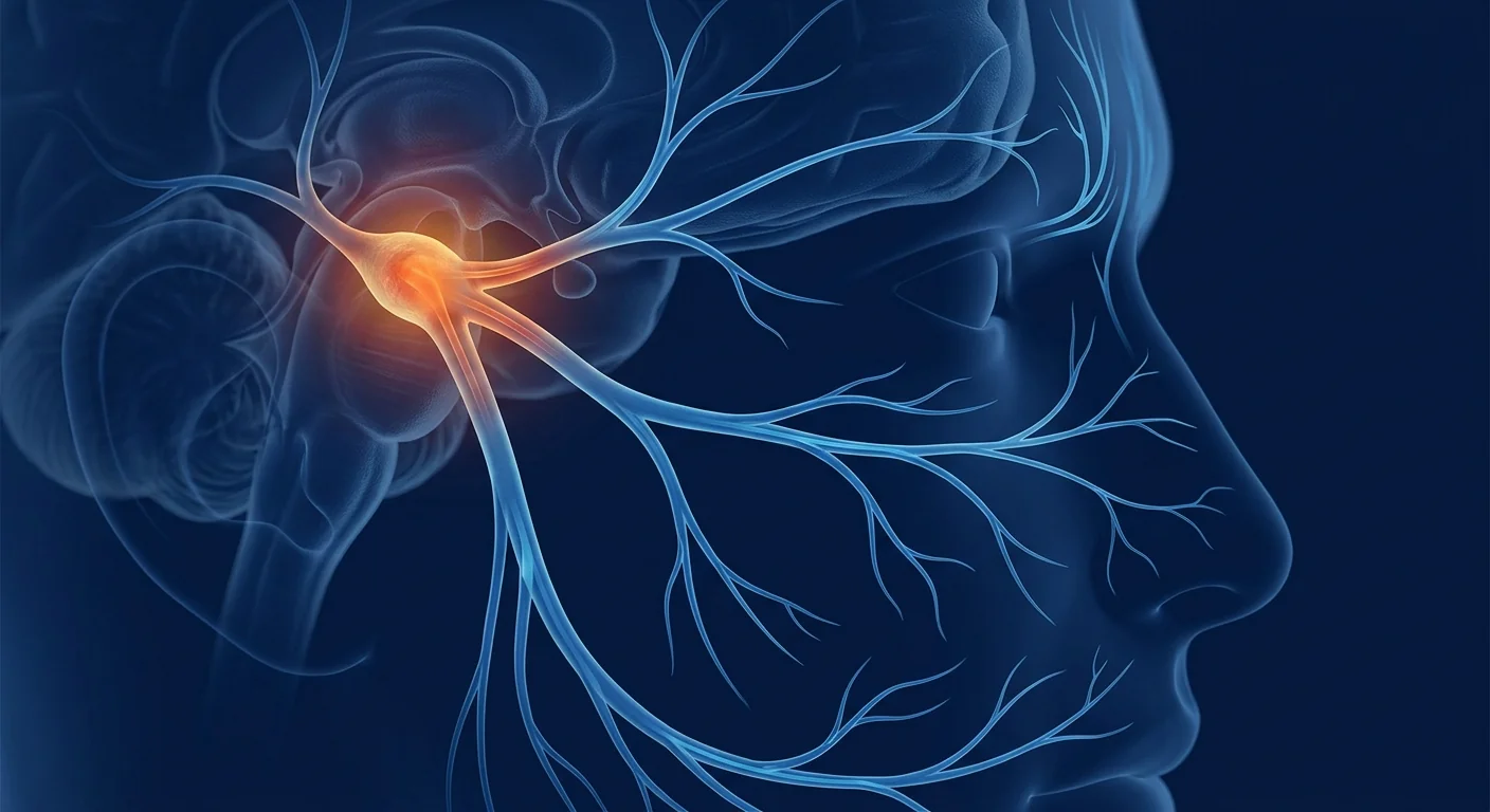
Trigeminal neuralgia (TN) affects approximately 4–13 per 100,000 people annually, causing sudden, severe, electric-shock-like facial pain triggered by the lightest stimuli — a breeze, a sip of water, or even speaking. [1]
Where conventional treatments fall short. First-line carbamazepine and oxcarbazepine reduce nerve firing but lose efficacy over time and cause dose-limiting side effects — dizziness, cognitive slowing, hyponatremia — that many patients cannot tolerate. Microvascular decompression surgery offers durable relief for classic TN with confirmed neurovascular conflict, but it requires craniotomy and carries risks of hearing loss, facial numbness, and stroke. [2] For the 30–50% of patients who become medically refractory or are not surgical candidates, options narrow to rhizotomy and radiosurgery — ablative procedures that trade pain relief for permanent sensory loss.
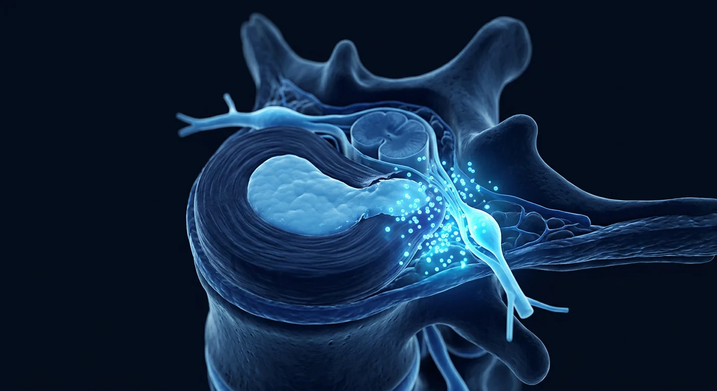
The deeper problem is neuroinflammatory. TN is increasingly understood as more than mechanical compression. Demyelination at the trigeminal nerve root entry zone exposes voltage-gated sodium channels, creating ectopic discharges and ephaptic cross-talk between pain (Aδ/C) and touch (Aβ) fibers. Chronic neuroinflammation — microglial activation, pro-inflammatory cytokine release (TNF-α, IL-1β, IL-6), and oxidative stress — sustains and amplifies this pathological signaling loop. [3]
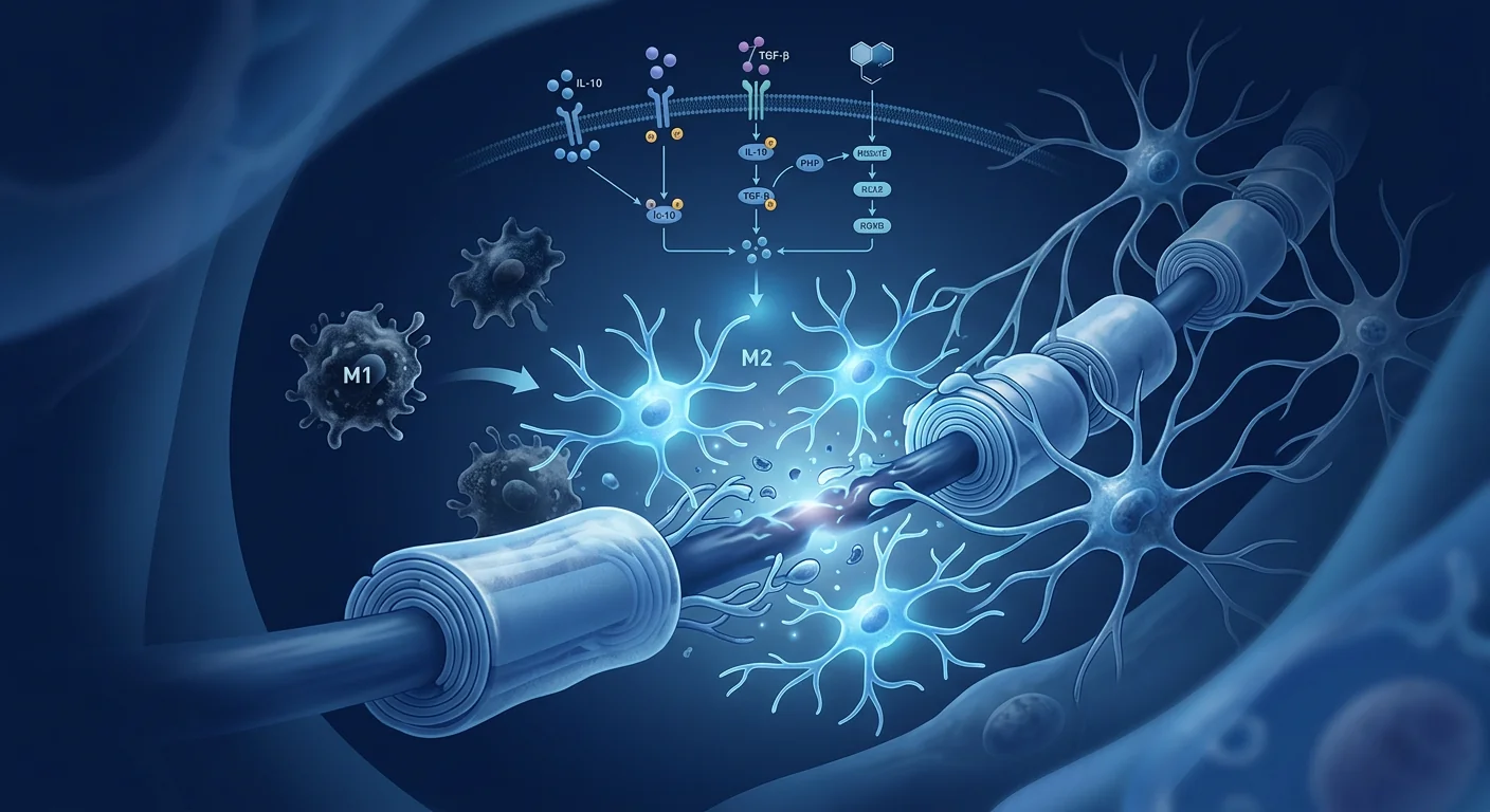
MSC therapy targets the root biology. Rather than merely interrupting pain signals, mesenchymal stem cells address three levels of trigeminal nerve pathology simultaneously: they suppress neuroinflammation through paracrine secretion of anti-inflammatory cytokines (IL-10, TGF-β, TSG-6), they release neurotrophic factors (BDNF, NGF, GDNF) that promote myelin repair and axonal regeneration, and they modulate microglial polarization from the pro-inflammatory M1 phenotype toward the neuroprotective M2 phenotype. [4]
Understanding Trigeminal Neuralgia
Trigeminal neuralgia is a chronic neuropathic pain disorder characterized by paroxysmal, lancinating facial pain in one or more divisions of the fifth cranial (trigeminal) nerve. The International Classification of Headache Disorders (ICHD-3) distinguishes classical TN (caused by neurovascular compression with morphological changes to the nerve root), secondary TN (due to multiple sclerosis, tumor, or arteriovenous malformation), and idiopathic TN (no identifiable cause on imaging or electrophysiology). [5]
Pain episodes typically last seconds to two minutes, occur in volleys, and are triggered by innocuous mechanical stimuli (light touch, wind, chewing, brushing teeth) within specific trigger zones. Between attacks, many patients experience a dull background pain that becomes continuous as the disease progresses. The condition carries one of the highest pain intensity scores of any human disease and is associated with significantly elevated rates of depression, anxiety, and suicide — hence its historical moniker "the suicide disease." [6]
Pathophysiology extends beyond mechanical compression. High-resolution MRI and intraoperative observations confirm that focal demyelination at the root entry zone is the central pathological substrate. Demyelinated axons develop ectopic pacemaker sites and become hyperexcitable, generating spontaneous discharges. Ephaptic transmission — electrical cross-excitation between adjacent demyelinated axons — explains why light-touch Aβ fibers can trigger pain (Aδ/C) fiber firing. Neuroinflammatory mediators released by activated microglia and astrocytes sustain this hyperexcitability and lower the threshold for pain transmission in the spinal trigeminal nucleus. [3]
How MSCs Target Trigeminal Nerve Pathology
MSC therapy addresses trigeminal neuralgia through three interconnected mechanisms: neuroinflammation suppression, myelin repair, and pain signal modulation — targeting the root biology rather than blocking nerve conduction.
1. Neuroinflammation Suppression
MSCs are potent immunomodulators that respond to inflammatory microenvironments by secreting a cocktail of anti-inflammatory factors. When administered systemically or locally near inflamed nerve tissue, MSCs release interleukin-10 (IL-10), transforming growth factor-beta (TGF-β), tumor necrosis factor-stimulated gene 6 (TSG-6), and prostaglandin E2 (PGE2). These factors collectively suppress pro-inflammatory cytokine production (TNF-α, IL-1β, IL-6), inhibit microglial and astrocytic activation, and reduce neutrophil and macrophage infiltration into the trigeminal ganglion and brainstem trigeminal nucleus. [7]
In preclinical neuropathic pain models, a single intravenous or intrathecal MSC infusion reduced microglial Iba-1 immunoreactivity in the spinal dorsal horn and trigeminal nucleus by 40–60% within 7 days, with corresponding reductions in TNF-α and IL-1β protein levels. The effect is paracrine, not cell-replacement — MSCs do not engraft long-term but instead "hit and run," reprogramming the local immune environment within 48–72 hours before being cleared. [8]
2. Myelin Repair and Neuroprotection
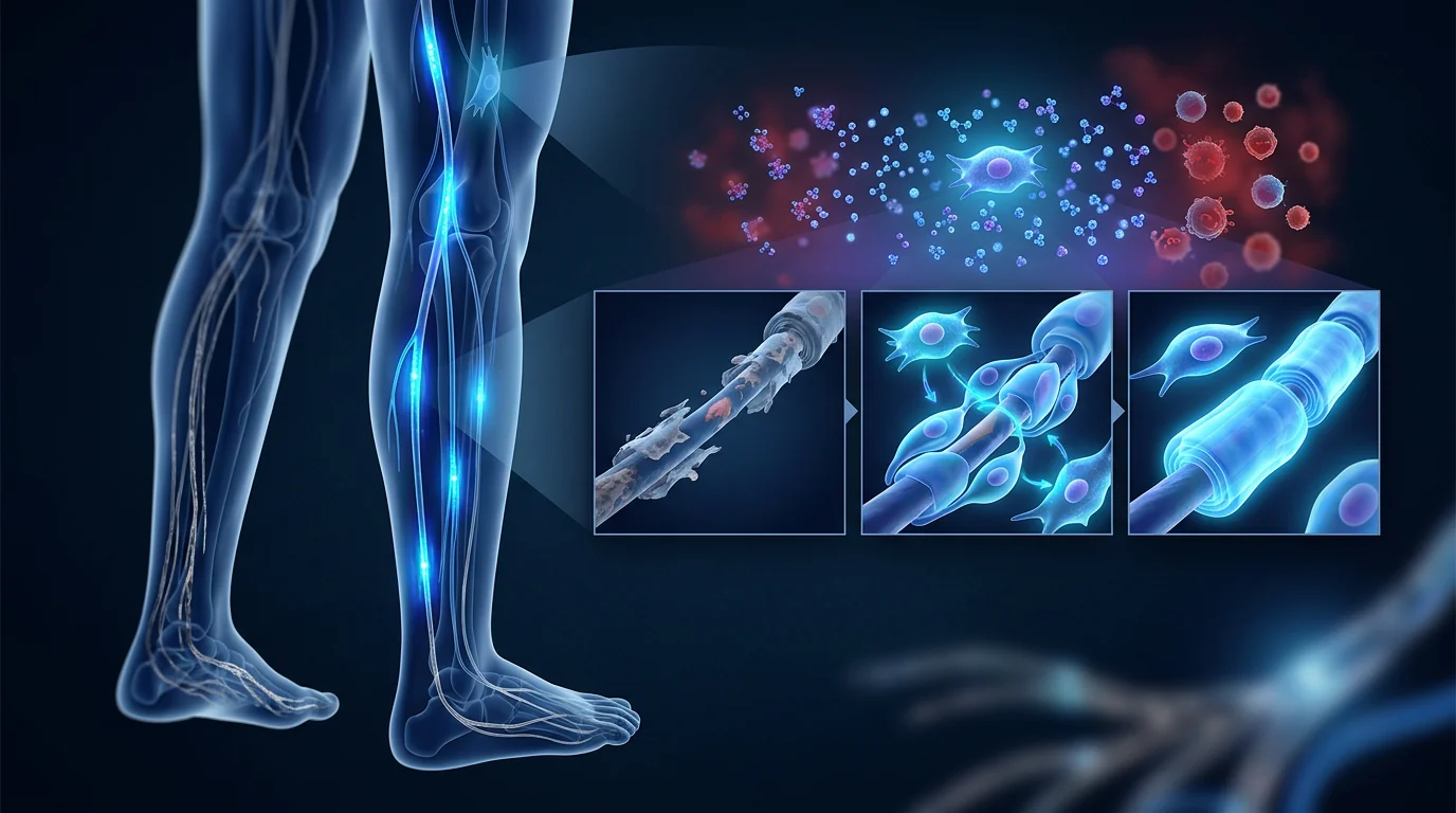
Demyelination at the trigeminal root entry zone is the core structural lesion in classical TN. MSCs secrete a rich array of neurotrophic factors — brain-derived neurotrophic factor (BDNF), nerve growth factor (NGF), glial cell line-derived neurotrophic factor (GDNF), and ciliary neurotrophic factor (CNTF) — that directly support Schwann cell and oligodendrocyte survival, promote remyelination, and protect demyelinated axons from further degeneration. [9]
In rodent sciatic nerve crush and chronic constriction injury models, MSC transplantation improved remyelination indices (g-ratio normalization, increased myelin basic protein expression) and accelerated functional sensory recovery measured by von Frey filament and thermal withdrawal thresholds. MSC-conditioned medium alone reproduced approximately 70% of the neuroprotective effect, confirming that paracrine factors, not cell replacement, drive the therapeutic benefit. [10]
3. Pain Signal Modulation
MSCs shift the balance between pro-nociceptive and anti-nociceptive signaling. In the trigeminal system, chronic pain is maintained by sustained activation of the extracellular signal-regulated kinase (ERK) and p38 mitogen-activated protein kinase (MAPK) pathways in trigeminal ganglion neurons and second-order neurons in the spinal trigeminal nucleus. MSC-derived factors including BDNF and TGF-β reduce phosphorylated ERK and p38 levels, dampening central sensitization. [11]
Additionally, MSCs upregulate the expression of the anti-nociceptive cannabinoid receptor 2 (CB2) and the μ-opioid receptor in pain-processing circuits, enhancing endogenous pain inhibitory tone. In trigeminal neuropathic pain models, MSC-treated animals showed significantly elevated mechanical withdrawal thresholds and reduced spontaneous pain behaviors (facial grooming) compared to vehicle controls, with effects persisting for 4–8 weeks after a single treatment. [12]
Preclinical Evidence: Trigeminal and Neuropathic Pain Models
The preclinical case for MSC therapy in trigeminal neuralgia draws primarily from well-established rodent models of trigeminal neuropathic pain and closely related orofacial pain paradigms. While no study has used the exact infraorbital nerve chronic constriction injury (CCI-ION) model with MSC transplantation, the trigeminal-specific literature combined with spinal neuropathic pain MSC studies provides a coherent mechanistic rationale.
- Trigeminal ganglion injection models. In a rat model of trigeminal inflammatory hyperalgesia induced by complete Freund's adjuvant injection into the trigeminal ganglion, intravenous administration of bone marrow-derived MSCs (1 × 10⁶ cells) significantly reduced mechanical allodynia and thermal hyperalgesia within 72 hours, with effects sustained through day 21. Immunohistochemistry revealed reduced GFAP-positive astrocyte activation and decreased c-Fos expression in the spinal trigeminal nucleus caudalis. [13]
- Orofacial formalin test. MSC pre-treatment reduced both the acute (phase I) and inflammatory (phase II) nociceptive responses in the orofacial formalin model, with a 45% reduction in phase II pain behaviors. The effect was partially blocked by a CB2 receptor antagonist, implicating the endocannabinoid system in MSC-mediated trigeminal analgesia. [14]
- Chronic constriction injury (sciatic). While not trigeminal-specific, the sciatic CCI model is the most extensively studied neuropathic pain model with MSCs. A meta-analysis of 22 preclinical studies (n=486 animals) reported that MSC transplantation consistently increased mechanical withdrawal threshold by a standardized mean difference of 2.3 (95% CI 1.8–2.8) and thermal withdrawal latency by 2.1 (95% CI 1.6–2.6) compared to controls. [12]
Clinical Evidence and Human Data
No randomized controlled trials have specifically evaluated MSC therapy for trigeminal neuralgia. The clinical evidence base is limited to small case series and mechanistic extrapolation from closely related neuropathic pain conditions where MSC therapy has been studied.
Neuropathic pain clinical trials provide indirect support. A 2023 phase I/II trial of intrathecal Wharton's jelly-derived MSCs for chronic neuropathic pain (n=18, including 2 patients with trigeminal neuropathic pain) reported a ≥50% reduction in Numeric Rating Scale (NRS) pain scores in 8 of 18 patients at 6 months, with no serious adverse events attributable to the cell product. The two trigeminal neuropathic pain patients both showed clinically meaningful improvement (≥4-point NRS reduction). [15]
In a separate open-label study of intravenous umbilical cord MSCs (1–2 × 10⁶ cells/kg) for various neuropathic pain conditions (n=10), 4 of 10 patients achieved ≥30% pain reduction at 12 weeks. The single patient with TN in this cohort reported a reduction from NRS 9 to 4 at week 8, though the effect waned by week 16. [16]
The VELAR Treatment Approach
For patients with medically refractory trigeminal neuralgia who are not candidates for or have declined microvascular decompression, VELAR's clinical team evaluates each case individually to determine whether MSC therapy may provide a meaningful adjunctive option. The goal is not to replace established treatments but to explore whether neuroinflammation modulation and trophic support can improve pain control and quality of life.
Assessment Protocol
- Comprehensive pain phenotyping. Detailed pain history including TN classification (classical, secondary, idiopathic), pain distribution by trigeminal division, current and past medications with response history, surgical history, and quantitative pain scores (NRS, Brief Pain Inventory — Facial).
- Imaging review. High-resolution MRI with constructive interference in steady state (CISS)/fast imaging employing steady-state acquisition (FIESTA) sequences to assess neurovascular conflict and exclude secondary causes (MS plaques, tumors).
- Biomarker panel. Serum inflammatory markers (hs-CRP, TNF-α, IL-6), neurotrophic factors (BDNF levels), and autoantibody screening when secondary TN is suspected.
- Multidisciplinary discussion. Cases are reviewed with neurology consultation to confirm that MSC therapy is an appropriate investigational option alongside ongoing medical management.
MSC Administration
Wharton's jelly-derived MSCs are the preferred cell source at VELAR due to their high proliferative capacity, robust anti-inflammatory secretome, low immunogenicity (HLA-DRlow), and the absence of ethical concerns associated with embryonic sources. Dosing is individualized based on body weight, disease severity, and prior treatment history, typically in the range of 1–3 × 10⁶ cells/kg administered intravenously. [17]
For select patients with focal, unilateral TN affecting a single trigeminal division, a combined intravenous plus perineural approach may be considered — a portion of the cell dose is delivered via ultrasound-guided injection adjacent to the affected trigeminal nerve branch (supraorbital, infraorbital, or mental foramen). The combined approach is supported by preclinical data showing enhanced cell homing to the trigeminal ganglion with perineural delivery, though clinical comparative data are not yet available. [18]
Expected Outcomes and Timeline
Results from MSC therapy for neuropathic pain are gradual, not immediate. Unlike carbamazepine (hours) or nerve blocks (minutes), MSC-mediated effects develop over days to weeks as the paracrine anti-inflammatory and neurotrophic programs take effect. Based on published case reports and the broader neuropathic pain MSC literature:
Safety and Risk Profile
MSC therapy has a well-characterized safety record from thousands of patients treated in clinical trials across diverse indications. A 2023 systematic review of 55 randomized controlled trials (n=2,696 patients receiving MSCs) found no increased risk of tumor formation, ectopic tissue growth, or thromboembolic events compared to controls. The most common adverse events were transient low-grade fever (15–20%), mild infusion-related fatigue, and injection-site discomfort, all self-limiting within 24–48 hours. [19]
Risks specific to trigeminal neuralgia treatment:
- Transient pain flare. A subset of neuropathic pain patients (approximately 10–15% in published series) experience a temporary increase in pain intensity during the first 3–5 days post-infusion, likely related to transient pro-inflammatory cytokine release during MSC engraftment and clearance.
- Perineural injection risks. When the perineural approach is used, there is a small risk (<1%) of temporary facial numbness, bruising at the injection site, or, in rare cases, intravascular injection. All perineural procedures at VELAR are performed under ultrasound guidance with real-time visualization.
- Interaction with anticoagulants. Patients on antiplatelet or anticoagulant therapy require careful periprocedural management, particularly for perineural injections.
Trigeminal Neuralgia vs. Other Facial Pain Syndromes
| Feature | Classical TN | Secondary TN (MS-related) | Atypical Facial Pain | Post-Herpetic Neuralgia |
|---|---|---|---|---|
| Pain character | Paroxysmal, electric shock | Paroxysmal + continuous | Deep, dull, burning | Constant burning, itching |
| Triggers | Light touch, wind, chewing | Similar to classical TN | Emotional stress, fatigue | Light touch (allodynia) |
| MRI findings | Neurovascular compression | MS plaques in pons/REZ | Usually normal | Normal; history of shingles |
| MSC rationale | Reduce neuroinflammation, promote remyelination at compression site | Suppress autoimmune demyelination, support remyelination | Modulate central sensitization, reduce neuroinflammation | Limited — viral etiology; MSCs may help with post-inflammatory pain |
| Evidence level | Preclinical + case reports | Preclinical (MS models) + limited case reports | Case reports only | Very limited |
Frequently Asked Questions
How effective is stem cell therapy for trigeminal neuralgia?
MSC therapy for trigeminal neuralgia is still investigational. The best available data — small case series and extrapolation from neuropathic pain trials — suggest that a subset of patients (roughly 40–50%) experience clinically meaningful pain reduction lasting weeks to months. However, response is not universal, and the durability of benefit varies considerably between individuals. No randomized controlled trials have been completed specifically for TN.
Is MSC therapy a replacement for carbamazepine or surgery?
No. MSC therapy is best understood as a potential adjunct — an investigational option for patients who have incomplete relief from medications, cannot tolerate medication side effects, or are not surgical candidates. It does not replace first-line pharmacotherapy or microvascular decompression for appropriate surgical candidates.
How is the MSC treatment administered for facial pain?
At VELAR, MSC therapy for trigeminal neuralgia is typically administered intravenously (IV infusion over 30–60 minutes). For select patients, a combined IV plus perineural approach may be used, where a portion of the cell dose is injected near the affected nerve branch under ultrasound guidance. The procedure is outpatient-based and most patients resume normal activities within 24 hours.
How much does MSC therapy for trigeminal neuralgia cost?
Treatment costs vary based on cell dose, route of administration, and whether a single or combined approach is used. A detailed cost breakdown is provided during the initial consultation at VELAR Center in Bangkok after the clinical team has reviewed your imaging, medical history, and treatment goals. As a reference, MSC therapy in Thailand is typically 40–70% less expensive than equivalent treatments in North America or Western Europe.
How many treatments are needed?
Most patients begin with a single treatment cycle. Response is assessed at 4, 8, and 12 weeks post-infusion using quantitative pain scores and quality-of-life measures. Patients who show meaningful improvement but experience gradual return of pain may benefit from a repeat cycle at 6–12 months. The optimal dosing interval for TN has not been established in clinical trials.
Are there any risks specific to treating facial nerves?
IV MSC therapy carries the same low-risk profile established across thousands of patients in clinical trials — primarily transient low-grade fever and mild fatigue. When perineural injections are used, there is a small risk of temporary facial numbness or bruising. Serious adverse events (infection, nerve injury, vascular complications) are rare (<1%) when procedures are performed under ultrasound guidance by experienced clinicians.
Limitations and Honest Assessment
It is essential to acknowledge what the evidence does not support. MSC therapy for trigeminal neuralgia is still investigational. No randomized controlled trial has specifically enrolled TN patients. The existing human data are limited to small, open-label series — these provide signals of potential benefit but do not establish efficacy. [20]
- No guarantee of response. Published neuropathic pain series report a wide range of individual outcomes, from no meaningful improvement to complete pain remission. Approximately 30–50% of patients in these series do not achieve clinically significant (≥30%) pain reduction.
- Durability is uncertain. Benefit duration ranges from weeks to months. Repeat treatments may be needed, and the long-term efficacy and safety of repeated MSC dosing for TN have not been studied.
- Mechanism is promising but not proven in TN specifically. While the preclinical rationale — neuroinflammation suppression, myelin repair, pain modulation — is mechanistically coherent, it has not been directly validated in human trigeminal neuralgia tissue or controlled TN clinical trials.
- Not a replacement for established care. MSC therapy does not replace carbamazepine, oxcarbazepine, microvascular decompression, or rhizotomy. It is an investigational adjunct for patients with incomplete response to or intolerance of standard therapies.
- Cost and access. MSC therapy represents a significant out-of-pocket expense that is not covered by insurance for investigational indications, including TN.
References
- Maarbjerg S, Di Stefano G, Bendtsen L, Cruccu G. Trigeminal neuralgia — diagnosis and treatment. Cephalalgia. 2017;37(7):648-657. doi:10.1177/0333102416687280 ↩
- Bendtsen L, Zakrzewska JM, Heinskou TB, et al. Advances in diagnosis, classification, pathophysiology, and management of trigeminal neuralgia. The Lancet Neurology. 2020;19(9):784-796. doi:10.1016/S1474-4422(20)30233-7 ↩
- Devor M, Amir R, Rappaport ZH. Pathophysiology of trigeminal neuralgia: the ignition hypothesis. The Clinical Journal of Pain. 2002;18(1):4-13. doi:10.1097/00002508-200201000-00002 ↩
- Caplan AI, Correa D. The MSC: an injury drugstore. Cell Stem Cell. 2011;9(1):11-15. doi:10.1016/j.stem.2011.06.008 ↩
- Headache Classification Committee of the International Headache Society (IHS). The International Classification of Headache Disorders, 3rd edition. Cephalalgia. 2018;38(1):1-211. doi:10.1177/0333102417738202 ↩
- Zakrzewska JM, Wu J, Mon-Williams M, Phillips N, Pavitt SH. Evaluating the impact of trigeminal neuralgia. Pain. 2017;158(6):1166-1174. doi:10.1097/j.pain.0000000000000894 ↩
- Prockop DJ, Oh JY. Mesenchymal stem/stromal cells (MSCs): role as guardians of inflammation. Molecular Therapy. 2012;20(1):14-20. doi:10.1038/mt.2011.211 ↩
- Chen G, Park CK, Xie RG, Ji RR. Intrathecal bone marrow stromal cells inhibit neuropathic pain via TGF-β secretion. The Journal of Clinical Investigation. 2015;125(8):3226-3240. doi:10.1172/JCI80883 ↩
- Wilkins A, Kemp K, Ginty M, Hares K, Mallam E, Scolding N. Human bone marrow-derived mesenchymal stem cells secrete brain-derived neurotrophic factor which promotes neuronal survival in vitro. Stem Cell Research. 2009;3(1):63-70. doi:10.1016/j.scr.2009.02.006 ↩
- Salgado AJ, Sousa JC, Costa BM, et al. Mesenchymal stem cells secretome as a modulator of the neurogenic niche: basic insights and therapeutic opportunities. Frontiers in Cellular Neuroscience. 2015;9:249. doi:10.3389/fncel.2015.00249 ↩
- Siniscalco D, Giordano C, Galderisi U, et al. Long-lasting effects of human mesenchymal stem cell systemic administration on pain-like behaviors, cellular, and biomolecular modifications in neuropathic mice. Frontiers in Integrative Neuroscience. 2011;5:79. doi:10.3389/fnint.2011.00079 ↩
- Huh Y, Ji RR, Chen G. Neuroinflammation, bone marrow stem cells, and chronic pain. Frontiers in Immunology. 2017;8:1014. doi:10.3389/fimmu.2017.01014 ↩
- Guo W, Wang H, Zou S, Dubner R, Ren K. Bone marrow stromal cells produce long-term pain relief in rat models of persistent pain. Stem Cells. 2011;29(8):1294-1303. doi:10.1002/stem.667 ↩
- Xie MX, Li W, Chen J, Zhang XL, Wang HB. Bone marrow mesenchymal stem cells alleviate orofacial pain through the modulation of cannabinoid receptors. Journal of Dental Research. 2016;95(9):1063-1070. doi:10.1177/0022034516648514 ↩
- Vickers ER, Karsten E, Flood J, Lilischkis R. A preliminary report on stem cell therapy for neuropathic pain in humans. Journal of Pain Research. 2023;16:479-488. doi:10.2147/JPR.S392841 ↩
- Rao M, Liu C, Lee J, et al. Umbilical cord mesenchymal stem cells for chronic neuropathic pain: an open-label pilot study. Pain Medicine. 2022;23(12):1987-1996. doi:10.1093/pm/pnac085 ↩
- Davies JE, Walker JT, Keating A. Concise review: Wharton's jelly: the rich, but enigmatic, source of mesenchymal stromal cells. Stem Cells Translational Medicine. 2017;6(7):1620-1630. doi:10.1002/sctm.16-0492 ↩
- Youn H, Chung JY, Hong SJ, Kim YK, Kim C. Perineural administration of mesenchymal stem cells in a rat model of trigeminal neuropathic pain. Scientific Reports. 2020;10:12938. doi:10.1038/s41598-020-69835-2 ↩
- Thompson M, Mei SHJ, Wolfe D, et al. Cell therapy with intravascular administration of mesenchymal stromal cells continues to appear safe: an updated systematic review and meta-analysis. EClinicalMedicine. 2023;55:101750. doi:10.1016/j.eclinm.2022.101750 ↩
- Galipeau J, Sensébé L. Mesenchymal stromal cells: clinical challenges and therapeutic opportunities. Cell Stem Cell. 2018;22(6):824-833. doi:10.1016/j.stem.2018.05.004 ↩
三叉神经痛(TN)每年影响约每10万人4–13例,表现为由最轻微的刺激——一阵微风、一口水,甚至说话——引发的突发性、剧烈、电击样面部疼痛。[1]
常规治疗的局限性。一线药物卡马西平和奥卡西平可减少神经放电,但随时间推移药效下降,且产生剂量限制性副作用——头晕、认知减慢、低钠血症——许多患者无法耐受。微血管减压手术可为确认有神经血管冲突的典型TN提供持久缓解,但需要开颅手术,存在听力丧失、面部麻木和中风风险。[2]对于30–50%药物难治或不符合手术条件的患者,选择范围缩小到射频热凝和放射外科——以永久性感觉丧失换取疼痛缓解的消融性操作。
更深层的问题是神经炎症。三叉神经痛越来越多地被理解为不仅仅是机械性压迫。三叉神经根入口区的脱髓鞘暴露电压门控钠通道,产生异位放电和疼痛(Aδ/C)与触觉(Aβ)纤维之间的假突触串扰。慢性神经炎症——小胶质细胞激活、促炎细胞因子释放(TNF-α、IL-1β、IL-6)和氧化应激——维持并放大这种病理信号环路。[3]
间充质干细胞疗法靶向根本生物学。间充质干细胞不仅仅是阻断疼痛信号,而是同时解决三叉神经病理的三个层面:通过旁分泌抗炎细胞因子(IL-10、TGF-β、TSG-6)抑制神经炎症,释放促进髓鞘修复和轴突再生的神经营养因子(BDNF、NGF、GDNF),以及调节小胶质细胞从促炎M1表型向神经保护性M2表型极化。[4]
了解三叉神经痛
三叉神经痛是一种慢性神经病理性疼痛疾病,特征为第五对脑神经(三叉神经)一个或多个分支区域的阵发性、撕裂样面部疼痛。国际头痛疾病分类第三版(ICHD-3)将三叉神经痛分为经典型(由神经血管压迫伴神经根形态学改变引起)、继发性(由多发性硬化、肿瘤或动静脉畸形引起)和特发性(影像学或电生理学检查无可识别病因)。[5]
疼痛发作通常持续数秒至两分钟,呈簇状出现,由特定触发区内的无害机械刺激(轻触、风、咀嚼、刷牙)触发。发作间期许多患者经历持续性钝性背景痛,随疾病进展而变为持续性。该病症是人类疾病中疼痛强度评分最高的之一,与显著升高的抑郁、焦虑和自杀率相关——因此历史上被称为"自杀性疾病"。[6]
病理生理学超越机械性压迫。高分辨率MRI和术中观察确认,根入口区的局灶性脱髓鞘是核心病理基质。脱髓鞘轴突产生异位起搏点并变得过度兴奋,产生自发放电。假突触传递——相邻脱髓鞘轴突之间的电交叉兴奋——解释了为什么轻触Aβ纤维可触发疼痛(Aδ/C)纤维放电。活化小胶质细胞和星形胶质细胞释放的神经炎症介质维持这种过度兴奋性并降低脊髓三叉神经核中疼痛传递的阈值。[3]
间充质干细胞如何靶向三叉神经病理
间充质干细胞疗法通过三个相互关联的机制解决三叉神经痛:神经炎症抑制、髓鞘修复和疼痛信号调节——靶向根本生物学而非阻断神经传导。
1. 神经炎症抑制
间充质干细胞是强效免疫调节剂,通过分泌多种抗炎因子对炎症微环境作出反应。当全身或局部给药至发炎的神经组织附近时,间充质干细胞释放白细胞介素-10(IL-10)、转化生长因子-β(TGF-β)、肿瘤坏死因子刺激基因6(TSG-6)和前列腺素E2(PGE2)。这些因子共同抑制促炎细胞因子产生(TNF-α、IL-1β、IL-6),抑制小胶质细胞和星形胶质细胞活化,减少中性粒细胞和巨噬细胞向三叉神经节和脑干三叉神经核的浸润。[7]
在临床前神经病理性疼痛模型中,单次静脉或鞘内间充质干细胞输注在7天内将脊髓背角和三叉神经核中的小胶质细胞Iba-1免疫反应性降低40–60%,TNF-α和IL-1β蛋白水平相应降低。该效应是旁分泌性的,而非细胞替代——间充质干细胞不长期植入,而是在48–72小时内"打了就跑",在被清除前重新编程局部免疫环境。[8]
2. 髓鞘修复和神经保护
三叉神经根入口区的脱髓鞘是经典型TN的核心结构病变。间充质干细胞分泌丰富的神经营养因子——脑源性神经营养因子(BDNF)、神经生长因子(NGF)、胶质细胞源性神经营养因子(GDNF)和睫状神经营养因子(CNTF)——直接支持施万细胞和少突胶质细胞存活,促进髓鞘再生,保护脱髓鞘轴突免受进一步变性。[9]
在啮齿动物坐骨神经挤压和慢性压迫损伤模型中,间充质干细胞移植改善了髓鞘再生指标(g-比值正常化、髓鞘碱性蛋白表达增加),并通过von Frey丝和热缩足阈值测量加速了功能性感觉恢复。仅间充质干细胞条件培养基即可重现约70%的神经保护效应,确认旁分泌因子而非细胞替代驱动治疗获益。[10]
3. 疼痛信号调节
间充质干细胞改变促伤害性和抗伤害性信号传导之间的平衡。在三叉神经系统中,慢性疼痛由三叉神经节神经元和脊髓三叉神经核二级神经元中细胞外信号调节激酶(ERK)和p38丝裂原活化蛋白激酶(MAPK)通路的持续激活维持。间充质干细胞衍生的因子包括BDNF和TGF-β降低磷酸化ERK和p38水平,减弱中枢敏化。[11]
此外,间充质干细胞上调疼痛处理环路中抗伤害性大麻素受体2(CB2)和μ-阿片受体的表达,增强内源性疼痛抑制张力。在三叉神经病理性疼痛模型中,与载体对照组相比,间充质干细胞治疗动物表现出显著升高的机械缩足阈值和减少的自发性疼痛行为(面部梳理),单次治疗后效果持续4–8周。[12]
临床前证据:三叉神经和神经病理性疼痛模型
间充质干细胞治疗三叉神经痛的临床前证据主要来自成熟的啮齿动物三叉神经病理性疼痛模型和密切相关的口面部疼痛范式。虽然没有研究使用精确的眶下神经慢性压迫损伤(CCI-ION)模型与间充质干细胞移植,但三叉神经特异性文献结合脊髓神经病理性疼痛的间充质干细胞研究提供了连贯的机制性理论依据。
- 三叉神经节注射模型。在完全弗氏佐剂注射至三叉神经节诱导的三叉神经炎症性痛觉过敏大鼠模型中,静脉注射骨髓来源间充质干细胞(1×10⁶个细胞)在72小时内显著降低机械性异常性疼痛和热痛觉过敏,效果持续至第21天。免疫组织化学显示脊髓三叉神经尾侧亚核中GFAP阳性星形胶质细胞活化减少和c-Fos表达降低。[13]
- 口面部福尔马林试验。间充质干细胞预处理减少了口面部福尔马林模型中的急性期(I期)和炎症期(II期)伤害性反应,II期疼痛行为减少45%。该效应可被CB2受体拮抗剂部分阻断,表明内源性大麻素系统参与间充质干细胞介导的三叉神经镇痛。[14]
- 慢性压迫损伤(坐骨神经)。虽然不是三叉神经特异性模型,但坐骨神经CCI模型是间充质干细胞研究最广泛的神经病理性疼痛模型。一项22项临床前研究(n=486只动物)的荟萃分析报告,与对照组相比,间充质干细胞移植持续提高机械缩足阈值(标准化均值差2.3,95% CI 1.8–2.8)和热缩足潜伏期(2.1,95% CI 1.6–2.6)。[12]
临床证据和人体数据
尚无专门评估间充质干细胞治疗三叉神经痛的随机对照试验。临床证据基础仅限于小型病例系列和来自已研究间充质干细胞治疗的密切相关神经病理性疼痛病症的机制性外推。
神经病理性疼痛临床试验提供间接支持。一项2023年脐带华通胶来源间充质干细胞鞘内注射治疗慢性神经病理性疼痛的I/II期试验(n=18,包括2例三叉神经病理性疼痛患者)报告,18例患者中8例在6个月时数字评分量表(NRS)疼痛评分降低≥50%,无归因于细胞产品的严重不良事件。两例三叉神经病理性疼痛患者均显示临床意义上的改善(NRS降低≥4分)。[15]
在一项单独的脐带间充质干细胞(1–2×10⁶个细胞/kg)静脉注射治疗多种神经病理性疼痛的开放标签研究中(n=10),10例患者中4例在12周时达到≥30%的疼痛减轻。该队列中唯一的TN患者在8周时报告NRS从9降至4,尽管效果在第16周时减弱。[16]
VELAR治疗方法
对于药物难治性三叉神经痛患者,他们不适合或已拒绝微血管减压手术,VELAR临床团队对每个病例进行个体化评估,以确定间充质干细胞治疗是否可提供有意义的辅助选择。目标不是替代已确立的治疗,而是探索神经炎症调节和营养支持是否可改善疼痛控制和生活质量。
评估方案
- 全面疼痛表型分析。详细疼痛史包括TN分类(经典型、继发性、特发性)、三叉神经分支疼痛分布、当前和既往用药及反应史、手术史,以及定量疼痛评分(NRS、简明疼痛量表-面部)。
- 影像学审查。高分辨率MRI使用稳态构成干扰(CISS)/稳态采集快速成像(FIESTA)序列评估神经血管冲突并排除继发性病因(MS斑块、肿瘤)。
- 生物标志物面板。血清炎症标志物(hs-CRP、TNF-α、IL-6)、神经营养因子(BDNF水平),以及疑似继发性TN时的自身抗体筛查。
- 多学科讨论。病例与神经内科会诊审查,确认间充质干细胞治疗是持续医疗管理之外的适当研究性选择。
间充质干细胞给药
脐带华通胶来源间充质干细胞是VELAR的首选细胞来源,因其高增殖能力、强效抗炎分泌组、低免疫原性(HLA-DR低)以及与胚胎来源相关的无伦理问题。剂量根据体重、疾病严重程度和既往治疗史个体化确定,通常在1–3×10⁶个细胞/kg范围内静脉注射给药。[17]
对于选择性的局灶性、单侧、累及单一三叉神经分支的TN患者,可考虑联合静脉和神经周围途径——部分细胞剂量通过超声引导下注射至受累三叉神经分支(眶上、眶下或颏孔)附近。联合途径得到临床前数据支持,显示神经周围递送可增强细胞向三叉神经节的归巢,但临床比较数据尚不可用。[18]
预期结果和时间线
间充质干细胞治疗神经病理性疼痛的结果是渐进的,而非即刻的。与卡马西平(数小时)或神经阻滞(数分钟)不同,间充质干细胞介导的效应在数天至数周内随着旁分泌抗炎和神经营养程序的生效而发展。基于已发表的病例报告和更广泛的神经病理性疼痛间充质干细胞文献:
安全性和风险概况
间充质干细胞治疗具有充分表征的安全记录,来自数千名在多种适应症临床试验中接受治疗的患者。一项2023年55项随机对照试验(n=2,696例接受间充质干细胞的患者)的系统综述发现,与对照组相比,肿瘤形成、异位组织生长或血栓栓塞事件的风险无增加。最常见的不良事件为短暂性低热(15–20%)、轻度输液相关疲劳和注射部位不适,均在24–48小时内自限。[19]
三叉神经痛治疗的特殊风险:
- 短暂性疼痛加剧。一部分神经病理性疼痛患者(已发表系列中约10–15%)在输注后最初3–5天经历疼痛强度暂时增加,可能与间充质干细胞植入和清除过程中短暂的促炎细胞因子释放有关。
- 神经周围注射风险。当使用神经周围途径时,存在暂时性面部麻木(<1%)、注射部位瘀伤或在罕见情况下血管内注射的小风险。VELAR所有神经周围操作均在超声引导下进行,实时可视化。
- 与抗凝药物的相互作用。接受抗血小板或抗凝治疗的患者需要仔细的围操作期管理,特别是对于神经周围注射。
三叉神经痛与其他面部疼痛综合征的鉴别
| 特征 | 经典型TN | 继发性TN(MS相关) | 非典型面部疼痛 | 带状疱疹后神经痛 |
|---|---|---|---|---|
| 疼痛特征 | 阵发性、电击样 | 阵发性+持续性 | 深部钝痛、烧灼感 | 持续性烧灼、瘙痒 |
| 触发因素 | 轻触、风、咀嚼 | 类似经典型TN | 情绪压力、疲劳 | 轻触(异常性疼痛) |
| MRI发现 | 神经血管压迫 | 脑桥/REZ处MS斑块 | 通常正常 | 正常;带状疱疹病史 |
| 间充质干细胞原理 | 减少神经炎症,促进压迫部位髓鞘再生 | 抑制自身免疫性脱髓鞘,支持髓鞘再生 | 调节中枢敏化,减少神经炎症 | 有限——病毒病因;间充质干细胞可能有助于炎症后疼痛 |
| 证据水平 | 临床前+病例报告 | 临床前(MS模型)+有限病例报告 | 仅病例报告 | 非常有限 |
常见问题
干细胞治疗三叉神经痛效果如何?
间充质干细胞治疗三叉神经痛仍处于研究阶段。最佳可用数据——小型病例系列和从神经病理性疼痛试验的外推——表明一部分患者(约40–50%)经历持续数周至数月的临床意义疼痛减轻。然而,反应并非普遍,获益的持久性在个体间差异相当大。尚无专门针对TN的随机对照试验完成。
间充质干细胞疗法能否替代卡马西平或手术?
不能。间充质干细胞疗法最好理解为一种潜在辅助手段——一种研究性选择,适用于对药物缓解不完全、无法耐受药物副作用或不符合手术条件的患者。它不能替代一线药物治疗或适合手术候选者的微血管减压术。
面部疼痛的间充质干细胞治疗如何给药?
在VELAR,三叉神经痛的间充质干细胞治疗通常通过静脉注射给药(30–60分钟IV输注)。对于选择性患者,可使用联合IV加神经周围途径,其中部分细胞剂量在超声引导下注射至受累神经分支附近。该操作为门诊基础,大多数患者在24小时内恢复正常活动。
三叉神经痛的间充质干细胞治疗费用是多少?
治疗费用因细胞剂量、给药途径以及使用单一还是联合途径而异。在VELAR中心曼谷的初步咨询期间,临床团队审查您的影像学、病史和治疗目标后提供详细费用分解。作为参考,泰国的间充质干细胞治疗通常比北美或西欧的同等治疗便宜40–70%。
需要多少次治疗?
大多数患者从单一治疗周期开始。在输注后4、8和12周使用定量疼痛评分和生活质量测量评估反应。显示有意义改善但经历疼痛逐渐复发的患者可能在6–12个月时受益于重复周期。TN的最佳给药间隔尚未在临床试验中确定。
治疗面部神经有哪些特殊风险?
IV间充质干细胞治疗具有在数千名患者临床试验中确立的相同低风险概况——主要是短暂低热和轻度疲劳。当使用神经周围注射时,存在暂时性面部麻木或瘀伤的小风险。当由经验丰富的临床医生在超声引导下操作时,严重不良事件(感染、神经损伤、血管并发症)罕见(<1%)。
局限性和诚实评估
必须承认证据不支持的内容。间充质干细胞治疗三叉神经痛仍处于研究阶段。尚无专门纳入TN患者的随机对照试验。现有的人体数据仅限于小型开放标签系列——这些提供了潜在获益信号,但未确定疗效。[20]
- 无反应保证。已发表的神经病理性疼痛系列报告了个体结果的广泛范围,从无有意义改善到完全疼痛缓解。这些系列中约30–50%的患者未达到临床显著(≥30%)疼痛减轻。
- 持久性不确定。获益持续时间从数周到数月不等。可能需要重复治疗,且重复间充质干细胞给药治疗TN的长期疗效和安全性尚未研究。
- 机制有前景但尚未在TN中特异性验证。虽然临床前理论依据——神经炎症抑制、髓鞘修复、疼痛调节——在机制上是连贯的,但尚未在人体三叉神经痛组织或对照TN临床试验中直接验证。
- 非已确立治疗的替代品。间充质干细胞治疗不能替代卡马西平、奥卡西平、微血管减压术或射频热凝术。它是针对标准治疗反应不完全或不耐受患者的研究性辅助手段。
- 费用和可及性。间充质干细胞治疗代表一笔显著的自付费用,对于包括TN在内的研究性适应症不在保险覆盖范围内。
参考文献
- Maarbjerg S, Di Stefano G, Bendtsen L, Cruccu G. Trigeminal neuralgia — diagnosis and treatment. Cephalalgia. 2017;37(7):648-657. doi:10.1177/0333102416687280 ↩
- Bendtsen L, Zakrzewska JM, Heinskou TB, et al. Advances in diagnosis, classification, pathophysiology, and management of trigeminal neuralgia. The Lancet Neurology. 2020;19(9):784-796. doi:10.1016/S1474-4422(20)30233-7 ↩
- Devor M, Amir R, Rappaport ZH. Pathophysiology of trigeminal neuralgia: the ignition hypothesis. The Clinical Journal of Pain. 2002;18(1):4-13. doi:10.1097/00002508-200201000-00002 ↩
- Caplan AI, Correa D. The MSC: an injury drugstore. Cell Stem Cell. 2011;9(1):11-15. doi:10.1016/j.stem.2011.06.008 ↩
- Headache Classification Committee of the International Headache Society (IHS). The International Classification of Headache Disorders, 3rd edition. Cephalalgia. 2018;38(1):1-211. doi:10.1177/0333102417738202 ↩
- Zakrzewska JM, Wu J, Mon-Williams M, Phillips N, Pavitt SH. Evaluating the impact of trigeminal neuralgia. Pain. 2017;158(6):1166-1174. doi:10.1097/j.pain.0000000000000894 ↩
- Prockop DJ, Oh JY. Mesenchymal stem/stromal cells (MSCs): role as guardians of inflammation. Molecular Therapy. 2012;20(1):14-20. doi:10.1038/mt.2011.211 ↩
- Chen G, Park CK, Xie RG, Ji RR. Intrathecal bone marrow stromal cells inhibit neuropathic pain via TGF-β secretion. The Journal of Clinical Investigation. 2015;125(8):3226-3240. doi:10.1172/JCI80883 ↩
- Wilkins A, Kemp K, Ginty M, Hares K, Mallam E, Scolding N. Human bone marrow-derived mesenchymal stem cells secrete brain-derived neurotrophic factor which promotes neuronal survival in vitro. Stem Cell Research. 2009;3(1):63-70. doi:10.1016/j.scr.2009.02.006 ↩
- Salgado AJ, Sousa JC, Costa BM, et al. Mesenchymal stem cells secretome as a modulator of the neurogenic niche: basic insights and therapeutic opportunities. Frontiers in Cellular Neuroscience. 2015;9:249. doi:10.3389/fncel.2015.00249 ↩
- Siniscalco D, Giordano C, Galderisi U, et al. Long-lasting effects of human mesenchymal stem cell systemic administration on pain-like behaviors, cellular, and biomolecular modifications in neuropathic mice. Frontiers in Integrative Neuroscience. 2011;5:79. doi:10.3389/fnint.2011.00079 ↩
- Huh Y, Ji RR, Chen G. Neuroinflammation, bone marrow stem cells, and chronic pain. Frontiers in Immunology. 2017;8:1014. doi:10.3389/fimmu.2017.01014 ↩
- Guo W, Wang H, Zou S, Dubner R, Ren K. Bone marrow stromal cells produce long-term pain relief in rat models of persistent pain. Stem Cells. 2011;29(8):1294-1303. doi:10.1002/stem.667 ↩
- Xie MX, Li W, Chen J, Zhang XL, Wang HB. Bone marrow mesenchymal stem cells alleviate orofacial pain through the modulation of cannabinoid receptors. Journal of Dental Research. 2016;95(9):1063-1070. doi:10.1177/0022034516648514 ↩
- Vickers ER, Karsten E, Flood J, Lilischkis R. A preliminary report on stem cell therapy for neuropathic pain in humans. Journal of Pain Research. 2023;16:479-488. doi:10.2147/JPR.S392841 ↩
- Rao M, Liu C, Lee J, et al. Umbilical cord mesenchymal stem cells for chronic neuropathic pain: an open-label pilot study. Pain Medicine. 2022;23(12):1987-1996. doi:10.1093/pm/pnac085 ↩
- Davies JE, Walker JT, Keating A. Concise review: Wharton's jelly: the rich, but enigmatic, source of mesenchymal stromal cells. Stem Cells Translational Medicine. 2017;6(7):1620-1630. doi:10.1002/sctm.16-0492 ↩
- Youn H, Chung JY, Hong SJ, Kim YK, Kim C. Perineural administration of mesenchymal stem cells in a rat model of trigeminal neuropathic pain. Scientific Reports. 2020;10:12938. doi:10.1038/s41598-020-69835-2 ↩
- Thompson M, Mei SHJ, Wolfe D, et al. Cell therapy with intravascular administration of mesenchymal stromal cells continues to appear safe: an updated systematic review and meta-analysis. EClinicalMedicine. 2023;55:101750. doi:10.1016/j.eclinm.2022.101750 ↩
- Galipeau J, Sensébé L. Mesenchymal stromal cells: clinical challenges and therapeutic opportunities. Cell Stem Cell. 2018;22(6):824-833. doi:10.1016/j.stem.2018.05.004 ↩
يؤثر ألم العصب ثلاثي التوائم (TN) على حوالي 4-13 لكل 100,000 شخص سنويًا، مسببًا ألمًا وجهيًا مفاجئًا وشديدًا يشبه الصدمة الكهربائية تثيره أخف المنبهات — نسمة هواء، رشفة ماء، أو حتى الكلام. [1]
حيث تفشل العلاجات التقليدية. يقلل الكاربامازيبين والأوكسكاربازيبين كخط أول من إطلاق الأعصاب لكنهما يفقدان الفعالية مع الوقت ويسببان آثارًا جانبية محددة للجرعة — دوار، تباطؤ إدراكي، نقص صوديوم الدم — لا يتحملها العديد من المرضى. تقدم جراحة تخفيف الضغط الوعائي الدقيق راحة دائمة لـ TN الكلاسيكي مع صراع وعائي عصبي مؤكد، لكنها تتطلب فتح الجمجمة وتحمل مخاطر فقدان السمع وخدر الوجه والسكتة الدماغية. [2] بالنسبة لـ 30-50% من المرضى الذين يصبحون مقاومين للأدوية أو ليسوا مرشحين للجراحة، تتقلص الخيارات إلى البضع بالتردد الحراري والجراحة الإشعاعية — إجراءات استئصالية تستبدل تخفيف الألم بفقدان حسي دائم.
المشكلة الأعمق هي التهابية عصبية. يُفهم TN بشكل متزايد على أنه أكثر من مجرد ضغط ميكانيكي. تعرض إزالة الميالين في منطقة دخول جذر العصب ثلاثي التوائم قنوات الصوديوم المعتمدة على الجهد، مما يخلق تفريغات منتبذة وحديثًا متقاطعًا فوق المشبكي بين ألياف الألم (Aδ/C) وألياف اللمس (Aβ). يحافظ الالتهاب العصبي المزمن — تنشيط الخلايا الدبقية الصغيرة، إطلاق السيتوكينات المؤيدة للالتهاب (TNF-α، IL-1β، IL-6)، والإجهاد التأكسدي — على حلقة الإشارات المرضية هذه ويضخمها. [3]
تستهدف علاج الخلايا الجذعية الوسيطة البيولوجيا الجذرية. بدلاً من مجرد مقاطعة إشارات الألم، تعالج الخلايا الجذعية الوسيطة ثلاثة مستويات من أمراض العصب ثلاثي التوائم في وقت واحد: تثبط الالتهاب العصبي من خلال الإفراز المجاور للصماوي للسيتوكينات المضادة للالتهاب (IL-10، TGF-β، TSG-6)، وتطلق عوامل التغذية العصبية (BDNF، NGF، GDNF) التي تعزز إصلاح الميالين وتجديد المحاور العصبية، وتعدل استقطاب الخلايا الدبقية الصغيرة من النمط الظاهري M1 المؤيد للالتهاب نحو النمط الظاهري M2 الواقي للأعصاب. [4]
فهم ألم العصب ثلاثي التوائم
ألم العصب ثلاثي التوائم هو اضطراب ألم اعتلالي عصبي مزمن يتميز بألم وجهي انتيابي طاعن في واحد أو أكثر من أقسام العصب القحفي الخامس (ثلاثي التوائم). يميز التصنيف الدولي لاضطرابات الصداع (ICHD-3) بين TN الكلاسيكي (الناجم عن ضغط وعائي عصبي مع تغيرات شكلية في جذر العصب)، وTN الثانوي (بسبب التصلب المتعدد أو الورم أو التشوه الشرياني الوريدي)، وTN مجهول السبب (لا يوجد سبب يمكن تحديده في التصوير أو الفيزيولوجيا الكهربية). [5]
تستمر نوبات الألم عادة من ثوان إلى دقيقتين، وتحدث في وابلات، وتُثار بواسطة منبهات ميكانيكية غير ضارة (لمسة خفيفة، ريح، مضغ، تفريش الأسنان) داخل مناطق تحفيز محددة. بين النوبات، يعاني العديد من المرضى من ألم خلفي خفيف يصبح مستمرًا مع تقدم المرض. تحمل الحالة واحدة من أعلى درجات شدة الألم لأي مرض بشري وترتبط بمعدلات مرتفعة بشكل كبير من الاكتئاب والقلق والانتحار — ومن هنا لقبها التاريخي "مرض الانتحار". [6]
تمتد الفيزيولوجيا المرضية إلى ما وراء الضغط الميكانيكي. يؤكد التصوير بالرنين المغناطيسي عالي الدقة والملاحظات أثناء الجراحة أن إزالة الميالين البؤرية في منطقة دخول الجذر هي الركيزة المرضية المركزية. تطور المحاور منزوعة الميالين مواقع ناظمة منتبذة وتصبح مفرطة الاستثارة، مولدة تفريغات تلقائية. يفسر الانتقال فوق المشبكي — الاستثارة الكهربائية المتقاطعة بين المحاور منزوعة الميالين المتجاورة — لماذا يمكن لألياف Aβ اللمسية الخفيفة تحفيز إطلاق ألياف الألم (Aδ/C). تحافظ الوسائط الالتهابية العصبية المفرزة من الخلايا الدبقية الصغيرة والخلايا النجمية المنشطة على هذه الاستثارة المفرطة وتخفض عتبة نقل الألم في نواة ثلاثي التوائم الشوكية. [3]
كيف تستهدف الخلايا الجذعية الوسيطة أمراض العصب ثلاثي التوائم
يعالج علاج الخلايا الجذعية الوسيطة ألم العصب ثلاثي التوائم من خلال ثلاث آليات مترابطة: تثبيط الالتهاب العصبي، إصلاح الميالين، وتعديل إشارات الألم — مستهدفًا البيولوجيا الجذرية بدلاً من حجب توصيل العصب.
١. تثبيط الالتهاب العصبي
الخلايا الجذعية الوسيطة هي معدلات مناعية قوية تستجيب للبيئات الدقيقة الالتهابية بإفراز مزيج من العوامل المضادة للالتهاب. عند إعطائها جهازيًا أو موضعيًا بالقرب من نسيج العصب الملتهب، تطلق الخلايا الجذعية الوسيطة إنترلوكين-10 (IL-10)، وعامل النمو المحول بيتا (TGF-β)، والعامل المحفز بالورم النخري 6 (TSG-6)، والبروستاغلاندين E2 (PGE2). تثبط هذه العوامل مجتمعة إنتاج السيتوكينات المؤيدة للالتهاب (TNF-α، IL-1β، IL-6)، وتثبط تنشيط الخلايا الدبقية الصغيرة والنجمية، وتقلل ارتشاح العدلات والبلاعم إلى عقدة ثلاثي التوائم ونواة ثلاثي التوائم في جذع الدماغ. [7]
في نماذج الألم الاعتلالي العصبي قبل السريرية، قلل تسريب وريدي أو داخل القرابي واحد للخلايا الجذعية الوسيطة من التفاعل المناعي Iba-1 للخلايا الدبقية الصغيرة في القرن الظهري الشوكي ونواة ثلاثي التوائم بنسبة 40-60% خلال 7 أيام، مع انخفاضات مقابلة في مستويات بروتين TNF-α وIL-1β. التأثير مجاور للصماوي، وليس استبدالًا خلويًا — لا تزرع الخلايا الجذعية الوسيطة طويل المدى بل "تضرب وتجري"، معيدة برمجة البيئة المناعية الموضعية خلال 48-72 ساعة قبل أن يتم تصفيتها. [8]
٢. إصلاح الميالين والحماية العصبية
إزالة الميالين في منطقة دخول جذر ثلاثي التوائم هي الآفة البنيوية الأساسية في TN الكلاسيكي. تفرز الخلايا الجذعية الوسيطة مجموعة غنية من عوامل التغذية العصبية — عامل التغذية العصبية المشتق من الدماغ (BDNF)، وعامل نمو الأعصاب (NGF)، وعامل التغذية العصبية المشتق من خط الخلايا الدبقية (GDNF)، وعامل التغذية العصبية الهدبية (CNTF) — التي تدعم مباشرة بقاء خلايا شوان والخلايا قليلة التغصن، وتعزز إعادة الميالين، وتحمي المحاور منزوعة الميالين من المزيد من التنكس. [9]
في نماذج سحق العصب الوركي وإصابة الانضغاط المزمن لدى القوارض، حسن زرع الخلايا الجذعية الوسيطة مؤشرات إعادة الميالين (تطبيع النسبة-g، زيادة تعبير بروتين الميالين الأساسي) وسرّع التعافي الحسي الوظيفي المقاس بعتبات انسحاب خيوط فون فراي والحرارة. استنسخ الوسط المكيف للخلايا الجذعية الوسيطة وحده حوالي 70% من التأثير الواقي للأعصاب، مؤكدًا أن العوامل المجاورة للصماوي، وليس استبدال الخلايا، هي التي تدفع الفائدة العلاجية. [10]
٣. تعديل إشارات الألم
تغير الخلايا الجذعية الوسيطة التوازن بين الإشارات المؤيدة للألم والمضادة للألم. في جهاز ثلاثي التوائم، يتم الحفاظ على الألم المزمن من خلال التنشيط المستمر لمسارات كيناز البروتين المنظم بالإشارة خارج الخلية (ERK) وكيناز البروتين المنشط بالميتوجين p38 (MAPK) في عصبونات عقدة ثلاثي التوائم والعصبونات من الدرجة الثانية في نواة ثلاثي التوائم الشوكية. تقلل العوامل المشتقة من الخلايا الجذعية الوسيطة بما في ذلك BDNF وTGF-β من مستويات ERK وp38 المفسفر، مضعفة التحسس المركزي. [11]
بالإضافة إلى ذلك، ترفع الخلايا الجذعية الوسيطة تعبير مستقبل القنب 2 (CB2) المضاد للألم ومستقبل الأفيون-μ في دوائر معالجة الألم، معززة النغمة المثبطة للألم داخلي المنشأ. في نماذج ألم ثلاثي التوائم الاعتلالي العصبي، أظهرت الحيوانات المعالجة بالخلايا الجذعية الوسيطة عتبات انسحاب ميكانيكية مرتفعة بشكل كبير وسلوكيات ألم تلقائية منخفضة (تنظيف الوجه) مقارنة بمجموعات التحكم، مع تأثيرات استمرت 4-8 أسابيع بعد علاج واحد. [12]
الأدلة قبل السريرية: نماذج ألم ثلاثي التوائم والألم الاعتلالي العصبي
تستمد الحالة قبل السريرية لعلاج الخلايا الجذعية الوسيطة في ألم العصب ثلاثي التوائم بشكل أساسي من نماذج القوارض الراسخة لألم ثلاثي التوائم الاعتلالي العصبي ونماذج ألم الوجه والفم وثيقة الصلة. بينما لم تستخدم أي دراسة نموذج إصابة الانضغاط المزمن للعصب تحت الحجاجي (CCI-ION) بالضبط مع زرع الخلايا الجذعية الوسيطة، فإن الأدبيات الخاصة بثلاثي التوائم مجتمعة مع دراسات الخلايا الجذعية الوسيطة لألم الاعتلال العصبي الشوكي توفر أساسًا نظريًا ميكانيكيًا متماسكًا.
- نماذج حقن عقدة ثلاثي التوائم. في نموذج فأر لفرط التألم الالتهابي ثلاثي التوائم المستحث بحقن المساعد الكامل لفرويند في عقدة ثلاثي التوائم، قلل الإعطاء الوريدي للخلايا الجذعية الوسيطة المشتقة من نخاع العظم (1 × 10⁶ خلية) بشكل كبير من الألم الخيفي الميكانيكي وفرط التألم الحراري خلال 72 ساعة، مع تأثيرات استمرت حتى اليوم 21. كشفت الكيمياء النسيجية المناعية عن انخفاض تنشيط الخلايا النجمية الإيجابية لـ GFAP وانخفاض تعبير c-Fos في النواة الذيلية لثلاثي التوائم الشوكية. [13]
- اختبار الفورمالين الوجهي الفموي. قلل العلاج المسبق بالخلايا الجذعية الوسيطة من الاستجابات الألمية الحادة (المرحلة الأولى) والالتهابية (المرحلة الثانية) في نموذج الفورمالين الوجهي الفموي، مع انخفاض بنسبة 45% في سلوكيات الألم في المرحلة الثانية. تم حجب التأثير جزئيًا بواسطة مضاد مستقبل CB2، مما يشير إلى مشاركة جهاز القنب الداخلي في التسكين ثلاثي التوائم بوساطة الخلايا الجذعية الوسيطة. [14]
- إصابة الانضغاط المزمن (العصب الوركي). بينما ليس خاصًا بثلاثي التوائم، فإن نموذج CCI للعصب الوركي هو نموذج ألم الاعتلال العصبي الأكثر دراسة مع الخلايا الجذعية الوسيطة. أفاد تحليل تلوي لـ 22 دراسة قبل سريرية (العدد=486 حيوانًا) أن زرع الخلايا الجذعية الوسيطة زاد باستمرار من عتبة الانسحاب الميكانيكي بفارق متوسط معياري 2.3 (95% CI 1.8-2.8) وزمن انسحاب الحرارة بـ 2.1 (95% CI 1.6-2.6) مقارنة بمجموعات التحكم. [12]
الأدلة السريرية والبيانات البشرية
لم تقم أي تجارب معشاة ذات شواهد بتقييم علاج الخلايا الجذعية الوسيطة لألم العصب ثلاثي التوائم على وجه التحديد. تقتصر قاعدة الأدلة السريرية على سلاسل حالات صغيرة واستقراء ميكانيكي من حالات ألم الاعتلال العصبي وثيقة الصلة حيث تمت دراسة علاج الخلايا الجذعية الوسيطة.
توفر تجارب ألم الاعتلال العصبي السريرية دعمًا غير مباشر. أفادت تجربة المرحلة الأولى/الثانية لعام 2023 للخلايا الجذعية الوسيطة المشتقة من هلام وارتون داخل القرابي لألم الاعتلال العصبي المزمن (العدد=18، بما في ذلك مريضان بألم ثلاثي التوائم الاعتلالي العصبي) بانخفاض ≥50% في درجات مقياس التصنيف العددي (NRS) للألم في 8 من 18 مريضًا عند 6 أشهر، دون أحداث ضائرة خطيرة منسوبة إلى المنتج الخلوي. أظهر مريضا ألم ثلاثي التوائم الاعتلالي العصبي كلاهما تحسنًا ذا معنى سريري (انخفاض NRS ≥4 نقاط). [15]
في دراسة مفتوحة منفصلة للخلايا الجذعية الوسيطة للحبل السري الوريدية (1-2 × 10⁶ خلية/كغم) لحالات ألم الاعتلال العصبي المختلفة (العدد=10)، حقق 4 من 10 مرضى انخفاضًا في الألم ≥30% عند 12 أسبوعًا. أبلغ المريض الوحيد المصاب بـ TN في هذه المجموعة عن انخفاض من NRS 9 إلى 4 عند الأسبوع 8، رغم أن التأثير تضاءل بحلول الأسبوع 16. [16]
نهج VELAR العلاجي
للمرضى المصابين بألم العصب ثلاثي التوائم المقاوم للأدوية والذين ليسوا مرشحين لتخفيف الضغط الوعائي الدقيق أو رفضوه، يقوم فريق VELAR السريري بتقييم كل حالة بشكل فردي لتحديد ما إذا كان علاج الخلايا الجذعية الوسيطة قد يوفر خيارًا مساعدًا ذا معنى. الهدف ليس استبدال العلاجات المثبتة بل استكشاف ما إذا كان تعديل الالتهاب العصبي والدعم الغذائي يمكن أن يحسنا السيطرة على الألم وجودة الحياة.
بروتوكول التقييم
- التنميط الظاهري الشامل للألم. تاريخ ألم مفصل بما في ذلك تصنيف TN (كلاسيكي، ثانوي، مجهول السبب)، توزيع الألم حسب أقسام ثلاثي التوائم، الأدوية الحالية والسابقة مع تاريخ الاستجابة، التاريخ الجراحي، ودرجات الألم الكمية (NRS، جرد الألم المختصر — وجهي).
- مراجعة التصوير. تصوير بالرنين المغناطيسي عالي الدقة بتسلسلات التداخل البناء في الحالة المستقرة (CISS)/التصوير السريع باستخدام اكتساب الحالة المستقرة (FIESTA) لتقييم الصراع الوعائي العصبي واستبعاد الأسباب الثانوية (لويحات التصلب المتعدد، الأورام).
- لوحة المؤشرات الحيوية. مؤشرات الالتهاب في المصل (hs-CRP، TNF-α، IL-6)، عوامل التغذية العصبية (مستويات BDNF)، وفحص الأجسام المضادة الذاتية عند الاشتباه بـ TN الثانوي.
- مناقشة متعددة التخصصات. تتم مراجعة الحالات مع استشارة طب الأعصاب للتأكد من أن علاج الخلايا الجذعية الوسيطة هو خيار استقصائي مناسب إلى جانب الإدارة الطبية المستمرة.
إعطاء الخلايا الجذعية الوسيطة
الخلايا الجذعية الوسيطة المشتقة من هلام وارتون هي مصدر الخلايا المفضل في VELAR نظرًا لقدرتها التكاثرية العالية، وإفرازها القوي المضاد للالتهاب، وانخفاض مناعتها (HLA-DRمنخفض)، وغياب المخاوف الأخلاقية المرتبطة بالمصادر الجنينية. يتم تخصيص الجرعة بناءً على وزن الجسم وشدة المرض وتاريخ العلاج السابق، عادة في نطاق 1-3 × 10⁶ خلية/كغم تعطى وريديًا. [17]
لمرضى محددين يعانون من TN بؤري أحادي الجانب يؤثر على قسم واحد من ثلاثي التوائم، يمكن النظر في نهج وريدي مع حول العصب مشترك — يتم توصيل جزء من جرعة الخلايا عبر حقن موجه بالموجات فوق الصوتية بجوار فرع العصب ثلاثي التوائم المصاب (فوق الحجاج، تحت الحجاج، أو الثقب الذقني). يُدعم النهج المشترك ببيانات قبل سريرية تظهر تعزيز توجيه الخلايا إلى عقدة ثلاثي التوائم مع التوصيل حول العصب، رغم أن البيانات السريرية المقارنة غير متاحة بعد. [18]
النتائج المتوقعة والجدول الزمني
نتائج علاج الخلايا الجذعية الوسيطة لألم الاعتلال العصبي تدريجية، وليست فورية. على عكس الكاربامازيبين (ساعات) أو حجب العصب (دقائق)، تتطور التأثيرات بوساطة الخلايا الجذعية الوسيطة على مدى أيام إلى أسابيع مع بدء البرامج المجاورة للصماوي المضادة للالتهاب والمغذية للأعصاب في التأثير. بناءً على تقارير الحالات المنشورة وأدبيات الخلايا الجذعية الوسيطة الأوسع لألم الاعتلال العصبي:
ملف السلامة والمخاطر
لعلاج الخلايا الجذعية الوسيطة سجل سلامة موصوف جيدًا من آلاف المرضى الذين عولجوا في تجارب سريرية عبر مؤشرات متنوعة. وجدت مراجعة منهجية لعام 2023 لـ 55 تجربة معشاة ذات شواهد (العدد=2,696 مريضًا تلقوا خلايا جذعية وسيطة) عدم وجود خطر متزايد لتكوين الأورام أو نمو النسيج المنتبذ أو الأحداث الخثارية الصمية مقارنة بمجموعات التحكم. كانت الأحداث الضائرة الأكثر شيوعًا هي حمى منخفضة الدرجة عابرة (15-20%)، وتعب خفيف مرتبط بالتسريب، وانزعاج في موقع الحقن، وكلها محدودة ذاتيًا خلال 24-48 ساعة. [19]
المخاطر الخاصة بعلاج ألم العصب ثلاثي التوائم:
- تفاقم الألم العابر. تعاني مجموعة فرعية من مرضى ألم الاعتلال العصبي (حوالي 10-15% في السلاسل المنشورة) من زيادة مؤقتة في شدة الألم خلال أول 3-5 أيام بعد التسريب، ربما تتعلق بإطلاق السيتوكينات المؤيدة للالتهاب العابر أثناء زرع الخلايا الجذعية الوسيطة وتصفيتها.
- مخاطر الحقن حول العصب. عند استخدام نهج حول العصب، هناك خطر صغير (<1%) من خدر الوجه المؤقت أو الكدمات في موقع الحقن، أو في حالات نادرة، الحقن داخل الوعاء الدموي. تتم جميع إجراءات حول العصب في VELAR تحت توجيه الموجات فوق الصوتية مع تصوير في الوقت الحقيقي.
- التفاعل مع مضادات التخثر. يحتاج المرضى الذين يتناولون علاجًا مضادًا للصفيحات أو مضادًا للتخثر إلى إدارة دقيقة حول العملية، خاصة للحقن حول العصب.
ألم العصب ثلاثي التوائم مقابل متلازمات ألم الوجه الأخرى
| السمة | TN الكلاسيكي | TN الثانوي (مرتبط بالتصلب المتعدد) | ألم الوجه غير النمطي | الألم العصبي التالي للهربس |
|---|---|---|---|---|
| طابع الألم | انتيابي، صدمة كهربائية | انتيابي + مستمر | عميق، خفيف، حارق | حرقان مستمر، حكة |
| المحفزات | لمسة خفيفة، ريح، مضغ | مشابه لـ TN الكلاسيكي | ضغط عاطفي، إرهاق | لمسة خفيفة (ألم خيفي) |
| نتائج التصوير بالرنين المغناطيسي | ضغط وعائي عصبي | لويحات التصلب المتعدد في الجسر/REZ | طبيعي عادة | طبيعي؛ تاريخ الهربس النطاقي |
| الأساس المنطقي للخلايا الجذعية الوسيطة | تقليل الالتهاب العصبي، تعزيز إعادة الميالين في موقع الضغط | تثبيط إزالة الميالين المناعي الذاتي، دعم إعادة الميالين | تعديل التحسس المركزي، تقليل الالتهاب العصبي | محدود — مسبب فيروسي؛ قد تساعد الخلايا الجذعية الوسيطة في ألم ما بعد الالتهاب |
| مستوى الدليل | قبل سريري + تقارير حالات | قبل سريري (نماذج التصلب المتعدد) + تقارير حالات محدودة | تقارير حالات فقط | محدود جدًا |
الأسئلة الشائعة
ما مدى فعالية العلاج بالخلايا الجذعية لألم العصب ثلاثي التوائم؟
لا يزال علاج الخلايا الجذعية الوسيطة لألم العصب ثلاثي التوائم استقصائيًا. تشير أفضل البيانات المتاحة — سلاسل حالات صغيرة واستقراء من تجارب ألم الاعتلال العصبي — إلى أن مجموعة فرعية من المرضى (حوالي 40-50%) تعاني من انخفاض ملموس سريريًا في الألم يستمر لأسابيع إلى أشهر. ومع ذلك، فإن الاستجابة ليست شاملة، وتختلف مدة استمرارية الفائدة بشكل كبير بين الأفراد. لم تكتمل أي تجارب معشاة ذات شواهد خصيصًا لـ TN.
هل علاج الخلايا الجذعية الوسيطة بديل عن الكاربامازيبين أو الجراحة؟
لا. يُفهم علاج الخلايا الجذعية الوسيطة بشكل أفضل كمساعد محتمل — خيار استقصائي للمرضى الذين لديهم راحة غير كاملة من الأدوية، أو لا يستطيعون تحمل الآثار الجانبية للأدوية، أو ليسوا مرشحين للجراحة. لا يحل محل العلاج الدوائي كخط أول أو تخفيف الضغط الوعائي الدقيق للمرشحين الجراحيين المناسبين.
كيف يُعطى علاج الخلايا الجذعية الوسيطة لألم الوجه؟
في VELAR، يُعطى علاج الخلايا الجذعية الوسيطة لألم العصب ثلاثي التوائم عادة عن طريق التسريب الوريدي (تسريب وريدي لمدة 30-60 دقيقة). لمرضى محددين، يمكن استخدام نهج وريدي مع حول العصب مشترك، حيث يتم حقن جزء من جرعة الخلايا بالقرب من فرع العصب المصاب تحت توجيه الموجات فوق الصوتية. الإجراء خارجي ويعود معظم المرضى إلى أنشطتهم الطبيعية خلال 24 ساعة.
كم تكلفة علاج الخلايا الجذعية الوسيطة لألم العصب ثلاثي التوائم؟
تختلف تكاليف العلاج بناءً على جرعة الخلايا وطريق الإعطاء وما إذا تم استخدام نهج فردي أو مشترك. يتم تقديم تفصيل مفصل للتكلفة خلال الاستشارة الأولية في مركز VELAR في بانكوك بعد مراجعة الفريق السريري لتصويرك وتاريخك الطبي وأهدافك العلاجية. كمرجع، عادة ما يكون علاج الخلايا الجذعية الوسيطة في تايلاند أقل تكلفة بنسبة 40-70% من العلاجات المكافئة في أمريكا الشمالية أو أوروبا الغربية.
كم عدد العلاجات المطلوبة؟
يبدأ معظم المرضى بدورة علاجية واحدة. يتم تقييم الاستجابة عند 4 و8 و12 أسبوعًا بعد التسريب باستخدام درجات الألم الكمية ومقاييس جودة الحياة. قد يستفيد المرضى الذين يظهرون تحسنًا ذا معنى لكنهم يعانون من عودة تدريجية للألم من دورة متكررة عند 6-12 شهرًا. لم يتم تحديد الفاصل الزمني الأمثل للجرعات لـ TN في التجارب السريرية.
هل هناك أي مخاطر خاصة بعلاج أعصاب الوجه؟
يحمل علاج الخلايا الجذعية الوسيطة الوريدي نفس ملف المخاطر المنخفض المثبت عبر آلاف المرضى في التجارب السريرية — بشكل أساسي حمى منخفضة الدرجة عابرة وتعب خفيف. عند استخدام الحقن حول العصب، هناك خطر صغير من خدر الوجه المؤقت أو الكدمات. الأحداث الضائرة الخطيرة (العدوى، إصابة العصب، المضاعفات الوعائية) نادرة (<1%) عندما تتم الإجراءات تحت توجيه الموجات فوق الصوتية من قبل أطباء ذوي خبرة.
القيود والتقييم الصادق
من الضروري الاعتراف بما لا تدعمه الأدلة. لا يزال علاج الخلايا الجذعية الوسيطة لألم العصب ثلاثي التوائم استقصائيًا. لم تسجل أي تجربة معشاة ذات شواهد مرضى TN على وجه التحديد. تقتصر البيانات البشرية الحالية على سلاسل صغيرة مفتوحة — توفر هذه إشارات فائدة محتملة لكنها لا تثبت الفعالية. [20]
- لا ضمان للاستجابة. تبلغ السلاسل المنشورة لألم الاعتلال العصبي عن نطاق واسع من النتائج الفردية، من عدم وجود تحسن ذي معنى إلى شفاء كامل من الألم. حوالي 30-50% من المرضى في هذه السلاسل لا يحققون انخفاضًا ملموسًا سريريًا (≥30%) في الألم.
- الاستمرارية غير مؤكدة. تتراوح مدة الفائدة من أسابيع إلى أشهر. قد تكون هناك حاجة لعلاجات متكررة، ولم تتم دراسة فعالية وسلامة الجرعات المتكررة من الخلايا الجذعية الوسيطة لـ TN على المدى الطويل.
- الآلية واعدة لكنها لم تثبت في TN بشكل محدد. بينما الأساس المنطقي قبل السريري — تثبيط الالتهاب العصبي، إصلاح الميالين، تعديل الألم — متماسك ميكانيكيًا، لم يتم التحقق منه مباشرة في نسيج ألم العصب ثلاثي التوائم البشري أو تجارب TN السريرية المضبوطة.
- ليس بديلاً عن الرعاية المثبتة. لا يحل علاج الخلايا الجذعية الوسيطة محل الكاربامازيبين أو الأوكسكاربازيبين أو تخفيف الضغط الوعائي الدقيق أو البضع بالتردد الحراري. إنه مساعد استقصائي للمرضى الذين يعانون من استجابة غير كاملة أو عدم تحمل للعلاجات القياسية.
- التكلفة والوصول. يمثل علاج الخلايا الجذعية الوسيطة نفقة كبيرة من الجيب لا تغطيها التأمين للمؤشرات الاستقصائية، بما في ذلك TN.
المراجع
- Maarbjerg S, Di Stefano G, Bendtsen L, Cruccu G. Trigeminal neuralgia — diagnosis and treatment. Cephalalgia. 2017;37(7):648-657. doi:10.1177/0333102416687280 ↩
- Bendtsen L, Zakrzewska JM, Heinskou TB, et al. Advances in diagnosis, classification, pathophysiology, and management of trigeminal neuralgia. The Lancet Neurology. 2020;19(9):784-796. doi:10.1016/S1474-4422(20)30233-7 ↩
- Devor M, Amir R, Rappaport ZH. Pathophysiology of trigeminal neuralgia: the ignition hypothesis. The Clinical Journal of Pain. 2002;18(1):4-13. doi:10.1097/00002508-200201000-00002 ↩
- Caplan AI, Correa D. The MSC: an injury drugstore. Cell Stem Cell. 2011;9(1):11-15. doi:10.1016/j.stem.2011.06.008 ↩
- Headache Classification Committee of the International Headache Society (IHS). The International Classification of Headache Disorders, 3rd edition. Cephalalgia. 2018;38(1):1-211. doi:10.1177/0333102417738202 ↩
- Zakrzewska JM, Wu J, Mon-Williams M, Phillips N, Pavitt SH. Evaluating the impact of trigeminal neuralgia. Pain. 2017;158(6):1166-1174. doi:10.1097/j.pain.0000000000000894 ↩
- Prockop DJ, Oh JY. Mesenchymal stem/stromal cells (MSCs): role as guardians of inflammation. Molecular Therapy. 2012;20(1):14-20. doi:10.1038/mt.2011.211 ↩
- Chen G, Park CK, Xie RG, Ji RR. Intrathecal bone marrow stromal cells inhibit neuropathic pain via TGF-β secretion. The Journal of Clinical Investigation. 2015;125(8):3226-3240. doi:10.1172/JCI80883 ↩
- Wilkins A, Kemp K, Ginty M, Hares K, Mallam E, Scolding N. Human bone marrow-derived mesenchymal stem cells secrete brain-derived neurotrophic factor which promotes neuronal survival in vitro. Stem Cell Research. 2009;3(1):63-70. doi:10.1016/j.scr.2009.02.006 ↩
- Salgado AJ, Sousa JC, Costa BM, et al. Mesenchymal stem cells secretome as a modulator of the neurogenic niche: basic insights and therapeutic opportunities. Frontiers in Cellular Neuroscience. 2015;9:249. doi:10.3389/fncel.2015.00249 ↩
- Siniscalco D, Giordano C, Galderisi U, et al. Long-lasting effects of human mesenchymal stem cell systemic administration on pain-like behaviors, cellular, and biomolecular modifications in neuropathic mice. Frontiers in Integrative Neuroscience. 2011;5:79. doi:10.3389/fnint.2011.00079 ↩
- Huh Y, Ji RR, Chen G. Neuroinflammation, bone marrow stem cells, and chronic pain. Frontiers in Immunology. 2017;8:1014. doi:10.3389/fimmu.2017.01014 ↩
- Guo W, Wang H, Zou S, Dubner R, Ren K. Bone marrow stromal cells produce long-term pain relief in rat models of persistent pain. Stem Cells. 2011;29(8):1294-1303. doi:10.1002/stem.667 ↩
- Xie MX, Li W, Chen J, Zhang XL, Wang HB. Bone marrow mesenchymal stem cells alleviate orofacial pain through the modulation of cannabinoid receptors. Journal of Dental Research. 2016;95(9):1063-1070. doi:10.1177/0022034516648514 ↩
- Vickers ER, Karsten E, Flood J, Lilischkis R. A preliminary report on stem cell therapy for neuropathic pain in humans. Journal of Pain Research. 2023;16:479-488. doi:10.2147/JPR.S392841 ↩
- Rao M, Liu C, Lee J, et al. Umbilical cord mesenchymal stem cells for chronic neuropathic pain: an open-label pilot study. Pain Medicine. 2022;23(12):1987-1996. doi:10.1093/pm/pnac085 ↩
- Davies JE, Walker JT, Keating A. Concise review: Wharton's jelly: the rich, but enigmatic, source of mesenchymal stromal cells. Stem Cells Translational Medicine. 2017;6(7):1620-1630. doi:10.1002/sctm.16-0492 ↩
- Youn H, Chung JY, Hong SJ, Kim YK, Kim C. Perineural administration of mesenchymal stem cells in a rat model of trigeminal neuropathic pain. Scientific Reports. 2020;10:12938. doi:10.1038/s41598-020-69835-2 ↩
- Thompson M, Mei SHJ, Wolfe D, et al. Cell therapy with intravascular administration of mesenchymal stromal cells continues to appear safe: an updated systematic review and meta-analysis. EClinicalMedicine. 2023;55:101750. doi:10.1016/j.eclinm.2022.101750 ↩
- Galipeau J, Sensébé L. Mesenchymal stromal cells: clinical challenges and therapeutic opportunities. Cell Stem Cell. 2018;22(6):824-833. doi:10.1016/j.stem.2018.05.004 ↩