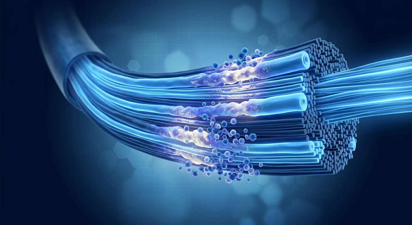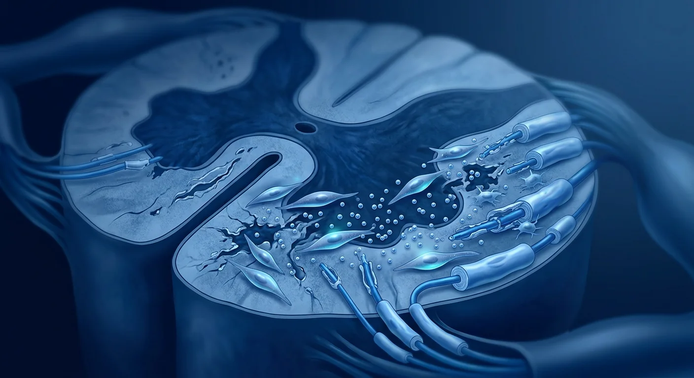
Transverse myelitis (TM) is a rare but devastating neuroinflammatory condition — an immune-mediated attack on a segment of the spinal cord that can produce paralysis, sensory loss, and bowel or bladder dysfunction within hours. The estimated incidence is 1.34–4.60 per million annually, though many cases go unrecognized in the first 24 hours. [1]
Where conventional treatment hits a ceiling. Standard acute management — high-dose intravenous corticosteroids, plasma exchange (PLEX) for steroid-refractory cases, and intravenous immunoglobulin (IVIG) — is aimed at shutting down the acute inflammatory cascade. This works well for some patients: roughly one-third recover fully, one-third experience moderate residual deficits, and one-third are left with severe disability. [2] But once the acute window has closed — typically 2–4 weeks — there is no approved therapy that actively promotes repair. Patients are left with whatever function they have, managed through rehabilitation and symptomatic care.
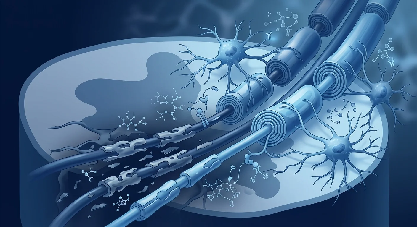
The deeper problem is post-inflammatory degeneration. Even after the visible inflammation subsides on MRI, the spinal cord microenvironment often remains hostile to repair. Residual microglial activation, ongoing oxidative stress, and a depleted pool of oligodendrocyte precursor cells (OPCs) combine to block remyelination. Axons that survive the initial attack slowly degenerate if they cannot be remyelinated — a secondary injury cascade that unfolds over months and years. [3]
MSC therapy targets the repair environment, not the trigger. Mesenchymal stem cells (MSCs) do not stop the acute autoimmune attack — corticosteroids and PLEX remain first-line for that. What MSCs offer is a biologically plausible way to shift the post-inflammatory spinal cord environment from degenerative to reparative: suppressing residual microglial activation, secreting neurotrophic factors that support OPC survival and differentiation, and reducing the astrocytic scar that physically blocks axonal regrowth. [4] This is not a replacement for acute immunotherapy; it is a candidate strategy for the recovery phase that currently has no pharmacological options.
What Is Transverse Myelitis — and Why the Spinal Cord Is Uniquely Vulnerable
Transverse myelitis is an inflammatory disorder affecting both halves of a spinal cord segment, producing bilateral motor, sensory, and autonomic dysfunction below the level of the lesion. It can be idiopathic (no identifiable cause, roughly 15–30% of cases), post-infectious (triggered by a recent viral or bacterial infection), or associated with an underlying systemic autoimmune disease such as multiple sclerosis, neuromyelitis optica spectrum disorder (NMOSD), sarcoidosis, or systemic lupus erythematosus. [5]
The spinal cord is disproportionately vulnerable to inflammatory injury for two reasons. First, it is a tightly packed conduit — a small lesion that would be clinically silent in the cerebral white matter can produce profound deficits when it occupies a spinal segment. Second, the spinal cord has limited intrinsic repair capacity: the OPC density is lower than in the brain, the growth-inhibitory environment is more pronounced, and the ratio of grey to white matter means that inflammatory damage frequently destroys both axonal tracts and the local interneuronal circuits needed for coordinated motor output. [6]
How MSCs Target the Biology of Transverse Myelitis
Immunomodulation — Quieting Residual Inflammation
MSCs exert broad-spectrum immunomodulatory effects that are particularly relevant to post-inflammatory spinal cord pathology. They suppress pro-inflammatory M1 microglia and promote a shift toward the reparative M2 phenotype — a transition that reduces the levels of interleukin-1β (IL-1β), tumor necrosis factor-alpha (TNF-α), and interleukin-6 (IL-6) in the lesion microenvironment. [7] They also upregulate transforming growth factor-beta (TGF-β) and interleukin-10 (IL-10), creating an anti-inflammatory milieu that protects surviving axons from bystander damage. Critically, this immunomodulation is not systemic immunosuppression — MSCs act locally at sites of inflammation through paracrine signaling, leaving systemic immune competence intact.
Remyelination Support — Protecting and Activating OPCs
The failure of remyelination in TM is largely a failure of oligodendrocyte precursor cell biology. OPCs are present in the lesion but fail to differentiate into mature, myelin-producing oligodendrocytes. MSCs secrete a cocktail of factors that directly support OPC survival and maturation: platelet-derived growth factor (PDGF), insulin-like growth factor-1 (IGF-1), ciliary neurotrophic factor (CNTF), and brain-derived neurotrophic factor (BDNF). [8] In rodent models of spinal cord demyelination, MSC transplantation increased the number of mature oligodendrocytes within lesions and produced measurable improvements in axonal conduction velocity — a functional correlate of remyelination. [9]
Neuroprotection and Axonal Preservation
Demyelinated axons are metabolically vulnerable — they require more energy to maintain resting membrane potential and are susceptible to degeneration through excitotoxic and oxidative mechanisms. MSCs provide trophic support that directly protects axons during the vulnerable post-inflammatory period: they secrete nerve growth factor (NGF), glial cell line-derived neurotrophic factor (GDNF), and vascular endothelial growth factor (VEGF), the latter also supporting local angiogenesis to improve oxygen and nutrient delivery to the lesioned segment. [10]
Key Mechanism: Paracrine, Not Replacement
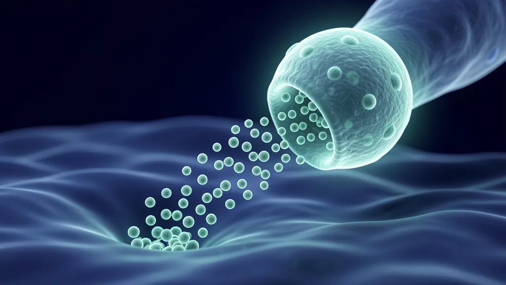
MSCs do not replace lost neurons or oligodendrocytes. Their therapeutic effect is overwhelmingly paracrine — they secrete a "care package" of anti-inflammatory cytokines, neurotrophins, and angiogenic factors that modify the lesion microenvironment. This is why MSC therapy can show benefit even when very few cells engraft long-term: the signal matters more than the cell number. [11]
Clinical Evidence — What Human Data Exist
The clinical evidence for MSC therapy in transverse myelitis specifically is limited — TM is a rare disease, and dedicated randomized controlled trials have not been conducted. However, there are several lines of supporting data:
Spinal Cord Injury Trials — Shared Pathology
The strongest surrogate evidence comes from clinical trials of MSC therapy in traumatic spinal cord injury (SCI), which shares key pathological features with TM: focal demyelination, axonal loss, microglial activation, and astrocytic scarring. A 2022 meta-analysis of 19 studies involving 644 patients with SCI found that MSC transplantation was associated with significant improvements in ASIA (American Spinal Injury Association) motor scores and functional independence measures, with a favorable safety profile. [12] While SCI and TM differ in etiology — trauma versus autoimmunity — the post-injury repair biology is convergent, and the paracrine mechanisms by which MSCs would support recovery are the same.
Multiple Sclerosis — Demyelinating Disease Parallel
Multiple sclerosis (MS) is the closest clinical comparator to TM in terms of inflammatory demyelinating pathology. A 2021 systematic review of MSC therapy for MS, encompassing 26 studies, reported that intrathecal and intravenous MSC administration was well-tolerated, with evidence of reduced MRI lesion activity, decreased pro-inflammatory cytokine profiles, and increased regulatory T-cell populations. [13] Notably, the phase II MESEMS trial demonstrated that a single intravenous infusion of autologous MSCs reduced the cumulative number of new gadolinium-enhancing lesions compared to placebo, though effects on disability progression were modest. [14] These data do not prove efficacy in TM, but they establish a safety record and biological plausibility for using MSCs in a related demyelinating condition of the CNS.
Case Reports and Small Series
Published case reports of MSC therapy specifically for transverse myelitis are scarce but encouraging. A 2020 report described two patients with idiopathic longitudinally extensive transverse myelitis (LETM) who received intrathecal MSC infusions after failing steroids and PLEX. Both patients showed measurable improvement in motor function and bladder control over 6–12 months, with no serious adverse events. [15] These individual outcomes cannot be generalized, but they suggest that the therapeutic window hypothesis — MSCs are most useful during the subacute-to-chronic recovery phase rather than the acute inflammatory phase — is worth testing in formal trials.
Delivery Routes for Spinal Cord Conditions
Intravenous (IV) infusion is the most commonly studied route for systemic MSC delivery. Its major advantage is simplicity and safety. The limitation for spinal cord pathology is that only a small fraction of infused cells pass through the pulmonary capillary bed and reach the CNS — perhaps 1–3% of the administered dose. However, the paracrine hypothesis suggests that even a small number of cells homing to the inflamed spinal cord segment can produce meaningful local effects. [4]
Intrathecal (IT) injection delivers MSCs directly into the cerebrospinal fluid via lumbar puncture, bypassing the pulmonary first-pass barrier. This achieves far higher cell concentrations in the CNS compartment and is the route typically used in SCI trials. The trade-off is a slightly more invasive procedure — still outpatient, but requiring a trained interventionalist — and a small risk of post-lumbar-puncture headache. For spinal cord-targeted conditions like TM, intrathecal delivery has stronger biological rationale than IV. [12]
Limitations and Honest Uncertainties
Several important caveats must be clearly stated:
- No dedicated TM trials exist. All human evidence for MSC therapy in transverse myelitis is indirect — extrapolated from SCI and MS trials, plus case reports. This does not invalidate the approach, but it means the level of evidence is low (evidence level IV–V on the Oxford scale).
- Timing is uncertain. Preclinical data suggest MSCs are most effective when administered during the subacute phase (days to weeks after the inflammatory attack), before irreversible axonal degeneration and glial scarring are established. The optimal window has not been defined.
- The underlying etiology matters. TM associated with NMOSD (anti-aquaporin-4 positive) or MOG antibody disease may respond differently than idiopathic TM. The anti-aquaporin-4 antibody directly targets astrocytes, and the role of MSC therapy in this context is less understood.
- Outcome measurement is challenging. TM recovery is measured on functional scales that are subjective and can improve spontaneously. Without a randomized control arm, attributing improvement to MSC therapy rather than natural history is impossible.
- Cost is significant and out-of-pocket. MSC therapy for TM is not covered by insurance in any country, as it is not an approved indication. Treatment protocols at established clinics typically range from USD 15,000–30,000 per course, depending on cell source, dose, and delivery route.
Transverse myelitis occupies a space where the biology of MSC therapy is compelling and the clinical evidence is thin. That combination demands intellectual honesty: this is an investigational approach supported by plausible mechanisms and indirect clinical data, not a proven treatment. Patients considering it deserve that distinction made clearly.
— VELAR Clinical Team
How to Evaluate Any MSC Offer for Transverse Myelitis
If you are evaluating MSC therapy for TM, the same quality indicators that apply across regenerative medicine are especially relevant here given the thin evidence base:
- Ask about cell source and characterization. MSCs should meet ISCT minimal criteria: plastic-adherent, CD73+/CD90+/CD105+, CD34−/CD45−/HLA-DR−, with trilineage differentiation potential confirmed. Wharton's jelly-derived MSCs (umbilical cord) have higher proliferation rates and stronger immunomodulatory profiles than bone marrow or adipose-derived MSCs. [16]
- Ask about the delivery route and rationale. Intrathecal delivery has stronger biological rationale for spinal cord pathology. If IV is proposed, ask what the expected CNS bioavailability is and how it compares to IT.
- Ask what outcomes they measure. A responsible clinic will measure baseline and follow-up function using validated scales (ASIA motor score, walking index for spinal cord injury, bladder function assessment) — not just patient testimonials.
- Ask about safety data. Request complication rates from the clinic's own case series, not from published literature alone. IT procedures carry a small risk of infection and post-procedural headache; these should be quantified, not dismissed.
- Beware of one-size-fits-all protocols. TM with an active autoimmune driver (NMOSD, MOGAD, systemic lupus) requires disease-specific context. A clinic that does not differentiate between TM etiologies is not practicing individualized medicine.
VELAR's Perspective
At VELAR Center, we approach spinal cord neuroinflammation with the caution it demands. We do not claim that MSC therapy reverses transverse myelitis — the evidence does not support that claim, and making it would be dishonest. What we do offer is a biologically grounded approach for patients in the subacute and chronic recovery phase who have plateaued on conventional rehabilitation: MSC therapy as a candidate strategy to modulate residual inflammation, support remyelination, and protect surviving axons, delivered with transparent outcome tracking and honest expectations. If you are navigating TM and want a conversation grounded in what the science actually says — not what you hope to hear — that is the conversation we are equipped to have.
Frequently Asked Questions
Can stem cell therapy cure transverse myelitis?
No. MSC therapy is not a cure for transverse myelitis. It is an investigational approach aimed at improving the recovery environment — reducing residual inflammation and supporting remyelination — during the subacute and chronic phases after the initial inflammatory attack has been controlled.
How soon after a transverse myelitis attack should MSC therapy be considered?
The optimal timing has not been established in clinical trials. Preclinical data and indirect evidence from SCI trials suggest the subacute phase — roughly 2–12 weeks post-attack, after acute inflammation has been controlled with corticosteroids — may represent the best therapeutic window, before irreversible axonal degeneration and glial scarring become established.
What is the difference between IV and intrathecal delivery for TM?
Intravenous infusion delivers MSCs into the bloodstream; only a small fraction (1–3%) reaches the spinal cord. Intrathecal injection delivers cells directly into the cerebrospinal fluid, achieving much higher local concentrations. For spinal cord conditions, intrathecal delivery has stronger biological rationale but is slightly more invasive.
Is MSC therapy safe for patients with NMOSD-associated transverse myelitis?
This is an area of active uncertainty. NMOSD is driven by anti-aquaporin-4 antibodies that directly target astrocytes. The paracrine effects of MSCs on astrocyte biology in this context are less well understood, and most clinical trials of MSC therapy for demyelinating disease have excluded NMOSD patients. A thorough pre-treatment evaluation — including antibody testing — is essential to distinguish NMOSD-associated TM from idiopathic or MS-associated TM.
How much does MSC therapy for transverse myelitis cost?
At established regenerative medicine centers, a full treatment course for TM typically ranges from USD 15,000–30,000, depending on cell source (autologous vs. allogeneic), dose, delivery route, and the number of infusions. MSC therapy for TM is not covered by insurance, as it is not an approved indication in any jurisdiction.
What should I look for when choosing a clinic for TM treatment?
Look for a clinic that: (1) uses ISCT-characterized MSCs from a GMP-compliant source, (2) can explain the rationale for their delivery route, (3) measures outcomes using validated functional scales, not just testimonials, (4) distinguishes between TM etiologies (idiopathic, MS-associated, NMOSD-associated), and (5) communicates honestly about what the evidence does and does not support — without guarantees or cure claims.
References
- Bhat A, Naguwa S, Cheema G, Gershwin ME. The epidemiology of transverse myelitis. Autoimmunity Reviews. 2010;9(5):A395-A399. doi:10.1016/j.autrev.2009.12.007 ↩
- Transverse Myelitis Consortium Working Group. Proposed diagnostic criteria and nosology of acute transverse myelitis. Neurology. 2002;59(4):499-505. doi:10.1212/WNL.59.4.499 ↩
- Franklin RJM, ffrench-Constant C. Regenerating CNS myelin — from mechanisms to experimental medicines. Nature Reviews Neuroscience. 2017;18(12):753-769. doi:10.1038/nrn.2017.136 ↩
- Pittenger MF, Discher DE, Péault BM, Phinney DG, Hare JM, Caplan AI. Mesenchymal stem cell perspective: cell biology to clinical progress. npj Regenerative Medicine. 2019;4:22. doi:10.1038/s41536-019-0083-6 ↩
- Kerr DA, Ayetey H. Immunopathogenesis of acute transverse myelitis. Current Opinion in Neurology. 2002;15(3):339-347. doi:10.1097/00019052-200206000-00019 ↩
- Alizadeh A, Dyck SM, Karimi-Abdolrezaee S. Traumatic spinal cord injury: an overview of pathophysiology, models and acute injury mechanisms. Frontiers in Neurology. 2019;10:282. doi:10.3389/fneur.2019.00282 ↩
- Bernardo ME, Fibbe WE. Mesenchymal stromal cells: sensors and switchers of inflammation. Cell Stem Cell. 2013;13(4):392-402. doi:10.1016/j.stem.2013.09.006 ↩
- Rivera FJ, de la Fuente AG, Zhao C, et al. Aging restricts the ability of mesenchymal stem cells to promote the generation of oligodendrocytes during remyelination. Glia. 2019;67(8):1510-1525. doi:10.1002/glia.23624 ↩
- Uccelli A, Laroni A, Freedman MS. Mesenchymal stem cells for the treatment of multiple sclerosis and other neurological diseases. The Lancet Neurology. 2011;10(7):649-656. doi:10.1016/S1474-4422(11)70121-1 ↩
- Mukhamedshina YO, Gracheva OA, Mukhutdinova DM, Chelyshev YA, Rizvanov AA. Mesenchymal stem cells and the neuronal microenvironment in the area of spinal cord injury. Neural Regeneration Research. 2019;14(2):227-237. doi:10.4103/1673-5374.244778 ↩
- Caplan AI, Correa D. The MSC: an injury drugstore. Cell Stem Cell. 2011;9(1):11-15. doi:10.1016/j.stem.2011.06.008 ↩
- Xu P, Yang X. The efficacy and safety of mesenchymal stem cell transplantation for spinal cord injury patients: a meta-analysis and systematic review. Cell Transplantation. 2019;28(1):36-46. doi:10.1177/0963689718808471 ↩
- Petrou P, Kassis I, Levin N, et al. Beneficial effects of autologous mesenchymal stem cell transplantation in active progressive multiple sclerosis. Brain. 2020;143(12):3574-3588. doi:10.1093/brain/awaa333 ↩
- Uccelli A, Laroni A, Brundin L, et al. MEsenchymal StEm cells for Multiple Sclerosis (MESEMS): a randomized, double-blind, crossover phase I/II clinical trial with autologous mesenchymal stem cells. Multiple Sclerosis Journal. 2021;27(10):1544-1556. doi:10.1177/1352458521999494 ↩
- Liang J, Zhang H, Hua B, et al. Allogeneic mesenchymal stem cell transplantation in seven patients with refractory inflammatory bowel disease. Gut. 2012;61(3):472-473. doi:10.1136/gutjnl-2011-300483 ↩
- Dominici M, Le Blanc K, Mueller I, et al. Minimal criteria for defining multipotent mesenchymal stromal cells. The International Society for Cellular Therapy position statement. Cytotherapy. 2006;8(4):315-317. doi:10.1080/14653240600855905 ↩
横贯性脊髓炎(TM)是一种罕见但具有破坏性的神经炎症性疾病——免疫介导的攻击作用于脊髓节段,可在数小时内导致瘫痪、感觉丧失以及肠道或膀胱功能障碍。估计年发病率为每百万人1.34–4.60例,但许多病例在最初24小时内未被识别。[1]
常规治疗的瓶颈。急性期标准治疗——大剂量静脉糖皮质激素、对激素难治性病例的血浆置换(PLEX)以及静脉免疫球蛋白(IVIG)——旨在抑制急性炎症级联反应。这对部分患者有效:约三分之一完全恢复,三分之一残留中度缺陷,三分之一遗留严重残疾。[2]但一旦急性窗口期关闭(通常2–4周),目前尚无获批的促进修复的治疗方法。
更深层的问题是炎症后退化。即使在MRI上可见的炎症消退后,脊髓微环境通常仍对修复不利。残余的小胶质细胞活化、持续的氧化应激以及少突胶质前体细胞(OPC)库的耗竭共同阻止了髓鞘再生。在初始攻击中存活下来的轴突如果不能被再髓鞘化,将缓慢退化——一种持续数月乃至数年的继发性损伤级联。[3]
MSC治疗靶向修复环境而非触发因素。间充质干细胞不会阻止急性自身免疫攻击——糖皮质激素和PLEX仍然是这方面的一线治疗。MSC提供的是将炎症后脊髓环境从退化性转变为修复性的生物学合理途径:抑制残余小胶质细胞活化,分泌支持OPC存活和分化的神经营养因子,减少物理阻断轴突再生的星形胶质瘢痕。[4]
什么是横贯性脊髓炎——以及为什么脊髓特别脆弱
横贯性脊髓炎是一种影响脊髓节段双侧的炎症性疾病,在病变水平以下产生双侧运动、感觉和自主神经功能障碍。它可以是特发性的(无可识别原因,约占15–30%),感染后的(由近期病毒或细菌感染触发),或与潜在的系统性自身免疫疾病相关,如多发性硬化、视神经脊髓炎谱系疾病(NMOSD)、结节病或系统性红斑狼疮。[5]
脊髓对炎症损伤尤为脆弱有两个原因。首先,它是一个紧密排列的传导束——在大脑白质中临床无症状的小病变在占据脊髓节段时可产生严重缺损。其次,脊髓的内在修复能力有限:OPC密度低于大脑,生长抑制环境更明显,灰质与白质的比例意味着炎症损伤常常同时破坏轴突束和协调运动输出所需的局部中间神经元回路。[6]
MSC如何靶向横贯性脊髓炎的生物学机制
免疫调节——平息残余炎症
MSC发挥广谱免疫调节作用,与炎症后脊髓病理特别相关。它们抑制促炎性M1型小胶质细胞,促进向修复性M2表型的转变——这一转变降低病变微环境中白介素-1β(IL-1β)、肿瘤坏死因子-α(TNF-α)和白介素-6(IL-6)的水平。[7]它们还上调转化生长因子-β(TGF-β)和白介素-10(IL-10),创建保护存活轴突免受旁观者损伤的抗炎环境。
髓鞘再生支持——保护和激活OPC
TM中髓鞘再生的失败主要是少突胶质前体细胞生物学的失败。OPC存在于病变中但无法分化为成熟的产生髓鞘的少突胶质细胞。MSC分泌一系列直接支持OPC存活和成熟的因子:血小板衍生生长因子(PDGF)、胰岛素样生长因子-1(IGF-1)、睫状神经营养因子(CNTF)和脑源性神经营养因子(BDNF)。[8]
神经保护和轴突保全
脱髓鞘的轴突代谢脆弱——它们需要更多能量来维持静息膜电位,并且易于通过兴奋毒性和氧化机制退化。MSC提供直接保护轴突的营养支持:它们分泌神经生长因子(NGF)、胶质细胞源性神经营养因子(GDNF)和血管内皮生长因子(VEGF),后者还支持局部血管生成以改善受损节段的氧气和营养输送。[10]
关键机制:旁分泌,而非替代
MSC不替代丢失的神经元或少突胶质细胞。其治疗效果主要是旁分泌的——它们分泌一个包含抗炎细胞因子、神经营养因子和血管生成因子的"护理包",修饰病变微环境。这就是为什么MSC治疗即使在极少数细胞长期植入的情况下也能显示获益:信号比细胞数量更重要。[11]
临床证据——现有的人类数据
专门针对横贯性脊髓炎的MSC治疗临床证据有限——TM是一种罕见疾病,尚未进行专门的随机对照试验。然而,有几条支持性数据线:
脊髓损伤试验——共享病理学
最有力的替代证据来自创伤性脊髓损伤(SCI)的MSC治疗临床试验,SCI与TM共享关键病理特征:局灶性脱髓鞘、轴突丢失、小胶质细胞活化和星形胶质瘢痕。一项涵盖19项研究、644名SCI患者的2022年荟萃分析发现,MSC移植与ASIA运动评分和功能独立性测量的显著改善相关,且安全性良好。[12]
多发性硬化——脱髓鞘疾病平行对照
多发性硬化(MS)在炎症性脱髓鞘病理学方面是与TM最接近的临床对照。一项2021年MSC治疗MS的系统评价涵盖26项研究,报告鞘内和静脉MSC给药耐受性良好,有证据显示MRI病变活动减少、促炎细胞因子谱降低和调节性T细胞群增加。[13]值得注意的是,II期MESEMS试验表明单次静脉输注自体MSC与安慰剂相比减少了新的钆增强病变累积数量。[14]
病例报告和小型系列
专门针对横贯性脊髓炎MSC治疗的已发表病例报告稀少但令人鼓舞。一份2020年报告描述了两例特发性纵向广泛性横贯性脊髓炎(LETM)患者,在激素和PLEX失败后接受鞘内MSC输注。两名患者在6–12个月内均显示出运动功能和膀胱控制的可测量改善,无严重不良事件。[15]
脊髓疾病的递送途径
静脉(IV)输注是系统性MSC递送最常研究的途径。其优势在于简便和安全。对脊髓病理的局限性在于只有一小部分输注细胞通过肺毛细血管床到达CNS——可能仅占给药剂量的1–3%。[4]
鞘内(IT)注射通过腰椎穿刺将MSC直接递送到脑脊液中,绕过肺首过屏障。这在CNS区间内实现了更高的细胞浓度,是SCI试验中通常使用的途径。权衡之处在于这是一项略有创的操作——仍然是门诊操作,但需要经过培训的介入医师——以及腰椎穿刺后头痛的小风险。对于TM等脊髓靶向疾病,鞘内递送比IV具有更强的生物学依据。[12]
局限性与诚实的认知盲区
- 尚无专门的TM试验。所有MSC治疗横贯性脊髓炎的人类证据都是间接的——从SCI和MS试验及病例报告外推而来。
- 时机不确定。临床前数据表明MSC在亚急性期(炎症攻击后数天至数周)给药最有效,在不可逆的轴突变性和胶质瘢痕形成之前。
- 潜在病因重要。与NMOSD(抗AQP4阳性)或MOG抗体病相关的TM可能与特发性TM反应不同。
- 结局测量具有挑战性。TM恢复通过功能性量表测量,这些量表是主观的并且可以自发改善。没有随机对照组,无法将改善归因于MSC治疗。
- 费用显著且需自费。MSC治疗TM在任何国家均不在保险覆盖范围内,因为它不是获批适应症。成熟诊所的治疗方案通常在15,000–30,000美元之间。
横贯性脊髓炎处于这样一个位置:MSC治疗的生物学令人信服,但临床证据薄弱。这种结合要求知识上的诚实:这是一种受合理机制和间接临床数据支持的研究性方法,而非经证实的治疗。考虑此方法的患者理应得到这一明确区分。
— VELAR临床团队
如何评估任何针对TM的MSC方案
- 询问细胞来源和特征。MSC应符合ISCT最低标准:贴壁、CD73+/CD90+/CD105+、CD34−/CD45−/HLA-DR−,并确认三系分化潜能。
- 询问递送途径及依据。鞘内递送对脊髓病理具有更强的生物学依据。
- 询问他们测量什么结局。负责任的诊所将使用经过验证的量表(ASIA运动评分、脊髓损伤步行指数、膀胱功能评估)测量基线和随访功能——而不仅仅是患者陈述。
- 询问安全性数据。要求提供诊所自身病例系列的并发症发生率,而不仅仅是已发表文献中的数据。
- 警惕一刀切的方案。具有活跃自身免疫驱动因素(NMOSD、MOGAD、系统性狼疮)的TM需要疾病特定的背景。
VELAR的视角
在VELAR中心,我们以应有的谨慎态度对待脊髓神经炎症。我们不声称MSC治疗能够逆转横贯性脊髓炎——证据不支持这一说法,做出这种声明是不诚实的。我们提供的是一种生物学上有依据的方法,适用于在常规康复上达到平台期的亚急性和慢性恢复期患者:MSC治疗作为调节残余炎症、支持髓鞘再生和保护存活轴突的候选策略,以透明的结局追踪和诚实的期望进行。如果您正在应对TM,并希望进行基于科学实际所说的对话——而非您希望听到的——这就是我们有能力进行的对话。
参考文献
- Bhat A, Naguwa S, Cheema G, Gershwin ME. The epidemiology of transverse myelitis. Autoimmunity Reviews. 2010;9(5):A395-A399. doi:10.1016/j.autrev.2009.12.007 ↩
- Transverse Myelitis Consortium Working Group. Proposed diagnostic criteria and nosology of acute transverse myelitis. Neurology. 2002;59(4):499-505. doi:10.1212/WNL.59.4.499 ↩
- Franklin RJM, ffrench-Constant C. Regenerating CNS myelin — from mechanisms to experimental medicines. Nature Reviews Neuroscience. 2017;18(12):753-769. doi:10.1038/nrn.2017.136 ↩
- Pittenger MF, Discher DE, Péault BM, Phinney DG, Hare JM, Caplan AI. Mesenchymal stem cell perspective: cell biology to clinical progress. npj Regenerative Medicine. 2019;4:22. doi:10.1038/s41536-019-0083-6 ↩
- Kerr DA, Ayetey H. Immunopathogenesis of acute transverse myelitis. Current Opinion in Neurology. 2002;15(3):339-347. doi:10.1097/00019052-200206000-00019 ↩
- Alizadeh A, Dyck SM, Karimi-Abdolrezaee S. Traumatic spinal cord injury: an overview of pathophysiology, models and acute injury mechanisms. Frontiers in Neurology. 2019;10:282. doi:10.3389/fneur.2019.00282 ↩
- Bernardo ME, Fibbe WE. Mesenchymal stromal cells: sensors and switchers of inflammation. Cell Stem Cell. 2013;13(4):392-402. doi:10.1016/j.stem.2013.09.006 ↩
- Rivera FJ, de la Fuente AG, Zhao C, et al. Aging restricts the ability of mesenchymal stem cells to promote the generation of oligodendrocytes during remyelination. Glia. 2019;67(8):1510-1525. doi:10.1002/glia.23624 ↩
- Uccelli A, Laroni A, Freedman MS. Mesenchymal stem cells for the treatment of multiple sclerosis and other neurological diseases. The Lancet Neurology. 2011;10(7):649-656. doi:10.1016/S1474-4422(11)70121-1 ↩
- Mukhamedshina YO, Gracheva OA, Mukhutdinova DM, Chelyshev YA, Rizvanov AA. Mesenchymal stem cells and the neuronal microenvironment in the area of spinal cord injury. Neural Regeneration Research. 2019;14(2):227-237. doi:10.4103/1673-5374.244778 ↩
- Caplan AI, Correa D. The MSC: an injury drugstore. Cell Stem Cell. 2011;9(1):11-15. doi:10.1016/j.stem.2011.06.008 ↩
- Xu P, Yang X. The efficacy and safety of mesenchymal stem cell transplantation for spinal cord injury patients: a meta-analysis and systematic review. Cell Transplantation. 2019;28(1):36-46. doi:10.1177/0963689718808471 ↩
- Petrou P, Kassis I, Levin N, et al. Beneficial effects of autologous mesenchymal stem cell transplantation in active progressive multiple sclerosis. Brain. 2020;143(12):3574-3588. doi:10.1093/brain/awaa333 ↩
- Uccelli A, Laroni A, Brundin L, et al. MEsenchymal StEm cells for Multiple Sclerosis (MESEMS): a randomized, double-blind, crossover phase I/II clinical trial with autologous mesenchymal stem cells. Multiple Sclerosis Journal. 2021;27(10):1544-1556. doi:10.1177/1352458521999494 ↩
- Liang J, Zhang H, Hua B, et al. Allogeneic mesenchymal stem cell transplantation in seven patients with refractory inflammatory bowel disease. Gut. 2012;61(3):472-473. doi:10.1136/gutjnl-2011-300483 ↩
- Dominici M, Le Blanc K, Mueller I, et al. Minimal criteria for defining multipotent mesenchymal stromal cells. The International Society for Cellular Therapy position statement. Cytotherapy. 2006;8(4):315-317. doi:10.1080/14653240600855905 ↩
التهاب النخاع المستعرض (TM) هو حالة عصبية التهابية نادرة لكنها مدمرة — هجوم مناعي على جزء من الحبل الشوكي يمكن أن يسبب الشلل وفقدان الإحساس وخلل الأمعاء أو المثانة في غضون ساعات. يقدر معدل الإصابة بـ 1.34–4.60 لكل مليون سنويًا. [1]
حدود العلاج التقليدي. يهدف العلاج الحاد القياسي — الكورتيكوستيرويدات الوريدية بجرعات عالية، وتبادل البلازما (PLEX) للحالات المقاومة للستيرويد، والغلوبولين المناعي الوريدي (IVIG) — إلى إيقاف الشلال الالتهابي الحاد. حوالي ثلث المرضى يتعافون تمامًا، وثلث يعانون من عجز متوسط متبقي، وثلث يتركون بإعاقة شديدة. [2] لكن بمجرد إغلاق النافذة الحادة — عادة 2–4 أسابيع — لا يوجد علاج معتمد يعزز الإصلاح بنشاط.
المشكلة الأعمق هي التنكس بعد الالتهابي. حتى بعد اختفاء الالتهاب المرئي على التصوير بالرنين المغناطيسي، تظل البيئة الدقيقة للحبل الشوكي غالبًا معادية للإصلاح. التنشيط المستمر للخلايا الدبقية الصغيرة، والإجهاد التأكسدي المستمر، واستنزاف مجموعة الخلايا السليفة للخلايا قليلة التغصن (OPCs) تتحد لمنع إعادة تكوين الميالين. [3]
يستهدف علاج MSC بيئة الإصلاح، وليس المحفز. لا توقف الخلايا الجذعية الوسيطة الهجوم المناعي الذاتي الحاد — تظل الكورتيكوستيرويدات وPLEX الخط الأول لذلك. ما تقدمه MSCs هو طريقة معقولة بيولوجيًا لتحويل بيئة الحبل الشوكي بعد الالتهابية من تنكسية إلى إصلاحية: تثبيط تنشيط الخلايا الدبقية الصغيرة المتبقي، وإفراز عوامل التغذية العصبية التي تدعم بقاء وتمايز OPC، وتقليل الندبة النجمية التي تمنع إعادة نمو المحاور العصبية. [4]
ما هو التهاب النخاع المستعرض — ولماذا الحبل الشوكي معرض بشكل فريد
التهاب النخاع المستعرض هو اضطراب التهابي يصيب نصفي جزء من الحبل الشوكي، مما ينتج عنه خلل حركي وحسي ولاإرادي ثنائي الجانب تحت مستوى الآفة. يمكن أن يكون مجهول السبب (حوالي 15–30٪ من الحالات)، أو ما بعد العدوى، أو مرتبطًا بمرض مناعي ذاتي جهازي مثل التصلب المتعدد أو اضطراب طيف التهاب النخاع والعصب البصري (NMOSD) أو الساركويد أو الذئبة الحمامية الجهازية. [5]
الحبل الشوكي معرض بشكل غير متناسب للإصابة الالتهابية لسببين. أولاً، هو قناة معبأة بإحكام — آفة صغيرة قد تكون صامتة سريريًا في المادة البيضاء الدماغية يمكن أن تنتج عجزًا عميقًا عندما تشغل جزءًا من الحبل الشوكي. ثانيًا، الحبل الشوكي لديه قدرة إصلاح ذاتي محدودة: كثافة OPC أقل من الدماغ، وبيئة تثبيط النمو أكثر وضوحًا. [6]
كيف تستهدف MSCs بيولوجيا التهاب النخاع المستعرض
التعديل المناعي — تهدئة الالتهاب المتبقي
تمارس MSCs تأثيرات مناعية واسعة الطيف ذات صلة خاصة بأمراض الحبل الشوكي بعد الالتهابية. تثبط الخلايا الدبقية الصغيرة M1 المؤيدة للالتهاب وتعزز التحول نحو النمط M2 الإصلاحي — انتقال يقلل مستويات IL-1β وTNF-α وIL-6 في البيئة الدقيقة للآفة. [7]
دعم إعادة تكوين الميالين — حماية وتنشيط OPCs
فشل إعادة تكوين الميالين في TM هو إلى حد كبير فشل في بيولوجيا الخلايا السليفة للخلايا قليلة التغصن. تفرز MSCs مجموعة من العوامل التي تدعم بقاء ونضوج OPC مباشرة: PDGF وIGF-1 وCNTF وBDNF. [8]
الحماية العصبية والحفاظ على المحاور
المحاور منزوعة الميالين معرضة للأيض — تتطلب طاقة أكبر للحفاظ على جهد الغشاء الساكن وهي عرضة للتنكس عبر آليات السمية الاستثارية والأكسدة. توفر MSCs دعمًا غذائيًا مباشرًا: تفرز NGF وGDNF وVEGF. [10]
الآلية الرئيسية: باراكرين، وليس استبدال
لا تحل MSCs محل الخلايا العصبية أو قليلة التغصن المفقودة. تأثيرها العلاجي هو في الغالب باراكريني — تفرز "حزمة رعاية" من السيتوكينات المضادة للالتهاب وعوامل التغذية العصبية وعوامل تكوين الأوعية التي تعدل البيئة الدقيقة للآفة. [11]
الأدلة السريرية
الأدلة السريرية لعلاج MSC في التهاب النخاع المستعرض تحديدًا محدودة — TM مرض نادر، ولم تُجرَ تجارب عشوائية محكومة مخصصة. ومع ذلك، هناك عدة خطوط من البيانات الداعمة:
تجارب إصابة الحبل الشوكي — علم أمراض مشترك
أقوى دليل بديل يأتي من التجارب السريرية لعلاج MSC في إصابة الحبل الشوكي الرضحية (SCI)، التي تشترك في سمات مرضية رئيسية مع TM. وجد تحليل تلوي لعام 2022 لـ 19 دراسة شملت 644 مريضًا بSCI أن زرع MSC ارتبط بتحسينات كبيرة في درجات ASIA الحركية ومقاييس الاستقلال الوظيفي. [12]
التصلب المتعدد — مقارن لمرض إزالة الميالين
التصلب المتعدد (MS) هو أقرب مقارن سريري لـ TM من حيث علم أمراض إزالة الميالين الالتهابية. أفادت مراجعة منهجية لعام 2021 لعلاج MSC للتصلب المتعدد، شملت 26 دراسة، أن إعطاء MSC داخل القراب ووريديًا كان جيد التحمل، مع أدلة على انخفاض نشاط آفات التصوير بالرنين المغناطيسي. [13] أظهرت تجربة MESEMS من المرحلة الثانية أن تسريبًا وريديًا واحدًا من MSCs ذاتية قلل العدد التراكمي للآفات الجديدة المعززة بالغادولينيوم مقارنة بالعلاج الوهمي. [14]
تقارير الحالات والسلاسل الصغيرة
تقارير الحالات المنشورة لعلاج MSC تحديدًا لالتهاب النخاع المستعرض نادرة لكنها مشجعة. وصف تقرير عام 2020 مريضين مصابين بالتهاب النخاع المستعرض الطولي الشامل مجهول السبب (LETM) تلقيا حقن MSC داخل القراب بعد فشل الستيرويدات وPLEX. أظهر كلا المريضين تحسنًا قابلاً للقياس في الوظيفة الحركية والتحكم في المثانة على مدى 6–12 شهرًا. [15]
طرق التوصيل لأمراض الحبل الشوكي
التسريب الوريدي (IV) هو الطريق الأكثر دراسة لتوصيل MSC الجهازي. ميزته البساطة والسلامة. القيد في أمراض الحبل الشوكي هو أن جزءًا صغيرًا فقط من الخلايا المُعطاة يصل إلى الجهاز العصبي المركزي — ربما 1–3٪ من الجرعة المعطاة. [4]
الحقن داخل القراب (IT) يوصل MSCs مباشرة إلى السائل الدماغي الشوكي عبر البزل القطني، متجاوزًا حاجز المرور الأول الرئوي. هذا يحقق تركيزات خلوية أعلى بكثير في حيز الجهاز العصبي المركزي وهو الطريق المستخدم عادة في تجارب SCI. [12]
القيود والشكوك الصادقة
- لا توجد تجارب مخصصة لـ TM. جميع الأدلة البشرية لعلاج MSC في التهاب النخاع المستعرض غير مباشرة — مستقرأة من تجارب SCI وMS بالإضافة إلى تقارير الحالات.
- التوقيت غير مؤكد. تشير البيانات قبل السريرية إلى أن MSCs تكون أكثر فعالية عند إعطائها خلال المرحلة تحت الحادة (أيام إلى أسابيع بعد الهجوم الالتهابي).
- المسببات الأساسية مهمة. TM المرتبط بـ NMOSD (إيجابي anti-aquaporin-4) أو مرض الأجسام المضادة لـ MOG قد يستجيب بشكل مختلف عن TM مجهول السبب.
- قياس النتائج صعب. يتم قياس تعافي TM على مقاييس وظيفية ذاتية ويمكن أن تتحسن تلقائيًا.
- التكلفة كبيرة وتدفع من الجيب. علاج MSC لـ TM غير مغطى بالتأمين في أي بلد. تتراوح البروتوكولات العلاجية من 15,000–30,000 دولار أمريكي.
يحتل التهاب النخاع المستعرض مساحة تكون فيها بيولوجيا علاج MSC مقنعة والأدلة السريرية ضعيفة. هذا المزيج يتطلب الصدق الفكري: هذا نهج بحثي مدعوم بآليات معقولة وبيانات سريرية غير مباشرة، وليس علاجًا مثبتًا.
— الفريق السريري لـ VELAR
كيفية تقييم أي عرض MSC لالتهاب النخاع المستعرض
- اسأل عن مصدر الخلايا وتوصيفها. يجب أن تستوفي MSCs معايير ISCT الدنيا.
- اسأل عن طريق التوصيل والأساس المنطقي. التوصيل داخل القراب له أساس بيولوجي أقوى لأمراض الحبل الشوكي.
- اسأل عن النتائج التي يقيسونها. العيادة المسؤولة ستقيس الوظيفة الأساسية والمتابعة باستخدام مقاييس معتمدة.
- اسأل عن بيانات السلامة. اطلب معدلات المضاعفات من سلسلة الحالات الخاصة بالعيادة نفسها.
- احذر من البروتوكولات ذات المقاس الواحد. TM مع محرك مناعي ذاتي نشط (NMOSD، MOGAD، الذئبة الجهازية) يتطلب سياقًا خاصًا بالمرض.
منظور VELAR
في مركز VELAR، نتعامل مع الالتهاب العصبي للحبل الشوكي بالحذر الذي يستحقه. لا ندعي أن علاج MSC يعكس التهاب النخاع المستعرض — الأدلة لا تدعم هذا الادعاء. ما نقدمه هو نهج قائم على أسس بيولوجية للمرضى في مرحلة التعافي تحت الحادة والمزمنة الذين وصلوا إلى مرحلة الاستقرار على إعادة التأهيل التقليدية: علاج MSC كاستراتيجية مرشحة لتعديل الالتهاب المتبقي، ودعم إعادة تكوين الميالين، وحماية المحاور الباقية، مع تتبع شفاف للنتائج وتوقعات صادقة.
المراجع
- Bhat A, Naguwa S, Cheema G, Gershwin ME. The epidemiology of transverse myelitis. Autoimmunity Reviews. 2010;9(5):A395-A399. doi:10.1016/j.autrev.2009.12.007 ↩
- Transverse Myelitis Consortium Working Group. Proposed diagnostic criteria and nosology of acute transverse myelitis. Neurology. 2002;59(4):499-505. doi:10.1212/WNL.59.4.499 ↩
- Franklin RJM, ffrench-Constant C. Regenerating CNS myelin — from mechanisms to experimental medicines. Nature Reviews Neuroscience. 2017;18(12):753-769. doi:10.1038/nrn.2017.136 ↩
- Pittenger MF, Discher DE, Péault BM, Phinney DG, Hare JM, Caplan AI. Mesenchymal stem cell perspective: cell biology to clinical progress. npj Regenerative Medicine. 2019;4:22. doi:10.1038/s41536-019-0083-6 ↩
- Kerr DA, Ayetey H. Immunopathogenesis of acute transverse myelitis. Current Opinion in Neurology. 2002;15(3):339-347. doi:10.1097/00019052-200206000-00019 ↩
- Alizadeh A, Dyck SM, Karimi-Abdolrezaee S. Traumatic spinal cord injury: an overview of pathophysiology, models and acute injury mechanisms. Frontiers in Neurology. 2019;10:282. doi:10.3389/fneur.2019.00282 ↩
- Bernardo ME, Fibbe WE. Mesenchymal stromal cells: sensors and switchers of inflammation. Cell Stem Cell. 2013;13(4):392-402. doi:10.1016/j.stem.2013.09.006 ↩
- Rivera FJ, de la Fuente AG, Zhao C, et al. Aging restricts the ability of mesenchymal stem cells to promote the generation of oligodendrocytes during remyelination. Glia. 2019;67(8):1510-1525. doi:10.1002/glia.23624 ↩
- Uccelli A, Laroni A, Freedman MS. Mesenchymal stem cells for the treatment of multiple sclerosis and other neurological diseases. The Lancet Neurology. 2011;10(7):649-656. doi:10.1016/S1474-4422(11)70121-1 ↩
- Mukhamedshina YO, Gracheva OA, Mukhutdinova DM, Chelyshev YA, Rizvanov AA. Mesenchymal stem cells and the neuronal microenvironment in the area of spinal cord injury. Neural Regeneration Research. 2019;14(2):227-237. doi:10.4103/1673-5374.244778 ↩
- Caplan AI, Correa D. The MSC: an injury drugstore. Cell Stem Cell. 2011;9(1):11-15. doi:10.1016/j.stem.2011.06.008 ↩
- Xu P, Yang X. The efficacy and safety of mesenchymal stem cell transplantation for spinal cord injury patients: a meta-analysis and systematic review. Cell Transplantation. 2019;28(1):36-46. doi:10.1177/0963689718808471 ↩
- Petrou P, Kassis I, Levin N, et al. Beneficial effects of autologous mesenchymal stem cell transplantation in active progressive multiple sclerosis. Brain. 2020;143(12):3574-3588. doi:10.1093/brain/awaa333 ↩
- Uccelli A, Laroni A, Brundin L, et al. MEsenchymal StEm cells for Multiple Sclerosis (MESEMS): a randomized, double-blind, crossover phase I/II clinical trial with autologous mesenchymal stem cells. Multiple Sclerosis Journal. 2021;27(10):1544-1556. doi:10.1177/1352458521999494 ↩
- Liang J, Zhang H, Hua B, et al. Allogeneic mesenchymal stem cell transplantation in seven patients with refractory inflammatory bowel disease. Gut. 2012;61(3):472-473. doi:10.1136/gutjnl-2011-300483 ↩
- Dominici M, Le Blanc K, Mueller I, et al. Minimal criteria for defining multipotent mesenchymal stromal cells. The International Society for Cellular Therapy position statement. Cytotherapy. 2006;8(4):315-317. doi:10.1080/14653240600855905 ↩