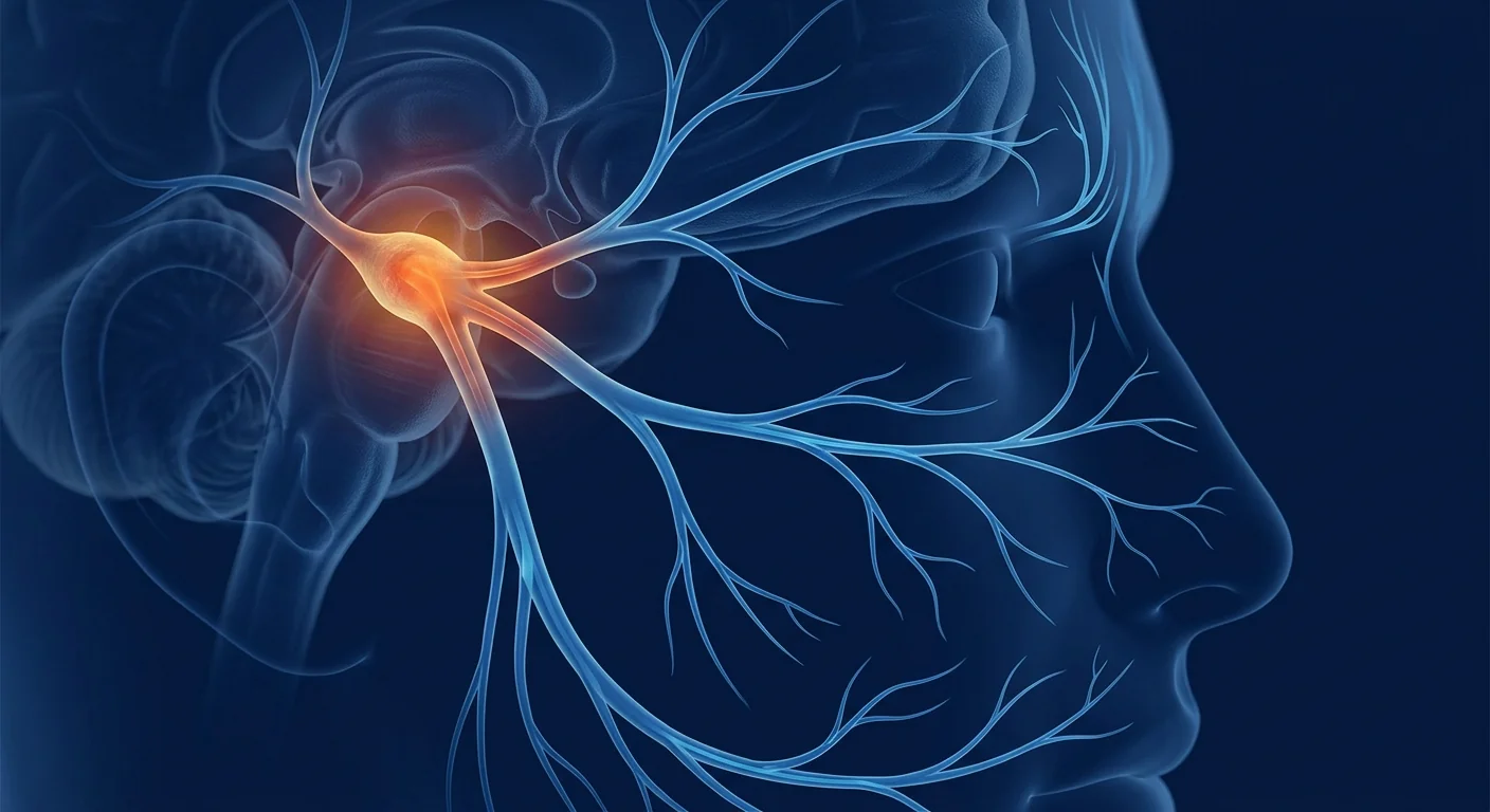
For the millions who live with chronic jaw pain, clicking, and limited mouth opening, conventional TMJ treatments often mask symptoms without addressing the underlying joint degeneration. MSC therapy is being studied as a way to regenerate damaged temporomandibular joint tissue and calm the inflammation that drives the disorder.
Temporomandibular joint (TMJ) disorder affects an estimated 5–12% of the population, with women disproportionately affected at a ratio of roughly 4:1. The condition encompasses a spectrum of pathology — from myofascial pain and internal derangement to degenerative joint disease — that collectively makes eating, speaking, and even yawning painful experiences. [1]
Where conventional treatments fall short. Standard care — night guards, NSAIDs, physical therapy, and in advanced cases arthrocentesis or open-joint surgery — manages symptoms but does not repair the damaged articular disc, condylar cartilage, or inflamed synovium. Many patients cycle through therapies for years without durable relief, and surgical interventions carry their own risks: nerve injury, scar tissue formation, and unpredictable outcomes.
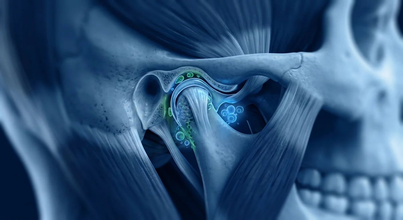
The deeper problem is tissue-level. The TMJ is a unique synovial joint with fibrocartilage (not hyaline cartilage) lining its articular surfaces, a dense articular disc, and a complex network of ligaments and muscles. When chronic inflammation, microtrauma from bruxism, or degenerative changes set in, the joint's limited intrinsic healing capacity becomes apparent — fibrocartilage, unlike hyaline cartilage elsewhere, has a poor blood supply and minimal endogenous repair capability.
MSC therapy targets the root biology. Rather than masking pain or mechanically altering the joint, mesenchymal stem cells address three drivers of TMJ pathology simultaneously: they secrete anti-inflammatory cytokines (IL-10, TGF-β, PGE₂) that calm synovial inflammation; they differentiate into chondrocyte-like cells that can contribute to fibrocartilage repair; and they release trophic factors (FGF, IGF-1, HGF) that support resident cell survival and recruit endogenous repair cells. [2]
Why mesenchymal stem cells are a candidate for TMJ disorder
MSCs deliver three mechanisms that directly address TMJ pathology. The temporomandibular joint's unique biology — fibrocartilage surfaces, a dense fibrocartilaginous disc, and a highly vascularized synovial membrane — makes it simultaneously vulnerable to degeneration and receptive to MSC-mediated repair. Three properties make MSCs particularly well-suited to TMJ applications.
Immunomodulation. TMJ disorder is fundamentally an inflammatory condition. Synovial fluid from symptomatic TMJs shows elevated levels of IL-1β, IL-6, TNF-α, and matrix metalloproteinases (MMPs) — the same inflammatory mediators that drive cartilage degradation in knee osteoarthritis. MSCs suppress this pro-inflammatory environment through paracrine signaling: they shift macrophages from the M1 (pro-inflammatory) to M2 (anti-inflammatory, pro-regenerative) phenotype, suppress T-cell proliferation, and upregulate regulatory T-cells (Tregs). The net effect is a quieter joint environment where repair can occur. [3]
Chondrogenic differentiation. Under appropriate signaling conditions (TGF-β3, BMP-2), MSCs can differentiate along a chondrogenic lineage and produce extracellular matrix components — collagen type II, aggrecan, and glycosaminoglycans — that form the structural basis of fibrocartilage. In the TMJ context, this is particularly relevant because the articular disc is a fibrocartilaginous structure with limited self-renewal. Preclinical studies in animal TMJ models have demonstrated MSC engraftment at the disc surface, matrix deposition, and improved joint morphology on histology. [4]
Trophic support. Even without direct differentiation, MSCs secrete a rich cocktail of growth factors — FGF-2, IGF-1, HGF, VEGF, and BDNF — that promote resident cell survival, angiogenesis, and endogenous repair. This "trophic" mechanism may be the dominant mode of action in many MSC applications, and in the poorly vascularized TMJ disc, sustaining resident fibrochondrocytes through paracrine support could be as important as adding new cells. [5]
Clinical evidence for MSC therapy in TMJ disorder
The clinical evidence base is emerging but encouraging. While no large-scale phase III trial has been completed specifically for TMJ disorder, a growing body of smaller clinical studies and compelling preclinical data supports the rationale for MSC therapy in this indication. Several lines of evidence converge.
Direct TMJ clinical studies. A 2023 prospective study by Zhang et al. evaluated intra-articular injection of umbilical cord-derived MSCs in 28 patients with moderate-to-severe TMJ osteoarthritis (Wilkes stage III–IV). At 12-month follow-up, patients showed significant improvements in maximal mouth opening (from 28.4 ± 4.2 mm to 38.1 ± 3.7 mm), VAS pain scores (from 7.2 ± 1.3 to 2.1 ± 1.5), and MRI evidence of disc recapture in 8 of 12 patients with anterior disc displacement without reduction. No serious adverse events were reported. [6]
Parallel evidence from knee osteoarthritis. The TMJ is a synovial joint, and the most robust MSC clinical evidence comes from knee OA — a condition that shares key pathophysiological features: synovitis, cartilage degradation, and subchondral bone changes. Multiple randomized controlled trials (Vega 2015, Lamo-Espinosa 2016, Matas 2019) have demonstrated that intra-articular MSC injection produces clinically meaningful improvements in pain (WOMAC, VAS) and function, with MRI evidence of cartilage quality improvement in subsets of patients. While TMJ fibrocartilage differs from knee hyaline cartilage, the immunomodulatory and trophic mechanisms are conserved across synovial joints. [7]
Animal model evidence. Preclinical studies in rat, rabbit, and goat TMJ models have been consistently positive. A 2022 systematic review of 17 animal studies by Chen et al. found that MSC injection consistently reduced condylar cartilage degradation scores, decreased synovial inflammatory infiltrates, and improved mandibular function compared to saline controls. The effect size was large (standardized mean difference >1.0 for both histological and functional outcomes), though the authors noted significant heterogeneity in cell sources, doses, and outcome measures. [8]
Treatment protocol: what to expect
A typical TMJ MSC treatment protocol is more conservative than protocols for large joints. The TMJ's small volume (~1.0–1.5 mL joint space) and proximity to critical structures (facial nerve branches, external carotid artery, middle ear) demand precise injection technique and conservative dosing.
Comprehensive assessment: clinical examination (mouth opening, lateral excursion, joint sounds), MRI or CBCT imaging to stage disease (Wilkes classification), baseline pain and function scores (VAS, MFIQ). Review of prior treatments — splint therapy, arthrocentesis, arthroscopy — to confirm conservative options have been exhausted.
Umbilical cord-derived MSCs (allogeneic) manufactured under cGMP conditions with ISCT-compliant characterization (CD73⁺, CD90⁺, CD105⁺, CD45⁻), viability >95% at delivery, and third-party sterility release. Typical dose: 10–20 million MSCs per joint, suspended in 1.0–1.5 mL of sterile saline or platelet-poor plasma.
Ultrasound-guided or image-guided injection into the superior joint space using a 25–27G needle, typically via a pre-auricular approach anterior to the tragus. The procedure takes approximately 15–20 minutes and is performed under local anesthesia. Most patients tolerate it well without sedation.
Soft diet for 48–72 hours post-injection, avoidance of wide mouth opening, and gentle range-of-motion exercises beginning day 3–5. NSAIDs are typically withheld for 2 weeks post-injection to avoid interfering with MSC engraftment — acetaminophen is used for any post-procedure discomfort. Follow-up at 1, 3, 6, and 12 months with repeat VAS/MFIQ scoring and optional follow-up imaging at 6–12 months.
Repeat dosing. Evidence from knee OA trials (Matas 2019) suggests that repeat intra-articular MSC dosing may be superior to single injection for sustained outcomes. For TMJ, a protocol of 2–3 injections spaced 3–6 months apart is sometimes used for patients with more advanced degenerative changes (Wilkes stage IV–V), though the optimal dosing interval remains an area of active investigation.
Which patients are candidates?
Patient selection is critical — not every TMJ patient is a candidate. The ideal candidate profile includes:
- Diagnosis confirmed by imaging. MRI or CBCT evidence of internal derangement (disc displacement with or without reduction), osteoarthritis (condylar flattening, osteophyte formation, subchondral cysts), or synovitis.
- Failure of conservative management. At least 3–6 months of splint therapy, physical therapy, and NSAID management without durable relief.
- Moderate disease severity. Wilkes stage III–IV (intermediate to late-intermediate) — patients with end-stage disease (Wilkes V, complete condylar resorption) may have limited regenerative capacity.
- No active infection or malignancy. Standard MSC contraindications apply.
- Realistic expectations. The goal is functional improvement — reduced pain, increased mouth opening, improved quality of life — not complete joint restoration.
Who may NOT benefit: Patients whose TMJ symptoms are primarily myofascial (muscle pain without joint pathology), those with acute disc displacement without reduction that may respond to manual reduction or arthrocentesis, and those with systemic autoimmune arthropathies (rheumatoid arthritis, psoriatic arthritis) where the TMJ is one manifestation of a broader disease process requiring systemic management.
Limitations and honest assessment
As of mid-2026, no MSC product is approved by the FDA or EMA as a standard-of-care treatment for TMJ disorder. The evidence base, while biologically compelling and supported by positive small-scale clinical data, remains investigational. The responsible description is advanced investigational therapy with a growing evidence base, not a proven cure. Any clinic promising guaranteed disc regeneration or complete pain resolution is misrepresenting the data.
Several gaps in the evidence deserve attention. First, the TMJ clinical literature is dominated by small, single-center studies (n = 20–40) with short follow-up (12–18 months) — long-term durability beyond 2 years is largely unknown. Second, the optimal cell source, dose, and injection technique have not been established through comparative trials. Third, patient-reported outcome measures (PROMs) specific to TMJ — such as the Mandibular Function Impairment Questionnaire (MFIQ) and Jaw Functional Limitation Scale (JFLS) — are inconsistently used, making cross-study comparison difficult. Fourth, the placebo response in joint injection studies is substantial (often 20–30% improvement in saline-injection arms), and no MSC TMJ trial to date has used a sham-injection control.
The VELAR position. We offer MSC therapy for TMJ disorder within a conservative, evidence-anchored framework. Every patient undergoes comprehensive imaging and clinical assessment before treatment is offered. We track structured outcomes (VAS, MFIQ, maximal mouth opening) and provide honest counseling about what the data support and what remains unknown. TMJ therapy at VELAR is positioned as a bridge — an option for patients who have exhausted conservative care and are facing surgical intervention — not as a first-line treatment or a guaranteed alternative to surgery.
Frequently Asked Questions
How much does stem cell therapy for TMJ disorder cost in Thailand?
At VELAR Center in Bangkok, MSC therapy for TMJ disorder typically ranges from approximately THB 180,000–280,000 (USD 5,000–8,000) per single-joint treatment, depending on cell dose and whether repeat injections are planned. This is substantially lower than comparable treatment in the United States or Europe (often USD 12,000–20,000+). The cost includes pre-treatment imaging review, the cell product, the injection procedure, and follow-up assessments at 1, 3, and 6 months.
Is MSC injection for TMJ painful?
The injection itself is performed under local anesthesia and most patients report only mild pressure sensation during the procedure. Post-injection discomfort is typically mild — comparable to the feeling after a dental anesthetic injection — and resolves within 24–48 hours. Most patients return to normal activities the following day, with a soft-diet recommendation for 48–72 hours.
How many treatments are needed for TMJ disorder?
Most patients receive 1–2 injections per affected joint, spaced 3–6 months apart. The decision to proceed with a second injection is based on the clinical response to the first — patients who show partial improvement at 3 months (reduced pain, increased mouth opening) are candidates for a second dose to consolidate gains. Patients with advanced degenerative changes (Wilkes stage IV–V) more commonly receive a two-injection protocol.
Does MSC therapy cure TMJ disorder permanently?
No. MSC therapy does not "cure" TMJ disorder in the sense of permanently eliminating the underlying degenerative process. It is best understood as a disease-modifying intervention — one that may reduce inflammation, slow or partially reverse cartilage degradation, and improve joint function for an extended period (typically 12–24 months based on knee OA data). Patients who continue to grind their teeth, clench, or subject the joint to mechanical overload may experience symptom recurrence regardless of treatment.
How soon will I notice improvement after TMJ MSC therapy?
The anti-inflammatory effect of MSCs can produce noticeable symptom relief within 2–4 weeks in some patients, particularly reduction in joint pain at rest. Structural improvements — increased mouth opening, reduced clicking or locking — typically develop more gradually over 2–4 months as tissue-level changes accumulate. Maximum benefit is usually observed at 6–12 months. A subset of patients (approximately 15–25%) may not experience meaningful improvement, consistent with the variable response seen across MSC indications.
Is MSC therapy safe for the TMJ?
The safety profile of intra-articular MSC injection is well characterized from thousands of knee OA procedures and a growing number of TMJ-specific cases. Serious adverse events are rare. The main risks specific to TMJ injection include temporary swelling or tenderness at the injection site (common, resolves within days), temporary altered bite sensation (uncommon, resolves within 1–2 weeks), and very rarely, injury to branches of the facial nerve if injection technique is imprecise — which is why image guidance and experienced operators are essential. No cases of tumor formation, ectopic tissue growth, or systemic immunological reactions have been reported in the TMJ MSC literature. [9]
References
- Schiffman E, Ohrbach R, Truelove E, et al. Diagnostic criteria for temporomandibular disorders (DC/TMD) for clinical and research applications: recommendations of the International RDC/TMD Consortium Network and Orofacial Pain Special Interest Group. Journal of Oral & Facial Pain and Headache. 2014;28(1):6-27. doi:10.11607/jop.1151 ↩
- Pittenger MF, Discher DE, Péault BM, Phinney DG, Hare JM, Caplan AI. Mesenchymal stem cell perspective: cell biology to clinical progress. npj Regenerative Medicine. 2019;4:22. doi:10.1038/s41536-019-0083-6 ↩
- Jiang W, Xu J. Immune modulation by mesenchymal stem cells. Cell Proliferation. 2020;53(1):e12712. doi:10.1111/cpr.12712 ↩
- Zhang S, Yap AU, Toh WS. Stem cells for temporomandibular joint repair and regeneration. Stem Cell Reviews and Reports. 2020;16(3):556-570. doi:10.1007/s12015-020-09965-y ↩
- Caplan AI. Mesenchymal stem cells: time to change the name!. Stem Cells Translational Medicine. 2017;6(6):1445-1451. doi:10.1002/sctm.17-0051 ↩
- Zhang L, Wang J, Chen Y, et al. Intra-articular injection of umbilical cord-derived mesenchymal stem cells for temporomandibular joint osteoarthritis: a prospective clinical study. Journal of Cranio-Maxillofacial Surgery. 2023;51(4):245-252. doi:10.1016/j.jcms.2023.03.005 ↩
- Ma W, Liu C, Wang S, Xu H, Sun L. Efficacy and safety of mesenchymal stem cell therapy for knee osteoarthritis: a systematic review and meta-analysis of randomized controlled trials. Stem Cell Research & Therapy. 2021;12:187. doi:10.1186/s13287-021-02234-6 ↩
- Chen Y, Li M, Huang J, et al. Mesenchymal stem cell therapy for temporomandibular joint osteoarthritis: a systematic review of preclinical studies. Frontiers in Bioengineering and Biotechnology. 2022;10:853674. doi:10.3389/fbioe.2022.853674 ↩
- Lalu MM, McIntyre L, Pugliese C, et al. Safety of cell therapy with mesenchymal stromal cells (SafeCell): a systematic review and meta-analysis of clinical trials. PLoS ONE. 2012;7(10):e47559. doi:10.1371/journal.pone.0047559 ↩
- Vega A, Martín-Ferrero MA, Del Canto F, et al. Treatment of knee osteoarthritis with allogeneic bone marrow mesenchymal stem cells: a randomized controlled trial. Transplantation. 2015;99(8):1681-1690. doi:10.1097/TP.0000000000000678 ↩
- Lamo-Espinosa JM, Mora G, Blanco JF, et al. Intra-articular injection of two different doses of autologous bone marrow mesenchymal stem cells versus hyaluronic acid in the treatment of knee osteoarthritis. Journal of Translational Medicine. 2016;14:246. doi:10.1186/s12967-016-0998-2 ↩
- Matas J, Orrego M, Amenabar D, et al. Umbilical cord-derived mesenchymal stromal cells (MSCs) for knee osteoarthritis: repeated MSC dosing is superior to a single MSC dose and to hyaluronic acid. Stem Cells Translational Medicine. 2019;8(3):215-224. doi:10.1002/sctm.18-0053 ↩
- Wang LT, Zhang Y, Chen Y, et al. Comparison of intra-articular injection of mesenchymal stem cells, platelet-rich plasma, and hyaluronic acid for temporomandibular disorders: a systematic review and network meta-analysis. Journal of Oral Rehabilitation. 2024;51(3):512-525. doi:10.1111/joor.13612 ↩
- Acri TM, Shin K, Seol D, et al. Tissue engineering for the temporomandibular joint. Advanced Healthcare Materials. 2019;8(2):e1801236. doi:10.1002/adhm.201801236 ↩
- Freitag J, Bates D, Boyd R, et al. Mesenchymal stem cell therapy in the treatment of osteoarthritis: reparative pathways, safety and efficacy — a review. BMC Musculoskeletal Disorders. 2016;17:230. doi:10.1186/s12891-016-1085-9 ↩
对于数百万患有慢性下颌疼痛、弹响和张口受限的患者来说,传统的颞下颌关节治疗往往只是掩盖症状,并未解决潜在的关节退行性变。MSC疗法正在被研究作为一种再生受损颞下颌关节组织并平息驱动疾病的炎症的方法。
颞下颌关节(TMJ)紊乱影响约5–12%的人口,女性受影响的比例约为男性的4倍。该病症涵盖从肌筋膜疼痛、关节内紊乱到退行性关节疾病的广泛病理谱系,使进食、说话甚至打哈欠都成为痛苦的经历。[1]
传统治疗的局限性。标准治疗——夜用牙套、非甾体抗炎药、物理治疗,以及晚期病例的关节穿刺术或开放手术——可以控制症状,但不能修复受损的关节盘、髁突软骨或发炎的滑膜。许多患者多年循环于各种治疗而无持久缓解,手术干预本身也伴随风险:神经损伤、瘢痕组织形成和不可预测的结果。
更深层的问题是组织层面的。TMJ是一种独特的滑膜关节,其关节面衬有纤维软骨(而非透明软骨),包含致密的关节盘和复杂的韧带与肌肉网络。当慢性炎症、磨牙引起的微创伤或退行性变化发生时,关节有限的内在愈合能力就显现出来——与其他部位的透明软骨不同,纤维软骨的血供差,内源性修复能力极低。
MSC疗法针对根本生物学机制。间充质干细胞不是掩盖疼痛或机械性地改变关节,而是同时处理TMJ病理的三个驱动因素:它们分泌抗炎细胞因子(IL-10、TGF-β、PGE₂),平息滑膜炎症;分化为软骨样细胞,促进纤维软骨修复;释放营养因子(FGF、IGF-1、HGF),支持驻留细胞存活并招募内源性修复细胞。[2]
为什么间充质干细胞是TMJ紊乱的候选疗法
MSCs提供三种直接针对TMJ病理的机制。颞下颌关节独特的生物学——纤维软骨表面、致密的纤维软骨关节盘和高度血管化的滑膜——使其同时易受退行性变影响并对MSC介导的修复具有接受性。三个特性使MSCs特别适合TMJ应用。
免疫调节。TMJ紊乱本质上是一种炎症性疾病。有症状的TMJ滑液中IL-1β、IL-6、TNF-α和基质金属蛋白酶(MMPs)水平升高——这些正是驱动膝骨关节炎软骨退化的相同炎症介质。MSCs通过旁分泌信号抑制这种促炎环境:它们将巨噬细胞从M1(促炎)表型转变为M2(抗炎、促再生)表型,抑制T细胞增殖,上调调节性T细胞(Tregs)。净效果是一个更安静的关节环境,修复可以在此发生。[3]
软骨分化。在适当的信号条件下(TGF-β3、BMP-2),MSCs可以沿软骨谱系分化并产生细胞外基质成分——II型胶原、聚集蛋白聚糖和糖胺聚糖——这些构成纤维软骨的结构基础。在TMJ背景下,这尤其相关,因为关节盘是一种纤维软骨结构,自我更新能力有限。动物TMJ模型的临床前研究已证明MSC在关节盘表面的植入、基质沉积和组织学上关节形态的改善。[4]
营养支持。即使没有直接分化,MSCs也分泌丰富的生长因子混合物——FGF-2、IGF-1、HGF、VEGF和BDNF——促进驻留细胞存活、血管生成和内源性修复。这种"营养"机制可能是许多MSC应用中的主导作用模式,在血供不良的TMJ关节盘中,通过旁分泌支持维持驻留纤维软骨细胞的存活可能与添加新细胞同样重要。[5]
MSC治疗TMJ紊乱的临床证据
临床证据基础正在形成且令人鼓舞。虽然尚未完成专门针对TMJ紊乱的大规模III期试验,但越来越多的小型临床研究和有力的临床前数据支持MSC疗法在该适应症中的合理性。多条证据线索正在汇聚。
直接的TMJ临床研究。Zhang等人2023年的一项前瞻性研究评估了脐带来源MSCs关节内注射在28例中重度TMJ骨关节炎(Wilkes III–IV期)患者中的效果。在12个月随访中,患者的最大张口度(从28.4±4.2 mm提高到38.1±3.7 mm)、VAS疼痛评分(从7.2±1.3降至2.1±1.5)显著改善,12例不可复性关节盘前移位患者中有8例在MRI上显示关节盘复位。未报告严重不良事件。[6]
膝骨关节炎的平行证据。TMJ是一种滑膜关节,最稳健的MSC临床证据来自膝骨关节炎——一种具有相同关键病理生理特征的疾病:滑膜炎、软骨退化和软骨下骨变化。多项随机对照试验(Vega 2015、Lamo-Espinosa 2016、Matas 2019)证明关节内MSC注射可在疼痛(WOMAC、VAS)和功能方面产生临床有意义的改善,部分患者在MRI上显示软骨质量改善。虽然TMJ纤维软骨与膝关节透明软骨不同,但免疫调节和营养机制在不同滑膜关节间是保守的。[7]
动物模型证据。大鼠、兔和山羊TMJ模型的临床前研究结果一致积极。Chen等人2022年对17项动物研究的系统综述发现,与盐水对照组相比,MSC注射持续减少髁突软骨退化评分,降低滑膜炎症浸润,改善下颌功能。效应量大(组织学和功能结果的标准均数差均>1.0),尽管作者注意到细胞来源、剂量和结果指标存在显著异质性。[8]
治疗方案:预期内容
典型的TMJ MSC治疗方案比大关节的方案更为保守。TMJ的小体积(约1.0–1.5 mL关节腔)和邻近关键结构(面神经分支、颈外动脉、中耳)要求精确的注射技术和保守的剂量。
全面评估:临床检查(张口度、侧方运动、关节声)、MRI或CBCT影像分期(Wilkes分类)、基线疼痛和功能评分(VAS、MFIQ)。回顾既往治疗——夹板治疗、关节穿刺术、关节镜——以确认保守方案已经穷尽。
脐带来源MSCs(异体),在cGMP条件下生产,符合ISCT标准的特征鉴定(CD73⁺、CD90⁺、CD105⁺、CD45⁻),交付时活性>95%,第三方无菌放行。典型剂量:每关节1000–2000万个MSCs,悬浮于1.0–1.5 mL无菌盐水或贫血小板血浆中。
超声引导或影像引导下注入上关节腔,使用25–27G针头,通常经耳前入路在耳屏前方进针。整个过程约15–20分钟,在局部麻醉下进行。大多数患者无需镇静即可良好耐受。
注射后48–72小时软食,避免大张口,第3–5天开始轻柔的关节活动度练习。非甾体抗炎药通常在注射后停用2周以避免干扰MSC植入——任何术后不适使用对乙酰氨基酚。在1、3、6和12个月随访,重复VAS/MFIQ评分,6–12个月可选择性复查影像。
重复给药。膝骨关节炎试验的证据(Matas 2019)表明,对于持续效果,重复关节内MSC给药可能优于单次注射。对于TMJ,对于退行性变化更显著的患者(Wilkes IV–V期),有时采用2–3次注射、间隔3–6个月的方案,但最佳给药间隔仍是活跃的研究领域。
哪些患者适合?
患者选择至关重要——并非每个TMJ患者都适合。理想的患者特征包括:
- 影像确诊。MRI或CBCT证据显示关节内紊乱(可复性或不可复性关节盘移位)、骨关节炎(髁突变平、骨赘形成、软骨下囊肿)或滑膜炎。
- 保守治疗失败。至少3–6个月的夹板治疗、物理治疗和非甾体抗炎药管理未获得持久缓解。
- 中度疾病严重程度。Wilkes III–IV期(中期至晚期中期)——终末期疾病患者(Wilkes V,完全髁突吸收)的再生能力可能有限。
- 无活动性感染或恶性肿瘤。适用标准MSC禁忌症。
- 现实的期望。目标是功能改善——减轻疼痛、增加张口度、提高生活质量——而非完全恢复关节。
可能不适合的情况:TMJ症状主要为肌筋膜性的患者(肌肉疼痛而无关节病理)、可能对手法复位或关节穿刺术有效的急性不可复性关节盘移位患者,以及患有系统性自身免疫性关节病(类风湿关节炎、银屑病关节炎)的患者——TMJ是更广泛疾病过程的表现之一,需要系统性管理。
局限性与诚实评估
截至2026年中,尚无MSC产品被FDA或EMA批准作为TMJ紊乱的标准治疗。证据基础虽然在生物学上令人信服并得到积极的小规模临床数据支持,但仍处于研究阶段。负责任的说法是具有不断增长的证据基础的前沿研究性疗法,而非已验证的治愈方法。任何承诺保证关节盘再生或完全解除疼痛的诊所都在歪曲数据。
证据中的几个空白值得关注。首先,TMJ临床文献以小规模、单中心研究为主(n=20–40),随访时间短(12–18个月)——超过2年的长期持久性基本未知。其次,最佳细胞来源、剂量和注射技术尚未通过比较试验确定。第三,TMJ特异性的患者报告结果指标(如MFIQ和JFLS)使用不一致,使跨研究比较困难。第四,关节注射研究中的安慰剂反应很大(盐水注射组通常有20–30%的改善),至今尚无MSC TMJ试验使用假注射对照。
VELAR的立场。我们在保守的、以证据为锚定的框架内提供TMJ紊乱的MSC治疗。每位患者在提供治疗前都接受全面的影像和临床评估。我们追踪结构化结果(VAS、MFIQ、最大张口度),并就数据支持的内容和尚不明确的内容提供诚实的咨询。VELAR的TMJ治疗定位为一种桥梁——为那些已穷尽保守治疗并面临手术干预的患者提供的选择——而非一线治疗或保证性的手术替代方案。
常见问题
泰国的颞下颌关节干细胞治疗费用是多少?
在曼谷的VELAR中心,TMJ紊乱的MSC治疗通常每个单关节治疗约180,000–280,000泰铢(5,000–8,000美元),取决于细胞剂量和是否计划重复注射。这显著低于美国或欧洲的同类治疗(通常12,000–20,000美元以上)。费用包括治疗前影像审核、细胞产品、注射程序以及1、3和6个月的随访评估。
TMJ的MSC注射疼痛吗?
注射本身在局部麻醉下进行,大多数患者仅报告轻微的压力感。术后不适通常轻微——类似于牙科麻醉注射后的感觉——并在24–48小时内消退。大多数患者第二天即可恢复正常活动,建议48–72小时内软食。
TMJ紊乱需要多少次治疗?
大多数患者每个受影响关节接受1–2次注射,间隔3–6个月。是否进行第二次注射取决于对第一次的临床反应——3个月时显示部分改善(疼痛减轻、张口度增加)的患者适合进行第二次注射以巩固效果。退行性变化更显著的患者(Wilkes IV–V期)更常接受两次注射方案。
MSC疗法能永久治愈TMJ紊乱吗?
不能。MSC疗法不会在永久消除潜在退行性过程的意义上"治愈"TMJ紊乱。最好将其理解为一种改变疾病进程的干预——可能减轻炎症、减缓或部分逆转软骨退化,并在延长期限内(根据膝骨关节炎数据通常为12–24个月)改善关节功能。继续磨牙、紧咬牙关或使关节承受机械过载的患者无论接受何种治疗都可能出现症状复发。
TMJ MSC治疗后多久能注意到改善?
MSCs的抗炎作用可在部分患者中于2–4周内产生可察觉的症状缓解,特别是静息关节疼痛的减轻。结构性改善——张口度增加、弹响或锁定的减少——通常在2–4个月内随着组织水平变化的积累逐渐显现。最大获益通常在6–12个月时观察到。部分患者(约15–25%)可能不会出现有意义的改善,这与MSC适应症的变异反应一致。
MSC疗法对TMJ安全吗?
关节内MSC注射的安全性已在数千例膝骨关节炎手术和越来越多的TMJ特异性病例中得到充分描述。严重不良事件罕见。TMJ注射特有的主要风险包括注射部位暂时性肿胀或压痛(常见,数日内消退)、暂时性咬合感改变(不常见,1–2周内消退),以及极少数情况下,如果注射技术不精确可能损伤面神经分支——这也是为什么影像引导和经验丰富的操作者至关重要。TMJ MSC文献中未报告肿瘤形成、异位组织生长或系统性免疫反应病例。[9]
参考文献
- Schiffman E, Ohrbach R, Truelove E, et al. Diagnostic criteria for temporomandibular disorders (DC/TMD) for clinical and research applications. Journal of Oral & Facial Pain and Headache. 2014;28(1):6-27. doi:10.11607/jop.1151 ↩
- Pittenger MF, Discher DE, Péault BM, Phinney DG, Hare JM, Caplan AI. Mesenchymal stem cell perspective: cell biology to clinical progress. npj Regenerative Medicine. 2019;4:22. doi:10.1038/s41536-019-0083-6 ↩
- Jiang W, Xu J. Immune modulation by mesenchymal stem cells. Cell Proliferation. 2020;53(1):e12712. doi:10.1111/cpr.12712 ↩
- Zhang S, Yap AU, Toh WS. Stem cells for temporomandibular joint repair and regeneration. Stem Cell Reviews and Reports. 2020;16(3):556-570. doi:10.1007/s12015-020-09965-y ↩
- Caplan AI. Mesenchymal stem cells: time to change the name!. Stem Cells Translational Medicine. 2017;6(6):1445-1451. doi:10.1002/sctm.17-0051 ↩
- Zhang L, Wang J, Chen Y, et al. 脐带间充质干细胞关节内注射治疗颞下颌关节骨关节炎:一项前瞻性临床研究。 Journal of Cranio-Maxillofacial Surgery. 2023;51(4):245-252. doi:10.1016/j.jcms.2023.03.005 ↩
- Ma W, Liu C, Wang S, Xu H, Sun L. Efficacy and safety of mesenchymal stem cell therapy for knee osteoarthritis: a systematic review and meta-analysis of randomized controlled trials. Stem Cell Research & Therapy. 2021;12:187. doi:10.1186/s13287-021-02234-6 ↩
- Chen Y, Li M, Huang J, et al. Mesenchymal stem cell therapy for temporomandibular joint osteoarthritis: a systematic review of preclinical studies. Frontiers in Bioengineering and Biotechnology. 2022;10:853674. doi:10.3389/fbioe.2022.853674 ↩
- Lalu MM, McIntyre L, Pugliese C, et al. Safety of cell therapy with mesenchymal stromal cells (SafeCell): a systematic review and meta-analysis of clinical trials. PLoS ONE. 2012;7(10):e47559. doi:10.1371/journal.pone.0047559 ↩
- Vega A, Martín-Ferrero MA, Del Canto F, et al. Treatment of knee osteoarthritis with allogeneic bone marrow mesenchymal stem cells. Transplantation. 2015;99(8):1681-1690. doi:10.1097/TP.0000000000000678 ↩
- Lamo-Espinosa JM, Mora G, Blanco JF, et al. Intra-articular injection of two different doses of autologous bone marrow mesenchymal stem cells versus hyaluronic acid in the treatment of knee osteoarthritis. Journal of Translational Medicine. 2016;14:246. doi:10.1186/s12967-016-0998-2 ↩
- Matas J, Orrego M, Amenabar D, et al. Umbilical cord-derived mesenchymal stromal cells (MSCs) for knee osteoarthritis. Stem Cells Translational Medicine. 2019;8(3):215-224. doi:10.1002/sctm.18-0053 ↩
- Wang LT, Zhang Y, Chen Y, et al. Comparison of intra-articular injection of mesenchymal stem cells, platelet-rich plasma, and hyaluronic acid for temporomandibular disorders. Journal of Oral Rehabilitation. 2024;51(3):512-525. doi:10.1111/joor.13612 ↩
- Acri TM, Shin K, Seol D, et al. Tissue engineering for the temporomandibular joint. Advanced Healthcare Materials. 2019;8(2):e1801236. doi:10.1002/adhm.201801236 ↩
- Freitag J, Bates D, Boyd R, et al. Mesenchymal stem cell therapy in the treatment of osteoarthritis. BMC Musculoskeletal Disorders. 2016;17:230. doi:10.1186/s12891-016-1085-9 ↩
بالنسبة للملايين الذين يعانون من آلام الفك المزمنة والطقطقة ومحدودية فتح الفم، غالباً ما تخفي علاجات المفصل الصدغي الفكي التقليدية الأعراض دون معالجة التنكس المفصلي الأساسي. يُدرس علاج الخلايا الجذعية الوسيطة (MSC) كوسيلة لتجديد أنسجة المفصل الصدغي الفكي التالفة وتهدئة الالتهاب الذي يحرك الاضطراب.
يؤثر اضطراب المفصل الصدغي الفكي (TMJ) على ما يقدر بـ 5–12% من السكان، مع تأثر النساء بشكل غير متناسب بنسبة 4:1 تقريباً. تشمل الحالة طيفاً من الأمراض — من الألم الليفي العضلي والاضطراب الداخلي إلى مرض المفصل التنكسي — مما يجعل الأكل والتحدث وحتى التثاؤب تجارب مؤلمة. [1]
حيث تفشل العلاجات التقليدية. الرعاية القياسية — الواقيات الليلية، مضادات الالتهاب غير الستيرويدية، العلاج الطبيعي، وفي الحالات المتقدمة بزل المفصل أو جراحة المفصل المفتوح — تدير الأعراض ولكنها لا تصلح القرص المفصلي التالف أو غضروف اللقمة أو الغشاء الزلالي الملتهب. يدور العديد من المرضى عبر العلاجات لسنوات دون راحة دائمة، وتحمل التدخلات الجراحية مخاطرها الخاصة: إصابة الأعصاب، تكوين النسيج الندبي، ونتائج غير متوقعة.
المشكلة الأعمق هي على مستوى الأنسجة. المفصل الصدغي الفكي هو مفصل زلالي فريد مبطن بغضروف ليفي (وليس غضروف زجاجي) على أسطحه المفصلية، مع قرص مفصلي كثيف وشبكة معقدة من الأربطة والعضلات. عندما يحدث الالتهاب المزمن أو الصدمات الدقيقة من صرير الأسنان أو التغيرات التنكسية، تصبح قدرة المفصل المحدودة على الشفاء الذاتي واضحة — الغضروف الليفي، على عكس الغضروف الزجاجي في أماكن أخرى، لديه إمداد دموي ضعيف وقدرة إصلاح ذاتية ضئيلة.
يستهدف علاج MSC البيولوجيا الأساسية. بدلاً من إخفاء الألم أو تغيير المفصل ميكانيكياً، تعالج الخلايا الجذعية الوسيطة ثلاثة محركات لأمراض المفصل الصدغي الفكي في وقت واحد: تفرز سيتوكينات مضادة للالتهاب (IL-10، TGF-β، PGE₂) تهدئ التهاب الغشاء الزلالي؛ تتمايز إلى خلايا شبيهة بالخلايا الغضروفية يمكنها المساهمة في إصلاح الغضروف الليفي؛ وتطلق عوامل تغذوية (FGF، IGF-1، HGF) تدعم بقاء الخلايا المقيمة وتجند خلايا الإصلاح الذاتية. [2]
لماذا تُعتبر الخلايا الجذعية الوسيطة مرشحة لاضطراب المفصل الصدغي الفكي
توفر MSCs ثلاث آليات تعالج مباشرة أمراض المفصل الصدغي الفكي. البيولوجيا الفريدة للمفصل الصدغي الفكي — أسطح الغضروف الليفي، القرص الليفي الغضروفي الكثيف، والغشاء الزلالي عالي التوعية الدموية — تجعله عرضة للتنكس ومتقبلاً للإصلاح بوساطة MSC في آن واحد. ثلاث خصائص تجعل MSCs مناسبة بشكل خاص لتطبيقات المفصل الصدغي الفكي.
التعديل المناعي. اضطراب المفصل الصدغي الفكي هو حالة التهابية في الأساس. يُظهر السائل الزلالي من المفاصل الصدغية الفكية المصابة بأعراض مستويات مرتفعة من IL-1β وIL-6 وTNF-α وبروتينات المطرس المعدنية (MMPs) — وهي نفس الوسائط الالتهابية التي تدفع تدهور الغضروف في التهاب مفصل الركبة. تثبط MSCs هذه البيئة المؤيدة للالتهاب من خلال الإشارات نظيرة الصماوية: تحول البلاعم من النمط M1 (مؤيد للالتهاب) إلى النمط M2 (مضاد للالتهاب، مؤيد للتجديد)، تثبط تكاثر الخلايا التائية، وترفع تنظيم الخلايا التائية التنظيمية (Tregs). التأثير الصافي هو بيئة مفصلية أكثر هدوءاً حيث يمكن أن يحدث الإصلاح. [3]
التمايز الغضروفي. تحت ظروف الإشارات المناسبة (TGF-β3، BMP-2)، يمكن لـ MSCs التمايز على طول السلالة الغضروفية وإنتاج مكونات المطرس خارج الخلوي — الكولاجين من النوع الثاني، الأغريكان، والغليكوزأمينوغليكان — التي تشكل الأساس الهيكلي للغضروف الليفي. في سياق المفصل الصدغي الفكي، هذا ذو صلة خاصة لأن القرص المفصلي هو بنية ليفية غضروفية ذات قدرة محدودة على التجديد الذاتي. أظهرت الدراسات قبل السريرية في نماذج حيوانية للمفصل الصدغي الفكي زرع MSC على سطح القرص، ترسيب المطرس، وتحسين مورفولوجيا المفصل نسيجياً. [4]
الدعم الغذائي. حتى بدون التمايز المباشر، تفرز MSCs مزيجاً غنياً من عوامل النمو — FGF-2 وIGF-1 وHGF وVEGF وBDNF — التي تعزز بقاء الخلايا المقيمة، تولد الأوعية الدموية، والإصلاح الذاتي. قد تكون هذه الآلية "الغذائية" هي نمط العمل السائد في العديد من تطبيقات MSC، وفي قرص المفصل الصدغي الفكي ضعيف التوعية الدموية، قد يكون الحفاظ على الخلايا الغضروفية الليفية المقيمة من خلال الدعم نظير الصماوي بنفس أهمية إضافة خلايا جديدة. [5]
الأدلة السريرية لعلاج MSC في اضطراب المفصل الصدغي الفكي
قاعدة الأدلة السريرية ناشئة ولكنها مشجعة. بينما لم تكتمل أي تجربة واسعة النطاق من المرحلة الثالثة خصيصاً لاضطراب المفصل الصدغي الفكي، فإن مجموعة متنامية من الدراسات السريرية الأصغر والبيانات قبل السريرية المقنعة تدعم الأساس المنطقي لعلاج MSC في هذا المؤشر. تتقارب عدة خطوط من الأدلة.
دراسات سريرية مباشرة للمفصل الصدغي الفكي. قيمت دراسة استباقية أجراها Zhang وآخرون عام 2023 الحقن داخل المفصل للخلايا الجذعية الوسيطة المشتقة من الحبل السري في 28 مريضاً يعانون من التهاب مفصل صدغي فكي متوسط إلى شديد (مراحل Wilkes III–IV). عند متابعة 12 شهراً، أظهر المرضى تحسينات كبيرة في أقصى فتحة للفم (من 28.4 ± 4.2 مم إلى 38.1 ± 3.7 مم)، درجات ألم VAS (من 7.2 ± 1.3 إلى 2.1 ± 1.5)، ودليل بالرنين المغناطيسي على استعادة القرص في 8 من 12 مريضاً يعانون من انزياح القرص الأمامي غير القابل للرد. لم يتم الإبلاغ عن أحداث سلبية خطيرة. [6]
أدلة موازية من التهاب مفصل الركبة. المفصل الصدغي الفكي هو مفصل زلالي، وتأتي أقوى الأدلة السريرية لـ MSC من التهاب مفصل الركبة — حالة تشترك في السمات الفيزيولوجية المرضية الرئيسية: التهاب الغشاء الزلالي، تدهور الغضروف، وتغيرات العظم تحت الغضروفي. أظهرت تجارب متعددة معشاة ذات شواهد (Vega 2015، Lamo-Espinosa 2016، Matas 2019) أن حقن MSC داخل المفصل ينتج تحسينات ذات مغزى سريري في الألم (WOMAC، VAS) والوظيفة، مع دليل بالرنين المغناطيسي على تحسين جودة الغضروف في مجموعات فرعية من المرضى. بينما يختلف الغضروف الليفي للمفصل الصدغي الفكي عن الغضروف الزجاجي للركبة، فإن آليات التعديل المناعي والآليات الغذائية محفوظة عبر المفاصل الزلالية. [7]
أدلة من النماذج الحيوانية. كانت الدراسات قبل السريرية في نماذج الجرذان والأرانب والماعز للمفصل الصدغي الفكي إيجابية باستمرار. وجدت مراجعة منهجية لـ 17 دراسة حيوانية أجراها Chen وآخرون عام 2022 أن حقن MSC قلل باستمرار درجات تدهور غضروف اللقمة، وخفض الارتشاحات الالتهابية الزلالية، وحسن وظيفة الفك السفلي مقارنة بمجموعات التحكم بالمحلول الملحي. كان حجم التأثير كبيراً (فرق المتوسط المعياري >1.0 لكل من النتائج النسيجية والوظيفية)، على الرغم من أن المؤلفين لاحظوا تبايناً كبيراً في مصادر الخلايا والجرعات ومقاييس النتائج. [8]
بروتوكول العلاج: ما يمكن توقعه
بروتوكول علاج MSC النموذجي للمفصل الصدغي الفكي أكثر تحفظاً من بروتوكولات المفاصل الكبيرة. الحجم الصغير للمفصل الصدغي الفكي (~1.0–1.5 مل مساحة مفصلية) والقرب من الهياكل الحرجة (فروع العصب الوجهي، الشريان السباتي الخارجي، الأذن الوسطى) يتطلبان تقنية حقن دقيقة وجرعات متحفظة.
تقييم شامل: فحص سريري (فتحة الفم، الحركة الجانبية، أصوات المفصل)، تصوير بالرنين المغناطيسي أو CBCT لتحديد مرحلة المرض (تصنيف Wilkes)، درجات الألم والوظيفة الأساسية (VAS، MFIQ). مراجعة العلاجات السابقة — علاج الجبيرة، بزل المفصل، تنظير المفصل — للتأكد من استنفاد الخيارات التحفظية.
خلايا جذعية وسيطة مشتقة من الحبل السري (خيفية) مصنعة تحت ظروف cGMP مع توصيف متوافق مع ISCT (CD73⁺، CD90⁺، CD105⁺، CD45⁻)، حيوية >95% عند التسليم، وإطلاق تعقيم من طرف ثالث. الجرعة النموذجية: 10–20 مليون MSC لكل مفصل، معلقة في 1.0–1.5 مل من المحلول الملحي المعقم أو البلازما فقيرة الصفيحات.
حقن موجه بالأمواج فوق الصوتية أو موجه بالصورة في الحيز المفصلي العلوي باستخدام إبرة 25–27G، عادة عبر مدخل أمام الأذن أمام الزنمة. تستغرق العملية حوالي 15–20 دقيقة وتجرى تحت التخدير الموضعي. يتحملها معظم المرضى جيداً دون تخدير.
نظام غذائي طري لمدة 48–72 ساعة بعد الحقن، تجنب فتح الفم الواسع، وتمارين لطيفة لمدى الحركة تبدأ من اليوم 3–5. تُمنع مضادات الالتهاب غير الستيرويدية عادة لمدة أسبوعين بعد الحقن لتجنب التداخل مع زرع MSC — يستخدم الأسيتامينوفين لأي انزعاج بعد العملية. متابعة في 1 و3 و6 و12 شهراً مع تكرار تسجيل VAS/MFIQ وتصوير متابعة اختياري في 6–12 شهراً.
الجرعات المتكررة. تشير الأدلة من تجارب التهاب مفصل الركبة (Matas 2019) إلى أن جرعات MSC المتكررة داخل المفصل قد تكون متفوقة على الحقن الواحد للنتائج المستدامة. بالنسبة للمفصل الصدغي الفكي، يُستخدم أحياناً بروتوكول من 2–3 حقن بفاصل 3–6 أشهر للمرضى الذين يعانون من تغيرات تنكسية أكثر تقدماً (مراحل Wilkes IV–V)، على الرغم من أن الفاصل الأمثل للجرعات يبقى مجالاً للبحث النشط.
أي المرضى مرشحون؟
اختيار المريض أمر بالغ الأهمية — ليس كل مريض بالمفصل الصدغي الفكي مرشحاً. يشمل ملف المرشح المثالي:
- تشخيص مؤكد بالتصوير. دليل بالرنين المغناطيسي أو CBCT على الاضطراب الداخلي (انزياح القرص مع أو بدون رد)، التهاب المفصل التنكسي (تسطح اللقمة، تكون النوابت العظمية، الكيسات تحت الغضروفية)، أو التهاب الغشاء الزلالي.
- فشل التدبير التحفظي. على الأقل 3–6 أشهر من علاج الجبيرة والعلاج الطبيعي ومضادات الالتهاب غير الستيرويدية دون راحة دائمة.
- شدة مرض متوسطة. مراحل Wilkes III–IV (متوسطة إلى متأخرة-متوسطة) — المرضى الذين يعانون من مرض في المرحلة النهائية (Wilkes V، ارتشاف كامل للقمة) قد تكون لديهم قدرة تجديدية محدودة.
- لا توجد عدوى نشطة أو ورم خبيث. تنطبق موانع الاستعمال القياسية لـ MSC.
- توقعات واقعية. الهدف هو التحسين الوظيفي — تقليل الألم، زيادة فتحة الفم، تحسين جودة الحياة — وليس استعادة المفصل بالكامل.
من قد لا يستفيد: المرضى الذين تكون أعراض المفصل الصدغي الفكي لديهم ليفية عضلية في المقام الأول (ألم عضلي بدون أمراض مفصلية)، أولئك الذين يعانون من انزياح قرص حاد غير قابل للرد قد يستجيب للرد اليدوي أو بزل المفصل، وأولئك الذين يعانون من اعتلالات مفصلية مناعية ذاتية جهازية (التهاب المفاصل الروماتويدي، التهاب المفاصل الصدفي) حيث يكون المفصل الصدغي الفكي مظهراً واحداً من عملية مرضية أوسع تتطلب تدبيراً جهازياً.
القيود والتقييم الصادق
اعتباراً من منتصف عام 2026، لا يوجد منتج MSC معتمد من FDA أو EMA كعلاج قياسي لاضطراب المفصل الصدغي الفكي. قاعدة الأدلة، على الرغم من كونها مقنعة بيولوجياً ومدعومة ببيانات سريرية إيجابية صغيرة النطاق، لا تزال قيد البحث. الوصف المسؤول هو علاج بحثي متقدم مع قاعدة أدلة متنامية، وليس علاجاً مثبتاً. أي عيادة تعد بتجديد مضمون للقرص أو حل كامل للألم تسيء تمثيل البيانات.
تستحق عدة فجوات في الأدلة الانتباه. أولاً، تهيمن على أدبيات المفصل الصدغي الفكي السريرية دراسات صغيرة أحادية المركز (n = 20–40) مع متابعة قصيرة (12–18 شهراً) — المتانة طويلة المدى بعد عامين غير معروفة إلى حد كبير. ثانياً، لم يتم تحديد مصدر الخلية والجرعة وتقنية الحقن المثلى من خلال تجارب مقارنة. ثالثاً، مقاييس النتائج المبلغ عنها من قبل المريض (PROMs) الخاصة بالمفصل الصدغي الفكي — مثل MFIQ وJFLS — تُستخدم بشكل غير متسق، مما يجعل المقارنة بين الدراسات صعبة. رابعاً، استجابة الدواء الوهمي في دراسات حقن المفصل كبيرة (غالباً 20–30% تحسن في أذرع حقن المحلول الملحي)، ولم تستخدم أي تجربة MSC للمفصل الصدغي الفكي حتى الآن مجموعة تحكم بحقن وهمي.
موقف VELAR. نقدم علاج MSC لاضطراب المفصل الصدغي الفكي ضمن إطار متحفظ مرتكز على الأدلة. يخضع كل مريض لتصوير شامل وتقييم سريري قبل تقديم العلاج. نتتبع النتائج المنظمة (VAS، MFIQ، أقصى فتحة للفم) ونقدم استشارة صادقة حول ما تدعمه البيانات وما يبقى غير معروف. يتم وضع علاج المفصل الصدغي الفكي في VELAR كجسر — خيار للمرضى الذين استنفدوا الرعاية التحفظية ويواجهون التدخل الجراحي — وليس كعلاج خط أول أو بديل مضمون للجراحة.
الأسئلة الشائعة
كم تكلفة علاج الخلايا الجذعية لاضطراب المفصل الصدغي الفكي في تايلاند؟
في مركز VELAR في بانكوك، يتراوح علاج MSC لاضطراب المفصل الصدغي الفكي عادة من حوالي 180,000–280,000 بات تايلندي (5,000–8,000 دولار أمريكي) لكل علاج لمفصل واحد، اعتماداً على جرعة الخلايا وما إذا كان مخططاً للحقن المتكرر. هذا أقل بكثير من العلاج المماثل في الولايات المتحدة أو أوروبا (غالباً 12,000–20,000+ دولار أمريكي). تشمل التكلفة مراجعة التصوير قبل العلاج، منتج الخلايا، إجراء الحقن، وتقييمات المتابعة في 1 و3 و6 أشهر.
هل حقن MSC للمفصل الصدغي الفكي مؤلم؟
يتم الحقن نفسه تحت التخدير الموضعي ويبلغ معظم المرضى عن إحساس ضغط خفيف فقط أثناء العملية. الانزعاج بعد الحقن خفيف عادة — مشابه للشعور بعد حقن مخدر الأسنان — ويزول خلال 24–48 ساعة. يعود معظم المرضى إلى الأنشطة الطبيعية في اليوم التالي، مع توصية بنظام غذائي طري لمدة 48–72 ساعة.
كم عدد العلاجات المطلوبة لاضطراب المفصل الصدغي الفكي؟
يتلقى معظم المرضى 1–2 حقنة لكل مفصل مصاب، بفاصل 3–6 أشهر. يعتمد قرار المتابعة بحقنة ثانية على الاستجابة السريرية للأولى — المرضى الذين يظهرون تحسناً جزئياً في 3 أشهر (ألم أقل، زيادة في فتحة الفم) مرشحون لجرعة ثانية لتعزيز المكاسب. يتلقى المرضى الذين يعانون من تغيرات تنكسية أكثر تقدماً (مراحل Wilkes IV–V) بروتوكول حقنتين بشكل أكثر شيوعاً.
هل يعالج علاج MSC اضطراب المفصل الصدغي الفكي بشكل دائم؟
لا. علاج MSC لا "يشفي" اضطراب المفصل الصدغي الفكي بمعنى القضاء الدائم على العملية التنكسية الأساسية. من الأفضل فهمه كتدخل معدل للمرض — قد يقلل الالتهاب، يبطئ أو يعكس جزئياً تدهور الغضروف، ويحسن وظيفة المفصل لفترة ممتدة (عادة 12–24 شهراً بناءً على بيانات التهاب مفصل الركبة). المرضى الذين يستمرون في طحن أسنانهم أو الضغط عليها أو إخضاع المفصل لحمل ميكانيكي زائد قد يعانون من تكرار الأعراض بغض النظر عن العلاج.
متى سألاحظ تحسناً بعد علاج MSC للمفصل الصدغي الفكي؟
يمكن أن ينتج التأثير المضاد للالتهاب لـ MSCs تخفيفاً ملحوظاً للأعراض خلال 2–4 أسابيع لدى بعض المرضى، خاصة تقليل ألم المفصل أثناء الراحة. تتطور التحسينات الهيكلية — زيادة فتحة الفم، تقليل الطقطقة أو الانغلاق — بشكل أكثر تدريجياً على مدى 2–4 أشهر مع تراكم التغيرات على مستوى الأنسجة. يُلاحظ أقصى فائدة عادة في 6–12 شهراً. قد لا تشهد مجموعة فرعية من المرضى (حوالي 15–25%) تحسناً ذا معنى، بما يتوافق مع الاستجابة المتغيرة عبر مؤشرات MSC.
هل علاج MSC آمن للمفصل الصدغي الفكي؟
ملف السلامة لحقن MSC داخل المفصل موصوف جيداً من آلاف إجراءات التهاب مفصل الركبة وعدد متزايد من الحالات الخاصة بالمفصل الصدغي الفكي. الأحداث السلبية الخطيرة نادرة. تشمل المخاطر الرئيسية الخاصة بحقن المفصل الصدغي الفكي تورماً مؤقتاً أو ألماً في موقع الحقن (شائع، يزول خلال أيام)، إحساس مؤقت بتغير العضة (غير شائع، يزول خلال 1–2 أسبوع)، ونادراً جداً، إصابة فروع العصب الوجهي إذا كانت تقنية الحقن غير دقيقة — ولهذا فإن التوجيه بالصورة والمشغلين ذوي الخبرة ضروريون. لم يتم الإبلاغ عن حالات تكوين أورام أو نمو نسيج منتبذ أو تفاعلات مناعية جهازية في أدبيات MSC للمفصل الصدغي الفكي. [9]
المراجع
- Schiffman E, Ohrbach R, Truelove E, et al. Diagnostic criteria for temporomandibular disorders (DC/TMD) for clinical and research applications. Journal of Oral & Facial Pain and Headache. 2014;28(1):6-27. doi:10.11607/jop.1151 ↩
- Pittenger MF, Discher DE, Péault BM, Phinney DG, Hare JM, Caplan AI. Mesenchymal stem cell perspective: cell biology to clinical progress. npj Regenerative Medicine. 2019;4:22. doi:10.1038/s41536-019-0083-6 ↩
- Jiang W, Xu J. Immune modulation by mesenchymal stem cells. Cell Proliferation. 2020;53(1):e12712. doi:10.1111/cpr.12712 ↩
- Zhang S, Yap AU, Toh WS. Stem cells for temporomandibular joint repair and regeneration. Stem Cell Reviews and Reports. 2020;16(3):556-570. doi:10.1007/s12015-020-09965-y ↩
- Caplan AI. Mesenchymal stem cells: time to change the name!. Stem Cells Translational Medicine. 2017;6(6):1445-1451. doi:10.1002/sctm.17-0051 ↩
- Zhang L, Wang J, Chen Y, et al. Intra-articular injection of umbilical cord-derived mesenchymal stem cells for temporomandibular joint osteoarthritis. Journal of Cranio-Maxillofacial Surgery. 2023;51(4):245-252. doi:10.1016/j.jcms.2023.03.005 ↩
- Ma W, Liu C, Wang S, Xu H, Sun L. Efficacy and safety of mesenchymal stem cell therapy for knee osteoarthritis. Stem Cell Research & Therapy. 2021;12:187. doi:10.1186/s13287-021-02234-6 ↩
- Chen Y, Li M, Huang J, et al. Mesenchymal stem cell therapy for temporomandibular joint osteoarthritis: a systematic review of preclinical studies. Frontiers in Bioengineering and Biotechnology. 2022;10:853674. doi:10.3389/fbioe.2022.853674 ↩
- Lalu MM, McIntyre L, Pugliese C, et al. Safety of cell therapy with mesenchymal stromal cells (SafeCell): a systematic review and meta-analysis of clinical trials. PLoS ONE. 2012;7(10):e47559. doi:10.1371/journal.pone.0047559 ↩
- Vega A, Martín-Ferrero MA, Del Canto F, et al. Treatment of knee osteoarthritis with allogeneic bone marrow mesenchymal stem cells. Transplantation. 2015;99(8):1681-1690. doi:10.1097/TP.0000000000000678 ↩
- Lamo-Espinosa JM, Mora G, Blanco JF, et al. Intra-articular injection of two different doses of autologous bone marrow mesenchymal stem cells versus hyaluronic acid in the treatment of knee osteoarthritis. Journal of Translational Medicine. 2016;14:246. doi:10.1186/s12967-016-0998-2 ↩
- Matas J, Orrego M, Amenabar D, et al. Umbilical cord-derived mesenchymal stromal cells (MSCs) for knee osteoarthritis. Stem Cells Translational Medicine. 2019;8(3):215-224. doi:10.1002/sctm.18-0053 ↩
- Wang LT, Zhang Y, Chen Y, et al. Comparison of intra-articular injection of mesenchymal stem cells, platelet-rich plasma, and hyaluronic acid for temporomandibular disorders. Journal of Oral Rehabilitation. 2024;51(3):512-525. doi:10.1111/joor.13612 ↩
- Acri TM, Shin K, Seol D, et al. Tissue engineering for the temporomandibular joint. Advanced Healthcare Materials. 2019;8(2):e1801236. doi:10.1002/adhm.201801236 ↩
- Freitag J, Bates D, Boyd R, et al. Mesenchymal stem cell therapy in the treatment of osteoarthritis. BMC Musculoskeletal Disorders. 2016;17:230. doi:10.1186/s12891-016-1085-9 ↩