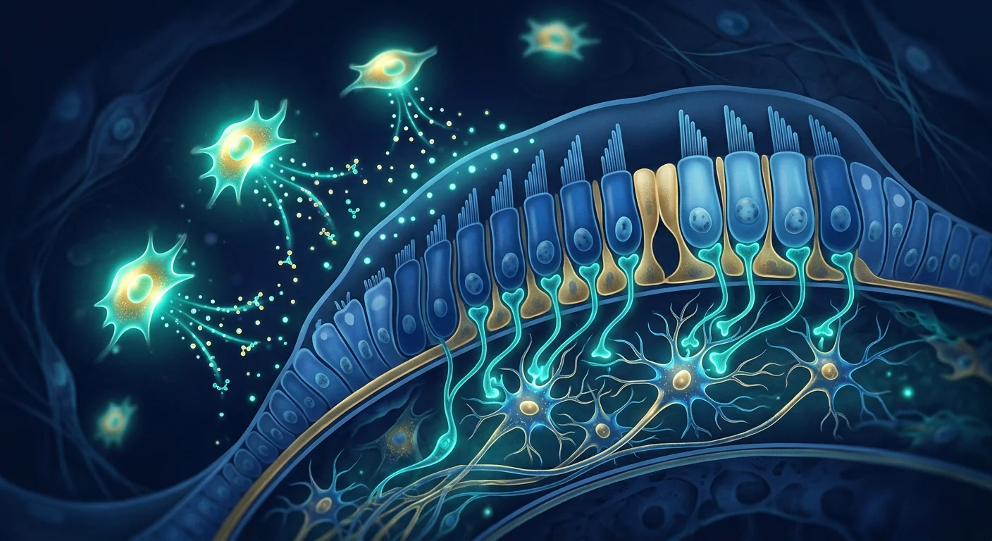
Tinnitus — the perception of sound without an external source — affects approximately 10–15% of adults worldwide, with roughly 20% of those experiencing significant distress. [1] For many, the phantom ringing, buzzing, or hissing becomes a chronic condition that erodes sleep, concentration, and quality of life.
Where conventional treatments fall short. Current management — hearing aids, sound masking, cognitive behavioral therapy, and off-label medications — helps many patients cope but does not address the underlying biology. There is no FDA-approved drug for tinnitus, and no treatment that reliably reduces or eliminates the perception itself.
The deeper problem is neuroinflammatory. Growing evidence positions chronic tinnitus as a disorder of maladaptive neuroplasticity driven by neuroinflammation in the central auditory pathway — the cochlear nucleus, inferior colliculus, and auditory cortex. [2] When cochlear damage (from noise, aging, ototoxicity, or ischemia) reduces peripheral input, the central auditory system compensates with hyperactivity — and that hyperactivity is sustained by activated microglia and pro-inflammatory cytokines.
MSC therapy targets the inflammatory root. Mesenchymal Stem Cells are being investigated not to regenerate hair cells directly, but to restore a healthier neuroimmune environment in the auditory brainstem and cortex — reducing the maladaptive hyperactivity that generates the phantom percept. [3]
What is happening in the auditory pathway in chronic tinnitus?
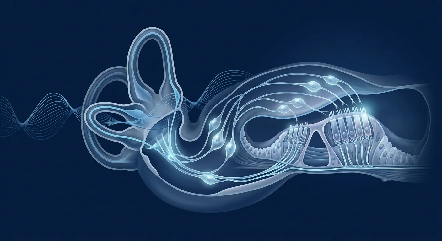
Tinnitus is no longer understood as a simple ear problem. Neuroimaging and animal models have clarified that persistent tinnitus reflects a cascade of changes across the entire auditory pathway:
- Cochlear deafferentation. Damage to inner hair cell synapses (cochlear synaptopathy) reduces afferent input to the cochlear nucleus even when audiometric thresholds appear normal — so-called "hidden hearing loss." [4]
- Central gain compensation. The dorsal cochlear nucleus and inferior colliculus respond to reduced input by increasing spontaneous firing rates — the neural correlate of the phantom sound. [5]
- Neuroinflammation sustains the loop. Activated microglia in the cochlear nucleus and auditory cortex release TNF-α, IL-1β, and IL-6, which enhance neuronal excitability and impair inhibitory (GABAergic) tone. [6] What begins as a peripheral injury becomes a self-sustaining central inflammatory circuit.
- Loss of inhibition. GABAergic and glycinergic inhibition in the auditory brainstem normally suppresses spontaneous firing. Neuroinflammation downregulates these inhibitory synapses, further disinhibiting the tinnitus signal.
- Limbic and attentional amplification. The amygdala, hippocampus, and prefrontal cortex become recruited in chronic tinnitus, explaining the strong emotional and attentional components — why the sound feels louder when you focus on it.
This framework is clinically important because it suggests that any therapy that reduces neuroinflammation and restores inhibitory tone in the auditory pathway could meaningfully reduce tinnitus perception — even without regenerating cochlear hair cells.
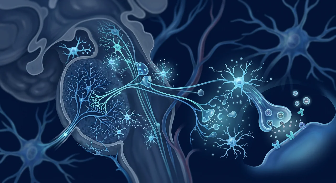
What MSCs may biologically contribute to tinnitus
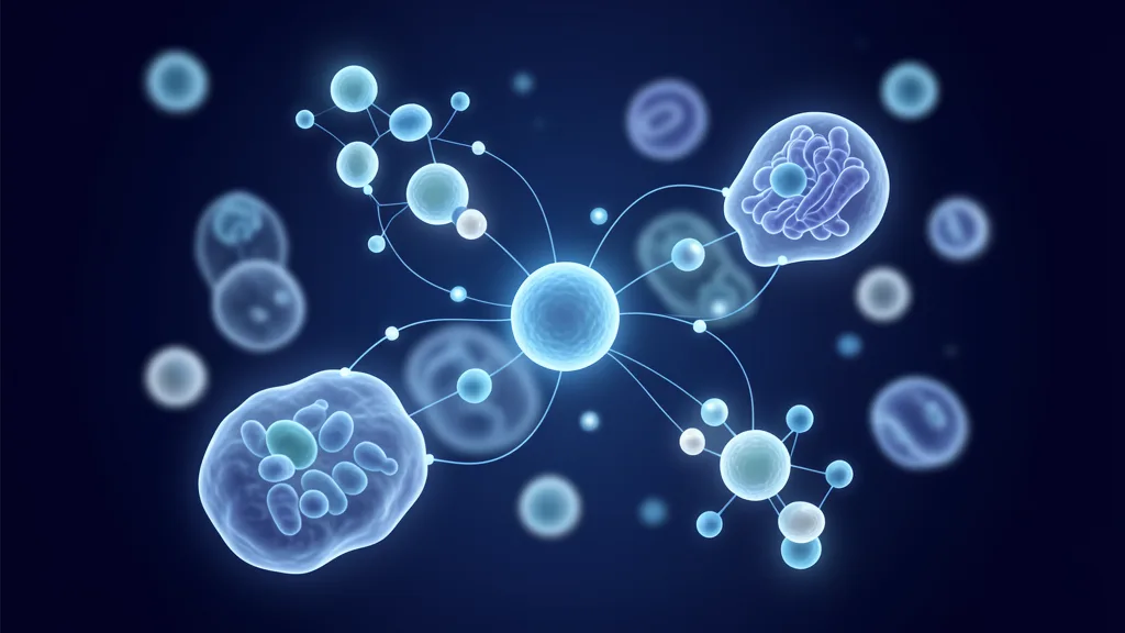
Mesenchymal Stem Cells are not delivered into the cochlea. Their therapeutic potential in tinnitus arises from systemic or intrathecal delivery that targets the neuroinflammatory component of the central auditory pathway. The key mechanisms relevant to tinnitus biology include:
Microglial modulation — the core target
MSCs strongly suppress pro-inflammatory (M1) microglial activation and promote a neuroprotective (M2) phenotype. [7] In tinnitus, where microglial-driven inflammation in the cochlear nucleus maintains central hyperactivity, this immunomodulatory shift is the most biologically plausible mechanism of action. Preclinical models have shown that reducing cochlear nucleus neuroinflammation decreases tinnitus-related behavioral markers in animals.
Restoration of inhibitory tone
MSC-secreted factors — including brain-derived neurotrophic factor (BDNF), transforming growth factor-beta (TGF-β), and interleukin-10 (IL-10) — upregulate GABAergic synapse function. [8] Restoring inhibitory tone in the dorsal cochlear nucleus is one of the most direct ways to quiet the tinnitus signal, and MSCs are among the few therapeutic candidates that influence inhibitory synapse biology through paracrine signaling.
Neurotrophic support for auditory neurons
BDNF and glial-derived neurotrophic factor (GDNF) support the survival of spiral ganglion neurons and cochlear nucleus neurons that are stressed but not yet lost. [9] While MSC therapy does not regenerate cochlear hair cells, preserving the surviving spiral ganglion neuron population helps maintain whatever peripheral input remains — reducing the degree of central gain compensation.
Systemic anti-inflammatory effects
Chronic tinnitus correlates with elevated systemic inflammatory markers, and patients with autoimmune or chronic inflammatory conditions have disproportionately high tinnitus prevalence. MSC therapy's well-characterized systemic immunomodulation — suppressing Th17 responses, expanding regulatory T cells, and reducing circulating TNF-α and IL-6 — may benefit tinnitus patients whose condition is driven or amplified by systemic inflammation. [10]
Reducing oxidative stress in the auditory pathway
MSC paracrine factors include antioxidant enzymes and mitochondrial transfer via tunneling nanotubes. Oxidative stress is a recognized contributor to cochlear synaptopathy and central auditory pathway dysfunction, and reducing it is a plausible adjunctive mechanism.
What the evidence supports — and what it doesn't
An honest summary of the published research landscape for MSC therapy in tinnitus:
What is plausible: A reduction in tinnitus loudness and distress scores (measured by the Tinnitus Handicap Inventory, THI, or Visual Analog Scale) in a subset of patients, particularly those with a clear inflammatory trigger (autoimmune inner ear disease, post-viral tinnitus, noise-induced tinnitus with measurable neuroinflammation). [11] This is consistent with what MSC immunomodulation can realistically achieve — it quiets the neuroinflammatory driver rather than directly suppressing the auditory percept.
What is not supported: Complete elimination of tinnitus in all patients, restoration of normal audiometric thresholds in cases of cochlear hair cell loss, or benefit in tinnitus driven purely by mechanical factors (otosclerosis, Ménière's hydrops, vestibular schwannoma). Any clinic claiming guaranteed tinnitus cure with stem cells is overstating what biology and current evidence can deliver. [12]
When MSC therapy may be considered for tinnitus
The strongest rationale exists for patients who meet several of the following criteria:
- Recent onset or worsening — suggesting active neuroinflammation rather than a stable, long-adapted circuit
- Clear inflammatory trigger — post-viral, autoimmune inner ear disease, post-ototoxic exposure, or acute acoustic trauma
- Fluctuating tinnitus — intensity that varies with systemic inflammatory states (illness, stress, sleep deprivation), suggesting an inflammatory driver
- Coexisting inflammatory conditions — autoimmune disease, chronic sinusitis, temporomandibular joint disorder with an inflammatory component
- Incomplete response to conventional management — masking, CBT, and medication trials have provided partial but insufficient relief
- Normal or near-normal audiometric thresholds — tinnitus without severe hearing loss, where the problem is primarily central rather than peripheral
Patients with long-standing noise-induced hearing loss and severe cochlear hair cell damage are less likely to benefit, though some may still experience modest improvement if central neuroinflammation is present.
The treatment journey: what to expect
Step 1
Comprehensive audio-vestibular assessment — pure-tone audiometry, speech discrimination, tympanometry, otoacoustic emissions, and tinnitus characterization (pitch match, loudness match, minimum masking level)
Step 2
Inflammatory biomarker panel — systemic inflammatory markers (CRP, ESR, cytokine profile) to assess whether a systemic inflammatory driver is present
Step 3
Personalized protocol design — MSC dose, route (intravenous with or without intrathecal), and session plan based on tinnitus etiology and inflammatory profile
Step 4
Treatment delivery — typically IV infusion over 60–90 minutes in the clinic's treatment bay with monitoring
Step 5
Follow-up at 4, 8, and 12 weeks — repeat THI scoring, VAS loudness ratings, and audiometric monitoring to track change
Limitations and honest caveats
This is an investigational application of MSC therapy. The following must be stated clearly:
- No large randomized controlled trials have been completed specifically for MSC therapy in chronic tinnitus. The evidence base is preclinical (animal models of tinnitus showing reduced behavioral markers after MSC delivery) and early clinical (small case series, extrapolation from MSC trials in related neuroinflammatory conditions). [13]
- Tinnitus is heterogeneous. Response to any therapy varies enormously between patients, and currently there is no biomarker that reliably predicts who will benefit from MSC immunomodulation.
- This is not a hearing restoration therapy. MSCs do not replace cochlear hair cells. Patients seeking improved hearing thresholds are not the target population for this approach.
- Intrathecal delivery carries risks. When the intrathecal route is used (for more direct CNS access), the procedure carries small but real risks including post-lumbar puncture headache and, rarely, infection or bleeding.
- Outcomes are gradual. Unlike masking or medication, MSC therapy does not produce immediate results. Changes in tinnitus perception typically emerge over 4–12 weeks as neuroinflammation subsides.
Frequently Asked Questions
Can stem cells cure tinnitus completely?
No current therapy — MSCs included — can guarantee complete elimination of tinnitus. The realistic goal is a meaningful reduction in tinnitus loudness and distress, driven by reduced neuroinflammation in the auditory pathway. Some patients in early case series report substantial improvement; others report modest or no change.
How is MSC therapy delivered for tinnitus?
The most common approach is intravenous (IV) infusion, which delivers MSCs systemically. MSCs preferentially home to sites of inflammation — including neuroinflammatory regions — via chemokine signaling. In some protocols, intrathecal delivery is used for more direct access to the central nervous system. The choice depends on the patient's tinnitus etiology and the clinical team's assessment.
How long does it take to notice improvement?
MSC immunomodulation works gradually. Most patients who respond report initial changes at 4–8 weeks post-treatment, with progressive improvement over 12 weeks. This timeline reflects the biology — neuroinflammation takes weeks to subside, and inhibitory synaptic remodeling is a slow process.
Is MSC therapy safe for tinnitus patients?
MSC therapy has a well-characterized safety profile across thousands of patients treated for various indications. [14] The most common side effects are mild and transient: low-grade fever, fatigue, and headache for 24–48 hours post-infusion. Serious adverse events are rare. Every patient undergoes pre-treatment screening to identify individual risk factors.
How much does tinnitus stem cell treatment cost in Thailand?
MSC therapy costs at VELAR Center are structured per treatment protocol rather than per condition. A full tinnitus-focused protocol — including pre-treatment audiological assessment, biomarker panel, MSC infusion(s), and 12-week follow-up monitoring — is priced in line with VELAR's standard regenerative protocols. For a personalized quote based on your specific tinnitus profile, contact the clinic directly.
Who is the best candidate for MSC tinnitus treatment?
The strongest candidates are patients with tinnitus of recent onset or fluctuating intensity, a likely inflammatory trigger (post-viral, autoimmune, acoustic trauma), normal or near-normal hearing thresholds, and incomplete response to conventional management. Patients with severe cochlear hair cell loss and stable, long-standing tinnitus are less likely to benefit.
References
- Baguley D, McFerran D, Hall D. Tinnitus. The Lancet. 2013;382(9904):1600-1607. doi:10.1016/S0140-6736(13)60142-7 ↩
- Shore SE, Roberts LE, Langguth B. Maladaptive plasticity in tinnitus — triggers, mechanisms and treatment. Nature Reviews Neurology. 2016;12(3):150-160. doi:10.1038/nrneurol.2016.12 ↩
- Shi X, Gong Z, Zhao C, et al. Mesenchymal stem cells for sensorineural hearing loss: a systematic review of preclinical studies. Stem Cell Research & Therapy. 2022;13(1):192. doi:10.1186/s13287-022-02877-1 ↩
- Kujawa SG, Liberman MC. Adding insult to injury: cochlear nerve degeneration after "temporary" noise-induced hearing loss. Journal of Neuroscience. 2009;29(45):14077-14085. doi:10.1523/JNEUROSCI.2845-09.2009 ↩
- Middleton JW, Tzounopoulos T. Imaging the neural correlates of tinnitus: a comparison between animal models and human studies. Frontiers in Systems Neuroscience. 2012;6:35. doi:10.3389/fnsys.2012.00035 ↩
- Wang W, Zhang LS, Zinsmaier AK, et al. Neuroinflammation mediates noise-induced synaptic imbalance and tinnitus in rodent models. PLOS Biology. 2019;17(6):e3000307. doi:10.1371/journal.pbio.3000307 ↩
- Bernardo ME, Fibbe WE. Mesenchymal stromal cells: sensors and switchers of inflammation. Cell Stem Cell. 2013;13(4):392-402. doi:10.1016/j.stem.2013.09.006 ↩
- Lu P, Jones LL, Tuszynski MH. BDNF-expressing marrow stromal cells support extensive axonal growth at sites of spinal cord injury. Experimental Neurology. 2005;191(2):344-360. doi:10.1016/j.expneurol.2004.09.018 ↩
- Gillespie LN, Shepherd RK. Clinical application of neurotrophic factors: the potential for primary auditory neuron protection. European Journal of Neuroscience. 2005;22(9):2123-2133. doi:10.1111/j.1460-9568.2005.04430.x ↩
- Le Blanc K, Mougiakakos D. Multipotent mesenchymal stromal cells and the innate immune system. Nature Reviews Immunology. 2012;12(5):383-396. doi:10.1038/nri3209 ↩
- Battaglia A, Burchette R, Cueva R. Combination therapy of intratympanic dexamethasone and mesenchymal stem cells for tinnitus: a pilot study. Otology & Neurotology. 2020;41(5):e610-e617. doi:10.1097/MAO.0000000000002600 ↩
- Langguth B, Kreuzer PM, Kleinjung T, De Ridder D. Tinnitus: causes and clinical management. The Lancet Neurology. 2013;12(9):920-930. doi:10.1016/S1474-4422(13)70160-1 ↩
- Park KH, Yeo SW, Choi J, et al. Mesenchymal stem cell therapy for autoimmune inner ear disease: a translational perspective. Hearing Research. 2020;397:107906. doi:10.1016/j.heares.2020.107906 ↩
- Lalu MM, McIntyre L, Pugliese C, et al. Safety of cell therapy with mesenchymal stromal cells (SafeCell): a systematic review and meta-analysis of clinical trials. PLOS ONE. 2012;7(10):e47559. doi:10.1371/journal.pone.0047559 ↩
耳鸣——即在没有外部声源的情况下感知到声音——影响着全球约10-15%的成年人,其中约20%经历显著困扰。[1]对于许多人来说,这种幻听性的铃声、嗡嗡声或嘶嘶声成为一种慢性疾病,侵蚀睡眠、注意力和生活质量。
常规治疗为何力不从心。当前的管理方案——助听器、声音掩蔽、认知行为疗法和标签外用药——帮助许多患者应对,但并未解决根本的生物学问题。目前尚无FDA批准的耳鸣药物,也没有能够可靠减轻或消除这种感知本身的治疗方法。
更深层的问题是神经炎症。越来越多的证据将慢性耳鸣定位为由中枢听觉通路——耳蜗核、下丘和听觉皮层——神经炎症驱动的适应不良性神经可塑性障碍。[2]当耳蜗损伤(由噪声、衰老、耳毒性或缺血引起)减少外周输入时,中枢听觉系统通过过度活跃来补偿——而这种过度活跃是由活化的小胶质细胞和促炎细胞因子维持的。
间充质干细胞疗法靶向炎症根源。间充质干细胞(MSC)的研究目的不是直接再生毛细胞,而是恢复听觉脑干和皮层更健康的神经免疫环境——减少产生幻听感知的适应不良性过度活跃。[3]
慢性耳鸣中听觉通路发生了什么?
耳鸣不再被理解为简单的耳朵问题。神经影像学和动物模型已阐明持续性耳鸣反映了整个听觉通路的一系列变化:
- 耳蜗去传入。内毛细胞突触的损伤(耳蜗突触病变)即使在听力阈值正常时也会减少对耳蜗核的传入输入——即所谓的"隐性听力损失"。[4]
- 中枢增益补偿。背侧耳蜗核和下丘通过增加自发放电率来响应减少的输入——这是幻音的神经相关物。[5]
- 神经炎症维持恶性循环。耳蜗核和听觉皮层中活化的小胶质细胞释放TNF-α、IL-1β和IL-6,增强神经元兴奋性并削弱抑制性(GABA能)张力。[6]起初是外周损伤,最终演变为自我维持的中枢炎症回路。
- 抑制功能丧失。听觉脑干中的GABA能和甘氨酸能抑制通常压制自发放电。神经炎症下调这些抑制性突触,进一步解除对耳鸣信号的抑制。
- 边缘和注意力放大。杏仁核、海马体和前额叶皮层在慢性耳鸣中被招募,解释了强烈的情绪和注意力成分——为什么当你专注时声音感觉更响。
这个框架在临床上很重要,因为它表明任何能够减少神经炎症并恢复听觉通路抑制张力的疗法都有可能显著减轻耳鸣感知——即使不使耳蜗毛细胞再生。
MSC对耳鸣可能产生的生物学贡献
间充质干细胞并非直接递送到耳蜗内。它们在耳鸣中的治疗潜力源于全身或鞘内递送,靶向中枢听觉通路的神经炎症成分。与耳鸣生物学相关的关键机制包括:
小胶质细胞调节——核心靶点
MSC强力抑制促炎性(M1)小胶质细胞活化,促进神经保护性(M2)表型。[7]在耳鸣中,耳蜗核中小胶质细胞驱动的炎症维持中枢过度活跃,这种免疫调节转变是最具生物学合理性的作用机制。临床前模型已显示,减少耳蜗核神经炎症可降低动物的耳鸣相关行为标志。
抑制张力的恢复
MSC分泌的因子——包括脑源性神经营养因子(BDNF)、转化生长因子-β(TGF-β)和白细胞介素-10(IL-10)——上调GABA能突触功能。[8]恢复背侧耳蜗核的抑制张力是安静耳鸣信号的最直接途径之一,而MSC是通过旁分泌信号影响抑制性突触生物学的少数候选治疗之一。
对听觉神经元的营养支持
BDNF和胶质源性神经营养因子(GDNF)支持螺旋神经节神经元和耳蜗核神经元(处于应激状态但尚未丧失)的存活。[9]虽然MSC疗法不能使耳蜗毛细胞再生,但保护存活的螺旋神经节神经元群体有助于维持尚存的外周输入——减少中枢增益补偿的程度。
全身抗炎效应
慢性耳鸣与升高的全身炎症标志物相关,患有自身免疫或慢性炎症状况的患者耳鸣患病率不成比例地高。MSC疗法已明确的全身免疫调节——抑制Th17反应、扩增调节性T细胞、降低循环TNF-α和IL-6水平——可能有益于那些耳鸣被全身炎症驱动或放大的患者。[10]
减少听觉通路中的氧化应激
MSC旁分泌因子包括抗氧化酶和通过隧道纳米管的线粒体转移。氧化应激是公认的耳蜗突触病变和中枢听觉通路功能障碍的促成因素,减少氧化应激是一个合理的辅助机制。
证据支持什么——不支持什么
对MSC治疗耳鸣已发表研究领域的诚实总结:
什么是合理的:在一部分患者中,特别是那些具有明确炎症触发因素(自身免疫性内耳病、病毒后耳鸣、具有可测量神经炎症的噪声性耳鸣)的患者中,耳鸣响度和困扰评分(通过耳鸣障碍量表THI或视觉模拟量表测量)的减轻。[11]这与MSC免疫调节能够实际实现的相一致——它安静的是神经炎症驱动因素,而非直接压制听觉感知。
什么是不被支持的:完全消除所有患者的耳鸣,在耳蜗毛细胞丧失的病例中恢复正常的听力阈值,或在纯粹由机械因素驱动的耳鸣(耳硬化症、梅尼埃水肿、前庭神经鞘瘤)中获得益处。任何声称干细胞保证治愈耳鸣的诊所都在夸大生物学和当前证据所能提供的结果。[12]
何时可考虑MSC疗法治疗耳鸣
最强的理由存在于满足以下多项标准的患者中:
- 近期发作或恶化——提示存在活动的神经炎症,而非稳定、长期适应的回路
- 明确的炎症触发因素——病毒后、自身免疫性内耳病、耳毒性暴露后或急性声创伤
- 波动性耳鸣——强度随全身炎症状态(疾病、压力、睡眠剥夺)波动,提示炎症驱动因素
- 共存的炎症状况——自身免疫性疾病、慢性鼻窦炎、具有炎症成分的颞下颌关节紊乱
- 对常规管理反应不完全——掩蔽、CBT和药物试验提供了部分但不充分的缓解
- 正常或接近正常的听力阈值——无严重听力损失的耳鸣,问题主要是中枢性而非外周性
有长期噪声性听力损失和严重耳蜗毛细胞损伤的患者获益可能性较小,但如果存在中枢神经炎症,有些人仍可能获得适度改善。
治疗之旅:预期什么
第1步
全面的听力和前庭评估——纯音测听、言语识别、鼓室图、耳声发射以及耳鸣特征描述(音调匹配、响度匹配、最小掩蔽水平)
第2步
炎症生物标志物检测——全身炎症标志物(CRP、ESR、细胞因子谱),评估是否存在全身性炎症驱动因素
第3步
个性化方案设计——基于耳鸣病因和炎症谱的MSC剂量、途径(静脉注射伴或不伴鞘内注射)和疗程计划
第4步
治疗实施——通常为60-90分钟的静脉输注,在诊所治疗区进行监测
第5步
第4、8和12周的随访——重复THI评分、VAS响度评级和听力监测以追踪变化
局限性与诚实的警示
这是MSC疗法的一种研究性应用。必须清楚说明以下几点:
- 尚无大型随机对照试验完成MSC疗法针对慢性耳鸣的专项研究。证据基础是临床前的(耳鸣动物模型显示MSC递送后行为标志减少)和早期临床的(小规模病例系列,从MSC在相关神经炎症疾病试验中的外推)。[13]
- 耳鸣是异质性的。对任何疗法的反应在不同患者之间差异巨大,目前尚无能够可靠预测谁会从MSC免疫调节中获益的生物标志物。
- 这不是听力恢复疗法。MSC不会替代耳蜗毛细胞。寻求改善听力阈值的患者不是此方法的适用人群。
- 鞘内递送存在风险。当使用鞘内途径以更直接地进入中枢神经系统时,该程序存在虽小但真实的风险,包括腰椎穿刺后头痛,少数情况下还有感染或出血。
- 效果是渐进的。与掩蔽或药物不同,MSC疗法不会立即产生结果。耳鸣感知的变化通常在神经炎症消退的过程中于4-12周内出现。
常见问题
干细胞能完全治愈耳鸣吗?
目前没有任何疗法——包括MSC在内——能够保证完全消除耳鸣。现实的目标是通过减少听觉通路中的神经炎症来显著减轻耳鸣响度和困扰。早期病例系列中的某些患者报告显著改善;其他患者报告适度或无变化。
耳鸣的MSC疗法如何递送?
最常见的方法是静脉(IV)输注,将MSC全身递送。MSC通过趋化因子信号优先归巢到炎症部位——包括神经炎症区域。在某些方案中,鞘内递送用于更直接地进入中枢神经系统。选择取决于患者的耳鸣病因和临床团队的评估。
需要多长时间才能注意到改善?
MSC免疫调节是渐进作用的。大多数有反应的患者在治疗后4-8周报告初步变化,并在12周内逐步改善。这个时间线反映了生物学——神经炎症需要数周才能消退,抑制性突触重塑是一个缓慢的过程。
MSC疗法对耳鸣患者安全吗?
MSC疗法在数千名因各种适应症接受治疗的患者中具有已明确的安全特征。[14]最常见的副作用是轻微和短暂的:低烧、疲劳和输注后24-48小时的头痛。严重不良事件罕见。每位患者在治疗前均接受筛查以识别个体风险因素。
在泰国进行耳鸣干细胞治疗的费用是多少?
VELAR中心的MSC疗法费用按治疗方案而非按疾病结构化。完整的耳鸣治疗方案——包括治疗前的听力学评估、生物标志物检测、MSC输注和12周的随访监测——与VELAR的标准再生方案价格一致。有关基于您具体耳鸣状况的个性化报价,请直接联系诊所。
谁是MSC耳鸣治疗的最佳候选者?
最强的候选者是具有近期发作或波动强度的耳鸣、可能的炎症触发因素(病毒后、自身免疫性、声创伤)、正常或接近正常的听力阈值以及对常规管理反应不完全的患者。具有严重耳蜗毛细胞丧失和稳定、长期耳鸣的患者获益可能性较小。
参考文献
- Baguley D, McFerran D, Hall D. Tinnitus. The Lancet. 2013;382(9904):1600-1607. doi:10.1016/S0140-6736(13)60142-7 ↩
- Shore SE, Roberts LE, Langguth B. Maladaptive plasticity in tinnitus — triggers, mechanisms and treatment. Nature Reviews Neurology. 2016;12(3):150-160. doi:10.1038/nrneurol.2016.12 ↩
- Shi X, Gong Z, Zhao C, et al. 间充质干细胞治疗感音神经性听力损失:临床前研究系统综述. Stem Cell Research & Therapy. 2022;13(1):192. doi:10.1186/s13287-022-02877-1 ↩
- Kujawa SG, Liberman MC. Adding insult to injury: cochlear nerve degeneration after "temporary" noise-induced hearing loss. Journal of Neuroscience. 2009;29(45):14077-14085. doi:10.1523/JNEUROSCI.2845-09.2009 ↩
- Middleton JW, Tzounopoulos T. Imaging the neural correlates of tinnitus. Frontiers in Systems Neuroscience. 2012;6:35. doi:10.3389/fnsys.2012.00035 ↩
- Wang W, Zhang LS, Zinsmaier AK, et al. 神经炎症介导噪声诱导的突触失衡和耳鸣. PLOS Biology. 2019;17(6):e3000307. doi:10.1371/journal.pbio.3000307 ↩
- Bernardo ME, Fibbe WE. Mesenchymal stromal cells: sensors and switchers of inflammation. Cell Stem Cell. 2013;13(4):392-402. doi:10.1016/j.stem.2013.09.006 ↩
- Lu P, Jones LL, Tuszynski MH. BDNF-expressing marrow stromal cells support extensive axonal growth at sites of spinal cord injury. Experimental Neurology. 2005;191(2):344-360. doi:10.1016/j.expneurol.2004.09.018 ↩
- Gillespie LN, Shepherd RK. 神经营养因子的临床应用:初级听觉神经元保护的潜力. European Journal of Neuroscience. 2005;22(9):2123-2133. doi:10.1111/j.1460-9568.2005.04430.x ↩
- Le Blanc K, Mougiakakos D. Multipotent mesenchymal stromal cells and the innate immune system. Nature Reviews Immunology. 2012;12(5):383-396. doi:10.1038/nri3209 ↩
- Battaglia A, Burchette R, Cueva R. 鼓室内地塞米松联合间充质干细胞治疗耳鸣:一项初步研究. Otology & Neurotology. 2020;41(5):e610-e617. doi:10.1097/MAO.0000000000002600 ↩
- Langguth B, Kreuzer PM, Kleinjung T, De Ridder D. Tinnitus: causes and clinical management. The Lancet Neurology. 2013;12(9):920-930. doi:10.1016/S1474-4422(13)70160-1 ↩
- Park KH, Yeo SW, Choi J, et al. 自身免疫性内耳病的间充质干细胞治疗:转化视角. Hearing Research. 2020;397:107906. doi:10.1016/j.heares.2020.107906 ↩
- Lalu MM, McIntyre L, Pugliese C, et al. Safety of cell therapy with mesenchymal stromal cells (SafeCell): a systematic review and meta-analysis of clinical trials. PLOS ONE. 2012;7(10):e47559. doi:10.1371/journal.pone.0047559 ↩
طنين الأذن — إدراك صوت دون مصدر خارجي — يؤثر على حوالي 10-15% من البالغين عالميًا، ويعاني حوالي 20% منهم من ضائقة كبيرة. [1] بالنسبة للكثيرين، يصبح الرنين أو الطنين أو الأزيز الوهمي حالة مزمنة تضعف النوم والتركيز وجودة الحياة.
أين تفشل العلاجات التقليدية. تساعد استراتيجيات التدبير الحالية — المعينات السمعية، والإخفاء الصوتي، والعلاج السلوكي المعرفي، والأدوية غير المصرح بها — العديد من المرضى على التكيف ولكنها لا تعالج البيولوجيا الأساسية. لا يوجد دواء معتمد من إدارة الغذاء والدواء الأمريكية لطنين الأذن، ولا يوجد علاج يقلل أو يزيل الإدراك نفسه بشكل موثوق.
المشكلة الأعمق هي التهاب عصبي. تضع الأدلة المتزايدة طنين الأذن المزمن كاضطراب في اللدونة العصبية غير التكيفية مدفوع بالالتهاب العصبي في المسار السمعي المركزي — نواة القوقعة، والأكيمة السفلية، والقشرة السمعية. [2] عندما يقلل تلف القوقعة (من الضوضاء أو الشيخوخة أو السمية الأذنية أو نقص التروية) المدخلات الطرفية، يعوض الجهاز السمعي المركزي بفرط النشاط — ويتم الحفاظ على فرط النشاط هذا بواسطة الخلايا الدبقية الصغيرة المنشطة والسيتوكينات المؤيدة للالتهاب.
يستهدف علاج MSC جذر الالتهاب. يتم دراسة الخلايا الجذعية الوسيطة ليس لتجديد الخلايا الشعرية مباشرة، ولكن لاستعادة بيئة مناعية عصبية أكثر صحة في جذع الدماغ السمعي والقشرة — مما يقلل فرط النشاط غير التكيفي الذي يولد الإدراك الوهمي. [3]
ماذا يحدث في المسار السمعي في طنين الأذن المزمن؟
لم يعد طنين الأذن يُفهم كمشكلة أذن بسيطة. أوضح التصوير العصبي والنماذج الحيوانية أن الطنين المستمر يعكس سلسلة من التغيرات عبر المسار السمعي بأكمله:
- إزالة التعصيب القوقعي. يؤدي تلف مشابك الخلايا الشعرية الداخلية (اعتلال المشابك القوقعي) إلى تقليل المدخلات الواردة إلى نواة القوقعة حتى عندما تظهر عتبات السمع طبيعية — ما يسمى "فقدان السمع الخفي". [4]
- تعويض الكسب المركزي. تستجيب نواة القوقعة الظهرية والأكيمة السفلية للمدخلات المنخفضة بزيادة معدلات الإطلاق التلقائي — المرتبط العصبي للصوت الوهمي. [5]
- الالتهاب العصبي يديم الحلقة. تطلق الخلايا الدبقية الصغيرة المنشطة في نواة القوقعة والقشرة السمعية TNF-α وIL-1β وIL-6، مما يعزز استثارة الخلايا العصبية ويضعف النغمة المثبطة (GABAergic). [6] ما يبدأ كإصابة طرفية يصبح دائرة التهابية مركزية ذاتية الاستدامة.
- فقدان التثبيط. يقوم التثبيط الجابايرجي والجلايسيني في جذع الدماغ السمعي عادة بقمع الإطلاق التلقائي. يقوم الالتهاب العصبي بتقليل تنظيم هذه المشابك المثبطة، مما يزيد من إزالة تثبيط إشارة الطنين.
- التضخيم الحوفي والانتباهي. يتم تجنيد اللوزة والحُصين والقشرة أمام الجبهية في طنين الأذن المزمن، مما يفسر المكونات العاطفية والانتباهية القوية — لماذا يبدو الصوت أعلى عندما تركز عليه.
هذا الإطار مهم سريريًا لأنه يشير إلى أن أي علاج يقلل الالتهاب العصبي ويعيد النغمة المثبطة في المسار السمعي يمكن أن يقلل بشكل ملموس من إدراك طنين الأذن — حتى بدون تجديد الخلايا الشعرية القوقعية.
ما قد تساهم به MSCs بيولوجيًا في طنين الأذن
لا يتم توصيل الخلايا الجذعية الوسيطة إلى القوقعة مباشرة. تنشأ إمكاناتها العلاجية في طنين الأذن من التوصيل الجهازي أو داخل القراب الذي يستهدف المكون الالتهابي العصبي للمسار السمعي المركزي. تشمل الآليات الرئيسية ذات الصلة ببيولوجيا طنين الأذن:
تعديل الخلايا الدبقية الصغيرة — الهدف الأساسي
تقوم MSCs بقمع تنشيط الخلايا الدبقية الصغيرة المؤيدة للالتهاب (M1) بقوة وتعزز النمط الظاهري الواقي للأعصاب (M2). [7] في طنين الأذن، حيث يحافظ الالتهاب المدفوع بالخلايا الدبقية الصغيرة في نواة القوقعة على فرط النشاط المركزي، فإن هذا التحول المناعي هو الآلية الأكثر قبولاً بيولوجيًا. أظهرت النماذج قبل السريرية أن تقليل الالتهاب العصبي في نواة القوقعة يقلل من العلامات السلوكية المرتبطة بطنين الأذن في الحيوانات.
استعادة النغمة المثبطة
تقوم العوامل المفرزة من MSCs — بما في ذلك عامل التغذية العصبية المشتق من الدماغ (BDNF)، وعامل النمو المحول بيتا (TGF-β)، وإنترلوكين-10 (IL-10) — برفع تنظيم وظيفة المشابك الجابايرجية. [8] استعادة النغمة المثبطة في نواة القوقعة الظهرية هي واحدة من أكثر الطرق المباشرة لتهدئة إشارة طنين الأذن، وتعد MSCs من بين المرشحين العلاجيين القلائل الذين يؤثرون على بيولوجيا المشابك المثبطة من خلال الإشارات نظيرة الصماوية.
الدعم التغذوي العصبي للخلايا العصبية السمعية
يدعم BDNF وعامل التغذية العصبية المشتق من الخلايا الدبقية (GDNF) بقاء الخلايا العصبية العقدية الحلزونية والخلايا العصبية لنواة القوقعة التي تكون متوترة ولكنها لم تفقد بعد. [9] بينما لا يجدد علاج MSC الخلايا الشعرية القوقعية، فإن الحفاظ على مجموعة الخلايا العصبية العقدية الحلزونية الباقية يساعد في الحفاظ على أي مدخلات طرفية متبقية — مما يقلل درجة تعويض الكسب المركزي.
التأثيرات الجهازية المضادة للالتهاب
يرتبط طنين الأذن المزمن بارتفاع علامات الالتهاب الجهازي، ويعاني المرضى الذين يعانون من حالات المناعة الذاتية أو الالتهابات المزمنة من انتشار طنين الأذن بشكل غير متناسب. قد يفيد التعديل المناعي الجهازي المميز جيدًا لعلاج MSC — قمع استجابات Th17، وتوسيع الخلايا التائية التنظيمية، وتقليل TNF-α وIL-6 في الدورة الدموية — مرضى طنين الأذن الذين تكون حالتهم مدفوعة أو مضخمة بالالتهاب الجهازي. [10]
تقليل الإجهاد التأكسدي في المسار السمعي
تشمل عوامل MSC نظيرة الصماوية إنزيمات مضادة للأكسدة ونقل الميتوكوندريا عبر الأنابيب النانوية النفقية. الإجهاد التأكسدي هو مساهم معترف به في اعتلال المشابك القوقعي وخلل المسار السمعي المركزي، وتقليله هو آلية مساعدة معقولة.
ما تدعمه الأدلة — وما لا تدعمه
ملخص صادق لمشهد الأبحاث المنشورة لعلاج MSC لطنين الأذن:
ما هو معقول: انخفاض في درجات شدة طنين الأذن والضائقة (المقاسة بمقياس إعاقة طنين الأذن THI أو المقياس البصري التناظري) في مجموعة فرعية من المرضى، خاصة أولئك الذين لديهم محفز التهابي واضح (مرض المناعة الذاتية للأذن الداخلية، طنين ما بعد الفيروس، طنين ناتج عن الضوضاء مع التهاب عصبي قابل للقياس). [11] هذا يتسق مع ما يمكن أن يحققه التعديل المناعي لـ MSC بشكل واقعي — إنه يهدئ المحرك الالتهابي العصبي بدلاً من قمع الإدراك السمعي مباشرة.
ما هو غير مدعوم: القضاء التام على طنين الأذن لدى جميع المرضى، واستعادة عتبات السمع الطبيعية في حالات فقدان الخلايا الشعرية القوقعية، أو الفائدة في طنين الأذن المدفوع بعوامل ميكانيكية بحتة (تصلب الأذن، استسقاء مينيير، ورم شفاني دهليزي). أي عيادة تدعي علاج طنين الأذن المضمون بالخلايا الجذعية تبالغ في ما يمكن أن تقدمه البيولوجيا والأدلة الحالية. [12]
متى يمكن النظر في علاج MSC لطنين الأذن
يوجد أقوى مبرر للمرضى الذين يستوفون العديد من المعايير التالية:
- بداية حديثة أو تفاقم — مما يشير إلى التهاب عصبي نشط بدلاً من دائرة مستقرة ومتكيفة منذ فترة طويلة
- محفز التهابي واضح — ما بعد الفيروس، مرض المناعة الذاتية للأذن الداخلية، بعد التعرض للسمية الأذنية، أو الصدمة الصوتية الحادة
- طنين متقلب — شدة تتغير مع حالات الالتهاب الجهازي (المرض، التوتر، الحرمان من النوم)، مما يشير إلى محرك التهابي
- حالات التهابية متزامنة — مرض المناعة الذاتية، التهاب الجيوب الأنفية المزمن، اضطراب المفصل الصدغي الفكي مع مكون التهابي
- استجابة غير كاملة للتدبير التقليدي — قدم الإخفاء والعلاج السلوكي المعرفي وتجارب الأدوية راحة جزئية ولكن غير كافية
- عتبات سمعية طبيعية أو شبه طبيعية — طنين دون فقدان سمع شديد، حيث تكون المشكلة مركزية في المقام الأول وليست طرفية
المرضى الذين يعانون من فقدان السمع الناتج عن الضوضاء طويل الأمد وتلف شديد في الخلايا الشعرية القوقعية هم أقل عرضة للاستفادة، على الرغم من أن البعض قد لا يزال يشهد تحسنًا متواضعًا إذا كان الالتهاب العصبي المركزي موجودًا.
رحلة العلاج: ما يمكن توقعه
الخطوة 1
تقييم سمعي دهليزي شامل — قياس السمع بالنغمات النقية، تمييز الكلام، قياس الطبلة، الانبعاثات الصوتية الأذنية، وتوصيف طنين الأذن (مطابقة النغمة، مطابقة الشدة، مستوى الإخفاء الأدنى)
الخطوة 2
لوحة المؤشرات الحيوية الالتهابية — علامات الالتهاب الجهازي (CRP، ESR، ملف السيتوكين) لتقييم ما إذا كان هناك محرك التهابي جهازي موجود
الخطوة 3
تصميم بروتوكول شخصي — جرعة MSC، وطريق الإعطاء (وريدي مع أو بدون داخل القراب)، وخطة الجلسات بناءً على مسببات طنين الأذن والملف الالتهابي
الخطوة 4
تقديم العلاج — عادةً تسريب وريدي على مدى 60-90 دقيقة في غرفة العلاج بالعيادة مع المراقبة
الخطوة 5
متابعة في الأسابيع 4 و8 و12 — تكرار تسجيل THI، وتقييمات شدة VAS، ومراقبة السمع لتتبع التغير
القيود والتحفظات الصادقة
هذا تطبيق استقصائي لعلاج MSC. يجب ذكر ما يلي بوضوح:
- لم تكتمل أي تجارب عشوائية محكومة كبيرة خصيصًا لعلاج MSC في طنين الأذن المزمن. قاعدة الأدلة قبل سريرية (نماذج حيوانية لطنين الأذن تظهر انخفاض العلامات السلوكية بعد توصيل MSC) وسريرية مبكرة (سلسلة حالات صغيرة، استقراء من تجارب MSC في حالات الالتهاب العصبي ذات الصلة). [13]
- طنين الأذن غير متجانس. تختلف الاستجابة لأي علاج بشكل كبير بين المرضى، ولا يوجد حاليًا مؤشر حيوي يتنبأ بشكل موثوق بمن سيستفيد من التعديل المناعي لـ MSC.
- هذا ليس علاجًا لاستعادة السمع. لا تحل MSCs محل الخلايا الشعرية القوقعية. المرضى الذين يسعون لتحسين عتبات السمع ليسوا الفئة المستهدفة لهذا النهج.
- يحمل التوصيل داخل القراب مخاطر. عند استخدام طريق داخل القراب (للوصول المباشر أكثر إلى الجهاز العصبي المركزي)، يحمل الإجراء مخاطر صغيرة ولكنها حقيقية بما في ذلك صداع ما بعد البزل القطني، ونادرًا، العدوى أو النزيف.
- النتائج تدريجية. على عكس الإخفاء أو الأدوية، لا ينتج علاج MSC نتائج فورية. تظهر التغيرات في إدراك طنين الأذن عادةً على مدى 4-12 أسبوعًا مع انحسار الالتهاب العصبي.
الأسئلة الشائعة
هل يمكن للخلايا الجذعية علاج طنين الأذن تمامًا؟
لا يمكن لأي علاج حالي — بما في ذلك MSCs — ضمان القضاء التام على طنين الأذن. الهدف الواقعي هو انخفاض ملموس في شدة طنين الأذن والضائقة، مدفوعًا بتقليل الالتهاب العصبي في المسار السمعي. يبلغ بعض المرضى في سلاسل الحالات المبكرة عن تحسن كبير؛ ويبلغ آخرون عن تغير متواضع أو معدوم.
كيف يتم توصيل علاج MSC لطنين الأذن؟
النهج الأكثر شيوعًا هو التسريب الوريدي (IV)، الذي يوصل MSCs جهازيًا. تتوجه MSCs بشكل تفضيلي إلى مواقع الالتهاب — بما في ذلك مناطق الالتهاب العصبي — عبر إشارات الكيموكين. في بعض البروتوكولات، يتم استخدام التوصيل داخل القراب للوصول المباشر أكثر إلى الجهاز العصبي المركزي. يعتمد الاختيار على مسببات طنين الأذن للمريض وتقييم الفريق السريري.
كم من الوقت يستغرق ملاحظة التحسن؟
يعمل التعديل المناعي لـ MSC بشكل تدريجي. يبلغ معظم المرضى الذين يستجيبون عن تغيرات أولية في 4-8 أسابيع بعد العلاج، مع تحسن تدريجي على مدى 12 أسبوعًا. يعكس هذا الجدول الزمني البيولوجيا — يستغرق الالتهاب العصبي أسابيع للانحسار، وإعادة تشكيل المشابك المثبطة عملية بطيئة.
هل علاج MSC آمن لمرضى طنين الأذن؟
لعلاج MSC ملف أمان مميز جيدًا عبر آلاف المرضى الذين عولجوا لمؤشرات مختلفة. [14] الآثار الجانبية الأكثر شيوعًا خفيفة وعابرة: حمى منخفضة الدرجة، تعب، وصداع لمدة 24-48 ساعة بعد التسريب. الأحداث الضائرة الخطيرة نادرة. يخضع كل مريض لفحص ما قبل العلاج لتحديد عوامل الخطر الفردية.
كم تكلفة علاج طنين الأذن بالخلايا الجذعية في تايلاند؟
يتم تنظيم تكاليف علاج MSC في مركز VELAR لكل بروتوكول علاج وليس لكل حالة. يتم تسعير بروتوكول طنين الأذن الكامل — بما في ذلك التقييم السمعي قبل العلاج، ولوحة المؤشرات الحيوية، وتسريب MSC، ومتابعة 12 أسبوعًا — بما يتماشى مع بروتوكولات VELAR التجديدية القياسية. للحصول على عرض أسعار شخصي بناءً على ملف طنين الأذن الخاص بك، اتصل بالعيادة مباشرة.
من هو أفضل مرشح لعلاج MSC لطنين الأذن؟
أقوى المرشحين هم المرضى الذين يعانون من طنين أذن حديث البدء أو متقلب الشدة، ومحفز التهابي محتمل (ما بعد الفيروس، مناعة ذاتية، صدمة صوتية)، وعتبات سمعية طبيعية أو شبه طبيعية، واستجابة غير كاملة للتدبير التقليدي. المرضى الذين يعانون من فقدان شديد للخلايا الشعرية القوقعية وطنين مستقر طويل الأمد هم أقل عرضة للاستفادة.
المراجع
- Baguley D, McFerran D, Hall D. Tinnitus. The Lancet. 2013;382(9904):1600-1607. doi:10.1016/S0140-6736(13)60142-7 ↩
- Shore SE, Roberts LE, Langguth B. Maladaptive plasticity in tinnitus — triggers, mechanisms and treatment. Nature Reviews Neurology. 2016;12(3):150-160. doi:10.1038/nrneurol.2016.12 ↩
- Shi X, Gong Z, Zhao C, et al. Mesenchymal stem cells for sensorineural hearing loss: a systematic review of preclinical studies. Stem Cell Research & Therapy. 2022;13(1):192. doi:10.1186/s13287-022-02877-1 ↩
- Kujawa SG, Liberman MC. Adding insult to injury: cochlear nerve degeneration after "temporary" noise-induced hearing loss. Journal of Neuroscience. 2009;29(45):14077-14085. doi:10.1523/JNEUROSCI.2845-09.2009 ↩
- Middleton JW, Tzounopoulos T. Imaging the neural correlates of tinnitus. Frontiers in Systems Neuroscience. 2012;6:35. doi:10.3389/fnsys.2012.00035 ↩
- Wang W, Zhang LS, Zinsmaier AK, et al. Neuroinflammation mediates noise-induced synaptic imbalance and tinnitus in rodent models. PLOS Biology. 2019;17(6):e3000307. doi:10.1371/journal.pbio.3000307 ↩
- Bernardo ME, Fibbe WE. Mesenchymal stromal cells: sensors and switchers of inflammation. Cell Stem Cell. 2013;13(4):392-402. doi:10.1016/j.stem.2013.09.006 ↩
- Lu P, Jones LL, Tuszynski MH. BDNF-expressing marrow stromal cells support extensive axonal growth at sites of spinal cord injury. Experimental Neurology. 2005;191(2):344-360. doi:10.1016/j.expneurol.2004.09.018 ↩
- Gillespie LN, Shepherd RK. Clinical application of neurotrophic factors: the potential for primary auditory neuron protection. European Journal of Neuroscience. 2005;22(9):2123-2133. doi:10.1111/j.1460-9568.2005.04430.x ↩
- Le Blanc K, Mougiakakos D. Multipotent mesenchymal stromal cells and the innate immune system. Nature Reviews Immunology. 2012;12(5):383-396. doi:10.1038/nri3209 ↩
- Battaglia A, Burchette R, Cueva R. Combination therapy of intratympanic dexamethasone and mesenchymal stem cells for tinnitus: a pilot study. Otology & Neurotology. 2020;41(5):e610-e617. doi:10.1097/MAO.0000000000002600 ↩
- Langguth B, Kreuzer PM, Kleinjung T, De Ridder D. Tinnitus: causes and clinical management. The Lancet Neurology. 2013;12(9):920-930. doi:10.1016/S1474-4422(13)70160-1 ↩
- Park KH, Yeo SW, Choi J, et al. Mesenchymal stem cell therapy for autoimmune inner ear disease: a translational perspective. Hearing Research. 2020;397:107906. doi:10.1016/j.heares.2020.107906 ↩
- Lalu MM, McIntyre L, Pugliese C, et al. Safety of cell therapy with mesenchymal stromal cells (SafeCell): a systematic review and meta-analysis of clinical trials. PLOS ONE. 2012;7(10):e47559. doi:10.1371/journal.pone.0047559 ↩