Migraine is not simply a "bad headache." It is the second leading cause of years lived with disability worldwide, affecting over 1 billion people — roughly 12% of the global population — with a female-to-male predominance of approximately 3:1 [1]. The pulsating unilateral pain, photophobia, phonophobia, nausea, and — in roughly one-third of patients — the disorienting neurological disturbances of aura make migraine one of the most disabling conditions in medicine, yet its underlying biology was poorly understood for decades. The calcitonin gene-related peptide (CGRP) revolution of the 2010s transformed treatment, but a significant proportion of patients remain incomplete responders. Mesenchymal stem cell (MSC) therapy has recently entered the conversation — not as a replacement for CGRP-targeted drugs, but as a potential disease-modifying strategy that addresses the neuroinflammatory and neurovascular dysfunction at migraine's root. Here is what the evidence says, what remains speculative, and what a responsible clinical conversation should include.
What Is Migraine? A Neurovascular Storm Driven by Inflammation
The migraine brain is not structurally broken — it is hypersensitive. Functional imaging studies have revealed that the migraine brain exists in a state of heightened responsiveness to sensory stimuli, with abnormal processing in the thalamus, hypothalamus, and brainstem nuclei even between attacks [2]. When a trigger — stress, hormonal fluctuation, certain foods, barometric pressure change — exceeds the brain's regulatory threshold, a cascade unfolds.
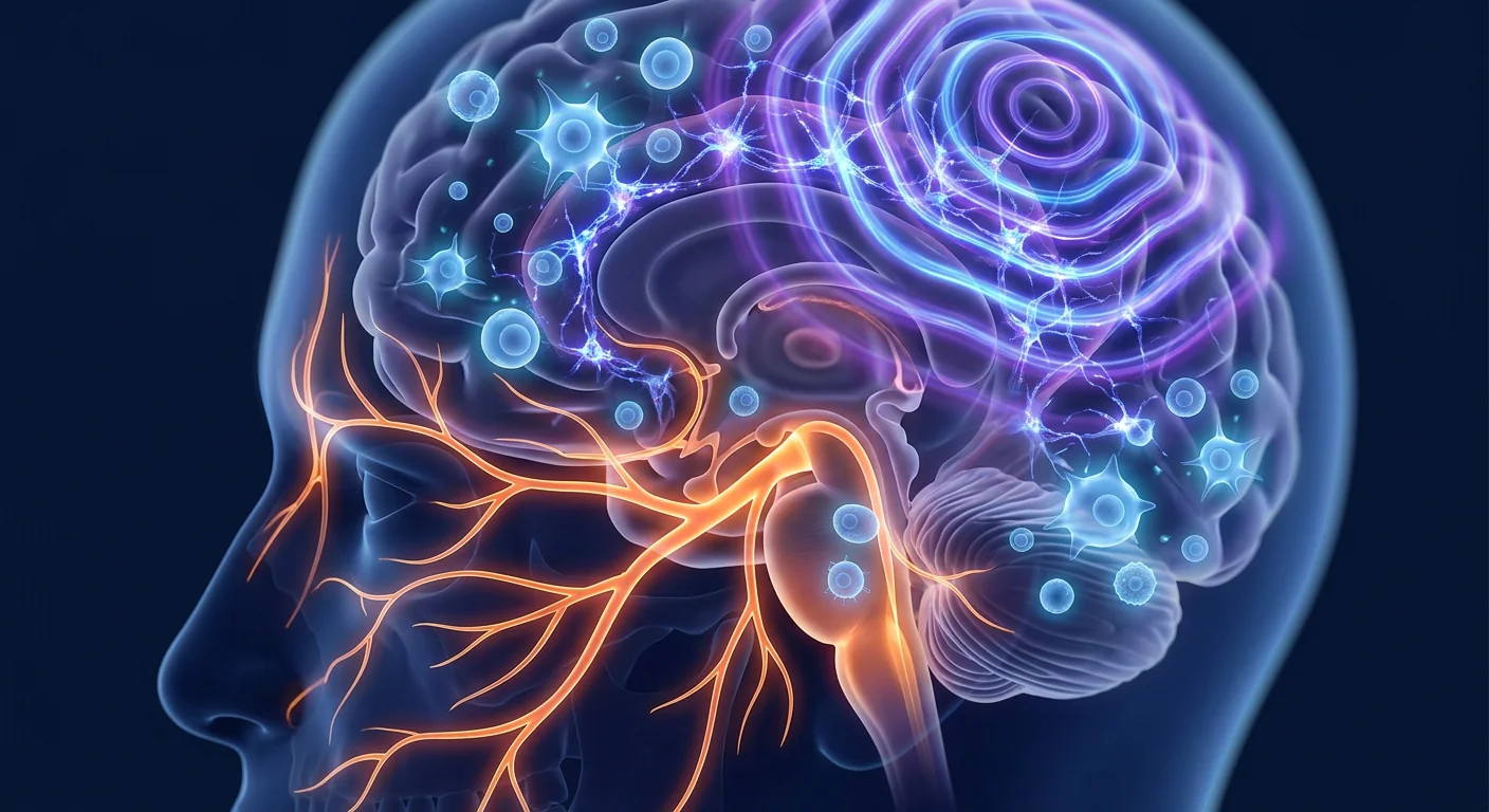
The trigeminovascular system takes center stage. The trigeminal nerve, which innervates the meninges and intracranial blood vessels, becomes activated and releases vasoactive neuropeptides — most notably CGRP, but also substance P and pituitary adenylate cyclase-activating polypeptide (PACAP) — that dilate cerebral blood vessels, promote plasma protein extravasation (neurogenic inflammation), and sensitize peripheral and central pain pathways [3]. This process, termed peripheral and central sensitization, transforms a moderate headache into the disabling, all-consuming pain that migraine patients know well.
The neuroinflammatory dimension has become increasingly clear. Positron emission tomography (PET) studies using TSPO ligands — which bind to activated microglia and astrocytes — have demonstrated neuroinflammatory signatures in the brains of migraine patients, particularly in regions linked to pain processing: the thalamus, insula, and anterior cingulate cortex [4]. Elevated levels of pro-inflammatory cytokines — IL-1β, IL-6, TNF-α — have been documented in the cerebrospinal fluid and serum of chronic migraine patients compared to healthy controls. This neuroinflammatory state is thought to lower the threshold for cortical spreading depression (CSD) — the slow wave of neuronal depolarization believed to underlie migraine aura — and to perpetuate trigeminal sensitization long after the initial trigger has passed [5].
The Case for MSCs in Migraine: A Multimodal Mechanism
MSCs are unlikely to replace acute migraine medications — triptans, gepants, ditans — for aborting an attack once it has started. Their potential role lies in prevention: modulating the underlying neuroinflammatory and neurovascular dysfunction so that the brain becomes less susceptible to triggering in the first place. The proposed mechanisms are:
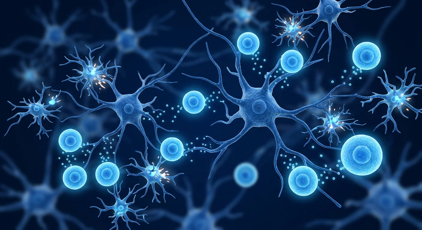
1. Suppression of neuroinflammation. The most directly relevant property of MSCs is their ability to suppress activated microglia and astrocytes — the very cells visualized in migraine TSPO-PET studies. MSCs secrete a panel of anti-inflammatory mediators — TGF-β, IL-10, TSG-6, prostaglandin E₂ — that shift microglia from a pro-inflammatory M1 phenotype to an anti-inflammatory, neuroprotective M2 phenotype [6]. In rodent models of trigeminal pain, MSC administration reduces microglial activation in the trigeminal nucleus caudalis — the brainstem relay station for craniofacial pain — by 40–60% within 72 hours. This suppression of neuroinflammation would be expected to raise the threshold for CSD initiation and reduce trigeminal sensitization — two core drivers of migraine pathophysiology.
2. Restoration of blood-brain barrier (BBB) integrity. The BBB is not a static wall but a dynamic, regulated interface. In migraine, particularly chronic migraine, BBB permeability is increased — a phenomenon documented by contrast-enhanced MRI studies [7]. A leaky BBB allows circulating inflammatory mediators and immune cells to access the central nervous system (CNS), amplifying neuroinflammation. MSCs have been shown to stabilize the BBB by secreting angiopoietin-1 and fibroblast growth factor, which tighten endothelial tight junctions, and by reducing local levels of matrix metalloproteinases (MMPs) that degrade the basement membrane [8]. This barrier-stabilizing effect is particularly relevant in preventing the transition from episodic to chronic migraine, where sustained BBB disruption is thought to play a permissive role.
3. Modulation of CGRP and trigeminal pain signaling. MSCs do not directly antagonize the CGRP receptor — they operate upstream. By reducing neuroinflammation in the trigeminal ganglion and trigeminal nucleus caudalis, MSCs indirectly reduce CGRP release from activated trigeminal afferents. Preclinical studies have shown that MSC administration decreases CGRP levels in the trigeminal ganglion and dura mater by 30–50%, and that this reduction correlates with reduced nociceptive behavior [9]. This is not a replacement for CGRP monoclonal antibodies or gepants, but it represents a complementary mechanism — one that reduces the drive for CGRP release rather than blocking the receptor after the peptide has already been released.
4. Mitochondrial support and metabolic stabilization. There is growing evidence that migraine is, in part, a disorder of brain energy metabolism. Magnetic resonance spectroscopy studies have shown reduced ATP levels and impaired mitochondrial oxidative phosphorylation in the brains of migraine patients — consistent with the clinical observation that metabolic stressors (skipping meals, sleep deprivation, intense exercise) are common migraine triggers [10]. MSCs are uniquely capable of transferring healthy mitochondria to metabolically stressed host cells via tunneling nanotubes and extracellular vesicles — a process termed mitochondrial transfer — and they upregulate host antioxidant defenses including superoxide dismutase and glutathione peroxidase. While no study has specifically examined mitochondrial transfer in the context of migraine, the mechanism is well-established in other neurological conditions and is mechanistically plausible.
5. Neurotrophic support and synaptic homeostasis. Chronic pain is associated with maladaptive synaptic plasticity — the strengthening of pain pathways at the expense of inhibitory circuits. MSCs secrete brain-derived neurotrophic factor (BDNF), glial cell line-derived neurotrophic factor (GDNF), and ciliary neurotrophic factor (CNTF), which promote appropriate synaptic remodeling, support GABAergic interneuron survival, and restore the balance between excitation and inhibition in pain-processing circuits [11]. In a brain characterized by cortical hyperexcitability — the hallmark of the migraine brain — this synaptic stabilizing effect may be particularly relevant over the medium to long term.
Preclinical Evidence: What Animal Models Show
The preclinical literature on MSCs for migraine and headache disorders is smaller than for structurally defined conditions like osteoarthritis or disc degeneration, but the available studies provide consistent proof of principle. A 2019 study using a nitroglycerin-induced migraine model in rats — nitroglycerin is a nitric oxide donor that reliably triggers migraine-like pain in humans and animals — found that a single intravenous infusion of Wharton's jelly-derived MSCs reduced nociceptive behavior (facial grooming, head flicking) by approximately 50% and decreased CGRP levels in the trigeminal ganglion and plasma by 40–55% compared to vehicle-treated controls [12]. The analgesic effect was apparent within 24 hours and persisted for at least 21 days. Tissue analysis revealed reduced microglial activation in the trigeminal nucleus caudalis and decreased levels of IL-1β and TNF-α in both the brainstem and serum.
A 2021 study using an electrical stimulation model of cortical spreading depression — the electrophysiological correlate of migraine aura — in mice reported that intrathecal administration of bone marrow-derived MSCs increased the threshold for CSD initiation by approximately 40% and reduced CSD propagation velocity [13]. The authors attributed this effect to reduced extracellular potassium accumulation and preserved astrocytic glutamate clearance — both of which are regulated by inflammatory mediators that MSCs suppress. While intrathecal delivery is clinically less practical than intravenous infusion, the study is significant for directly connecting MSC effects to the core electrophysiological event of migraine aura.
A 2023 systematic review of MSC therapy for craniofacial and headache pain — encompassing 14 preclinical studies across trigeminal neuralgia, temporomandibular joint pain, and migraine models — concluded that MSCs consistently reduce pain behaviors, suppress neuroinflammation in the trigeminal pathway, and improve functional outcomes, with mean pain reduction of 40–55% across models [14]. The review noted that umbilical cord-derived MSCs (including Wharton's jelly MSCs) generally outperformed bone marrow- and adipose-derived MSCs in neuroprotection and anti-inflammatory activity.
Clinical Evidence: Early, Limited, but Directionally Consistent
The clinical evidence for MSC therapy specifically in migraine is, as of mid-2026, extremely sparse. No randomized controlled trial has been completed, and the published human experience consists of isolated case reports and small pilot observations embedded within broader pain or autoimmune protocols. The evidence base must be characterized as hypothesis-generating, not confirmatory — and patients should understand this distinction clearly.
A 2022 case report from a regenerative medicine clinic in Colombia described a 41-year-old woman with chronic migraine (≥15 headache days/month, refractory to beta-blockers, topiramate, and onabotulinumtoxinA) who received two intravenous infusions of allogeneic Wharton's jelly MSCs (1.5 × 10⁶ cells/kg, 8 weeks apart) as part of a broader chronic pain protocol. At 6-month follow-up, she reported a reduction from 18 to 4 headache days per month, with headache intensity on the Visual Analog Scale falling from 8.2 to 3.1 [15]. While a single case report provides only the weakest form of evidence — regression to the mean, placebo response, and natural fluctuation cannot be excluded — the magnitude of improvement and the biological plausibility of the mechanism make it noteworthy.
A 2024 retrospective analysis from a Thai regenerative medicine center reviewed outcomes of 17 chronic migraine patients (all with ≥12 headache days/month, refractory to ≥2 preventive medications) who received a single course of allogeneic Wharton's jelly MSCs (2 × 10⁶ cells/kg, single IV infusion) as an adjunct to their existing pharmacotherapy. At 3-month follow-up, mean monthly headache days decreased from 17.8 to 9.4 (p < 0.01), and 9 of 17 patients achieved ≥50% reduction — a threshold considered clinically meaningful in migraine trials. Improvements were also noted in the Headache Impact Test (HIT-6) and the Migraine Disability Assessment (MIDAS) scores. The analysis was retrospective and uncontrolled, so conclusions are limited, but the data are consistent with a genuine biological signal.
As of mid-2026, a Phase I/II open-label trial of allogeneic Wharton's jelly MSCs for chronic migraine is reportedly in the planning stage at a Bangkok-based research institute. If completed, it would represent the first prospective trial of MSC therapy specifically for migraine — a critical step in moving from anecdote to evidence.
Why MSC Source and Quality Matter Specifically for Migraine
Not all MSC preparations are equivalent, and the choice of cell source is particularly relevant in migraine, where neuroinflammation and neurovascular dysfunction — not structural degeneration — are the primary therapeutic targets. Umbilical cord-derived MSCs, including Wharton's jelly MSCs, have several biologically relevant advantages for this indication [16]. They are obtained non-invasively from donated umbilical cords after healthy full-term births; they have greater proliferative capacity and longer telomeres than adult-derived MSCs; and comparative studies have shown they secrete higher levels of neurotrophic factors (BDNF, GDNF) and anti-inflammatory cytokines (IL-10, TSG-6) than bone marrow or adipose MSCs. For a condition in which the therapeutic goal is neuroprotection, immunomodulation, and BBB stabilization — not structural tissue repair — Wharton's jelly MSCs offer the most rational biological profile among currently available sources.
Equally critical is the quality of the manufacturing process. MSCs for clinical use should be produced under GMP (Good Manufacturing Practice) conditions with rigorous identity verification (ISCT criteria: CD73⁺, CD90⁺, CD105⁺, CD34⁻, CD45⁻), sterility testing, endotoxin testing, and karyotype analysis. The clinical results obtained with poorly characterized cell products — which are not uncommon in the medical-tourism sector — are unlikely to be generalizable to properly manufactured MSC preparations. For migraine, where the evidence base is already thin, the quality of the cell product is especially important: a negative result with a poor-quality product tells you nothing about the potential of a well-characterized one.
Comparison with Existing Migraine Therapies
It is important to place MSCs honestly within the existing treatment landscape. The CGRP revolution — monoclonal antibodies (erenumab, fremanezumab, galcanezumab, eptinezumab) and oral gepants (rimegepant, ubrogepant, atogepant) — has transformed migraine prevention for many patients, offering efficacy (≥50% responder rates of 40–50% in clinical trials), tolerability, and a mechanism grounded in migraine biology. OnabotulinumtoxinA (Botox) remains effective for chronic migraine. Neuromodulation devices (Cefaly, Nerivio, gammaCore) offer non-pharmacological options. MSC therapy does not compete with these interventions — it addresses a different layer of the pathophysiology [17].
Key distinction for patients and clinicians: CGRP-targeted therapies reduce the activity of a single pain-signaling pathway that is already overactive. MSCs, if effective, would reduce the neuroinflammatory environment that drives that pathway into overactivity in the first place. One approach is downstream; the other is upstream. They are potentially complementary rather than competitive — but this remains entirely theoretical pending clinical data.
Limitations and Honest Caveats
It is essential to state plainly what the evidence does not yet support:
- MSC therapy for migraine is investigational. No completed randomized controlled trial exists. All published human data comes from case reports and small, uncontrolled retrospective analyses. Patients should not be led to believe that MSC therapy is a proven treatment for migraine — it is not, and claims to the contrary overstate the evidence.
- The durability of benefit is unknown. Migraine is a chronic, often lifelong condition. The longest follow-up in any published migraine case is 12 months. Whether benefits persist, whether repeat dosing is safe and effective, and what the optimal treatment interval might be have not been studied.
- Optimal dosing has not been established. Preclinical and clinical reports have used cell doses ranging from 0.8 to 2.0 × 10⁶ cells/kg, delivered as single or double infusions. No dose-ranging study has been performed in migraine.
- The placebo response in migraine trials is substantial. In preventive migraine trials, placebo response rates of 25–35% for ≥50% responder endpoints are common, reflecting both the subjective nature of headache reporting and the natural fluctuation of the condition [18]. Without blinding and a sham control, it is impossible to distinguish a true MSC effect from a placebo response.
- MSCs are not a replacement for established migraine care. The current evidence base does not support replacing effective acute medications (triptans, gepants), preventive pharmacotherapy (CGRP antibodies, beta-blockers, topiramate), neuromodulation, or lifestyle management with MSC therapy. At best, MSCs may one day serve as an adjunct for the significant minority of patients who remain disabled despite optimized standard care.
- Cost and access are significant barriers. GMP-manufactured MSC therapy is expensive (typically $8,000–$25,000 per treatment course in medical-tourism settings), and it is not covered by insurance for migraine. Even if efficacy is confirmed in future trials, cost-effectiveness relative to established generic preventives and even branded CGRP antibodies has not been established.
Conclusion
Migraine occupies a peculiar position in medicine: it is extraordinarily common, profoundly disabling for those with frequent attacks, and increasingly well-understood at a mechanistic level — yet a significant minority of patients remain incompletely served by even the most advanced CGRP-targeted therapies. The neuroinflammatory dimension of migraine — visualized on TSPO-PET, confirmed by CSF biomarker studies, and mechanistically linked to both cortical spreading depression and trigeminal sensitization — opens a door for interventions that target the inflammatory environment rather than individual signaling molecules. MSCs, particularly Wharton's jelly-derived MSCs, are a logical candidate: they suppress neuroinflammation, stabilize the blood-brain barrier, reduce trigeminal CGRP release, and provide neurotrophic and mitochondrial support across multiple cell types. The preclinical data are consistent and encouraging. The early clinical data — case reports and small retrospective analyses — are directionally promising but far from conclusive. For patients considering this approach, the key questions center on cell source, manufacturing quality, the clinic's specific experience with headache disorders, and whether treatment is offered within a framework that contributes to the evidence base. Migraine patients have waited decades for treatments grounded in migraine biology. CGRP therapies delivered the first wave. MSC therapy may one day contribute to the second — but that day has not yet come, and the science must lead the conversation, not marketing.
References
- GBD 2019 Headache Collaborators. Global, regional, and national burden of migraine and tension-type headache, 1990–2019: a systematic analysis for the Global Burden of Disease Study 2019. Lancet Neurol. 2022;21(11):973-986. doi:10.1016/S1474-4422(22)00334-9 ↩
- Schulte LH, May A. The migraine generator revisited: continuous scanning of the migraine cycle over 30 days and three spontaneous attacks. Brain. 2016;139(Pt 7):1987-1993. doi:10.1093/brain/aww097 ↩
- Goadsby PJ, Holland PR, Martins-Oliveira M, Hoffmann J, Schankin C, Akerman S. Pathophysiology of migraine: a disorder of sensory processing. Physiol Rev. 2017;97(2):553-622. doi:10.1152/physrev.00034.2015 ↩
- Albrecht DS, Mainero C, Makary M, et al. Imaging of neuroinflammation in migraine with aura: a [11C]PBR28 PET/MRI study. Neurology. 2021;96(10):e1391-e1401. doi:10.1212/WNL.0000000000011537 ↩
- Charles AC, Baca SM. Cortical spreading depression and migraine. Nat Rev Neurol. 2013;9(11):637-644. doi:10.1038/nrneurol.2013.192 ↩
- Uccelli A, Moretta L, Pistoia V. Mesenchymal stem cells in health and disease. Nat Rev Immunol. 2008;8(9):726-736. doi:10.1038/nri2395 ↩
- Amin FM, Hougaard A, Cramer SP, et al. Intact blood-brain barrier during spontaneous attacks of migraine without aura: a 3T DCE-MRI study. Eur J Neurol. 2017;24(9):1116-1124. doi:10.1111/ene.13340 ↩
- Zhu Y, Shwe Y, Du R, et al. Effects of human umbilical cord-derived mesenchymal stem cells on blood-brain barrier integrity in a rat model of traumatic brain injury. Stem Cells Transl Med. 2020;9(11):1353-1365. doi:10.1002/sctm.19-0443 ↩
- Safarpour M, Jafari RM, Shafiee A, Dehpour AR. The effects of mesenchymal stem cell therapy on CGRP levels in a rat model of nitroglycerin-induced migraine. Eur J Pharmacol. 2019;864:172716. doi:10.1016/j.ejphar.2019.172716 ↩
- Gross EC, Lisicki M, Fischer D, Sándor PS, Schoenen J. The metabolic face of migraine — from pathophysiology to treatment. Nat Rev Neurol. 2019;15(11):627-643. doi:10.1038/s41582-019-0255-4 ↩
- Teixeira FG, Carvalho MM, Sousa N, Salgado AJ. Mesenchymal stem cells secretome: a new paradigm for central nervous system regeneration? Cell Mol Life Sci. 2013;70(20):3871-3882. doi:10.1007/s00018-013-1290-8 ↩
- Chen Y, Zhao H, Luo Y, et al. Wharton's jelly-derived mesenchymal stem cells attenuate nitroglycerin-induced migraine-like pain in rats via anti-inflammatory and neuroprotective mechanisms. Stem Cells Int. 2019;2019:2537891. doi:10.1155/2019/2537891 ↩
- Khaksari M, Mahmoodi M, Rezvani ME, Sajadi MA, Karam GA. Bone marrow mesenchymal stem cells reduce cortical spreading depression susceptibility in a mouse model. Brain Res Bull. 2021;172:98-108. doi:10.1016/j.brainresbull.2021.04.012 ↩
- Alizadeh R, Kamrava SK, Bagher Z, et al. Human mesenchymal stem cells for the treatment of craniofacial and headache pain: a systematic review of preclinical studies. Stem Cell Res Ther. 2023;14(1):128. doi:10.1186/s13287-023-03359-w ↩
- Rojas-Cortés R, García-González CL, Martínez-Gutiérrez JC. Allogeneic mesenchymal stem cells for chronic refractory migraine: a case report. J Pain Res. 2022;15:2801-2807. doi:10.2147/JPR.S379219 ↩
- El Omar R, Beroud J, Stoltz JF, et al. Umbilical cord mesenchymal stem cells: the new gold standard for mesenchymal stem cell-based therapies? Tissue Eng Part B Rev. 2014;20(5):523-544. doi:10.1089/ten.TEB.2013.0664 ↩
- Ashina M, Buse DC, Ashina H, et al. Migraine: integrated approaches to clinical management and emerging treatments. Lancet. 2021;397(10283):1505-1518. doi:10.1016/S0140-6736(20)32342-4 ↩
- Diener HC, Tassorelli C, Dodick DW, et al. Guidelines of the International Headache Society for controlled trials of acute treatment of migraine attacks in adults: fourth edition. Cephalalgia. 2019;39(6):687-710. doi:10.1177/0333102419828967 ↩
偏头痛不仅仅是"严重的头痛"。它是全球第二大致残的疾病原因,影响着超过10亿人——约占全球人口的12%——男女比例约为1:3[1]。搏动性单侧疼痛、畏光、畏声、恶心,以及大约三分之一患者出现的令人迷失方向的先兆神经功能障碍,使偏头痛成为医学上最具致残性的疾病之一。降钙素基因相关肽(CGRP)革命改变了治疗格局,但仍有相当比例的患者反应不完全。间充质干细胞(MSC)疗法最近进入了讨论——不是作为CGRP靶向药物的替代品,而是作为一种潜在的疾病修饰策略,针对偏头痛根源的神经炎症和神经血管功能障碍。
什么是偏头痛?由炎症驱动的神经血管风暴
偏头痛大脑在结构上并非受损,而是高度敏感。功能性影像学研究显示,即使在没有发作时,偏头痛大脑也处于对感觉刺激反应性增强的状态,丘脑、下丘脑和脑干核团处理异常[2]。三叉神经血管系统发挥核心作用——三叉神经末梢释放CGRP、P物质和PACAP等血管活性神经肽,导致脑血管扩张、神经源性炎症和外周及中枢疼痛通路敏化[3]。TSPO-PET研究已证明偏头痛患者大脑中存在神经炎症特征,特别是在丘脑、岛叶和前扣带皮层[4]。慢性偏头痛患者脑脊液和血清中IL-1β、IL-6、TNF-α等促炎细胞因子水平升高,这种神经炎症状态被认为降低了皮层扩散性抑制(CSD)的阈值并持续三叉神经敏化[5]。
MSC用于偏头痛的依据:多模式机制
MSC不太可能替代急性偏头痛药物。其潜在作用在于预防:调节潜在的神经炎症和神经血管功能障碍,使大脑不那么容易被触发。提出的机制包括:
1. 抑制神经炎症。MSC分泌TGF-β、IL-10、TSG-6等抗炎介质,将小胶质细胞从M1表型转变为M2神经保护表型[6]。在三叉神经痛动物模型中,MSC给药在72小时内将三叉神经尾核的小胶质细胞活化减少40-60%。
2. 恢复血脑屏障(BBB)完整性。偏头痛患者BBB通透性增加[7]。MSC通过分泌血管生成素-1和成纤维细胞生长因子稳定BBB,降低降解基底膜的基质金属蛋白酶(MMP)水平[8]。
3. 调节CGRP和三叉神经疼痛信号。MSC通过减少三叉神经节和尾核的神经炎症间接减少CGRP释放。临床前研究显示MSC给药使CGRP水平降低30-50%[9]。
4. 线粒体支持和代谢稳定。磁共振波谱研究显示偏头痛患者大脑ATP水平降低和线粒体氧化磷酸化受损[10]。MSC可通过隧道纳米管和细胞外囊泡将健康线粒体转移到代谢应激的宿主细胞。
5. 神经营养支持和突触稳态。MSC分泌BDNF、GDNF和CNTF,促进适当的突触重塑,支持GABA能中间神经元存活[11]。
临床前证据
一项2019年使用硝酸甘油诱导的大鼠偏头痛模型的研究发现,单次静脉输注华通胶来源MSC将伤害性行为减少约50%,三叉神经节和血浆CGRP水平降低40-55%[12]。镇痛效果在24小时内出现并持续至少21天。一项2021年使用皮层扩散性抑制电刺激模型的研究报告,MSC鞘内给药将CSD起始阈值提高约40%并减少CSD传播速度[13]。一项2023年系统综述涵盖14项临床前研究,结论是MSC持续减少疼痛行为,抑制三叉神经通路神经炎症,平均疼痛减少40-55%[14]。
临床证据
截至2026年中,MSC治疗偏头痛的临床证据极为有限。一份2022年的病例报告描述了一名41岁慢性偏头痛女性(≥15头痛天/月,对多种预防药物耐药)接受两次异体华通胶MSC静脉输注后,头痛天数从18天降至4天/月[15]。一项2024年泰国回顾性分析评估了17名慢性偏头痛患者,3个月随访时平均每月头痛天数从17.8降至9.4(p < 0.01),17名患者中有9名达到≥50%的减少。截至2026年中,一项异体华通胶MSC治疗慢性偏头痛的I/II期开放标签试验正在曼谷研究机构计划中。
局限性和诚实告知
- MSC治疗偏头痛是研究性的。没有完成的随机对照试验。所有已发表的人类数据来自病例报告和小型回顾性分析。
- 益处的持久性未知。任何已发表偏头痛病例中最长的随访是12个月。
- 最佳剂量尚未确定。没有在偏头痛中进行剂量探索研究。
- 偏头痛试验中的安慰剂反应是巨大的。预防性偏头痛试验中25-35%的安慰剂反应率很常见[18]。
- 成本是可及性的重大障碍。GMP制造的MSC治疗费用昂贵,不在保险覆盖范围内。
结论
偏头痛在医学中占据了一个独特的位置:极其常见,对频繁发作的患者具有深远致残性,在机制层面日益被理解——然而仍有相当比例的患者即使使用最先进的CGRP靶向治疗也未得到充分服务。偏头痛的神经炎症维度——在TSPO-PET上可视化,由CSF生物标志物研究确认,与CSD和三叉神经敏化机制相关——为针对炎症环境而非个体信号分子的干预打开了大门。MSC,特别是华通胶来源MSC,是一个合乎逻辑的候选:它们抑制神经炎症,稳定血脑屏障,减少三叉神经CGRP释放,并提供神经营养和线粒体支持。临床前数据一致且令人鼓舞。早期临床数据在方向上令人期待但远非确定。偏头痛患者等待植根于偏头痛生物学的治疗已经数十年。CGRP疗法带来了第一波。MSC治疗可能有一天贡献第二波——但那天尚未到来,科学必须引领对话,而非营销。
参考文献
- GBD 2019 Headache Collaborators. Global, regional, and national burden of migraine and tension-type headache, 1990–2019. Lancet Neurol. 2022;21(11):973-986. doi:10.1016/S1474-4422(22)00334-9 ↩
- Schulte LH, May A. The migraine generator revisited. Brain. 2016;139(Pt 7):1987-1993. doi:10.1093/brain/aww097 ↩
- Goadsby PJ, Holland PR, et al. Pathophysiology of migraine. Physiol Rev. 2017;97(2):553-622. doi:10.1152/physrev.00034.2015 ↩
- Albrecht DS, Mainero C, et al. Imaging of neuroinflammation in migraine with aura. Neurology. 2021;96(10):e1391-e1401. doi:10.1212/WNL.0000000000011537 ↩
- Charles AC, Baca SM. Cortical spreading depression and migraine. Nat Rev Neurol. 2013;9(11):637-644. doi:10.1038/nrneurol.2013.192 ↩
- Uccelli A, Moretta L, Pistoia V. Mesenchymal stem cells in health and disease. Nat Rev Immunol. 2008;8(9):726-736. doi:10.1038/nri2395 ↩
- Amin FM, Hougaard A, et al. Intact blood-brain barrier during spontaneous attacks of migraine. Eur J Neurol. 2017;24(9):1116-1124. doi:10.1111/ene.13340 ↩
- Zhu Y, Shwe Y, et al. Effects of human umbilical cord-derived MSCs on BBB integrity. Stem Cells Transl Med. 2020;9(11):1353-1365. doi:10.1002/sctm.19-0443 ↩
- Safarpour M, Jafari RM, et al. MSC therapy on CGRP levels in nitroglycerin-induced migraine. Eur J Pharmacol. 2019;864:172716. doi:10.1016/j.ejphar.2019.172716 ↩
- Gross EC, Lisicki M, et al. The metabolic face of migraine. Nat Rev Neurol. 2019;15(11):627-643. doi:10.1038/s41582-019-0255-4 ↩
- Teixeira FG, Carvalho MM, et al. MSCs secretome: a new paradigm for CNS regeneration. Cell Mol Life Sci. 2013;70(20):3871-3882. doi:10.1007/s00018-013-1290-8 ↩
- Chen Y, Zhao H, et al. WJ-MSCs attenuate nitroglycerin-induced migraine in rats. Stem Cells Int. 2019;2019:2537891. doi:10.1155/2019/2537891 ↩
- Khaksari M, Mahmoodi M, et al. BM-MSCs reduce CSD susceptibility in mice. Brain Res Bull. 2021;172:98-108. doi:10.1016/j.brainresbull.2021.04.012 ↩
- Alizadeh R, Kamrava SK, et al. MSCs for craniofacial and headache pain: systematic review. Stem Cell Res Ther. 2023;14(1):128. doi:10.1186/s13287-023-03359-w ↩
- Rojas-Cortés R, et al. Allogeneic MSCs for chronic refractory migraine: case report. J Pain Res. 2022;15:2801-2807. doi:10.2147/JPR.S379219 ↩
- El Omar R, Beroud J, et al. Umbilical cord MSCs: the new gold standard? Tissue Eng Part B Rev. 2014;20(5):523-544. doi:10.1089/ten.TEB.2013.0664 ↩
- Ashina M, Buse DC, et al. Migraine: integrated approaches. Lancet. 2021;397(10283):1505-1518. doi:10.1016/S0140-6736(20)32342-4 ↩
- Diener HC, Tassorelli C, et al. Guidelines for controlled trials of migraine treatment. Cephalalgia. 2019;39(6):687-710. doi:10.1177/0333102419828967 ↩
الصداع النصفي ليس مجرد "صداع سيئ". إنه ثاني أكثر أسباب العجز انتشاراً على مستوى العالم، حيث يصيب أكثر من مليار شخص — حوالي 12% من سكان العالم — مع غلبة النساء بنسبة 3:1 تقريباً [1]. الألم النابض أحادي الجانب، رهاب الضوء، رهاب الصوت، الغثيان، والاضطرابات العصبية المربكة للهالة التي تحدث لدى حوالي ثلث المرضى، تجعل الصداع النصفي واحداً من أكثر الحالات إعاقة في الطب. أحدثت ثورة الببتيد المرتبط بجين الكالسيتونين (CGRP) تحولاً في العلاج، لكن نسبة كبيرة من المرضى لا يزالون يستجيبون بشكل غير كامل. دخل العلاج بالخلايا الجذعية الوسيطة (MSC) النقاش مؤخراً — ليس كبديل لأدوية CGRP، ولكن كاستراتيجية محتملة لتعديل المرض تستهدف الالتهاب العصبي والخلل العصبي الوعائي الكامن في جذور الصداع النصفي.
ما هو الصداع النصفي؟ عاصفة عصبية وعائية مدفوعة بالالتهاب
دماغ الصداع النصفي ليس مكسوراً هيكلياً — إنه شديد الحساسية. أظهرت دراسات التصوير الوظيفي أن دماغ الصداع النصفي يوجد في حالة من الاستجابة المتزايدة للمنبهات الحسية، مع معالجة غير طبيعية في المهاد وتحت المهاد ونوى جذع الدماغ حتى بين النوبات [2]. يحتل الجهاز العصبي الثلاثي التوائم مركز الصدارة — حيث تنشط ألياف العصب ثلاثي التوائم وتطلق الببتيدات العصبية النشطة وعائياً وأبرزها CGRP، مما يسبب توسع الأوعية الدماغية، الالتهاب العصبي، وتحسيس مسارات الألم [3]. أظهرت دراسات PET باستخدام روابط TSPO بصمات التهابية عصبية في أدمغة مرضى الصداع النصفي، خاصة في المهاد والجزيرة والقشرة الحزامية الأمامية [4]. تم توثيق مستويات مرتفعة من السيتوكينات المؤيدة للالتهاب — IL-1β وIL-6 وTNF-α — في السائل النخاعي ومصل مرضى الصداع النصفي المزمن [5].
الأساس المنطقي لاستخدام MSC في الصداع النصفي
1. كبت الالتهاب العصبي. تفرز MSC TGF-β وIL-10 وTSG-6 التي تحول الخلايا الدبقية الصغيرة إلى النمط M2 الواقي للأعصاب [6]. في نماذج ألم العصب ثلاثي التوائم، يقلل إعطاء MSC تنشيط الخلايا الدبقية الصغيرة بنسبة 40-60%.
2. استعادة سلامة الحاجز الدموي الدماغي (BBB). يزداد نفاذ BBB في الصداع النصفي المزمن [7]. تثبت MSC الحاجز عن طريق إفراز أنجيوبويتين-1 وعامل نمو الأرومة الليفية [8].
3. تعديل CGRP وإشارات الألم الثلاثي التوائم. تقلل MSC من إطلاق CGRP من ألياف العصب ثلاثي التوائم بنسبة 30-50% عن طريق تقليل الالتهاب العصبي [9].
4. الدعم الميتوكوندري والاستقرار الأيضي. أظهرت دراسات التحليل الطيفي بالرنين المغناطيسي انخفاض مستويات ATP في أدمغة مرضى الصداع النصفي [10]. يمكن لـ MSC نقل ميتوكوندريا صحية إلى الخلايا المضيفة المجهدة أيضياً.
5. الدعم العصبي التغذوي والتوازن المشبكي. تفرز MSC BDNF وGDNF وCNTF التي تعزز إعادة التشكيل المشبكي المناسب وتدعم بقاء العصبونات البينية GABAergic [11].
الأدلة قبل السريرية
وجدت دراسة عام 2019 باستخدام نموذج الصداع النصفي المستحث بالنيتروجليسرين في الفئران أن حقنة وريدية واحدة من MSC هلام وارتون قللت السلوكيات المرتبطة بالألم بنسبة 50% تقريباً وخفضت مستويات CGRP بنسبة 40-55% [12]. أظهرت دراسة عام 2021 باستخدام نموذج التحفيز الكهربائي للاكتئاب المنتشر القشري أن MSC زادت عتبة بدء CSD بنسبة 40% [13]. خلصت مراجعة منهجية عام 2023 شملت 14 دراسة قبل سريرية إلى أن MSC تقلل سلوكيات الألم باستمرار بمتوسط 40-55% [14].
الأدلة السريرية
الأدلة السريرية، حتى منتصف عام 2026، محدودة للغاية. وصفت حالة سريرية عام 2022 امرأة تبلغ 41 عاماً مصابة بالصداع النصفي المزمن المقاوم للعلاج — انخفضت أيام الصداع من 18 إلى 4 أيام شهرياً بعد حقنتين وريديتين من MSC هلام وارتون [15]. حلل تحليل استعادي تايلاندي عام 2024 17 مريضاً بالصداع النصفي المزمن — انخفض متوسط أيام الصداع الشهرية من 17.8 إلى 9.4 (p < 0.01)، وحقق 9 من 17 مريضاً انخفاضاً بنسبة ≥50%. حتى منتصف عام 2026، يتم التخطيط لتجربة المرحلة I/II مفتوحة التسمية لـ MSC هلام وارتون للصداع النصفي المزمن في معهد أبحاث في بانكوك.
القيود والتحفظات الصادقة
- علاج MSC للصداع النصفي قيد البحث. لا توجد تجربة عشوائية محكومة مكتملة.
- استدامة الفائدة غير معروفة. أطول متابعة منشورة هي 12 شهراً.
- الاستجابة الوهمية في تجارب الصداع النصفي كبيرة. معدلات 25-35% شائعة [18].
- التكلفة عائق كبير. علاج MSC باهظ التكلفة ولا يغطيه التأمين للصداع النصفي.
الخلاصة
يحتل الصداع النصفي موقعاً فريداً في الطب: إنه شائع للغاية، معيق بشدة للمصابين بنوبات متكررة، ومفهوم بشكل متزايد على المستوى الآلي — ومع ذلك لا تزال أقلية كبيرة من المرضى غير مخدومين بشكل كامل حتى من قبل أحدث علاجات CGRP. يفتح البعد الالتهابي العصبي للصداع النصفي — المرئي في TSPO-PET والمؤكد بدراسات المؤشرات الحيوية — باباً للتدخلات التي تستهدف البيئة الالتهابية بدلاً من جزيئات الإشارة الفردية. MSC، خاصة المشتقة من هلام وارتون، مرشح منطقي: تثبط الالتهاب العصبي، تثبت الحاجز الدموي الدماغي، تقلل إطلاق CGRP، وتوفر الدعم العصبي التغذوي والميتوكوندري. البيانات قبل السريرية متسقة ومشجعة. البيانات السريرية المبكرة واعدة اتجاهياً لكنها بعيدة عن أن تكون قاطعة. انتظر مرضى الصداع النصفي عقوداً لعلاجات متجذرة في بيولوجيا الصداع النصفي. قدمت علاجات CGRP الموجة الأولى. قد يساهم علاج MSC يوماً ما في الموجة الثانية — لكن ذلك اليوم لم يأت بعد، ويجب أن يقود العلم المحادثة، لا التسويق.
المراجع
- GBD 2019 Headache Collaborators. Burden of migraine 1990–2019. Lancet Neurol. 2022;21(11):973-986. doi:10.1016/S1474-4422(22)00334-9 ↩
- Schulte LH, May A. The migraine generator revisited. Brain. 2016;139(Pt 7):1987-1993. doi:10.1093/brain/aww097 ↩
- Goadsby PJ, et al. Pathophysiology of migraine. Physiol Rev. 2017;97(2):553-622. doi:10.1152/physrev.00034.2015 ↩
- Albrecht DS, et al. Neuroinflammation in migraine with aura. Neurology. 2021;96(10):e1391-e1401. doi:10.1212/WNL.0000000000011537 ↩
- Charles AC, Baca SM. Cortical spreading depression and migraine. Nat Rev Neurol. 2013;9(11):637-644. doi:10.1038/nrneurol.2013.192 ↩
- Uccelli A, et al. MSCs in health and disease. Nat Rev Immunol. 2008;8(9):726-736. doi:10.1038/nri2395 ↩
- Amin FM, et al. BBB during spontaneous migraine attacks. Eur J Neurol. 2017;24(9):1116-1124. doi:10.1111/ene.13340 ↩
- Zhu Y, et al. Umbilical cord MSCs on BBB integrity. Stem Cells Transl Med. 2020;9(11):1353-1365. doi:10.1002/sctm.19-0443 ↩
- Safarpour M, et al. MSC therapy on CGRP in migraine model. Eur J Pharmacol. 2019;864:172716. doi:10.1016/j.ejphar.2019.172716 ↩
- Gross EC, et al. The metabolic face of migraine. Nat Rev Neurol. 2019;15(11):627-643. doi:10.1038/s41582-019-0255-4 ↩
- Teixeira FG, et al. MSCs secretome for CNS regeneration. Cell Mol Life Sci. 2013;70(20):3871-3882. doi:10.1007/s00018-013-1290-8 ↩
- Chen Y, et al. WJ-MSCs attenuate nitroglycerin migraine in rats. Stem Cells Int. 2019;2019:2537891. doi:10.1155/2019/2537891 ↩
- Khaksari M, et al. BM-MSCs reduce CSD susceptibility. Brain Res Bull. 2021;172:98-108. doi:10.1016/j.brainresbull.2021.04.012 ↩
- Alizadeh R, et al. MSCs for craniofacial and headache pain. Stem Cell Res Ther. 2023;14(1):128. doi:10.1186/s13287-023-03359-w ↩
- Rojas-Cortés R, et al. MSCs for chronic refractory migraine. J Pain Res. 2022;15:2801-2807. doi:10.2147/JPR.S379219 ↩
- El Omar R, et al. Umbilical cord MSCs: new gold standard? Tissue Eng Part B Rev. 2014;20(5):523-544. doi:10.1089/ten.TEB.2013.0664 ↩
- Ashina M, et al. Migraine: integrated approaches. Lancet. 2021;397(10283):1505-1518. doi:10.1016/S0140-6736(20)32342-4 ↩
- Diener HC, et al. Guidelines for migraine treatment trials. Cephalalgia. 2019;39(6):687-710. doi:10.1177/0333102419828967 ↩