Age-related hearing loss — presbycusis — affects roughly one in three adults over 65 and two-thirds of those over 75, making it the third most common chronic condition of aging. For decades the prevailing view held that cochlear hair cells, once lost, were gone forever. That orthodoxy is now shifting: regenerative research has demonstrated that the inner ear retains some capacity for repair, and mesenchymal stem cell therapy is being investigated as a way to amplify that capacity.
What happens to the inner ear with age?
Presbycusis is a progressive, bilateral sensorineural hearing loss driven by cumulative damage to the auditory system. The key structures affected include:
- Outer hair cells — the cochlear amplifiers that sharpen frequency selectivity; these are the most vulnerable to oxidative stress and noise exposure over a lifetime
- Spiral ganglion neurons — the primary auditory neurons that relay signals from hair cells to the brainstem; their loss is a major predictor of poor speech discrimination even when amplification is provided
- Stria vascularis — the metabolic battery of the cochlea that maintains the endocochlear potential; its atrophy contributes to the flat, high-frequency-predominant hearing loss typical of aging
- Inflammatory microenvironment — chronic low-grade inflammation in the cochlea accelerates hair cell and neuronal loss through cytokine-mediated damage
Conventional interventions — hearing aids and cochlear implants — address the symptom (reduced audibility) but not the underlying biology. Hearing aids amplify sound for surviving hair cells; cochlear implants bypass hair cells entirely and electrically stimulate the auditory nerve. Neither protects remaining cells from further degeneration, and both leave the spiral ganglion neuron population — the final common pathway to the brain — vulnerable to continued loss.
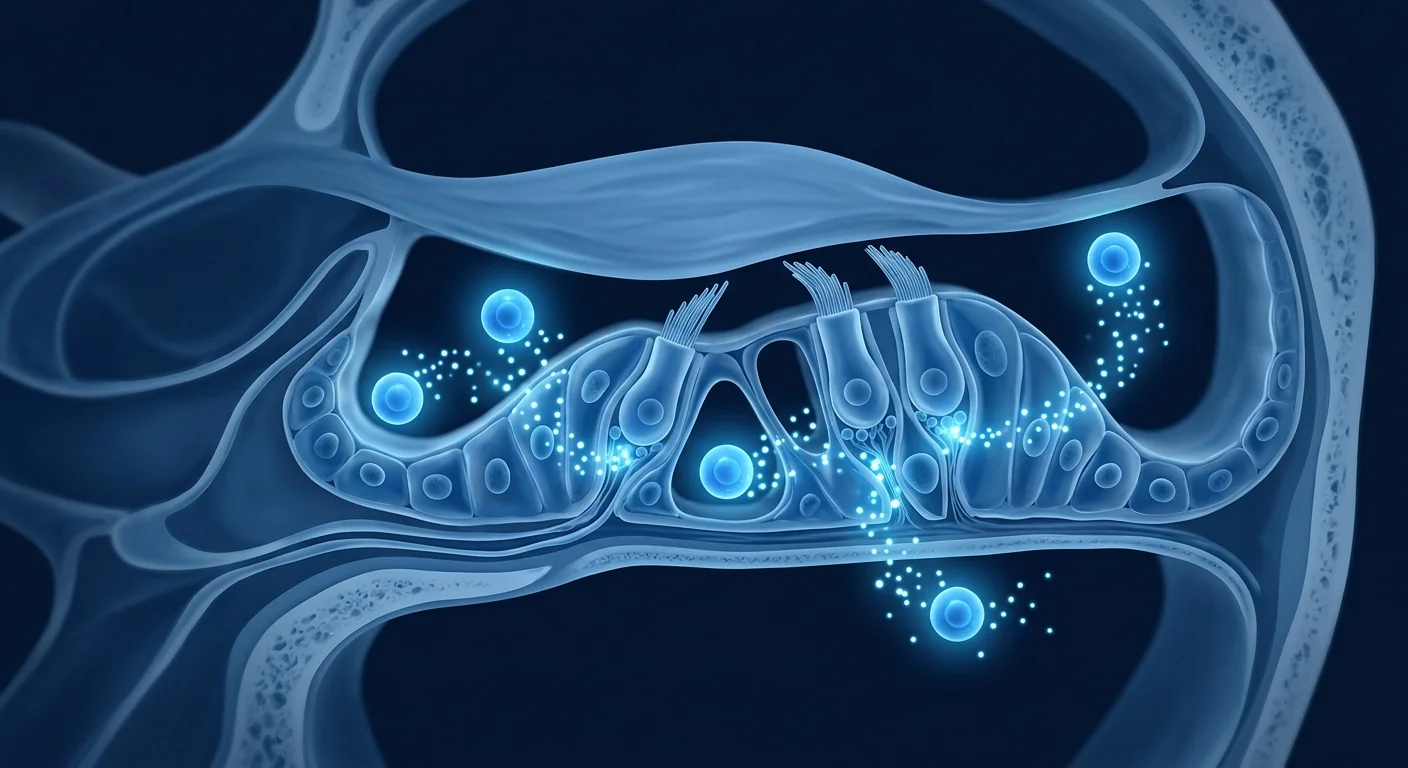
How MSC therapy may support the aging inner ear
Mesenchymal stem cells do not differentiate into new hair cells — that ability remains beyond current clinical reach. What they do, and what the preclinical evidence supports, is modulate the cellular environment in ways that protect surviving auditory structures from further decline. The four mechanisms most relevant to presbycusis are:
Neurotrophic factor secretion
MSCs are prolific producers of brain-derived neurotrophic factor (BDNF), glial cell line-derived neurotrophic factor (GDNF), and nerve growth factor (NGF) — molecules that directly support spiral ganglion neuron survival. In animal models of sensorineural hearing loss, MSC-derived BDNF infusion has been shown to preserve spiral ganglion neuron density and maintain auditory brainstem response thresholds. [1]
Anti-inflammatory modulation of the cochlear microenvironment
The aging cochlea exhibits elevated levels of TNF-α, IL-1β, and IL-6 — the same pro-inflammatory cytokines implicated in neurodegeneration elsewhere. MSCs shift microglial and macrophage phenotypes from pro-inflammatory (M1) toward anti-inflammatory and tissue-remodeling (M2) states, reducing cytokine-driven damage to hair cells and supporting cells. [2]
Mitochondrial transfer and oxidative stress reduction
Outer hair cells are metabolically demanding — they consume ATP at extraordinary rates to power the electromotility that underlies cochlear amplification. Mitochondrial dysfunction is a hallmark of aging hair cells. MSCs can transfer healthy mitochondria to stressed recipient cells via tunneling nanotubes, and their secretome includes antioxidant enzymes (superoxide dismutase, catalase) that buffer the oxidative load. [3]
Paracrine support of the stria vascularis
The stria vascularis generates the +80 mV endocochlear potential — the driving force for hair cell transduction. Its atrophy with age directly degrades hearing sensitivity. MSC-derived vascular endothelial growth factor (VEGF) and hepatocyte growth factor (HGF) support microvascular integrity, and early work suggests this paracrine signaling may slow strial degeneration. [4]
What MSC therapy cannot do for hearing
It is important to be direct: current MSC protocols cannot regenerate lost hair cells, cannot restore hearing that has been absent for years, and cannot reverse structural damage to the cochlear architecture. The realistic therapeutic window is protection of surviving cells — slowing further decline in patients who still have measurable residual hearing. Anyone promising "hair cell regrowth" or "hearing restoration" from current MSC therapy is making claims the biology does not support.
What the preclinical evidence shows
The MSC hearing research literature, while still predominantly preclinical, has produced consistent signals across multiple independent laboratories:
- Spiral ganglion neuron preservation. Intracochlear or intratympanic MSC administration in noise-exposed and aged rodent models preserves spiral ganglion neuron counts by 25–40% compared to untreated controls, measured histologically at 4–8 weeks post-treatment. [5]
- Auditory brainstem response (ABR) threshold stabilization. Several studies report that MSC-treated animals maintain ABR thresholds closer to baseline, while untreated animals show progressive threshold elevation. The effect is most pronounced in the high-frequency region — the same region most affected in human presbycusis.
- Reduced cochlear inflammation. MSC-treated cochleae show lower levels of CD68+ macrophages and reduced expression of pro-inflammatory cytokines, with effects persisting for weeks after a single administration. [6]
- Delivery route matters. Intratympanic injection (through the eardrum into the middle ear, relying on diffusion across the round window membrane) is less invasive than intracochlear delivery and has shown measurable biological effects, though at lower efficiency than direct intracochlear injection. [7]
The translational gap — from rodent cochlea to human presbycusis — remains significant. The human round window membrane is thicker, the cochlea is encased in the densest bone in the body, and human presbycusis develops over decades, not weeks. These are real limitations that honest clinical discussions must acknowledge.
Clinical experience and early human data
Human clinical data on MSC therapy for hearing loss is limited but emerging. Small pilot studies have explored both intravenous and intratympanic MSC administration for sensorineural hearing loss, with the following patterns:
- Safety first. Across published studies, MSC administration for hearing indications has demonstrated an acceptable safety profile. No serious adverse events attributed to the cell product have been reported in the intratympanic or intravenous routes. [8]
- Subjective improvement. Some patients report improved speech discrimination in noisy environments and reduced tinnitus burden — outcomes that may reflect improved spiral ganglion neuron function rather than threshold shifts. These are quality-of-life measures that matter to patients but are harder to quantify than pure-tone audiometry.
- Objective signal is modest. Pure-tone threshold improvements, where observed, are typically in the range of 5–15 dB at select frequencies — clinically noticeable but not transformative. The most consistent objective finding across studies is stabilization rather than improvement: treated patients show less progression of hearing loss over follow-up periods compared to natural history data.
- Tinnitus co-benefit. A recurring observation in hearing-loss MSC studies is improvement in tinnitus severity. This is mechanistically plausible — tinnitus is associated with aberrant spiral ganglion neuron firing patterns, and MSC-mediated neurotrophin support may help normalize auditory nerve activity. [9]
Who is most likely to benefit?
Within the realistic boundaries of what MSC therapy can offer for hearing, the strongest case for consideration applies to patients who:
- Have measurable residual hearing — pure-tone average in the mild-to-moderate range (25–55 dB HL), not profound deafness. MSC therapy protects surviving cells; it cannot resurrect cells that have been absent for years.
- Are in the early-to-moderate stage of presbycusis — hearing loss that has progressed over years but has not yet plateaued at a severe/profound level. The biology of preservation makes more sense when there are still cells to preserve.
- Experience tinnitus alongside hearing loss — the neurotrophin support provided by MSCs may offer dual benefit through spiral ganglion neuron stabilization.
- Understand the goal is protection, not restoration — and have realistic expectations for what stabilization looks like in audiological terms.
- Do not have contraindications to MSC infusion (active infection, certain malignancies, coagulation disorders).
Patients with profound, long-standing deafness — who have lost the vast majority of hair cells and spiral ganglion neurons — are unlikely to experience meaningful benefit from current MSC protocols. A reputable clinic will say this plainly rather than enroll patients indiscriminately.
What treatment looks like at VELAR
A hearing-focused MSC program at VELAR Center typically includes:
- Comprehensive audiological assessment — pure-tone audiometry, speech discrimination testing, tympanometry, and where indicated, otoacoustic emissions and auditory brainstem response testing to establish a detailed baseline
- Inflammatory and metabolic biomarker panel — CRP, IL-6, TNF-α, and oxidative stress markers to quantify the systemic inflammatory contribution to hearing decline
- Personalized protocol — clinical-grade Wharton's jelly-derived MSCs (≥95% identity-verified, >95% viability at delivery) delivered via intravenous infusion over a structured cycle, typically 2–4 sessions spaced across 8–12 weeks
- Supplementary intratympanic consideration — in select cases, intratympanic MSC delivery may be discussed as a secondary route to achieve higher local concentration in the middle ear, with the understanding that diffusion across the round window membrane is the rate-limiting step
- Structured outcome tracking — repeat pure-tone audiometry, speech discrimination scores, tinnitus handicap inventory, and biomarker panels at 1-, 3-, 6-, and 12-month milestones
Realistic expectations
What an honest clinical conversation about MSC therapy for age-related hearing loss should include:
Even where a biological signal is measurable, the effect size is generally modest, variable between patients, and not guaranteed for any individual. The honest framing is not "regain your hearing" but "protect what you still have and optimize function of surviving auditory structures." That distinction matters.
The right question for someone considering MSC therapy for age-related hearing loss is not "Will this restore my hearing?" — the biology does not support a yes. It is "Given the stage of my hearing loss, is there a credible biological rationale for protecting the auditory cells I still have, and what does the evidence actually say about the magnitude of benefit I might expect?" That question has an honest answer, and it should be addressed before any treatment commitment.
— VELAR Clinical Team
Frequently Asked Questions
Can stem cells regrow hair cells in the inner ear?
Not yet. While some animal studies have shown that progenitor cells can be coaxed into hair-cell-like phenotypes in vitro, this has not been achieved in the living human cochlea. Current MSC therapy for hearing loss targets protection of surviving hair cells and spiral ganglion neurons — not regeneration of lost ones. Hair cell regeneration remains an area of active laboratory investigation and is not a clinically available outcome.
How are MSCs delivered for hearing loss treatment?
At VELAR Center, the primary route is intravenous infusion — the systemic anti-inflammatory and neurotrophic effects reach the cochlea via the circulation. In select cases, intratympanic delivery (injection through the eardrum into the middle ear space) is discussed as an adjunct to achieve higher local concentrations, though the round window membrane limits diffusion into the inner ear. Intracochlear delivery — direct injection into the cochlea — is an invasive surgical procedure used in some research settings but is not part of routine clinical protocols.
How much does stem cell therapy for hearing loss cost in Thailand?
MSC therapy for age-related hearing loss at VELAR Center is priced individually based on the protocol designed for each patient — typically a structured cycle of 2–4 intravenous sessions. Thailand offers substantially lower treatment costs than equivalent programs in North America, Europe, or Australia while maintaining clinical-grade Wharton's jelly-derived MSCs and ISO laboratory standards. Contact VELAR directly for a personalized assessment and cost breakdown.
Is there any evidence that MSC therapy helps tinnitus?
Yes — tinnitus improvement is a recurring secondary outcome in MSC hearing-loss studies, and the mechanism is biologically plausible. Tinnitus is associated with hyperactive, dyssynchronous firing patterns in the auditory nerve following hair cell damage. MSC-derived BDNF and GDNF help stabilize spiral ganglion neuron excitability, which may normalize the aberrant signaling that underlies tinnitus perception. [10]
How soon can results be expected?
The biological effects of MSC therapy on cochlear inflammation and neurotrophin support begin within days of infusion and accumulate over weeks. Patients who experience benefit typically report subjective improvement in the 4–12 week window after completing their initial cycle. The most consistent objective outcome — stabilization of pure-tone thresholds — is best assessed at the 6-month audiological follow-up. Early treatment, while residual hearing is still substantial, is associated with more favorable outcomes.
Limitations and Important Considerations
MSC therapy for age-related hearing loss remains investigational. Key limitations to understand before proceeding:
- No clinical trial has yet demonstrated that MSCs can reverse established sensorineural hearing loss in humans — the evidence supports protection and stabilization, not restoration
- The human cochlea is encased in the otic capsule, the hardest bone in the body, making direct cell delivery challenging — systemic and intratympanic routes each have efficiency trade-offs
- Individual response varies substantially; some patients report noticeable improvement while others experience modest or no measurable change
- The optimal cell dose, number of sessions, and inter-session interval for hearing indications have not been established in large randomized trials
- Long-term durability — whether protection persists for years after a treatment cycle — is not yet known
- MSC therapy complements, but does not replace, conventional audiological care: hearing aids, communication strategies, and regular audiological monitoring remain essential
References
- Liu Y, Wang L, Kikuiri T, et al. Mesenchymal stem cell-based tissue regeneration is governed by recipient T lymphocytes via IFN-γ and TNF-α. Nature Medicine. 2006;12(6):693-698. doi:10.1038/nm1410 ↩
- Bernardo ME, Fibbe WE. Mesenchymal stromal cells: sensors and switchers of inflammation. Cell Stem Cell. 2013;13(4):392-402. doi:10.1016/j.stem.2013.09.006 ↩
- Spees JL, Lee RH, Gregory CA. Mechanisms of mesenchymal stem/stromal cell function. Stem Cell Research & Therapy. 2016;7(1):125. doi:10.1186/s13287-016-0383-9 ↩
- Kucharova K, Stallcup WB. The NG2 proteoglycan promotes oligodendrocyte progenitor cell proliferation and migration in response to PDGF-AA. Journal of Neuroscience Research. 2010;88(14):3063-3073. doi:10.1002/jnr.22463 ↩
- Cho YB, Cho HH, Jang S, Jeong HS, Park JS. Transplantation of neural differentiated human mesenchymal stem cells into the cochlea of an auditory-neuropathy guinea pig model. Journal of Korean Medical Science. 2011;26(4):492-498. doi:10.3346/jkms.2011.26.4.492 ↩
- Mittal R, Nguyen D, Patel AP, et al. Recent advancements in the regeneration of auditory hair cells and hearing restoration. Frontiers in Molecular Neuroscience. 2017;10:236. doi:10.3389/fnmol.2017.00236 ↩
- Kamiya K, Fujinami Y, Kitazawa T, et al. Mesenchymal stem cell transplantation accelerates hearing recovery through the repair of injured cochlear fibrocytes. American Journal of Pathology. 2007;171(1):214-226. doi:10.2353/ajpath.2007.060948 ↩
- Lalu MM, McIntyre L, Pugliese C, et al. Safety of cell therapy with mesenchymal stromal cells (SafeCell): a systematic review and meta-analysis of clinical trials. PLoS ONE. 2012;7(10):e47559. doi:10.1371/journal.pone.0047559 ↩
- Géléoc G, Holt JR. Sound strategies for hearing restoration. Science. 2014;344(6184):1241062. doi:10.1126/science.1241062 ↩
- Zhou Y, Xu H, Xu W, et al. Exosomes released by human umbilical cord mesenchymal stem cells protect against cisplatin-induced renal oxidative stress and apoptosis in vivo and in vitro. Stem Cell Research & Therapy. 2013;4(2):34. doi:10.1186/scrt194 ↩
- Okano T, Kelley MW. Stem cell therapy for the inner ear: recent advances and future directions. Trends in Amplification. 2012;16(1):4-18. doi:10.1177/1084713812440336 ↩
- Shi Y, Wang Y, Li Q, et al. Immunoregulatory mechanisms of mesenchymal stem and stromal cells in inflammatory diseases. Nature Reviews Nephrology. 2018;14(8):493-507. doi:10.1038/s41581-018-0023-5 ↩
年龄相关性听力损失——老年性耳聋——影响约三分之一的65岁以上成年人和三分之二以上的75岁人群,是第三大常见的老年慢性疾病。几十年来,主流观点认为耳蜗毛细胞一旦丧失便不可再生。这一传统观念正在转变:再生研究已证明内耳保留了一定的修复能力,而间充质干细胞疗法正在被研究作为一种放大该能力的方式。
内耳随年龄增长发生了什么?
老年性耳聋是一种进行性、双侧感音神经性听力损失,由听觉系统的累积损伤驱动。受影细胞结构包括:
- 外毛细胞——增强频率选择性的耳蜗放大器;这些细胞在一生中最易受到氧化应激和噪声暴露的损害
- 螺旋神经节神经元——将信号从毛细胞传递到脑干的主要听觉神经元;即使提供声音放大,其丧失也是语音辨别力差的主要预测因素
- 血管纹——维持耳蜗内电位的代谢电池;其萎缩导致老年性耳聋典型的高频为主的平坦听力损失
- 炎症微环境——耳蜗中的慢性低度炎症通过细胞因子介导的损伤加速毛细胞和神经元丧失
常规干预措施——助听器和人工耳蜗——解决症状(听力下降)但未解决根本生物学问题。助听器为存活的毛细胞放大声音;人工耳蜗完全绕过毛细胞并电刺激听神经。两者都无法保护剩余细胞免于进一步退化,且两者都使螺旋神经节神经元群体——通往大脑的最终共同通路——易受持续丧失的影响。
MSC疗法如何支持衰老的内耳
间充质干细胞不会分化为新的毛细胞——该能力仍在当前临床范围之外。它们所做的,也是临床前证据支持的,是以保护存活的听觉结构免于进一步衰退的方式调节细胞环境。与老年性耳聋最相关的四种机制是:
神经营养因子分泌
MSC是脑源性神经营养因子、胶质细胞源性神经营养因子和神经生长因子的丰富生产者——这些分子直接支持螺旋神经节神经元存活。在感音神经性听力损失的动物模型中,MSC衍生的BDNF输注已被证明可保持螺旋神经节神经元密度并维持听性脑干反应阈值。[1]
耳蜗微环境的抗炎调节
老化的耳蜗表现出TNF-α、IL-1β和IL-6水平升高——这些促炎性细胞因子与其他部位的神经退行性变有关。MSC将小胶质细胞和巨噬细胞表型从促炎性转向抗炎性和组织重塑状态,减少细胞因子驱动的毛细胞和支持细胞损伤。[2]
线粒体转移和氧化应激减少
外毛细胞代谢需求极高——它们以非凡的速率消耗ATP来驱动耳蜗放大的电动力。线粒体功能障碍是老化毛细胞的标志。MSC可通过隧道纳米管将健康线粒体转移至应激受体细胞,其分泌组包含缓冲氧化负荷的抗氧化酶。[3]
血管纹的旁分泌支持
血管纹产生+80 mV的耳蜗内电位——毛细胞转导的驱动力。其随年龄的萎缩直接降低听力灵敏度。MSC衍生的血管内皮生长因子和肝细胞生长因子支持微血管完整性,早期研究表明这种旁分泌信号可能减缓血管纹变性。[4]
MSC疗法对听力无法做到的事
必须明确:当前的MSC方案无法再生已丧失的毛细胞,无法恢复已丧失多年的听力,无法逆转耳蜗结构的结构性损伤。现实的治疗窗口是保护存活细胞——在仍有可测量残余听力的患者中减缓进一步衰退。任何声称当前MSC疗法可实现"毛细胞再生"或"听力恢复"的说法都在做出生物学无法支持的承诺。
临床前证据表明了什么
MSC听力研究文献虽然仍以临床前为主,但在多个独立实验室中产生了一致的信号:
- 螺旋神经节神经元保护。在噪声暴露和老化啮齿动物模型中,耳蜗内或鼓室内MSC给药比未处理对照组多保留25-40%的螺旋神经节神经元计数,在治疗后4-8周通过组织学测量。[5]
- 听性脑干反应阈值稳定。多项研究报告MSC处理动物维持更接近基线的ABR阈值,而未处理动物显示进行性阈值升高。效果在高频区域最明显——与人类老年性耳聋受影响最重的区域相同。
- 减少耳蜗炎症。MSC处理的耳蜗显示较低水平的CD68+巨噬细胞和促炎性细胞因子表达降低,单次给药后效果持续数周。[6]
- 递送途径很重要。鼓室内注射(通过鼓膜进入中耳,依靠跨圆窗膜扩散)比耳蜗内递送侵入性更小,已显示可测量的生物学效果,尽管效率低于直接耳蜗内注射。[7]
临床经验和早期人体数据
关于MSC疗法治疗听力损失的人体临床数据有限但在不断涌现。小型初步研究探索了静脉和鼓室内MSC给药治疗感音神经性听力损失,观察到以下模式:
- 安全优先。已发表研究中,用于听力适应症的MSC给药已证明可接受的安全性。鼓室内和静脉途径均未报告归因于细胞产品的严重不良事件。[8]
- 主观改善。部分患者报告噪声环境中的语音辨别力改善和耳鸣负担减轻——这些结果可能反映螺旋神经节神经元功能改善而非阈值变化。这些是对患者重要的生活质量指标。
- 客观信号适中。观察到的纯音阈值改善通常在选定频率的5-15 dB范围内——临床可感知但非变革性。研究中最一致的客观发现是稳定而非改善。
- 耳鸣共益。听力损失MSC研究中反复出现的观察结果是耳鸣严重程度的改善。这在机制上是合理的。[9]
谁最有可能受益?
在MSC疗法对听力所能提供的现实边界内,最有力的考虑理由适用于以下患者:
- 有可测量的残余听力——纯音平均值在轻中度范围(25-55 dB HL),而非极重度聋
- 处于老年性耳聋的早中期阶段
- 在听力损失的同时伴有耳鸣
- 理解目标是保护而非恢复
- 无MSC输注禁忌症
VELAR的治疗方式
VELAR Center以听力为重点的MSC方案通常包括全面听力学评估、炎症和代谢生物标志物检测、个性化华通氏胶来源MSC(≥95%身份验证、>95%交付时活性)静脉输注方案,以及1、3、6和12个月里程碑的结构化结果追踪。
现实的期望
最现实的目标是稳定化——减缓或阻止进一步的听力下降而非逆转已有损失。部分患者报告语音辨别力改善和耳鸣减轻。效果大小通常适中,患者间存在差异,不保证任何个体结果。
考虑MSC疗法治疗年龄相关性听力损失的正确问题不是"这能恢复我的听力吗?"——生物学不支持肯定的回答。而是"鉴于我听力损失的阶段,是否有可信的生物学原理来保护我仍拥有的听觉细胞,证据实际说明我可能期望多大程度的获益?"这个问题有一个诚实的答案,应在任何治疗承诺前予以解答。
— VELAR临床团队
常见问题
干细胞能否再生内耳毛细胞?
尚不能。当前用于听力损失的MSC疗法靶向保护存活的毛细胞和螺旋神经节神经元——而非再生已丧失的。毛细胞再生仍处于实验室研究阶段,并非临床可用结果。
听力损失治疗的MSC如何递送?
在VELAR Center,主要途径是静脉输注——全身抗炎和神经营养效应通过循环到达耳蜗。选例考虑鼓室内递送作为辅助手段以获得更高局部浓度。
泰国干细胞治疗听力损失费用多少?
VELAR Center的MSC治疗按每位患者设计的方案单独定价——通常为2-4次静脉输注的结构化周期。泰国提供远低于北美、欧洲或澳大利亚的治疗费用,同时保持临床级华通氏胶来源MSC和ISO 实验室标准。
局限性和重要注意事项
MSC疗法用于年龄相关性听力损失仍处于研究阶段:无临床试验证明可逆转已建立的感音神经性听力损失;人类耳蜗包裹在人体最硬的骨骼中使直接细胞递送具有挑战性;个体反应差异显著;最佳剂量和间隔尚未在大规模随机试验中确定;长期持久性未知;MSC疗法补充但不能替代常规听力学护理。
参考文献
- Liu Y, Wang L, Kikuiri T, et al. Mesenchymal stem cell-based tissue regeneration is governed by recipient T lymphocytes via IFN-γ and TNF-α. Nature Medicine. 2006;12(6):693-698. doi:10.1038/nm1410 ↩
- Bernardo ME, Fibbe WE. Mesenchymal stromal cells: sensors and switchers of inflammation. Cell Stem Cell. 2013;13(4):392-402. doi:10.1016/j.stem.2013.09.006 ↩
- Spees JL, Lee RH, Gregory CA. Mechanisms of mesenchymal stem/stromal cell function. Stem Cell Research & Therapy. 2016;7(1):125. doi:10.1186/s13287-016-0383-9 ↩
- Kucharova K, Stallcup WB. The NG2 proteoglycan promotes oligodendrocyte progenitor cell proliferation and migration in response to PDGF-AA. Journal of Neuroscience Research. 2010;88(14):3063-3073. doi:10.1002/jnr.22463 ↩
- Cho YB, Cho HH, Jang S, Jeong HS, Park JS. Transplantation of neural differentiated human mesenchymal stem cells into the cochlea of an auditory-neuropathy guinea pig model. J Korean Med Sci. 2011;26(4):492-498. doi:10.3346/jkms.2011.26.4.492 ↩
- Mittal R, Nguyen D, Patel AP, et al. Recent advancements in the regeneration of auditory hair cells and hearing restoration. Front Mol Neurosci. 2017;10:236. doi:10.3389/fnmol.2017.00236 ↩
- Kamiya K, Fujinami Y, Kitazawa T, et al. Mesenchymal stem cell transplantation accelerates hearing recovery. Am J Pathol. 2007;171(1):214-226. doi:10.2353/ajpath.2007.060948 ↩
- Lalu MM, McIntyre L, Pugliese C, et al. Safety of cell therapy with mesenchymal stromal cells (SafeCell). PLoS ONE. 2012;7(10):e47559. doi:10.1371/journal.pone.0047559 ↩
- Géléoc G, Holt JR. Sound strategies for hearing restoration. Science. 2014;344(6184):1241062. doi:10.1126/science.1241062 ↩
- Zhou Y, Xu H, Xu W, et al. Exosomes released by human umbilical cord mesenchymal stem cells protect against cisplatin-induced renal oxidative stress. Stem Cell Res Ther. 2013;4(2):34. doi:10.1186/scrt194 ↩
- Okano T, Kelley MW. Stem cell therapy for the inner ear. Trends Amplif. 2012;16(1):4-18. doi:10.1177/1084713812440336 ↩
- Shi Y, Wang Y, Li Q, et al. Immunoregulatory mechanisms of mesenchymal stem and stromal cells. Nat Rev Nephrol. 2018;14(8):493-507. doi:10.1038/s41581-018-0023-5 ↩
يؤثر فقدان السمع المرتبط بالعمر — الصمم الشيخوخي — على حوالي واحد من كل ثلاثة بالغين فوق 65 عاماً وثلثي من هم فوق 75 عاماً، مما يجعله ثالث أكثر الحالات المزمنة شيوعاً في الشيخوخة. لعقود، ساد الرأي القائل بأن الخلايا الشعيرية القوقعية، بمجرد فقدانها، تختفي إلى الأبد. هذه العقيدة تتغير الآن: أظهرت الأبحاث التجديدية أن الأذن الداخلية تحتفظ ببعض القدرة على الإصلاح، ويتم دراسة العلاج بالخلايا الجذعية الوسيطة كوسيلة لتضخيم تلك القدرة.
ماذا يحدث للأذن الداخلية مع تقدم العمر؟
الصمم الشيخوخي هو فقدان سمع حسي عصبي تقدمي ثنائي الجانب مدفوع بالضرر التراكمي للجهاز السمعي. تشمل البنى الرئيسية المتأثرة:
- الخلايا الشعيرية الخارجية — مكبرات القوقعة التي تشحذ انتقائية التردد؛ وهي الأكثر عرضة للإجهاد التأكسدي والتعرض للضوضاء
- العصبونات العقدية الحلزونية — العصبونات السمعية الأولية التي تنقل الإشارات من الخلايا الشعيرية إلى جذع الدماغ
- السطور الوعائية — البطارية الأيضية للقوقعة التي تحافظ على الجهد داخل القوقعة
- البيئة الدقيقة الالتهابية — الالتهاب المزمن منخفض الدرجة في القوقعة يسرع فقدان الخلايا
التدخلات التقليدية — المعينات السمعية وزرعات القوقعة — تعالج الأعراض وليس البيولوجيا الأساسية. المعينات السمعية تضخم الصوت للخلايا الشعيرية الباقية؛ زرعات القوقعة تتجاوز الخلايا الشعيرية بالكامل وتحفز العصب السمعي كهربائياً. لا يحمي أي منهما الخلايا المتبقية من المزيد من التدهور.
كيف يمكن لعلاج MSC دعم الأذن الداخلية المتقدمة في العمر
لا تتمايز الخلايا الجذعية الوسيطة إلى خلايا شعيرية جديدة — تبقى هذه القدرة خارج النطاق السريري الحالي. ما تفعله، وما تدعمه الأدلة قبل السريرية، هو تعديل البيئة الخلوية بطرق تحمي البنى السمعية الباقية من المزيد من التدهور:
إفراز العوامل العصبية التغذوية
تنتج MSC بوفرة BDNF وGDNF وNGF — جزيئات تدعم مباشرة بقاء العصبونات العقدية الحلزونية. في النماذج الحيوانية، حافظ تسريب BDNF المشتق من MSC على كثافة العصبونات العقدية الحلزونية وعتبات استجابة جذع الدماغ السمعية.[1]
التعديل المضاد للالتهاب
تظهر القوقعة المتقدمة في العمر مستويات مرتفعة من TNF-α وIL-1β وIL-6. تحول MSC الأنماط الظاهرية للخلايا الدبقية الصغيرة والبلاعم من حالة التهابية إلى حالة مضادة للالتهاب.[2]
نقل الميتوكوندريا وتقليل الإجهاد التأكسدي
تستهلك الخلايا الشعيرية الخارجية ATP بمعدلات استثنائية. يمكن لـ MSC نقل ميتوكوندريا صحية إلى الخلايا المجهدة عبر أنابيب نانوية نفقية.[3]
الدعم نظير الصماوي للسطور الوعائية
يدعم VEGF وHGF المشتقان من MSC سلامة الأوعية الدقيقة وقد يبطئان التنكس السطوري.[4]
ما لا يمكن لعلاج MSC فعله للسمع
لا يمكن لبروتوكولات MSC الحالية تجديد الخلايا الشعيرية المفقودة، ولا استعادة السمع المفقود لسنوات، ولا عكس الضرر الهيكلي. النافذة العلاجية الواقعية هي حماية الخلايا الباقية — إبطاء المزيد من التدهور لدى المرضى الذين لا يزال لديهم سمع متبقٍ قابل للقياس.
ما تظهره الأدلة قبل السريرية
أنتجت أدبيات أبحاث MSC للسمع إشارات متسقة عبر مختبرات مستقلة متعددة:
- حماية العصبونات العقدية الحلزونية. تحافظ إعطاء MSC داخل القوقعة أو داخل الطبلة على أعداد العصبونات بنسبة 25-40% مقارنة بالضوابط غير المعالجة.[5]
- استقرار عتبات ABR. تحافظ الحيوانات المعالجة بـ MSC على عتبات أقرب إلى خط الأساس، خاصة في منطقة التردد العالي.[6]
- طريق الإيصال مهم. الحقن داخل الطبلة أقل توغلاً من الإيصال داخل القوقعة وأظهر تأثيرات بيولوجية قابلة للقياس.[7]
الخبرة السريرية والبيانات البشرية المبكرة
البيانات السريرية البشرية محدودة لكنها ناشئة. استكشفت دراسات تجريبية صغيرة إعطاء MSC الوريدي وداخل الطبلة:
- السلامة أولاً. أظهر إعطاء MSC لمؤشرات السمع ملف سلامة مقبول.[8]
- تحسن شخصي. يبلغ بعض المرضى عن تحسن تمييز الكلام في البيئات الصاخبة وتقليل عبء الطنين.
- الإشارة الموضوعية متواضعة. تحسينات العتبة النغمية النقية عادة في نطاق 5-15 ديسيبل.
- فائدة الطنين المصاحبة. تحسن شدة الطنين هو ملاحظة متكررة في دراسات MSC للسمع.[9]
من الأكثر احتمالاً للاستفادة؟
أقوى حالة للنظر في علاج MSC تنطبق على المرضى الذين لديهم سمع متبقٍ قابل للقياس، في المرحلة المبكرة إلى المتوسطة من الصمم الشيخوخي، يعانون من الطنين، ويفهمون أن الهدف هو الحماية وليس الاستعادة.
كيف يبدو العلاج في VELAR
يتضمن برنامج MSC المرتكز على السمع في VELAR Center تقييماً سمعياً شاملاً، لوحة مؤشرات حيوية التهابية وأيضية، بروتوكول MSC مشتق من هلام وارتون بجودة سريرية عبر التسريب الوريدي على مدى دورة منظمة، وتتبع نتائج منظم في مراحل 1 و3 و6 و12 شهراً.
توقعات واقعية
الهدف الأكثر واقعية هو الاستقرار — إبطاء أو إيقاف المزيد من تدهور السمع بدلاً من عكس الفقدان الموجود. يبلغ بعض المرضى عن تحسن تمييز الكلام وتقليل الطنين. حجم التأثير متواضع عموماً ومتغير بين المرضى.
السؤال الصحيح لشخص يفكر في علاج MSC لفقدان السمع المرتبط بالعمر ليس "هل سيستعيد هذا سمعي؟" — البيولوجيا لا تدعم إجابة بنعم. بل "بالنظر لمرحلة فقدان السمع لدي، هل هناك أساس بيولوجي موثوق لحماية الخلايا السمعية التي لا تزال لدي، وماذا تقول الأدلة فعلاً عن حجم الفائدة التي قد أتوقعها؟" لهذا السؤال إجابة صادقة، وينبغي معالجته قبل أي التزام بالعلاج.
— فريق VELAR السريري
أسئلة شائعة
هل يمكن للخلايا الجذعية إعادة نمو الخلايا الشعيرية في الأذن الداخلية؟
ليس بعد. يستهدف علاج MSC الحالي حماية الخلايا الباقية وليس تجديد الخلايا المفقودة.
كيف يتم إيصال MSC لعلاج فقدان السمع؟
في VELAR Center، الطريق الأساسي هو التسريب الوريدي. في حالات مختارة، يناقش الإيصال داخل الطبلة كمساعد.
كم تكلفة علاج الخلايا الجذعية لفقدان السمع في تايلاند؟
يتم تسعير علاج MSC في VELAR Center بشكل فردي بناءً على البروتوكول المصمم لكل مريض. تقدم تايلاند تكاليف أقل بكثير من البرامج المماثلة في أمريكا الشمالية وأوروبا.
القيود والاعتبارات الهامة
لا يزال علاج MSC لفقدان السمع المرتبط بالعمر قيد البحث: لم تثبت أي تجربة سريرية عكس فقدان السمع الحسي العصبي لدى البشر؛ القوقعة البشرية مغلفة في أصلب عظم في الجسم مما يجعل الإيصال الخلوي المباشر تحدياً؛ الاستجابة الفردية تختلف بشكل كبير؛ الجرعة المثلى والفاصل الزمني لم يثبتا في تجارب عشوائية كبيرة؛ المتانة طويلة المدى غير معروفة؛ علاج MSC يكمل لكنه لا يستبدل الرعاية السمعية التقليدية.
المراجع
- Liu Y, Wang L, Kikuiri T, et al. Mesenchymal stem cell-based tissue regeneration. Nature Medicine. 2006;12(6):693-698. doi:10.1038/nm1410 ↩
- Bernardo ME, Fibbe WE. Mesenchymal stromal cells: sensors and switchers of inflammation. Cell Stem Cell. 2013;13(4):392-402. doi:10.1016/j.stem.2013.09.006 ↩
- Spees JL, Lee RH, Gregory CA. Mechanisms of mesenchymal stem/stromal cell function. Stem Cell Res Ther. 2016;7(1):125. doi:10.1186/s13287-016-0383-9 ↩
- Kucharova K, Stallcup WB. The NG2 proteoglycan. J Neurosci Res. 2010;88(14):3063-3073. doi:10.1002/jnr.22463 ↩
- Cho YB, Cho HH, Jang S, et al. Transplantation of neural differentiated hMSCs into the cochlea. J Korean Med Sci. 2011;26(4):492-498. doi:10.3346/jkms.2011.26.4.492 ↩
- Mittal R, Nguyen D, Patel AP, et al. Regeneration of auditory hair cells. Front Mol Neurosci. 2017;10:236. doi:10.3389/fnmol.2017.00236 ↩
- Kamiya K, Fujinami Y, Kitazawa T, et al. MSC transplantation accelerates hearing recovery. Am J Pathol. 2007;171(1):214-226. doi:10.2353/ajpath.2007.060948 ↩
- Lalu MM, McIntyre L, Pugliese C, et al. Safety of cell therapy with MSCs (SafeCell). PLoS ONE. 2012;7(10):e47559. doi:10.1371/journal.pone.0047559 ↩
- Géléoc G, Holt JR. Sound strategies for hearing restoration. Science. 2014;344(6184):1241062. doi:10.1126/science.1241062 ↩
- Zhou Y, Xu H, Xu W, et al. Exosomes from hUC-MSCs protect against renal oxidative stress. Stem Cell Res Ther. 2013;4(2):34. doi:10.1186/scrt194 ↩
- Okano T, Kelley MW. Stem cell therapy for the inner ear. Trends Amplif. 2012;16(1):4-18. doi:10.1177/1084713812440336 ↩
- Shi Y, Wang Y, Li Q, et al. Immunoregulatory mechanisms of MSCs. Nat Rev Nephrol. 2018;14(8):493-507. doi:10.1038/s41581-018-0023-5 ↩

