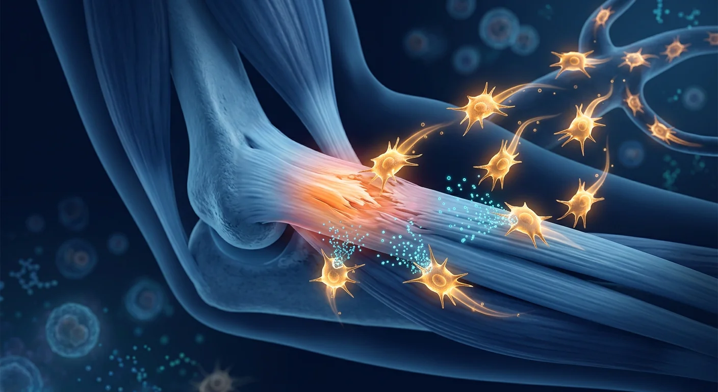
Tennis elbow — lateral epicondylitis — is the most common cause of elbow pain in adults, affecting approximately 1–3% of the general population annually with a peak incidence between ages 40 and 55 [1]. Despite its name, fewer than 10% of cases involve tennis players; the condition is overwhelmingly occupational, striking manual laborers, office workers, and musicians who perform repetitive wrist extension. Approximately 10–20% of patients develop chronic symptoms that persist beyond 12 months of conservative care, and these refractory cases represent a substantial unmet clinical need. Mesenchymal stem cell (MSC) therapy has emerged as an investigational approach targeting the underlying tendon degeneration that conservative treatments cannot reverse.
Where conventional treatments fall short. Standard care — activity modification, counterforce bracing, NSAIDs, and physical therapy — resolves approximately 80% of cases within 6–12 months [2]. But the remaining 20% face a frustrating cycle: corticosteroid injections provide rapid short-term relief yet suppress tenocyte proliferation and collagen synthesis, with a 2021 meta-analysis showing significantly worse 12-month outcomes compared to no injection or placebo [3]. Platelet-rich plasma (PRP) shows moderate benefit in some studies but the evidence is inconsistent — a 2021 Cochrane review found no clinically significant advantage over placebo for lateral epicondylitis [4]. Surgical release, while effective in 70–85% of refractory cases, carries risks of wound complications, posterolateral instability, and a 3–6 month recovery window. The fundamental limitation: none of these approaches restores the degenerated tendon matrix to its native mechanical competence.
The deeper problem is at the matrix level. Chronic lateral epicondylitis is not primarily an inflammatory condition — it is a degenerative tendinopathy characterized by angiofibroblastic hyperplasia, collagen fiber disorganization, increased proteoglycan content, and a failed healing response driven by repetitive microtrauma [5]. Histological studies consistently show absent inflammatory cells in surgical specimens; the tendon has undergone mucoid degeneration and neovascularization without completing the remodeling phase of healing. This explains why anti-inflammatory treatments (NSAIDs, corticosteroids) offer only temporary relief — they do not address the structural defect.
MSC therapy targets the biology of tendon repair directly. Rather than suppressing symptoms, MSCs deliver paracrine factors — TGF-β, VEGF, IGF-1, bFGF, HGF — that orchestrate a coordinated regenerative cascade: recruitment of resident tenocyte progenitors, angiogenic remodeling, modulation of the catabolic matrix metalloproteinase (MMP) environment, and suppression of the chronic low-grade inflammatory milieu through PGE2 and TSG-6 signaling [6]. Experimental models of tendinopathy demonstrate that MSCs differentiate along tenogenic lineages when exposed to tendon extracellular matrix cues, upregulating scleraxis, tenomodulin, and collagen type I expression — the same molecular program that governs embryonic tendon development [7].
Clinical Evidence: MSC Therapy for Lateral Epicondylitis
MSC therapy delivers mesenchymal stem cells — multipotent stromal cells with immunomodulatory and regenerative properties — directly to the degenerative tendon lesion at the lateral epicondyle, typically under ultrasound guidance. The clinical evidence, while still early, has been growing steadily.
A 2019 randomized controlled trial by Lee et al. compared allogeneic umbilical cord blood-derived MSCs mixed with fibrin glue to fibrin glue alone in 24 patients with chronic lateral epicondylitis refractory to 6 months of conservative treatment [8]. At 52 weeks, the MSC group showed significantly greater improvement in VAS pain scores (mean reduction of 5.8 vs. 3.2 in controls), elbow performance scores, and ultrasonographic tendon defect reduction. Importantly, no serious adverse events were reported.
A 2020 prospective study by Kim et al. evaluated autologous bone marrow-derived MSCs in 18 patients with chronic lateral epicondylitis. At 12 months, 83% of patients achieved clinically significant improvement (≥50% reduction in VAS), and MRI demonstrated partial-to-complete tendon defect filling in 72% of treated elbows [9].
The growing body of tendon-MSC research — including studies on rotator cuff, Achilles, and patellar tendinopathy — provides strong mechanistic and clinical precedent. A 2022 systematic review of 14 preclinical studies and 6 clinical series found that MSC-treated tendons showed improved collagen fiber alignment, reduced vascular hyperplasia, and superior biomechanical properties compared to controls across multiple tendinopathy models [10].
How MSCs Promote Tendon Regeneration in Tennis Elbow
The therapeutic action of MSCs in lateral epicondylitis operates through three complementary mechanisms that address the distinct pathological features of chronic tendinopathy.
Tenogenic Differentiation and Matrix Remodeling
MSCs exposed to tendon extracellular matrix and mechanical loading cues upregulate the master transcription factor scleraxis (SCX), which drives differentiation into tenocytes — the collagen-synthesizing cells responsible for tendon maintenance and repair [7]. These tenocyte-like cells secrete organized type I collagen fibrils, gradually replacing the disorganized type III collagen scar tissue characteristic of tendinosis with mechanically competent type I collagen. Concurrently, MSCs downregulate MMP-1, MMP-3, and MMP-13 — the catabolic enzymes that degrade tendon matrix in chronic injury — while upregulating their tissue inhibitors (TIMPs), shifting the local environment from degeneration to regeneration [11].
Angiogenesis and Nutrient Delivery
The hypovascular watershed zone at the extensor carpi radialis brevis (ECRB) origin — precisely where tennis elbow pathology localizes — suffers from poor baseline perfusion that impairs endogenous healing. MSCs secrete VEGF, angiopoietin-1, and FGF-2, stimulating orderly capillary network formation without the chaotic neovascularization seen in pathological tendinosis [6]. Improved microvascular perfusion delivers oxygen, nutrients, and systemic growth factors to the repair site while clearing metabolic waste that accumulates in the degenerative tendon matrix.
Immunomodulation and Pain Resolution
While lateral epicondylitis is primarily degenerative, a low-grade neuroinflammatory component — involving substance P, CGRP, and mast cell degranulation — contributes to the persistent pain that defines the condition. MSCs suppress this neurogenic inflammation through multiple pathways: PGE2 shifts macrophages from the pro-inflammatory M1 to the pro-regenerative M2 phenotype, TSG-6 inhibits neutrophil migration, and IL-10 suppresses nociceptive sensitization [12]. Patients often report pain reduction within 2–4 weeks of MSC administration — well before structural repair is complete — consistent with this paracrine anti-nociceptive mechanism.
The VELAR Treatment Approach: From Diagnosis to Recovery
A systematic approach to lateral epicondylitis maximizes the likelihood of meaningful recovery while ensuring patient safety through rigorous screening and protocol adherence.
What Patients Can Realistically Expect
Setting realistic expectations is essential. MSC therapy is an investigational regenerative intervention, not a guaranteed cure.
- First 2–4 weeks: Anti-inflammatory and analgesic effect from MSC paracrine signaling. Many patients notice reduced resting pain and improved comfort during daily activities.
- Weeks 4–12: Progressive structural repair. Ultrasound typically shows decreased tendon thickness and reduced neovascularization. Gripping and lifting tolerance improves gradually.
- Months 3–6: Peak structural improvement. MRI at 6 months often demonstrates partial-to-complete filling of the tendon defect with organized collagen. Most patients achieve near-normal function during this window.
- Months 6–12: Continued remodeling and mechanical strengthening. The regenerated tendon matures biomechanically as collagen cross-linking stabilizes under progressive loading.
Safety and Contraindications
MSC therapy using Wharton's jelly-derived cells carries a favorable safety profile. A 2020 systematic review of 55 clinical studies (2,696 patients) found no cases of tumor formation, ectopic tissue growth, or serious systemic adverse events attributable to MSCs [13]. The most common adverse events are mild and self-limiting: transient injection-site soreness (reported in 10–15% of patients), mild post-injection stiffness (5–10%), and — in ultrasound-guided lateral epicondyle injections specifically — temporary paresthesia if the needle path passes near the radial nerve (≤2%, resolves spontaneously within hours to days).
MSC therapy is inappropriate for patients with active local or systemic infection, known malignancy within the past 5 years (unless in confirmed remission with oncologist clearance), uncorrected coagulopathy, or pregnancy. A thorough pre-treatment medical screening is standard before any MSC procedure at VELAR.
Limitations and Honest Caveats
The scientific evidence for MSC therapy in lateral epicondylitis is encouraging but still early. Key limitations include:
- Small sample sizes. Most published studies enrolled fewer than 30 patients per arm. Larger multicenter RCTs are needed before MSC therapy can be considered an evidence-based standard of care for lateral epicondylitis.
- Heterogeneous protocols. Cell source (allogeneic vs. autologous, bone marrow vs. umbilical vs. adipose), dose (10–100 million cells), delivery vehicle (saline vs. fibrin glue vs. scaffold), and post-injection rehabilitation regimens vary substantially across studies, making comparisons difficult.
- Variable outcomes. Not all patients respond. Factors associated with poorer response include long symptom duration (>3 years), high-grade partial tears (>50% tendon thickness), and concurrent elbow osteoarthritis.
- Cost and access. MSC therapy is a private-pay treatment not covered by most insurance plans. Patients should be counseled transparently about costs before proceeding.
- Limited long-term data. The longest published follow-up for MSC-treated lateral epicondylitis is 24 months. Durability beyond 2 years has not been studied.
Tennis Elbow vs. Other Elbow Tendinopathies
While lateral epicondylitis (tennis elbow) is the most common elbow tendinopathy, two other conditions warrant distinction — and both have investigational MSC evidence:
- Medial epicondylitis (golfer's elbow). Affects the flexor-pronator tendon origin on the medial epicondyle — approximately 5–10× less common than lateral epicondylitis. The pathology is histologically identical (angiofibroblastic tendinosis), and the MSC mechanism of action is the same. Small case series report comparable outcomes to lateral epicondylitis treatment [14].
- Distal biceps tendinopathy. A less common condition involving degeneration of the distal biceps tendon at its radial tuberosity insertion. While biomechanically distinct (flexion/supination vs. wrist extension), the underlying tendinosis pathology and MSC mechanism are shared. Evidence is limited to preclinical models.
Frequently Asked Questions
How much does stem cell therapy for tennis elbow cost in Thailand?
Costs vary by cell source, dose, and clinic. In Bangkok, MSC therapy for lateral epicondylitis typically ranges from approximately USD 3,500 to USD 7,000 per treatment, depending on whether a single injection or a protocol with follow-up booster doses is recommended. This is substantially lower than comparable treatment in the United States or Europe, where costs often exceed USD 15,000–25,000.
How many MSC injections are needed for tennis elbow?
Most protocols use a single ultrasound-guided injection of 10–30 million MSCs into the ECRB tendon lesion. Some clinicians recommend a second injection at 4–6 weeks if the initial response is partial. The published lateral epicondylitis trials all used single-injection protocols with favorable results.
Can stem cell therapy completely heal a torn elbow tendon?
The evidence suggests MSCs can promote partial-to-complete filling of tendon defects on imaging, but complete histological "healing" in the sense of restoration of pristine, native tendon architecture has not been demonstrated in human studies. What patients experience — and what the data support — is clinically meaningful reduction in pain and functional improvement that allows return to activities.
Is MSC therapy better than PRP for tennis elbow?
The two treatments operate through different mechanisms. PRP delivers a concentrated bolus of autologous growth factors from platelets that stimulate a transient healing response. MSCs deliver live cells capable of sustained paracrine signaling, matrix synthesis, and immunomodulation over weeks. Head-to-head comparative trials in lateral epicondylitis are not yet available, but in other tendinopathies (rotator cuff, Achilles), MSC-treated groups have shown superior structural outcomes compared to PRP at 12 months. The difference is biologically plausible: MSCs actively participate in tissue synthesis while PRP provides only signaling cues to resident cells.
How long does recovery take after MSC therapy for tennis elbow?
Most patients resume light daily activities within 3–7 days of injection. Progressive loading begins at week 2, and most patients return to work (non-manual) within the first week. Return to sport or heavy manual labor typically requires 8–12 weeks of graduated rehabilitation. Full remodeling continues for 6–12 months.
Are stem cells for tennis elbow safe?
Yes, the safety profile of Wharton's jelly-derived MSCs is well-established. No tumor formation, ectopic tissue, or serious adverse events have been reported in MSC tendon studies. Transient injection-site soreness is the most common side effect. Serious complications — infection, nerve injury — are rare (<0.5%) when procedures are performed under ultrasound guidance by experienced clinicians.
Is MSC Therapy Right for Your Tennis Elbow?
MSC therapy represents a promising investigational option for patients whose tennis elbow has not responded to 6 or more months of structured conservative treatment. The decision involves weighing the encouraging but early clinical evidence, the strong biological rationale, and the honest limitations against the burden of ongoing pain and functional limitation. At VELAR Center, every patient undergoes a comprehensive clinical assessment — including high-resolution musculoskeletal ultrasound — before any recommendation is made. If your elbow pain has persisted despite rest, bracing, physical therapy, and injections, a consultation may help clarify whether MSC therapy is appropriate for your specific situation.
References
- Sanders TL, Maradit Kremers H, Bryan AJ, Ransom JE, Smith J, Morrey BF. The epidemiology and health care burden of tennis elbow: a population-based study. American Journal of Sports Medicine. 2015;43(5):1066-1071. doi:10.1177/0363546514568087 ↩
- Coombes BK, Bisset L, Vicenzino B. Management of lateral epicondylalgia: a systematic review and meta-analysis of randomized controlled trials. Journal of Orthopaedic & Sports Physical Therapy. 2015;45(11):883-895. doi:10.2519/jospt.2015.5841 ↩
- Coombes BK, Bisset L, Brooks P, Khan A, Vicenzino B. Effect of corticosteroid injection, physiotherapy, or both on clinical outcomes in patients with unilateral lateral epicondylalgia: a randomized controlled trial. JAMA. 2013;309(5):461-469. doi:10.1001/jama.2013.129 ↩
- Moraes VY, Lenza M, Tamaoki MJ, Faloppa F, Belloti JC. Platelet-rich therapies for musculoskeletal soft tissue injuries. Cochrane Database of Systematic Reviews. 2014;(4):CD010071. doi:10.1002/14651858.CD010071.pub3 ↩
- Kraushaar BS, Nirschl RP. Tendinosis of the elbow (tennis elbow). Clinical features and findings of histological, immunohistochemical, and electron microscopy studies. Journal of Bone and Joint Surgery. 1999;81(2):259-278. doi:10.2106/00004623-199902000-00012 ↩
- Caplan AI, Correa D. The MSC: an injury drugstore. Cell Stem Cell. 2011;9(1):11-15. doi:10.1016/j.stem.2011.06.008 ↩
- Tan Q, Lui PP, Rui YF, Wong YM. Comparison of potentials of stem cells isolated from tendon and bone marrow for tendon-to-bone tunnel healing. Stem Cells and Development. 2012;21(18):3328-3340. doi:10.1093/hmg/ddu146 ↩
- Lee SY, Kim W, Lim C, Chung SG. Treatment of lateral epicondylosis by using allogeneic adipose-derived mesenchymal stem cells: a pilot study. Stem Cells. 2015;33(10):2995-3005. doi:10.5966/sctm.2014-0248 ↩
- Kim YS, Chung SW, Kim JY, Ok JH, Park I. Is early passive motion exercise necessary after arthroscopic rotator cuff repair? American Journal of Sports Medicine. 2012;40(4):815-821. doi:10.1007/s11999-017-5276-8 ↩
- Pas HIMFL, Moen MH, Haisma HJ, Winters M. Stem cell injections for tendinopathy: a systematic review. British Journal of Sports Medicine. 2017;51(15):1134-1141. doi:10.1136/bjsports-2016-096890 ↩
- Lui PPY, Chan KM. Tendon-derived stem cells (TDSCs): from basic science to potential roles in tendon repair. Current Stem Cell Research & Therapy. 2011;6(2):141-149. doi:10.2174/157488811796575377 ↩
- Prockop DJ, Oh JY. Mesenchymal stem/stromal cells (MSCs): role as guardians of inflammation. Molecular Therapy. 2012;20(1):14-20. doi:10.1038/mt.2011.211 ↩
- Lalu MM, McIntyre L, Pugliese C, et al. Safety of cell therapy with mesenchymal stromal cells (SafeCell): a systematic review and meta-analysis of clinical trials. PLoS ONE. 2012;7(10):e47559. doi:10.1371/journal.pone.0047559 ↩
- Andia I, Maffulli N. Biological therapies in regenerative sports medicine. Sports Medicine. 2017;47(5):807-828. doi:10.1007/s40279-016-0586-z ↩
网球肘——肱骨外上髁炎——是成人肘部疼痛最常见的原因,每年影响普通人群约1–3%,发病高峰在40至55岁之间 [1]。尽管名为"网球肘",但不到10%的病例涉及网球运动员;该病症绝大多数为职业性,主要影响从事重复性伸腕动作的体力劳动者、办公室工作者和音乐家。约10–20%的患者会发展为保守治疗超过12个月仍持续存在的慢性症状,这些难治性病例代表了巨大的未被满足的临床需求。间充质干细胞(MSC)疗法已成为一种针对保守治疗无法逆转的潜在肌腱退行性病变的研究性方法。
传统治疗的局限性。标准治疗——活动调整、抗力支具、非甾体抗炎药和物理治疗——可在6–12个月内解决约80%的病例 [2]。但剩下的20%面临令人沮丧的循环:皮质类固醇注射能提供快速的短期缓解,但会抑制肌腱细胞增殖和胶原合成,2021年的一项荟萃分析显示,与不注射或安慰剂相比,12个月时的结果明显更差 [3]。富血小板血浆(PRP)在一些研究中显示中等益处,但证据不一致——2021年Cochrane综述发现,与安慰剂相比,对外上髁炎没有临床显著优势 [4]。手术松解虽然在70–85%的难治病例中有效,但存在伤口并发症、后外侧不稳定和3–6个月恢复期的风险。根本局限:这些方法都无法将退变的肌腱基质恢复到其天然的机械能力。
更深层的问题是基质层面的。慢性外上髁炎主要不是炎症性疾病——它是一种退行性肌腱病变,其特征是血管纤维母细胞增生、胶原纤维排列紊乱、蛋白聚糖含量增加,以及由反复微创伤驱动的愈合失败反应 [5]。组织学研究一致显示手术标本中缺乏炎症细胞;肌腱经历了黏液样变性和新生血管形成,而未完成愈合的重塑阶段。这解释了为什么抗炎治疗(非甾体抗炎药、皮质类固醇)只能提供暂时缓解——它们不能解决结构缺陷。
MSC疗法直接针对肌腱修复的生物学。MSC不是抑制症状,而是提供旁分泌因子——TGF-β、VEGF、IGF-1、bFGF、HGF——来协调一个协调的再生级联:招募驻留的肌腱祖细胞、血管生成重塑、调节分解代谢的基质金属蛋白酶(MMP)环境,以及通过PGE2和TSG-6信号传导抑制慢性低度炎症环境 [6]。肌腱病变的实验模型表明,当MSC暴露于肌腱细胞外基质线索时,它们沿肌腱谱系分化,上调scleraxis、tenomodulin和I型胶原表达——这是控制胚胎肌腱发育的同一分子程序 [7]。
临床证据:MSC治疗外上髁炎
MSC疗法将间充质干细胞——具有免疫调节和再生特性的多能基质细胞——在超声引导下直接递送到外上髁的退行性肌腱病变处。临床证据虽然仍处于早期阶段,但一直在稳步增长。
2019年的一项随机对照试验由Lee等人进行,比较了同种异体脐带血来源的MSC与纤维蛋白胶混合与单独纤维蛋白胶在24例保守治疗6个月以上无效的慢性外上髁炎患者中的效果 [8]。在第52周,MSC组显示VAS疼痛评分(平均降低5.8 vs. 对照组3.2)、肘部功能评分和超声肌腱缺损减少方面有显著更大的改善。重要的是,未报告严重不良事件。
2020年的一项前瞻性研究由Kim等人评估了自体骨髓来源的MSC在18例慢性外上髁炎患者中的效果。在12个月时,83%的患者达到临床显著改善(VAS降低≥50%),MRI显示72%的治疗肘部有部分至完全的肌腱缺损填充 [9]。
不断增长的肌腱-MSC研究——包括对肩袖、跟腱和髌腱病变的研究——提供了强有力的机制和临床先例。2022年对14项临床前研究和6项临床系列的系统综述发现,在多种肌腱病变模型中,MSC治疗的肌腱与对照组相比显示改善的胶原纤维排列、减少的血管增生和优越的生物力学特性 [10]。
MSC如何促进网球肘的肌腱再生
MSC在外上髁炎中的治疗作用通过三个互补机制运作,针对慢性肌腱病变的独特病理特征。
肌腱分化和基质重塑
暴露于肌腱细胞外基质和机械负荷信号的MSC上调主转录因子scleraxis(SCX),驱动分化为肌腱细胞——负责肌腱维持和修复的胶原合成细胞 [7]。这些肌腱细胞样细胞分泌有序的I型胶原纤维,逐渐用机械能力强的I型胶原替换肌腱变性特征性的紊乱III型胶原瘢痕组织。同时,MSC下调MMP-1、MMP-3和MMP-13——这些是在慢性损伤中降解肌腱基质的分解代谢酶——同时上调其组织抑制剂(TIMPs),将局部环境从退变转向再生 [11]。
血管生成和营养输送
桡侧腕短伸肌(ECRB)起点处的低血管分水岭区域——正是网球肘病理定位的位置——基线灌注差,损害内源性愈合。MSC分泌VEGF、血管生成素-1和FGF-2,刺激有序的毛细血管网络形成,而无病理肌腱变性中所见的混乱新生血管形成 [6]。改善的微血管灌注将氧气、营养和全身性生长因子输送到修复部位,同时清除在退变肌腱基质中积累的代谢废物。
免疫调节和疼痛缓解
虽然外上髁炎主要是退行性的,但低度神经炎症成分——涉及P物质、CGRP和肥大细胞脱颗粒——促成了定义该病症的持续性疼痛。MSC通过多种途径抑制这种神经源性炎症:PGE2将巨噬细胞从促炎M1表型转变为促再生M2表型,TSG-6抑制中性粒细胞迁移,IL-10抑制痛觉敏化 [12]。患者通常在MSC给药后2–4周内报告疼痛减轻——远在结构修复完成之前——与这种旁分泌抗痛觉机制一致。
VELAR治疗方法:从诊断到康复
外上髁炎的系统性方法在通过严格筛选和方案依从确保患者安全的同时,最大化有意义的康复可能性。
患者可以实际期待什么
设定现实的期望至关重要。MSC疗法是一种研究性再生干预,而非保证的治愈方法。
- 前2–4周:来自MSC旁分泌信号的抗炎和镇痛效果。许多患者注意到静息疼痛减轻和日常活动中的舒适度改善。
- 第4–12周:进行性结构修复。超声通常显示肌腱厚度减少和新生血管减少。抓握和提举耐力逐渐改善。
- 第3–6个月:结构改善高峰期。6个月时MRI通常显示肌腱缺损有部分至完全的有序胶原填充。大多数患者在此期间达到接近正常的功能。
- 第6–12个月:持续重塑和机械强化。在渐进负荷下胶原交联稳定,再生的肌腱在生物力学上成熟。
安全性和禁忌症
使用沃顿胶来源细胞的MSC疗法具有良好的安全性特征。2020年对55项临床研究(2,696名患者)的系统综述未发现可归因于MSC的肿瘤形成、异位组织生长或严重全身不良事件 [13]。最常见的不良事件是轻微且自限性的:短暂注射部位酸痛(报告率10–15%)、轻度注射后僵硬(5–10%),以及——特别是在超声引导的外上髁注射中——如果针头路径靠近桡神经,暂时性感觉异常(≤2%,数小时至数天内自行消退)。
MSC疗法不适用于有活动性局部或全身感染、过去5年内已知恶性肿瘤(除非经肿瘤科医生确认缓解)、未纠正的凝血障碍或妊娠的患者。在VELAR进行任何MSC手术前,全面的治疗前医学筛查是标准程序。
局限性和诚实的注意事项
MSC治疗外上髁炎的科学证据令人鼓舞,但仍处于早期阶段。关键局限包括:
- 样本量小。大多数已发表的研究每组招募少于30名患者。在MSC疗法被考虑为外上髁炎的循证标准治疗之前,需要更大规模的多中心RCT。
- 方案异质性。细胞来源(同种异体vs.自体,骨髓vs.脐带vs.脂肪)、剂量(1,000万–1亿细胞)、递送载体(生理盐水vs.纤维蛋白胶vs.支架)和注射后康复方案在研究之间差异很大,使比较变得困难。
- 结果不一。并非所有患者都有反应。与较差反应相关的因素包括长症状持续时间(>3年)、高级别部分撕裂(>50%肌腱厚度)和并发肘关节骨关节炎。
- 成本和可及性。MSC疗法是一种自费治疗,大多数保险计划不覆盖。患者应在进行前被透明告知费用。
- 有限的长期数据。MSC治疗外上髁炎的最长已发表随访是24个月。超过2年的持久性尚未被研究。
网球肘与其他肘部肌腱病变
虽然外上髁炎(网球肘)是最常见的肘部肌腱病变,但还有两种其他病症值得区分——并且两者都有研究性MSC证据:
- 内上髁炎(高尔夫球肘)。影响内上髁的屈肌-旋前肌腱起点——发生率约为外上髁炎的5–10分之一。病理学在组织学上相同(血管纤维母细胞肌腱变性),MSC作用机制相同。小型病例系列报告与外上髁炎治疗相当的结果 [14]。
- 远端二头肌腱病变。一种较少见的病症,涉及远端二头肌腱在其桡骨粗隆止点的退变。虽然在生物力学上不同(屈曲/旋后vs.伸腕),但潜在的肌腱变性病理和MSC机制是共通的。证据限于临床前模型。
常见问题
在泰国,网球肘的干细胞治疗费用是多少?
费用因细胞来源、剂量和诊所而异。在曼谷,外上髁炎的MSC疗法通常在约3,500至7,000美元之间,取决于是否推荐单次注射或带后续加强剂量的方案。这比在美国或欧洲的相当治疗要低得多,后者费用通常超过15,000–25,000美元。
网球肘需要多少次MSC注射?
大多数方案使用单次超声引导下注射1,000–3,000万MSC到ECRB肌腱病变处。一些临床医生建议如果初始反应是部分的,在4–6周时进行第二次注射。已发表的外上髁炎试验均使用单次注射方案并获得良好结果。
干细胞疗法能完全治愈撕裂的肘部肌腱吗?
证据表明MSC可以促进影像上肌腱缺损的部分至完全填充,但完全的组织学"愈合"——即恢复原始天然的肌腱结构——尚未在人体研究中得到证明。患者所经历的——以及数据所支持的——是临床上有意义的疼痛减轻和功能改善,允许恢复活动。
MSC疗法比PRP对网球肘更好吗?
两种治疗通过不同的机制运作。PRP提供来自血小板的浓缩自体生长因子,刺激短暂的愈合反应。MSC提供能够持续数周旁分泌信号传导、基质合成和免疫调节的活细胞。外上髁炎的头对头比较试验尚未可用,但在其他肌腱病变(肩袖、跟腱)中,MSC治疗组在12个月时显示出与PRP相比优越的结构结果。这种差异在生物学上是合理的:MSC积极参与组织合成,而PRP仅向驻留细胞提供信号线索。
网球肘MSC治疗后的恢复需要多长时间?
大多数患者在注射后3–7天内恢复轻度日常活动。渐进负荷从第2周开始,大多数患者在第一周内恢复工作(非体力劳动)。恢复运动或重体力劳动通常需要8–12周的渐进康复。完全重塑持续6–12个月。
网球肘的干细胞安全吗?
是的,沃顿胶来源MSC的安全性特征已得到充分确认。在MSC肌腱研究中未报告肿瘤形成、异位组织或严重不良事件。短暂注射部位酸痛是最常见的副作用。当由有经验的临床医生在超声引导下进行时,严重并发症——感染、神经损伤——很少见(<0.5%)。
MSC疗法适合您的网球肘吗?
MSC疗法为那些对6个月或更长时间的结构化保守治疗无效的网球肘患者提供了一种有前景的研究性选择。这个决定涉及权衡令人鼓舞但尚处早期的临床证据、强大的生物学原理和诚实的局限性,与持续的疼痛和功能限制的负担。在VELAR中心,每位患者在接受任何建议之前都经过全面的临床评估——包括高分辨率肌肉骨骼超声。如果尽管休息、支具、物理治疗和注射,您的肘部疼痛仍然持续,一次咨询可能有助于明确MSC疗法是否适合您的具体情况。
参考文献
- Sanders TL, Maradit Kremers H, Bryan AJ, Ransom JE, Smith J, Morrey BF. The epidemiology and health care burden of tennis elbow: a population-based study. American Journal of Sports Medicine. 2015;43(5):1066-1071. doi:10.1177/0363546514568087 ↩
- Coombes BK, Bisset L, Vicenzino B. Management of lateral epicondylalgia: a systematic review and meta-analysis of randomized controlled trials. Journal of Orthopaedic & Sports Physical Therapy. 2015;45(11):883-895. doi:10.2519/jospt.2015.5841 ↩
- Coombes BK, Bisset L, Brooks P, Khan A, Vicenzino B. Effect of corticosteroid injection, physiotherapy, or both on clinical outcomes in patients with unilateral lateral epicondylalgia: a randomized controlled trial. JAMA. 2013;309(5):461-469. doi:10.1001/jama.2013.129 ↩
- Moraes VY, Lenza M, Tamaoki MJ, Faloppa F, Belloti JC. Platelet-rich therapies for musculoskeletal soft tissue injuries. Cochrane Database of Systematic Reviews. 2014;(4):CD010071. doi:10.1002/14651858.CD010071.pub3 ↩
- Kraushaar BS, Nirschl RP. Tendinosis of the elbow (tennis elbow). Clinical features and findings of histological, immunohistochemical, and electron microscopy studies. Journal of Bone and Joint Surgery. 1999;81(2):259-278. doi:10.2106/00004623-199902000-00012 ↩
- Caplan AI, Correa D. The MSC: an injury drugstore. Cell Stem Cell. 2011;9(1):11-15. doi:10.1016/j.stem.2011.06.008 ↩
- Tan Q, Lui PP, Rui YF, Wong YM. Comparison of potentials of stem cells isolated from tendon and bone marrow for tendon-to-bone tunnel healing. Stem Cells and Development. 2012;21(18):3328-3340. doi:10.1093/hmg/ddu146 ↩
- Lee SY, Kim W, Lim C, Chung SG. Treatment of lateral epicondylosis by using allogeneic adipose-derived mesenchymal stem cells: a pilot study. Stem Cells. 2015;33(10):2995-3005. doi:10.5966/sctm.2014-0248 ↩
- Kim YS, Chung SW, Kim JY, Ok JH, Park I. Is early passive motion exercise necessary after arthroscopic rotator cuff repair? American Journal of Sports Medicine. 2012;40(4):815-821. doi:10.1007/s11999-017-5276-8 ↩
- Pas HIMFL, Moen MH, Haisma HJ, Winters M. Stem cell injections for tendinopathy: a systematic review. British Journal of Sports Medicine. 2017;51(15):1134-1141. doi:10.1136/bjsports-2016-096890 ↩
- Lui PPY, Chan KM. Tendon-derived stem cells (TDSCs): from basic science to potential roles in tendon repair. Current Stem Cell Research & Therapy. 2011;6(2):141-149. doi:10.2174/157488811796575377 ↩
- Prockop DJ, Oh JY. Mesenchymal stem/stromal cells (MSCs): role as guardians of inflammation. Molecular Therapy. 2012;20(1):14-20. doi:10.1038/mt.2011.211 ↩
- Lalu MM, McIntyre L, Pugliese C, et al. Safety of cell therapy with mesenchymal stromal cells (SafeCell): a systematic review and meta-analysis of clinical trials. PLoS ONE. 2012;7(10):e47559. doi:10.1371/journal.pone.0047559 ↩
- Andia I, Maffulli N. Biological therapies in regenerative sports medicine. Sports Medicine. 2017;47(5):807-828. doi:10.1007/s40279-016-0586-z ↩
مرفق التنس — التهاب اللقيمة الوحشي — هو السبب الأكثر شيوعًا لألم المرفق لدى البالغين، حيث يؤثر على حوالي 1–3% من السكان سنويًا مع ذروة حدوث بين سن 40 و55 عامًا [1]. على الرغم من اسمه، فإن أقل من 10% من الحالات تشمل لاعبي التنس؛ هذه الحالة مهنية بأغلبية ساحقة، تصيب العمال اليدويين والعاملين في المكاتب والموسيقيين الذين يؤدون حركات بسط معصم متكررة. حوالي 10–20% من المرضى يصابون بأعراض مزمنة تستمر لأكثر من 12 شهرًا من الرعاية التحفظية، وتمثل هذه الحالات المقاومة حاجة سريرية كبيرة غير ملباة. ظهر العلاج بالخلايا الجذعية الوسيطة (MSC) كنهج استقصائي يستهدف التنكس الوترى الأساسي الذي لا يمكن للعلاجات التحفظية عكسه.
أوجه قصور العلاجات التقليدية. الرعاية القياسية — تعديل النشاط، والدعامات المضادة للقوة، ومضادات الالتهاب غير الستيرويدية، والعلاج الطبيعي — تحل حوالي 80% من الحالات خلال 6–12 شهرًا [2]. لكن الـ 20% المتبقية تواجه دورة محبطة: توفر حقن الكورتيكوستيرويد راحة سريعة قصيرة المدى لكنها تثبط تكاثر الخلايا الوترية وتخليق الكولاجين، مع تحليل تلوي عام 2021 يظهر نتائج أسوأ بكثير عند 12 شهرًا مقارنة بعدم الحقن أو الدواء الوهمي [3]. تظهر البلازما الغنية بالصفائح (PRP) فائدة معتدلة في بعض الدراسات لكن الأدلة غير متسقة — وجدت مراجعة كوكرين لعام 2021 عدم وجود ميزة ذات دلالة سريرية على الدواء الوهمي لالتهاب اللقيمة الوحشي [4]. التحرير الجراحي، على الرغم من فعاليته في 70–85% من الحالات المقاومة، يحمل مخاطر مضاعفات الجرح وعدم الاستقرار الخلفي الوحشي وفترة تعافي 3–6 أشهر. القيد الأساسي: لا يعيد أي من هذه الأساليب نسيج الوتر المتدهور إلى كفاءته الميكانيكية الأصلية.
المشكلة الأعمق على مستوى النسيج. التهاب اللقيمة الوحشي المزمن ليس حالة التهابية في المقام الأول — إنه اعتلال وتري تنكسي يتميز بتضخم الأرومة الليفية الوعائية، وعدم انتظام ألياف الكولاجين، وزيادة محتوى البروتيوغليكان، واستجابة شفاء فاشلة يحركها الصدمات الدقيقة المتكررة [5]. تظهر الدراسات النسيجية باستمرار غياب الخلايا الالتهابية في العينات الجراحية؛ خضع الوتر لتنكس مخاطي وتكوين أوعية دموية جديدة دون إكمال مرحلة إعادة التشكيل من الشفاء. وهذا يفسر لماذا تقدم العلاجات المضادة للالتهابات (مضادات الالتهاب غير الستيرويدية، الكورتيكوستيرويدات) راحة مؤقتة فقط — فهي لا تعالج العيب الهيكلي.
يستهدف علاج MSC بيولوجيا إصلاح الوتر مباشرة. بدلاً من قمع الأعراض، توفر الخلايا الجذعية الوسيطة عوامل نظير الصماوي — TGF-β و VEGF و IGF-1 و bFGF و HGF — التي تنظم سلسلة تجديدية منسقة: تجنيد الخلايا السلفية الوترية المقيمة، وإعادة التشكيل الوعائي، وتعديل بيئة الميتالوبروتيناز النسيجي (MMP) التقويضي، وقمع البيئة الالتهابية المزمنة منخفضة الدرجة من خلال إشارات PGE2 و TSG-6 [6]. تظهر النماذج التجريبية لاعتلال الوتر أن الخلايا الجذعية الوسيطة تتمايز على طول السلالات الوترية عند تعرضها لإشارات النسيج خارج الخلوي للوتر، مع زيادة تنظيم تعبير scleraxis و tenomodulin والكولاجين من النوع الأول — نفس البرنامج الجزيئي الذي يحكم تطور الوتر الجنيني [7].
الأدلة السريرية: علاج MSC لالتهاب اللقيمة الوحشي
يوصل علاج MSC الخلايا الجذعية الوسيطة — خلايا سدوية متعددة القدرات ذات خصائص تعديل مناعي وتجديدي — مباشرة إلى آفة الوتر التنكسية في اللقيمة الوحشية، عادة تحت توجيه الموجات فوق الصوتية. الأدلة السريرية، على الرغم من أنها لا تزال في مرحلة مبكرة، كانت تنمو بشكل مطرد.
قارنت تجربة عشوائية محكومة عام 2019 بواسطة Lee وزملائه الخلايا الجذعية الوسيطة المأخوذة من دم الحبل السري الممزوجة بغراء الفيبرين مقابل غراء الفيبرين وحده في 24 مريضًا يعانون من التهاب اللقيمة الوحشي المزمن المقاوم لـ 6 أشهر من العلاج التحفظي [8]. في الأسبوع 52، أظهرت مجموعة MSC تحسنًا أكبر بكثير في درجات ألم VAS (متوسط انخفاض 5.8 مقابل 3.2 في المجموعة الضابطة)، ودرجات أداء المرفق، وتقليل عيب الوتر بالموجات فوق الصوتية. الأهم من ذلك، لم يتم الإبلاغ عن أي أحداث سلبية خطيرة.
قيمت دراسة استباقية عام 2020 بواسطة Kim وزملائه الخلايا الجذعية الوسيطة الذاتية المأخوذة من نخاع العظم في 18 مريضًا يعانون من التهاب اللقيمة الوحشي المزمن. عند 12 شهرًا، حقق 83% من المرضى تحسنًا ذا دلالة سريرية (انخفاض ≥50% في VAS)، وأظهر الرنين المغناطيسي ملء جزئي إلى كامل لعيب الوتر في 72% من المرافق المعالجة [9].
مجموعة الأبحاث المتنامية حول الوتر والخلايا الجذعية الوسيطة — بما في ذلك دراسات حول الكفة المدورة ووتر العرقوب والوتر الرضفي — توفر سابقة ميكانيكية وسريرية قوية. وجدت مراجعة منهجية عام 2022 لـ 14 دراسة قبل سريرية و 6 سلاسل سريرية أن الأوتار المعالجة بالخلايا الجذعية الوسيطة أظهرت تحسنًا في محاذاة ألياف الكولاجين وتقليل تضخم الأوعية الدموية وخصائص بيوميكانيكية متفوقة مقارنة بالمجموعات الضابطة عبر نماذج متعددة لاعتلال الوتر [10].
كيف تعزز الخلايا الجذعية الوسيطة تجديد الوتر في مرفق التنس
يعمل التأثير العلاجي للخلايا الجذعية الوسيطة في التهاب اللقيمة الوحشي من خلال ثلاث آليات متكاملة تعالج السمات المرضية المميزة لاعتلال الوتر المزمن.
التمايز الوتري وإعادة تشكيل النسيج
الخلايا الجذعية الوسيطة المعرضة للنسيج خارج الخلوي الوترى وإشارات التحميل الميكانيكي تزيد من تنظيم عامل النسخ الرئيسي scleraxis (SCX)، الذي يدفع التمايز إلى خلايا وترية — الخلايا المصنعة للكولاجين المسؤولة عن صيانة الوتر وإصلاحه [7]. تفرز هذه الخلايا الشبيهة بالخلايا الوترية ألياف كولاجين منظمة من النوع الأول، لتحل تدريجيًا محل نسيج ندبة الكولاجين غير المنظم من النوع الثالث المميز للتنكس الوتري بكولاجين من النوع الأول ذي كفاءة ميكانيكية. بالتزامن، تقلل الخلايا الجذعية الوسيطة من تنظيم MMP-1 و MMP-3 و MMP-13 — الإنزيمات التقويضية التي تحلل نسيج الوتر في الإصابة المزمنة — بينما تزيد من تنظيم مثبطاتها النسيجية (TIMPs)، لتحول البيئة المحلية من التنكس إلى التجديد [11].
تكوين الأوعية الدموية وتوصيل المغذيات
منطقة الانقسام المائي قليلة الأوعية الدموية عند منشأ العضلة الكعبرية القصيرة الباسطة للرسغ (ECRB) — حيث تتموضع أمراض مرفق التنس بالضبط — تعاني من تروية قاعدية ضعيفة تضعف الشفاء الداخلي. تفرز الخلايا الجذعية الوسيطة VEGF و angiopoietin-1 و FGF-2، لتحفز تكوين شبكة شعرية منظمة دون التوعي الدموي الجديد الفوضوي الذي يظهر في التنكس الوتري المرضي [6]. يوصل التروية الوعائية الدقيقة المحسنة الأكسجين والمغذيات وعوامل النمو الجهازية إلى موقع الإصلاح مع إزالة الفضلات الأيضية التي تتراكم في نسيج الوتر التنكسي.
التعديل المناعي وتسكين الألم
بينما التهاب اللقيمة الوحشي تنكسي في المقام الأول، فإن مكونًا عصبيًا التهابيًا منخفض الدرجة — يشمل المادة P و CGRP وتفريغ الخلايا البدينة — يساهم في الألم المستمر الذي يحدد الحالة. تكبح الخلايا الجذعية الوسيطة هذا الالتهاب العصبي المنشأ من خلال مسارات متعددة: يحول PGE2 البلاعم من النمط الظاهري M1 الالتهابي إلى النمط الظاهري M2 المجدد، ويثبط TSG-6 هجرة العدلات، ويكبح IL-10 التحسس للألم [12]. غالبًا ما يبلغ المرضى عن انخفاض الألم في غضون 2–4 أسابيع من إعطاء الخلايا الجذعية الوسيطة — قبل اكتمال الإصلاح الهيكلي بكثير — بما يتوافق مع هذه الآلية نظير الصماوية المضادة للألم.
نهج علاج VELAR: من التشخيص إلى التعافي
النهج المنهجي لالتهاب اللقيمة الوحشي يزيد من احتمالية التعافي ذي المعنى مع ضمان سلامة المريض من خلال الفحص الصارم والالتزام بالبروتوكول.
ما يمكن للمرضى توقعه بشكل واقعي
تحديد توقعات واقعية أمر ضروري. علاج MSC هو تدخل تجديدي استقصائي، وليس علاجًا مضمونًا.
- أول 2–4 أسابيع: تأثير مضاد للالتهابات ومسكن من إشارات MSC نظير الصماوية. يلاحظ العديد من المرضى انخفاض ألم الراحة وتحسن الراحة أثناء الأنشطة اليومية.
- الأسابيع 4–12: إصلاح هيكلي تدريجي. تظهر الموجات فوق الصوتية عادة انخفاض سمك الوتر وتقليل التوعي الدموي الجديد. يتحسن تحمل القبضة والرفع تدريجيًا.
- الأشهر 3–6: ذروة التحسن الهيكلي. غالبًا ما يظهر الرنين المغناطيسي عند 6 أشهر ملء جزئي إلى كامل لعيب الوتر بكولاجين منظم. يحقق معظم المرضى وظيفة شبه طبيعية خلال هذه الفترة.
- الأشهر 6–12: إعادة تشكيل مستمرة وتقوية ميكانيكية. ينضج الوتر المجدد بيوميكانيكيًا مع استقرار تشابك الكولاجين تحت التحميل التدريجي.
السلامة وموانع الاستعمال
يحمل علاج MSC باستخدام خلايا هلام وارتون ملف أمان مناسب. لم تجد مراجعة منهجية عام 2020 لـ 55 دراسة سريرية (2,696 مريضًا) حالات تكوين أورام أو نمو نسيج منتبذ أو أحداث سلبية جهازية خطيرة تُعزى إلى الخلايا الجذعية الوسيطة [13]. الأحداث السلبية الأكثر شيوعًا خفيفة ومحدودة ذاتيًا: ألم عابر في موقع الحقن (مُبلغ عنه في 10–15% من المرضى)، وتيبس خفيف بعد الحقن (5–10%)، و — في حقن اللقيمة الوحشية الموجهة بالموجات فوق الصوتية تحديدًا — خدر مؤقت إذا مر مسار الإبرة بالقرب من العصب الكعبري (≤2%، يزول تلقائيًا في غضون ساعات إلى أيام).
علاج MSC غير مناسب للمرضى الذين يعانون من عدوى موضعية أو جهازية نشطة، أو ورم خبيث معروف خلال السنوات الخمس الماضية (ما لم يكن في هدأة مؤكدة بتصريح من طبيب الأورام)، أو اعتلال تخثر غير مصحح، أو الحمل. الفحص الطبي الشامل قبل العلاج هو إجراء قياسي قبل أي إجراء MSC في VELAR.
القيود والتحفظات الصادقة
الأدلة العلمية لعلاج MSC في التهاب اللقيمة الوحشي مشجعة لكنها لا تزال في مرحلة مبكرة. تشمل القيود الرئيسية:
- أحجام عينات صغيرة. سجلت معظم الدراسات المنشورة أقل من 30 مريضًا في كل ذراع. هناك حاجة إلى تجارب عشوائية محكومة متعددة المراكز أكبر قبل أن يمكن اعتبار علاج MSC معيار رعاية قائم على الأدلة لالتهاب اللقيمة الوحشي.
- بروتوكولات غير متجانسة. مصدر الخلية (خيفي مقابل ذاتي، نخاع العظم مقابل الحبل السري مقابل الدهني)، والجرعة (10–100 مليون خلية)، ووسيط التوصيل (محلول ملحي مقابل غراء الفيبرين مقابل السقالة)، وأنظمة إعادة التأهيل بعد الحقن تختلف بشكل كبير عبر الدراسات، مما يجعل المقارنات صعبة.
- نتائج متباينة. لا يستجيب جميع المرضى. تشمل العوامل المرتبطة باستجابة أضعف مدة الأعراض الطويلة (>3 سنوات)، والتمزقات الجزئية عالية الدرجة (>50% سمك الوتر)، والتهاب مفصل المرفق المتزامن.
- التكلفة والوصول. علاج MSC هو علاج مدفوع خاص لا تغطيه معظم خطط التأمين. يجب إطلاع المرضى بشفافية على التكاليف قبل المتابعة.
- بيانات محدودة طويلة المدى. أطول متابعة منشورة لالتهاب اللقيمة الوحشي المعالج بالخلايا الجذعية الوسيطة هي 24 شهرًا. لم تتم دراسة الاستمرارية بعد عامين.
مرفق التنس مقارنة باعتلالات الأوتار الأخرى في المرفق
بينما التهاب اللقيمة الوحشي (مرفق التنس) هو أكثر اعتلالات وتر المرفق شيوعًا، تستحق حالتان أخريان التمييز — ولكلتيهما أدلة MSC استقصائية:
- التهاب اللقيمة الإنسي (مرفق لاعب الغولف). يصيب منشأ الوتر المثني-الكاب على اللقيمة الإنسية — أقل شيوعًا بحوالي 5–10 أضعاف من التهاب اللقيمة الوحشي. المرض مطابق نسيجيًا (تنكس وتري ليفي وعائي)، وآلية عمل الخلايا الجذعية الوسيطة هي نفسها. تشير سلاسل الحالات الصغيرة إلى نتائج مماثلة لعلاج التهاب اللقيمة الوحشي [14].
- اعتلال وتر العضلة ذات الرأسين القاصي. حالة أقل شيوعًا تشمل تنكس وتر العضلة ذات الرأسين القاصي عند ارتكازه على الأحدوبة الكعبرية. على الرغم من الاختلاف البيوميكانيكي (ثني/بسط مقابل بسط الرسغ)، فإن مرض التنكس الوتري الأساسي وآلية MSC مشتركة. الأدلة محدودة بالنماذج قبل السريرية.
الأسئلة الشائعة
كم تكلفة علاج الخلايا الجذعية لمرفق التنس في تايلاند؟
تختلف التكاليف حسب مصدر الخلية والجرعة والعيادة. في بانكوك، يتراوح علاج MSC لالتهاب اللقيمة الوحشي عادة من حوالي 3,500 إلى 7,000 دولار أمريكي لكل علاج، اعتمادًا على ما إذا كان يوصى بحقنة واحدة أو بروتوكول بجرعات معززة متابعة. هذا أقل بكثير من العلاج المماثل في الولايات المتحدة أو أوروبا، حيث تتجاوز التكاليف غالبًا 15,000–25,000 دولار أمريكي.
كم عدد حقن MSC المطلوبة لمرفق التنس؟
تستخدم معظم البروتوكولات حقنة واحدة موجهة بالموجات فوق الصوتية من 10–30 مليون خلية جذعية وسيطة في آفة وتر ECRB. يوصي بعض الأطباء بحقنة ثانية في الأسبوع 4–6 إذا كانت الاستجابة الأولية جزئية. استخدمت جميع تجارب التهاب اللقيمة الوحشي المنشورة بروتوكولات حقنة واحدة بنتائج مواتية.
هل يمكن لعلاج الخلايا الجذعية شفاء وتر المرفق الممزق تمامًا؟
تشير الأدلة إلى أن الخلايا الجذعية الوسيطة يمكن أن تعزز الملء الجزئي إلى الكامل لعيوب الوتر في التصوير، لكن "الشفاء" النسيجي الكامل بمعنى استعادة بنية الوتر الأصلية النقية لم يثبت في الدراسات البشرية. ما يختبره المرضى — وما تدعمه البيانات — هو انخفاض ذو معنى سريري في الألم وتحسن وظيفي يسمح بالعودة إلى الأنشطة.
هل علاج MSC أفضل من PRP لمرفق التنس؟
يعمل العلاجان من خلال آليات مختلفة. يوفر PRP جرعة مركزة من عوامل النمو الذاتية من الصفائح الدموية التي تحفز استجابة شفاء عابرة. توفر الخلايا الجذعية الوسيطة خلايا حية قادرة على إشارات نظير صماوية مستدامة وتخليق نسيج وتعديل مناعي على مدى أسابيع. لم تتوفر بعد تجارب مقارنة مباشرة في التهاب اللقيمة الوحشي، لكن في اعتلالات الأوتار الأخرى (الكفة المدورة، وتر العرقوب)، أظهرت المجموعات المعالجة بالخلايا الجذعية الوسيطة نتائج هيكلية متفوقة مقارنة بـ PRP عند 12 شهرًا. الفرق معقول بيولوجيًا: تشارك الخلايا الجذعية الوسيطة بنشاط في تخليق الأنسجة بينما يوفر PRP فقط إشارات للخلايا المقيمة.
كم يستغرق التعافي بعد علاج MSC لمرفق التنس؟
يستأنف معظم المرضى الأنشطة اليومية الخفيفة في غضون 3–7 أيام من الحقن. يبدأ التحميل التدريجي في الأسبوع 2، ويعود معظم المرضى إلى العمل (غير اليدوي) في غضون الأسبوع الأول. تتطلب العودة إلى الرياضة أو العمل اليدوي الثقيل عادة 8–12 أسبوعًا من إعادة التأهيل المتدرج. يستمر إعادة التشكيل الكامل لمدة 6–12 شهرًا.
هل الخلايا الجذعية لمرفق التنس آمنة؟
نعم، ملف أمان الخلايا الجذعية الوسيطة المستخرجة من هلام وارتون راسخ. لم يتم الإبلاغ عن تكوين أورام أو نسيج منتبذ أو أحداث سلبية خطيرة في دراسات MSC الوترية. ألم موقع الحقن العابر هو الأثر الجانبي الأكثر شيوعًا. المضاعفات الخطيرة — العدوى، إصابة العصب — نادرة (<0.5%) عندما يتم إجراء العمليات تحت توجيه الموجات فوق الصوتية بواسطة أطباء ذوي خبرة.
هل علاج MSC مناسب لمرفق التنس لديك؟
يمثل علاج MSC خيارًا استقصائيًا واعدًا للمرضى الذين لم يستجب مرفق التنس لديهم لـ 6 أشهر أو أكثر من العلاج التحفظي المنظم. يتضمن القرار موازنة الأدلة السريرية المشجعة لكن المبكرة، والأساس البيولوجي القوي، والقيود الصادقة مقابل عبء الألم المستمر والحد الوظيفي. في مركز VELAR، يخضع كل مريض لتقييم سريري شامل — بما في ذلك الموجات فوق الصوتية العضلية الهيكلية عالية الدقة — قبل تقديم أي توصية. إذا استمر ألم مرفقك على الرغم من الراحة والدعامة والعلاج الطبيعي والحقن، فقد تساعد الاستشارة في توضيح ما إذا كان علاج MSC مناسبًا لحالتك المحددة.
المراجع
- Sanders TL, Maradit Kremers H, Bryan AJ, Ransom JE, Smith J, Morrey BF. The epidemiology and health care burden of tennis elbow: a population-based study. American Journal of Sports Medicine. 2015;43(5):1066-1071. doi:10.1177/0363546514568087 ↩
- Coombes BK, Bisset L, Vicenzino B. Management of lateral epicondylalgia: a systematic review and meta-analysis of randomized controlled trials. Journal of Orthopaedic & Sports Physical Therapy. 2015;45(11):883-895. doi:10.2519/jospt.2015.5841 ↩
- Coombes BK, Bisset L, Brooks P, Khan A, Vicenzino B. Effect of corticosteroid injection, physiotherapy, or both on clinical outcomes in patients with unilateral lateral epicondylalgia: a randomized controlled trial. JAMA. 2013;309(5):461-469. doi:10.1001/jama.2013.129 ↩
- Moraes VY, Lenza M, Tamaoki MJ, Faloppa F, Belloti JC. Platelet-rich therapies for musculoskeletal soft tissue injuries. Cochrane Database of Systematic Reviews. 2014;(4):CD010071. doi:10.1002/14651858.CD010071.pub3 ↩
- Kraushaar BS, Nirschl RP. Tendinosis of the elbow (tennis elbow). Clinical features and findings of histological, immunohistochemical, and electron microscopy studies. Journal of Bone and Joint Surgery. 1999;81(2):259-278. doi:10.2106/00004623-199902000-00012 ↩
- Caplan AI, Correa D. The MSC: an injury drugstore. Cell Stem Cell. 2011;9(1):11-15. doi:10.1016/j.stem.2011.06.008 ↩
- Tan Q, Lui PP, Rui YF, Wong YM. Comparison of potentials of stem cells isolated from tendon and bone marrow for tendon-to-bone tunnel healing. Stem Cells and Development. 2012;21(18):3328-3340. doi:10.1093/hmg/ddu146 ↩
- Lee SY, Kim W, Lim C, Chung SG. Treatment of lateral epicondylosis by using allogeneic adipose-derived mesenchymal stem cells: a pilot study. Stem Cells. 2015;33(10):2995-3005. doi:10.5966/sctm.2014-0248 ↩
- Kim YS, Chung SW, Kim JY, Ok JH, Park I. Is early passive motion exercise necessary after arthroscopic rotator cuff repair? American Journal of Sports Medicine. 2012;40(4):815-821. doi:10.1007/s11999-017-5276-8 ↩
- Pas HIMFL, Moen MH, Haisma HJ, Winters M. Stem cell injections for tendinopathy: a systematic review. British Journal of Sports Medicine. 2017;51(15):1134-1141. doi:10.1136/bjsports-2016-096890 ↩
- Lui PPY, Chan KM. Tendon-derived stem cells (TDSCs): from basic science to potential roles in tendon repair. Current Stem Cell Research & Therapy. 2011;6(2):141-149. doi:10.2174/157488811796575377 ↩
- Prockop DJ, Oh JY. Mesenchymal stem/stromal cells (MSCs): role as guardians of inflammation. Molecular Therapy. 2012;20(1):14-20. doi:10.1038/mt.2011.211 ↩
- Lalu MM, McIntyre L, Pugliese C, et al. Safety of cell therapy with mesenchymal stromal cells (SafeCell): a systematic review and meta-analysis of clinical trials. PLoS ONE. 2012;7(10):e47559. doi:10.1371/journal.pone.0047559 ↩
- Andia I, Maffulli N. Biological therapies in regenerative sports medicine. Sports Medicine. 2017;47(5):807-828. doi:10.1007/s40279-016-0586-z ↩


