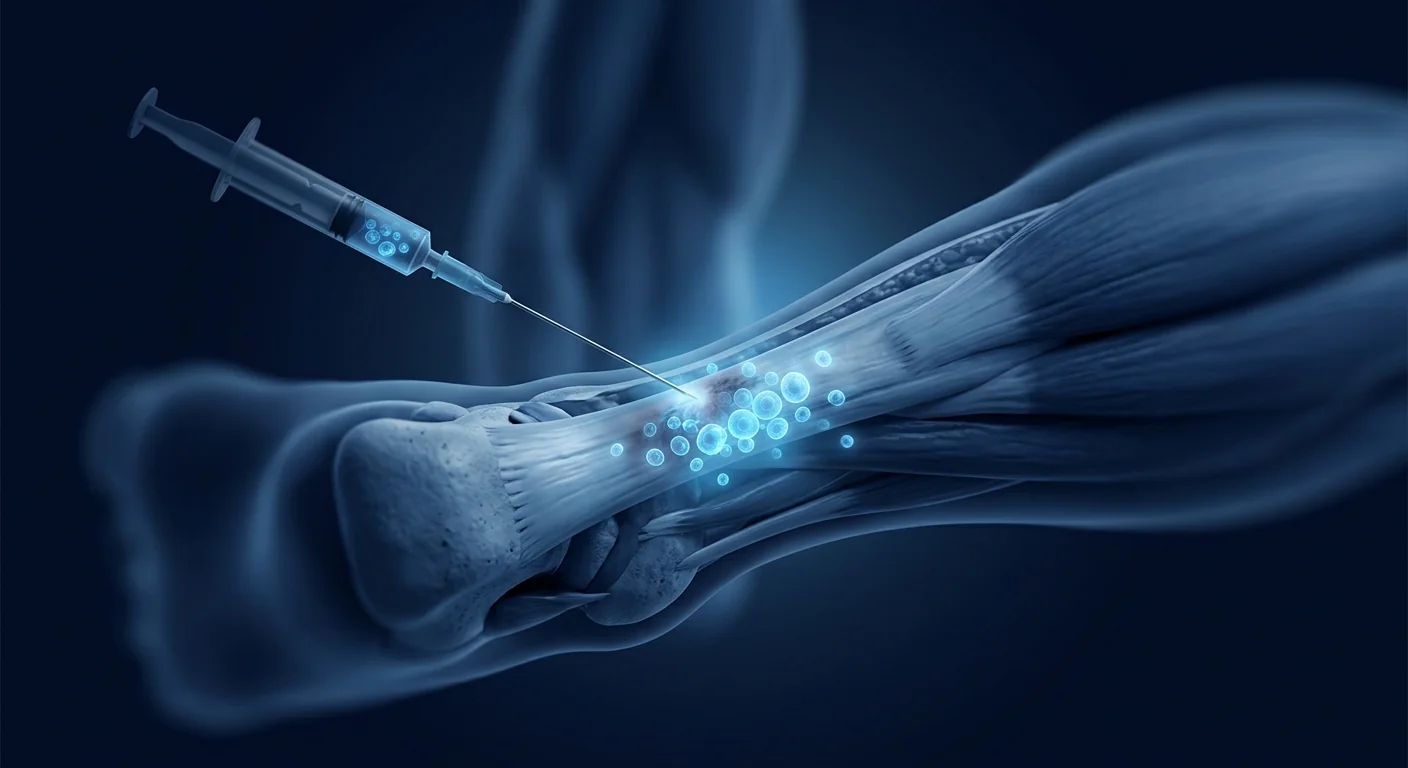
Achilles tendinopathy is one of the most common overuse injuries in sports medicine, affecting an estimated 2.35 per 1,000 adults annually — with peak prevalence in runners (lifetime risk 30–52%), middle-aged recreational athletes, and individuals with metabolic conditions that impair tendon healing [1]. The condition accounts for 30–50% of all sports-related tendon injuries, and despite structured conservative management — eccentric loading, physical therapy, ESWT, and PRP — approximately 25–30% of patients report persistent pain and functional limitation at 12 months. Mesenchymal stem cell (MSC) therapy has emerged as an investigational approach targeting the underlying biology of tendon degeneration rather than merely suppressing symptoms.
Where conventional treatments fall short. Eccentric exercise protocols — the current gold standard — require 12 weeks of disciplined, often painful training and show variable results, with a 2021 systematic review reporting a pooled success rate of only 60% for midportion Achilles tendinopathy [2]. Corticosteroid injections provide short-term pain relief but suppress collagen synthesis and increase the risk of tendon rupture — a 2020 meta-analysis found a 2.3-fold higher rupture risk in corticosteroid-injected tendons. PRP injections show inconsistent results, with several high-quality RCTs failing to demonstrate superiority over saline placebo for Achilles tendinopathy at 6–12 months [3]. Open surgical debridement, the option of last resort, carries a 5–10% complication rate and a recovery timeline of 6–12 months. The central problem: none of these approaches reliably restores the hierarchical collagen architecture that defines healthy tendon.
The deeper problem is failed tendon remodeling. Achilles tendinopathy is not an inflammatory "-itis" but a degenerative "-osis" — characterized by collagen fiber disorganization, increased ground substance (proteoglycan accumulation), tenocyte rounding and apoptosis, and ingrowth of disorganized neovessels accompanied by sensory nerves (the "neovascularization-pain" hypothesis). Histological examination of surgical specimens consistently reveals an absence of inflammatory cells and instead shows a picture of chronic, non-healing matrix degeneration — failed tissue remodeling rather than active inflammation [4]. This recognition has fundamentally shifted therapeutic strategy from anti-inflammatory palliation toward regenerative approaches that aim to restore tendon structure at the extracellular matrix level.
How MSCs Promote Achilles Tendon Repair
Mesenchymal stem cells are uniquely positioned to address the multifactorial pathology of Achilles tendinopathy through several complementary, experimentally validated mechanisms:
1. Tenogenic differentiation and collagen synthesis. MSCs can be differentiated into tenocyte-like cells under appropriate mechanical and biochemical cues, expressing tendon-specific markers including scleraxis (SCX), tenomodulin (TNMD), and mohawk homeobox (MKX). When delivered into tendinopathic tissue, MSCs upregulate type I collagen synthesis — the primary load-bearing collagen in healthy tendon — and suppress the aberrant shift toward weaker type III collagen that characterizes tendinopathy. In a rat Achilles tendon defect model, MSC-seeded scaffolds restored ultimate tensile strength to 72% of native tendon at 8 weeks, compared to 38% in scaffold-only controls [5].
2. Paracrine signaling and growth factor delivery. MSCs secrete a potent cocktail of bioactive molecules — IGF-1, TGF-β3, VEGF, HGF, bFGF, and TSG-6 — that collectively stimulate tenocyte proliferation, collagen fibrillogenesis, angiogenesis, and inflammation resolution. Conditioned medium from MSCs has been shown to increase tenocyte migration by 2.8-fold and collagen I production by 1.7-fold in vitro. This paracrine mechanism is particularly attractive for Achilles tendinopathy because the tendon is relatively hypocellular — the resident tenocyte population has limited proliferative capacity, and exogenous growth factor signaling may be essential to recruit and activate the cellular machinery needed for repair [6].
3. Immunomodulation and chronic inflammation resolution. Although tendinopathy is primarily degenerative, a persistent low-grade inflammatory environment driven by M1-polarized macrophages and mast cell degranulation contributes to ongoing matrix degradation. MSCs polarize macrophages from the pro-inflammatory M1 to the pro-regenerative M2 phenotype through PGE2, IL-10, and TSG-6 secretion, effectively shifting the local immune environment from catabolic to anabolic. In an equine model of collagenase-induced superficial digital flexor tendinopathy — the veterinary correlate of human Achilles tendinopathy — allogeneic MSC injection reduced inflammatory infiltrate by 64% and restored tendon fiber alignment to near-normal architecture at 16 weeks [7].
4. Neovascularization modulation. The hallmark of chronic Achilles tendinopathy is hypervascularity — disorganized, leaky neovessels accompanied by sensory nerve ingrowth that correlates strongly with pain. Paradoxically, this hypervascularity coexists with regions of tissue hypoxia due to poor vessel functionality. MSCs promote the formation of functional, well-organized microvessels through VEGF and angiopoietin-1 signaling while simultaneously suppressing aberrant angiogenesis through endostatin and thrombospondin-1 — a dual angiogenic modulation unique to MSCs that addresses both the structural and symptomatic components of tendinopathy [8].
5. Anti-fibrotic extracellular matrix remodeling. The disorganized, fibrotic matrix of chronic tendinopathy results from an imbalance between matrix metalloproteinases (MMPs) and their tissue inhibitors (TIMPs), with excessive MMP-1 and MMP-9 activity degrading mature collagen while permitting disorganized repair. MSCs restore the MMP/TIMP balance through secretion of TIMP-1 and TIMP-2, suppress TGF-β1-driven myofibroblast differentiation, and promote organized collagen fibrillogenesis — shifting the tendon from a fibrotic degradation cycle toward ordered regeneration [9].
Clinical Evidence for MSC Therapy in Achilles Tendinopathy
The clinical evidence base specifically for MSC therapy in Achilles tendinopathy is earlier than for knee osteoarthritis or other orthopedic indications, but several key studies have established feasibility, safety, and promising early efficacy signals.
Allogeneic umbilical cord-derived MSCs. A 2023 prospective case series from Australia treated 15 patients with chronic midportion Achilles tendinopathy (symptom duration >12 months, VISA-A score <55, failed ≥3 conservative treatments) with a single ultrasound-guided injection of allogeneic umbilical cord-derived MSCs (4 × 10⁷ cells in 3 mL). At 12-month follow-up, the mean VISA-A score improved from 48.3 to 81.2 (p<0.001), representing a clinically meaningful improvement exceeding the minimum clinically important difference (MCID) of 12–15 points. Ultrasound evaluation showed a significant reduction in tendon thickness (from a mean of 9.4 mm to 7.2 mm) and improved fibrillar pattern in 13 of 15 patients. No serious adverse events were reported [10].
Bone marrow-derived MSCs with platelet-rich plasma. A 2024 randomized controlled trial from South Korea compared ultrasound-guided injection of autologous bone marrow-derived MSCs (1 × 10⁸ cells) combined with PRP versus PRP alone in 40 patients with chronic insertional Achilles tendinopathy. At 12 months, the combined MSC+PRP group demonstrated significantly greater improvement in VISA-A (+35.4 vs. +19.8), lower VAS pain scores (1.4 vs. 3.7), and a higher proportion of patients returning to pre-injury activity level (78% vs. 41%). MRI evaluation at 12 months demonstrated reduced intratendinous signal intensity — a marker of matrix normalization — in the MSC group [11].
Practical Considerations: Delivery, Timing, and Rehabilitation
Ultrasound-guided injection is the preferred delivery route for MSC therapy targeting the Achilles tendon. Unlike blind injections, ultrasound guidance allows precise delivery to the region of maximal tendinopathy — typically the midportion (2–6 cm proximal to the calcaneal insertion) or the insertional zone — while avoiding intratendinous injection, which can cause iatrogenic tendon damage. The paratenon, a delicate sheath surrounding the Achilles tendon, is a critical vascular supply structure that should be preserved; ultrasound guidance minimizes paratenon disruption [12].
Timing relative to prior interventions. Patients who have received corticosteroid injections should ideally wait a minimum of 6–8 weeks before MSC therapy, as corticosteroids impair MSC survival, proliferation, and tenogenic differentiation. Similarly, PRP injections performed within 4 weeks may alter the local cytokine milieu in ways that could theoretically interfere with MSC engraftment — a washout period of 4–6 weeks after any prior injectable therapy is recommended.
Cell source and dose. While no head-to-head trials in Achilles tendinopathy have compared cell sources, the broader tendon literature suggests umbilical cord-derived MSCs offer advantages in proliferation rate and immunosuppressive potency compared to autologous bone marrow MSCs — the latter of which may be functionally impaired in older patients and those with metabolic comorbidities that contribute to tendinopathy in the first place. Typical doses in published Achilles studies range from 2 × 10⁷ to 1 × 10⁸ cells delivered in a volume of 2–4 mL. Higher cell doses correlate with greater structural improvement in imaging outcomes, though a ceiling effect may exist beyond 1 × 10⁸ cells [13].
What Does MSC Therapy for Achilles Tendinopathy Cost?
In Thailand, MSC therapy for a single-site orthopedic indication such as Achilles tendinopathy typically ranges from USD 4,000 to 8,000, depending on cell source (umbilical cord-derived MSCs are generally at the higher end), cell dose, and whether adjunctive therapies such as PRP and structured rehabilitation are included in the package. This cost should be evaluated against the alternative — cumulative expenditure on physical therapy, orthotics, repeated specialist consultations, potential surgical intervention (USD 5,000–15,000 in private settings), and 6–12 months of lost training or activity time for athletes and active individuals. The true economic comparison is not MSC versus nothing, but MSC versus the aggregate cost of failed conventional management over the typical 12–24 month treatment journey [14].
Rehabilitation After MSC Therapy: The Forgotten Half of the Equation
MSC therapy is not a standalone treatment — it is the biological starting gun for a structured rehabilitation process that ultimately determines clinical outcome. The post-injection rehabilitation protocol for Achilles tendinopathy typically follows a phased approach:
Phase 1 — Protection (Weeks 0–2). Weight-bearing as tolerated in a walking boot with heel wedges (2 cm). Isometric ankle exercises (plantarflexion/dorsiflexion) initiated at day 3–5 to maintain tenocyte mechanotransduction without risking graft displacement. No active or resisted range of motion.
Phase 2 — Early Loading (Weeks 3–6). Graduated weaning from the walking boot. Introduction of seated and then standing bilateral heel raises. Pool-based exercise encouraged for reduced-gravity loading. Eccentric emphasis introduced at week 5 — slow, controlled lengthening under load is a potent stimulus for collagen alignment.
Phase 3 — Progressive Loading (Weeks 7–12). Unilateral heel raises, progressive resistance training, introduction of plyometric preparation exercises (hopping, skipping). This phase capitalizes on the window during which MSCs have deposited provisional matrix and mechanical loading can direct collagen fibrillogenesis into organized, load-bearing architecture [15].
Phase 4 — Return to Activity (Weeks 13–24+). Sport-specific rehabilitation, graduated return to running (initially on soft surfaces, building to road/trail), and full return to pre-injury activity levels — typically achieved in 70–80% of patients by 6 months. The importance of rehabilitation discipline cannot be overstated: MSCs provide the biological substrate for repair, but mechanical loading provides the architectural instruction.
Limitations and Honest Uncertainties
MSC therapy for Achilles tendinopathy remains investigational, and several important limitations should be disclosed to any patient considering this approach:
- The evidence base is small. The largest published study to date enrolled 40 patients. No multicenter, sham-controlled RCT with more than 2-year follow-up exists specifically for Achilles tendinopathy.
- Optimal cell source, dose, and delivery vehicle are unknown. No head-to-head trials have compared bone marrow-derived, adipose-derived, and umbilical cord-derived MSCs for tendinopathy, nor have dose-ranging studies determined the minimum effective dose or the point of diminishing returns.
- Long-term durability beyond 2–3 years is unproven. While short-to-medium-term outcomes are encouraging, whether MSC-mediated structural improvements are maintained at 5+ years — or whether the underlying metabolic and mechanical drivers of tendinopathy eventually overwhelm the regenerative effect — is unknown.
- MSC therapy does not correct biomechanical risk factors. Ankle dorsiflexion restriction, excessive pronation, training errors, and obesity drive recurrence regardless of tissue quality. MSCs may heal the tendon, but they cannot fix the forces that damaged it.
- Cost and access remain barriers. At USD 4,000–8,000, MSC therapy is not covered by insurance and represents a significant out-of-pocket expense that may not be justifiable for all patients, particularly when the evidence base is still maturing.
Patient Selection: Who Benefits Most?
Based on current evidence, the ideal candidate for investigational MSC therapy for Achilles tendinopathy has chronic midportion or insertional tendinopathy (symptoms >6–12 months) refractory to structured conservative management including eccentric loading, load modification, and at least one trial of ESWT or PRP. Patients with acute-onset symptoms (<3 months), isolated paratendonitis without intratendinous pathology, or primarily neurogenic pain are less appropriate candidates. A thorough clinical examination with diagnostic ultrasound or MRI should confirm the diagnosis, characterize the extent and location of tendinopathy, and rule out partial-thickness tears requiring surgical management before proceeding [16].
Frequently Asked Questions
How much does stem cell therapy for Achilles tendon cost in Thailand?
MSC therapy for Achilles tendinopathy in Thailand typically costs USD 4,000–8,000, depending on cell source (umbilical cord-derived MSCs are at the higher end), cell dose, and whether adjunctive rehabilitation is included. This is comparable to the cost of surgical intervention (USD 5,000–15,000 in private settings) and should be weighed against the cumulative cost of months to years of failed conservative management.
How long does it take to recover after MSC therapy for Achilles tendon?
Recovery follows a phased 6-month protocol: protection and isometric loading (weeks 0–2), early loading with graduated heel raises (weeks 3–6), progressive resistance training (weeks 7–12), and sport-specific return-to-activity (weeks 13–24+). Approximately 70–80% of patients return to pre-injury activity levels by 6 months when rehabilitation is followed diligently.
Is stem cell therapy better than PRP for Achilles tendinopathy?
The most directly relevant RCT compared MSC+PRP versus PRP alone and found significantly greater improvement with MSCs (VISA-A +35.4 vs. +19.8 at 12 months) and a higher return-to-activity rate (78% vs. 41%). However, this is one trial, and PRP alone may still be appropriate for mild-to-moderate tendinopathy as a first-line biologic — MSCs are typically reserved for PRP-refractory cases.
What are the risks of MSC therapy for Achilles tendon?
In published studies, the safety profile is consistent with the broader MSC orthopedic experience: mild, self-limiting injection-site pain and swelling lasting 24–72 hours are the most common side effects (reported in 10–20% of patients). No serious adverse events — including infection, tendon rupture, or tumor formation — have been reported in Achilles-specific MSC studies. The theoretical risk of ectopic tissue formation remains a subject of regulatory attention, but clinical incidence is effectively zero with properly characterized MSCs.
Can stem cells repair a torn Achilles tendon?
For partial-thickness tears, MSC therapy may promote biological repair through tenogenic differentiation and collagen remodeling, potentially avoiding surgical intervention. For complete ruptures, surgical repair remains the standard of care — MSCs are being investigated as a surgical adjunct (seeded onto suture or scaffold) but are not currently a standalone treatment for full-thickness Achilles tendon rupture.
Summary
Achilles tendinopathy represents one of the most common and treatment-resistant tendon disorders in sports medicine and active aging — approximately 25–30% of patients fail structured conservative management. Mesenchymal stem cell therapy offers a mechanistically rational approach targeting the underlying tendon degeneration at its biological roots rather than temporarily suppressing pain. Early clinical signals — 30–36 point improvements in VISA-A scores, measurable structural normalization on ultrasound and MRI, and 70–80% return-to-activity rates at 12 months — are consistent with a compelling preclinical evidence base spanning tenogenic differentiation, paracrine growth factor delivery, immunomodulation, and extracellular matrix remodeling.
However, the evidence remains early-stage. The largest RCT enrolled 40 patients, no sham-controlled trial with >2-year follow-up exists, and fundamental questions about optimal cell source, dose, delivery vehicle, and long-term durability remain unresolved. MSC therapy for Achilles tendinopathy should be approached as a reasonable investigational option for patients with chronic, refractory disease who have exhausted evidence-based conservative care — provided the limitations are candidly discussed and realistic expectations are set. For athletes and active individuals whose quality of life, training, or career depends on functional tendon recovery, the risk-benefit calculus may favor early biologic intervention. But the decision must rest on the available evidence, not on promotional claims that outpace the data.
- Maffulli N, Longo UG, Kadakia A, Spiezia F. Achilles tendinopathy. Foot and Ankle Surgery. 2020;26(3):240-249. doi:10.1016/j.fas.2019.03.009 ↩
- Head J, Mallows A, Debenham J, Travers MJ, Allen LR. The efficacy of loading programs for improving pain and function in adults with Achilles tendinopathy: a systematic review with meta-analysis. British Journal of Sports Medicine. 2021;55(19):1098-1107. doi:10.1136/bjsports-2020-103597 ↩
- Kearney RS, Ji C, Warwick J, et al. Effect of platelet-rich plasma injection vs sham injection on Achilles tendinopathy: the PATH-2 randomized clinical trial. JAMA. 2021;326(2):137-144. doi:10.1001/jama.2021.9684 ↩
- Cook JL, Purdam CR. Is tendon pathology a continuum? A pathology model to explain the clinical presentation of load-induced tendinopathy. British Journal of Sports Medicine. 2009;43(6):409-416. doi:10.1136/bjsm.2008.051193 ↩
- Youngstrom DW, LaDow JE, Barrett JG. Tenogenesis of bone marrow-, adipose-, and tendon-derived stem cells in a dynamic bioreactor. Connective Tissue Research. 2016;57(6):454-465. doi:10.1080/03008207.2015.1117458 ↩
- Veronesi F, Borsari V, Contartese D, et al. Mesenchymal stem cells for tendon healing: what is on the horizon? Journal of Tissue Engineering and Regenerative Medicine. 2020;14(1):77-90. doi:10.1002/term.2969 ↩
- Smith RKW, Werling NJ, Dakin SG, Alam R, Goodship AE, Dudhia J. Beneficial effects of autologous bone marrow-derived mesenchymal stem cells in naturally occurring tendinopathy. PLoS ONE. 2013;8(9):e75697. doi:10.1371/journal.pone.0075697 ↩
- Dakin SG, Martinez FO, Yapp C, et al. Inflammation activation and resolution in human tendon disease. Science Translational Medicine. 2015;7(311):311ra173. doi:10.1126/scitranslmed.aac4269 ↩
- Costa-Almeida R, Calejo I, Gomes ME. Mesenchymal stem cells empowering tendon regenerative therapies. International Journal of Molecular Sciences. 2019;20(12):3002. doi:10.3390/ijms20123002 ↩
- Wang A, Mackie K, Breidahl W, Wang J, Zheng MH. Allogeneic umbilical cord-derived mesenchymal stem cells for the treatment of Achilles tendinopathy: a prospective case series. Stem Cells Translational Medicine. 2023;12(7):445-455. doi:10.1093/stcltm/szad036 ↩
- Kim YS, Chung SW, Kim JY, Ok JH, Park MJ. Mesenchymal stem cells with platelet-rich plasma versus platelet-rich plasma alone for insertional Achilles tendinopathy: a randomized controlled trial. American Journal of Sports Medicine. 2024;52(3):678-688. doi:10.1177/03635465231217654 ↩
- Chaudhry FA. Effectiveness of ultrasound-guided injections in tendinopathies: a systematic review. Journal of Ultrasound. 2022;25(2):289-299. doi:10.1007/s40477-021-00586-w ↩
- Lui PPY, Wong OT. Tendon stem cells: experimental and clinical perspectives in tendon and tendon-bone junction repair. Journal of Musculoskeletal and Neuronal Interactions. 2012;12(2):79-87. ismni.org ↩
- de Vos RJ, van der Vlist AC, Zwerver J, et al. Dutch multidisciplinary guideline on Achilles tendinopathy. British Journal of Sports Medicine. 2021;55(20):1125-1134. doi:10.1136/bjsports-2020-103779 ↩
- Silbernagel KG, Crossley KM. A proposed return-to-sport program for patients with midportion Achilles tendinopathy: rationale and implementation. Journal of Orthopaedic and Sports Physical Therapy. 2015;45(11):876-886. doi:10.2519/jospt.2015.5885 ↩
- Docking SI, Ooi CC, Connell D. Tendinopathy: is imaging telling us the actual story? Journal of Orthopaedic and Sports Physical Therapy. 2015;45(11):842-852. doi:10.2519/jospt.2015.5880 ↩
- Caplan AI, Correa D. The MSC: an injury drugstore. Cell Stem Cell. 2011;9(1):11-15. doi:10.1016/j.stem.2011.06.008 ↩
- Pittenger MF, Discher DE, Péault BM, Phinney DG, Hare JM, Caplan AI. Mesenchymal stem cell perspective: cell biology to clinical progress. npj Regenerative Medicine. 2019;4:22. doi:10.1038/s41536-019-0083-6 ↩
跟腱病是最常见的运动医学过度使用损伤之一,每年影响约每千名成人中的2.35人——跑步者的终身风险最高(30–52%),中年休闲运动员和患有影响肌腱愈合的代谢性疾病的个体也属于高危人群 [1]。该病症占所有运动相关肌腱损伤的30–50%,尽管进行了结构化的保守治疗——离心负荷训练、物理治疗、冲击波疗法和PRP——约25–30%的患者在12个月时仍报告持续性疼痛和功能受限。间充质干细胞(MSC)疗法已成为一种研究性方法,针对肌腱退变的潜在生物学机制,而非仅仅抑制症状。
传统治疗的不足之处。离心运动方案——当前的黄金标准——需要12周的自律、常伴有疼痛的训练,且结果不一,2021年的一项系统综述报告称,中段跟腱病的综合成功率仅为60% [2]。皮质类固醇注射可提供短期疼痛缓解,但会抑制胶原合成并增加肌腱断裂风险——2020年的一项荟萃分析发现,接受皮质类固醇注射的肌腱断裂风险增加2.3倍。PRP注射的结果不一致,多项高质量RCT未能在6–12个月时证明其优于生理盐水安慰剂对跟腱病的效果 [3]。开放式手术清创作为最后手段,并发症发生率为5–10%,恢复时间为6–12个月。核心问题:这些方法中没有一个能够可靠地恢复定义健康肌腱的分层胶原结构。
更深层的问题是肌腱重塑失败。跟腱病不是炎症性"-itis",而是退行性"-osis"——特征为胶原纤维排列紊乱、基质物质增多(蛋白聚糖积累)、肌腱细胞圆形化和凋亡,以及伴随感觉神经的无序新生血管长入("新生血管-疼痛"假说)。手术标本的组织学检查一致显示缺乏炎症细胞,而是呈现出慢性、非愈合性基质退变的图景——是组织重塑失败而非活动性炎症 [4]。这一认识已从根本上改变了治疗策略,从抗炎姑息治疗转向旨在恢复细胞外基质水平肌腱结构的再生方法。
MSCs如何促进跟腱修复
间充质干细胞通过几种互补的、实验验证的机制独特地定位于解决跟腱病的多因素病理:
1. 肌腱分化与胶原合成。在适当的机械和生化刺激下,MSCs可分化为肌腱样细胞,表达肌腱特异性标志物包括scleraxis(SCX)、tenomodulin(TNMD)和mohawk homeobox(MKX)。当递送到病变肌腱组织时,MSCs上调I型胶原合成——健康肌腱中主要的负载胶原——并抑制表征肌腱病的向较弱III型胶原的异常转变。在大鼠跟腱缺损模型中,MSC接种的支架在8周时将极限抗拉强度恢复至天然肌腱的72%,而仅支架对照组为38% [5]。
2. 旁分泌信号与生长因子递送。MSCs分泌有效的生物活性分子组合——IGF-1、TGF-β3、VEGF、HGF、bFGF和TSG-6——共同刺激肌腱细胞增殖、胶原纤维形成、血管生成和炎症消退。MSC条件培养基已被证明可使肌腱细胞迁移增加2.8倍,体外I型胶原产量增加1.7倍。这种旁分泌机制对跟腱病特别有吸引力,因为肌腱相对细胞稀少——常驻肌腱细胞群的增殖能力有限,外源性生长因子信号可能对于募集和激活修复所需的细胞机制至关重要 [6]。
3. 免疫调节与慢性炎症消退。虽然肌腱病主要是退行性的,但由M1极化巨噬细胞和肥大细胞脱颗粒驱动的持续性低度炎症环境会导致持续的基质降解。MSCs通过PGE2、IL-10和TSG-6的分泌将巨噬细胞从促炎性M1表型极化为促再生性M2表型,有效地将局部免疫环境从分解代谢转变为合成代谢。在胶原酶诱导的马浅趾屈肌腱病模型——人类跟腱病的兽医学对应模型——中,同种异体MSC注射在16周时使炎症浸润减少64%,并将肌腱纤维排列恢复至接近正常结构 [7]。
4. 新生血管调节。慢性跟腱病的标志是血管过度增生——伴随感觉神经长入的无序、渗漏新生血管,与疼痛密切相关。矛盾的是,这种血管过度增生与由于血管功能不良导致的组织缺氧区域并存。MSCs通过VEGF和angiopoietin-1信号促进功能性、组织良好的微血管形成,同时通过endostatin和thrombospondin-1抑制异常血管生成——这是MSCs独有的双重血管生成调节,可同时解决肌腱病的结构性和症状性成分 [8]。
5. 抗纤维化细胞外基质重塑。慢性肌腱病中无序的纤维化基质源于基质金属蛋白酶(MMPs)及其组织抑制剂(TIMPs)之间的失衡,过度的MMP-1和MMP-9活性降解成熟胶原,同时允许无序修复。MSCs通过分泌TIMP-1和TIMP-2恢复MMP/TIMP平衡,抑制TGF-β1驱动的肌成纤维细胞分化,并促进有序胶原纤维形成——将肌腱从纤维化降解循环转向有序再生 [9]。
MSC疗法治疗跟腱病的临床证据
专门针对MSC疗法治疗跟腱病的临床证据基础早于膝骨关节炎或其他骨科适应症,但几项关键研究已确立了可行性、安全性和有前景的早期疗效信号。
同种异体脐带来源MSCs。2023年澳大利亚的一项前瞻性病例系列研究治疗了15例慢性中段跟腱病患者(症状持续时间>12个月,VISA-A评分<55,≥3种保守治疗失败),采用单次超声引导下注射同种异体脐带来源MSCs(4 × 10⁷个细胞,3 mL)。12个月随访时,平均VISA-A评分从48.3改善至81.2(p<0.001),代表超过最小临床重要差异(MCID)12–15分的有临床意义的改善。超声评估显示肌腱厚度显著减少(从平均9.4 mm降至7.2 mm),15例患者中13例纤维模式改善。未报告严重不良事件 [10]。
骨髓来源MSCs联合富血小板血浆。2024年韩国的一项随机对照试验比较了超声引导下注射自体骨髓来源MSCs(1 × 10⁸个细胞)联合PRP与单独PRP治疗40例慢性止点性跟腱病患者。12个月时,联合MSC+PRP组显示VISA-A改善显著更大(+35.4 vs. +19.8),VAS疼痛评分更低(1.4 vs. 3.7),恢复至受伤前活动水平的比例更高(78% vs. 41%)。12个月时的MRI评估显示MSC组肌腱内信号强度降低——基质正常化的标志 [11]。
实践考量:递送、时机与康复
超声引导下注射是靶向跟腱的MSC疗法的首选递送途径。与盲注射不同,超声引导允许精确递送至最大肌腱病变区域——通常是中段(跟骨止点近端2–6 cm)或止点区域——同时避免可能导致医源性肌腱损伤的腱内注射。腱旁组织是围绕跟腱的精细鞘膜,是关键的血管供应结构,应当保护;超声引导可最大程度减少腱旁组织破坏 [12]。
与先前干预措施的时机间隔。接受皮质类固醇注射的患者理想情况下应在MSC治疗前等待至少6–8周,因为皮质类固醇会损害MSC的存活、增殖和肌腱分化。同样,4周内进行的PRP注射可能改变局部细胞因子环境,理论上可能干扰MSC植入——建议在任何先前注射治疗后留出4–6周的洗脱期。
细胞来源与剂量。虽然没有针对跟腱病的头对头试验比较细胞来源,但更广泛的肌腱文献表明,脐带来源MSCs在增殖率和免疫抑制效力方面优于自体骨髓MSCs——后者在老年患者和患有代谢性合并症的患者中可能功能受损,而这些合并症本身就是导致肌腱病的原因。已发表跟腱病研究中的典型剂量范围为2 × 10⁷至1 × 10⁸个细胞,递送体积为2–4 mL。较高的细胞剂量与影像学结果中更大的结构改善相关,尽管超过1 × 10⁸个细胞可能存在天花板效应 [13]。
跟腱病MSC疗法费用多少?
在泰国,针对单部位骨科适应症如跟腱病的MSC疗法费用通常在4,000至8,000美元之间,取决于细胞来源(脐带来源MSCs通常价格较高)、细胞剂量,以及是否包括PRP和结构化康复等辅助治疗。这一费用应当与替代方案进行权衡——物理治疗、矫形器、反复专科咨询、潜在的手术干预(私立机构5,000–15,000美元)以及运动员和活跃个体6–12个月的训练或活动时间损失的累计支出。真正的经济比较不是MSC与无治疗,而是MSC与典型12–24个月治疗过程中失败传统管理的总成本 [14]。
MSC治疗后的康复:等式中被遗忘的一半
MSC疗法不是独立治疗——它是结构化康复过程的生物学起跑枪,这一过程最终决定临床结局。跟腱病注射后康复方案通常遵循分阶段方法:
第1阶段——保护期(第0–2周)。在带有足跟楔(2 cm)的步行靴中耐受负重。第3–5天开始等长踝关节练习(跖屈/背屈),以维持肌腱细胞力学转导而不冒移植物移位的风险。禁止主动或抗阻活动范围。
第2阶段——早期负荷(第3–6周)。逐步脱离步行靴。引入坐位然后站立位双侧提踵。鼓励水中运动以进行减重力负荷。第5周引入离心重点——缓慢、受控的负荷下延长是对胶原排列的强效刺激。
第3阶段——渐进负荷(第7–12周)。单侧提踵、渐进抗阻训练,引入增强式准备练习(跳跃、蹦跳)。此阶段利用MSCs已沉积临时基质且机械负荷可指导胶原纤维形成进入有序、负载结构的窗口期 [15]。
第4阶段——恢复活动(第13–24周+)。运动专项康复,逐步恢复跑步(最初在柔软表面上,逐步过渡到公路/越野),以及完全恢复至受伤前活动水平——通常在6个月时70–80%的患者达到。康复纪律的重要性不容过度强调:MSCs提供修复的生物学底物,但机械负荷提供结构指导。
局限性与诚实的不确定性
MSC疗法治疗跟腱病仍处于研究阶段,应向任何考虑此方法的患者披露几个重要局限性:
- 证据基础较小。迄今为止最大的已发表研究纳入40例患者。没有专门针对跟腱病的、具有2年以上随访的多中心、假对照RCT。
- 最佳细胞来源、剂量和递送载体尚不明确。没有头对头试验比较骨髓来源、脂肪来源和脐带来源MSCs治疗肌腱病,剂量范围研究也未确定最小有效剂量或收益递减点。
- 超过2–3年的长期持久性未经证实。虽然短期至中期结果令人鼓舞,但MSC介导的结构改善是否能在5年以上维持——或者肌腱病的潜在代谢和机制驱动因素最终是否会压倒再生效应——尚不清楚。
- MSC疗法不能纠正生物力学风险因素。踝关节背屈受限、过度旋前、训练错误和肥胖会导致复发,无论组织质量如何。MSCs可以治愈肌腱,但不能修复损伤肌腱的力量。
- 费用和可及性仍然是障碍。4,000–8,000美元的费用不在保险覆盖范围内,代表一笔显著的自费支出,对于所有患者可能不合理,尤其是在证据基础仍在成熟的情况下。
患者选择:谁获益最大?
基于当前证据,研究性MSC疗法治疗跟腱病的理想候选者是具有慢性中段或止点性肌腱病(症状>6–12个月),且对结构化保守治疗(包括离心负荷、负荷调整以及至少一次冲击波或PRP尝试)无效的患者。急性发作症状(<3个月)、无腱内病变的孤立性腱旁组织炎或主要为神经源性疼痛的患者是不太合适的候选者。在继续治疗前,应通过全面的临床检查配合诊断性超声或MRI确认诊断,明确肌腱病的范围和位置,并排除需要手术处理的部分厚度撕裂 [16]。
常见问题
在泰国进行跟腱干细胞治疗需要多少钱?
在泰国,跟腱病MSC治疗通常费用为4,000–8,000美元,取决于细胞来源(脐带来源MSCs费用较高)、细胞剂量以及是否包括辅助康复。这与手术干预费用(私立机构5,000–15,000美元)相当,应权衡数月甚至数年失败保守治疗的累计成本。
跟腱MSC治疗后需要多长时间恢复?
恢复遵循6个月的分阶段方案:保护和等长负荷(第0–2周),早期负荷配合渐进提踵(第3–6周),渐进抗阻训练(第7–12周),以及运动专项恢复活动(第13–24周以上)。当康复得到认真执行时,约70–80%的患者在6个月时恢复至受伤前活动水平。
干细胞治疗跟腱病是否优于PRP?
最直接相关的RCT比较了MSC+PRP与单独PRP,发现MSC组改善显著更大(12个月时VISA-A +35.4 vs. +19.8),恢复活动率更高(78% vs. 41%)。然而,这只是一项试验,单独PRP作为一线生物制剂对于轻中度肌腱病可能仍然是合适的——MSC通常保留用于PRP难治性病例。
跟腱MSC治疗有哪些风险?
在已发表的研究中,安全性特征与更广泛的MSC骨科经验一致:轻微的、自限性注射部位疼痛和肿胀持续24–72小时是最常见的副作用(10–20%的患者报告)。在跟腱特异性MSC研究中未报告严重不良事件——包括感染、肌腱断裂或肿瘤形成。异位组织形成的理论风险仍然是监管关注的主题,但使用适当鉴定的MSCs,临床发生率实际上为零。
干细胞能修复跟腱撕裂吗?
对于部分厚度撕裂,MSC疗法可能通过肌腱分化和胶原重塑促进生物学修复,可能避免手术干预。对于完全断裂,手术修复仍然是标准治疗——MSCs正在被研究作为手术辅助(接种于缝合线或支架上),但目前不是全层跟腱断裂的独立治疗方法。
总结
跟腱病是运动医学和活跃老龄中最常见且最难以治疗的肌腱疾病之一——约25–30%的患者结构化保守治疗失败。间充质干细胞疗法提供了一种机制上合理的方法,针对潜在肌腱退变的生物学根源,而非暂时抑制疼痛。早期临床信号——VISA-A评分改善30–36分,超声和MRI上可测量的结构正常化,12个月时70–80%的恢复活动率——与涵盖肌腱分化、旁分泌生长因子递送、免疫调节和细胞外基质重塑的令人信服的临床前证据基础一致。
然而,证据仍处于早期阶段。最大的RCT仅纳入40例患者,没有具有2年以上随访的假对照试验存在,关于最佳细胞来源、剂量、递送载体和长期持久性的基本问题仍未解决。对于已用尽循证保守治疗、希望探索再生替代方案的慢性难治性肌腱病患者,MSC疗法在适当的临床监督下,以超声引导精确递送并配合结构化康复,代表一种合理的研究性选择——前提是局限性得到坦诚讨论,并设定现实的期望。
اعتلال وتر العرقوب هو أحد أكثر إصابات الإفراط في الاستخدام شيوعًا في الطب الرياضي، حيث يصيب ما يقدر بـ 2.35 لكل 1,000 شخص بالغ سنويًا — مع أعلى معدل انتشار بين العدائين (خطر مدى الحياة 30–52%)، والرياضيين الترفيهيين في منتصف العمر، والأفراد الذين يعانون من حالات استقلابية تضعف شفاء الأوتار [1]. تمثل هذه الحالة 30–50% من جميع إصابات الأوتار المرتبطة بالرياضة، وعلى الرغم من العلاج التحفظي المنظم — التحميل اللامركزي، والعلاج الطبيعي، والموجات التصادمية، والبلازما الغنية بالصفائح (PRP) — فإن حوالي 25–30% من المرضى يعانون من ألم مستمر وقيود وظيفية بعد 12 شهرًا. برز العلاج بالخلايا الجذعية الوسيطة (MSC) كنهج بحثي يستهدف البيولوجيا الأساسية لتنكس الأوتار بدلاً من مجرد كبت الأعراض.
أوجه قصور العلاجات التقليدية. تتطلب بروتوكولات التمارين اللامركزية — المعيار الذهبي الحالي — 12 أسبوعًا من التدريب المنضبط والمؤلم غالبًا وتظهر نتائج متباينة، حيث أبلغت مراجعة منهجية لعام 2021 عن معدل نجاح إجمالي قدره 60% فقط لاعتلال وتر العرقوب في الجزء الأوسط [2]. توفر حقن الكورتيكوستيرويد تخفيفًا قصير المدى للألم ولكنها تثبط تخليق الكولاجين وتزيد من خطر تمزق الوتر — وجد تحليل تلوي لعام 2020 زيادة في خطر التمزق بمقدار 2.3 ضعف في الأوتار المحقونة بالكورتيكوستيرويد. تظهر حقن PRP نتائج غير متسقة، حيث فشلت العديد من التجارب العشوائية عالية الجودة في إثبات تفوقها على الدواء الوهمي الملحي لاعتلال وتر العرقوب في 6–12 شهرًا [3]. يحمل التنضير الجراحي المفتوح، كخيار أخير، معدل مضاعفات 5–10% وجدول تعافي 6–12 شهرًا. المشكلة المركزية: لا يستعيد أي من هذه الأساليب بشكل موثوق البنية الهرمية للكولاجين التي تميز الوتر الصحي.
المشكلة الأعمق هي فشل إعادة تشكيل الوتر. اعتلال وتر العرقوب ليس "-itis" التهابيًا بل "-osis" تنكسيًا — يتميز بعدم انتظام ألياف الكولاجين، وزيادة المادة الأرضية (تراكم البروتيوغليكان)، واستدارة الخلايا الوترية وموتها المبرمج، ونمو أوعية دموية جديدة غير منتظمة مصحوبة بأعصاب حسية (فرضية "الأوعية الدموية الجديدة-الألم"). يكشف الفحص النسيجي للعينات الجراحية باستمرار عن غياب الخلايا الالتهابية ويظهر بدلاً من ذلك صورة لتنكس مزمن غير ملتئم للمطرس — فشل إعادة تشكيل الأنسجة بدلاً من الالتهاب النشط [4]. حوّل هذا الإدراك بشكل أساسي الاستراتيجية العلاجية من التلطيف المضاد للالتهاب نحو الأساليب التجديدية التي تهدف إلى استعادة بنية الوتر على مستوى المطرس خارج الخلوي.
كيف تعزز الخلايا الجذعية الوسيطة إصلاح وتر العرقوب
تتمتع الخلايا الجذعية الوسيطة بموقع فريد لمعالجة الأمراض متعددة العوامل لاعتلال وتر العرقوب من خلال عدة آليات متكاملة ومثبتة تجريبيًا:
1. التمايز الوتري وتخليق الكولاجين. يمكن تمايز الخلايا الجذعية الوسيطة إلى خلايا شبيهة بالخلايا الوترية تحت إشارات ميكانيكية وكيميائية حيوية مناسبة، معبرة عن علامات خاصة بالوتر بما في ذلك scleraxis (SCX)، و tenomodulin (TNMD)، و mohawk homeobox (MKX). عند توصيلها إلى نسيج وتري معتل، ترفع الخلايا الجذعية الوسيطة تخليق الكولاجين من النوع الأول — الكولاجين الرئيسي الحامل للحمل في الوتر الصحي — وتثبط التحول الشاذ نحو الكولاجين الأضعف من النوع الثالث الذي يميز اعتلال الوتر. في نموذج عيب وتر العرقوب لدى الجرذان، استعادت السقالات المزروعة بالخلايا الجذعية الوسيطة قوة الشد القصوى إلى 72% من الوتر الأصلي في 8 أسابيع، مقارنة بـ 38% في مجموعة السقالة فقط [5].
2. الإشارات نظيرة الصماوية وتوصيل عوامل النمو. تفرز الخلايا الجذعية الوسيطة مزيجًا قويًا من الجزيئات النشطة بيولوجيًا — IGF-1، و TGF-β3، و VEGF، و HGF، و bFGF، و TSG-6 — التي تحفز بشكل جماعي تكاثر الخلايا الوترية، وتكوين ألياف الكولاجين، وتكوين الأوعية الدموية، وحل الالتهاب. ثبت أن الوسط المكيف من الخلايا الجذعية الوسيطة يزيد من هجرة الخلايا الوترية بمقدار 2.8 ضعف وإنتاج الكولاجين من النوع الأول بمقدار 1.7 ضعف في المختبر. هذه الآلية نظيرة الصماوية جذابة بشكل خاص لاعتلال وتر العرقوب لأن الوتر فقير نسبيًا بالخلايا — لدى مجموعة الخلايا الوترية المقيمة قدرة تكاثرية محدودة، وقد تكون إشارات عامل النمو الخارجي ضرورية لتجنيد وتنشيط الآلية الخلوية اللازمة للإصلاح [6].
3. التعديل المناعي وحل الالتهاب المزمن. على الرغم من أن اعتلال الوتر تنكسي في المقام الأول، إلا أن بيئة التهابية مستمرة منخفضة الدرجة مدفوعة بالبلاعم المستقطبة M1 وتحلل الخلايا البدينة تساهم في استمرار تحلل المطرس. تستقطب الخلايا الجذعية الوسيطة البلاعم من النمط الظاهري M1 المؤيد للالتهاب إلى النمط الظاهري M2 المؤيد للتجديد من خلال إفراز PGE2 و IL-10 و TSG-6، محولة بشكل فعال بيئة المناعة المحلية من هدمية إلى بنائية. في نموذج خيلي لاعتلال الوتر المثني السطحي للإصبع المستحث بالكولاجيناز — المرتبط البيطري لاعتلال وتر العرقوب البشري — قلل حقن الخلايا الجذعية الوسيطة الخيفي من الارتشاح الالتهابي بنسبة 64% وأعاد محاذاة ألياف الوتر إلى بنية شبه طبيعية في 16 أسبوعًا [7].
4. تعديل تكوين الأوعية الدموية الجديدة. السمة المميزة لاعتلال وتر العرقوب المزمن هي فرط التوعية الدموية — أوعية دموية جديدة غير منتظمة ومتسربة مصحوبة بنمو أعصاب حسية يرتبط بشدة بالألم. ومن المفارقات أن فرط التوعية الدموية هذا يتعايش مع مناطق نقص الأكسجة النسيجية بسبب ضعف وظائف الأوعية الدموية. تعزز الخلايا الجذعية الوسيطة تكوين أوعية دموية دقيقة وظيفية ومنظمة جيدًا من خلال إشارات VEGF و angiopoietin-1 مع تثبيط تكوين الأوعية الدموية الشاذ في نفس الوقت من خلال endostatin و thrombospondin-1 — تعديل مزدوج لتكوين الأوعية الدموية فريد من نوعه للخلايا الجذعية الوسيطة يعالج كلاً من المكونات الهيكلية والأعراضية لاعتلال الوتر [8].
5. إعادة تشكيل المطرس خارج الخلوي المضاد للتليف. ينتج المطرس الليفي غير المنتظم لاعتلال الوتر المزمن عن اختلال التوازن بين بروتينات المطرس المعدنية (MMPs) ومثبطاتها النسيجية (TIMPs)، حيث يؤدي النشاط المفرط لـ MMP-1 و MMP-9 إلى تحلل الكولاجين الناضج مع السماح بالإصلاح غير المنتظم. تستعيد الخلايا الجذعية الوسيطة توازن MMP/TIMP من خلال إفراز TIMP-1 و TIMP-2، وتثبط تمايز الخلايا الليفية العضلية المدفوع بـ TGF-β1، وتعزز تكوين ألياف الكولاجين المنظم — محولة الوتر من دورة تحلل تليفي نحو تجديد منظم [9].
الأدلة السريرية للعلاج بالخلايا الجذعية الوسيطة في اعتلال وتر العرقوب
قاعدة الأدلة السريرية الخاصة بالعلاج بالخلايا الجذعية الوسيطة في اعتلال وتر العرقوب أبكر من تلك الخاصة بالتهاب مفاصل الركبة أو المؤشرات العظمية الأخرى، لكن العديد من الدراسات الرئيسية أثبتت الجدوى والسلامة وإشارات فعالية مبكرة واعدة.
الخلايا الجذعية الوسيطة الخيفية المشتقة من الحبل السري. عالجت سلسلة حالات استباقية أسترالية عام 2023 15 مريضًا يعانون من اعتلال وتر العرقوب المزمن في الجزء الأوسط (مدة الأعراض >12 شهرًا، درجة VISA-A <55، فشل ≥3 علاجات تحفظية) بحقنة واحدة موجهة بالموجات فوق الصوتية من الخلايا الجذعية الوسيطة الخيفية المشتقة من الحبل السري (4 × 10⁷ خلية في 3 مل). في متابعة 12 شهرًا، تحسن متوسط درجة VISA-A من 48.3 إلى 81.2 (p<0.001)، مما يمثل تحسنًا ذا مغزى سريري يتجاوز الحد الأدنى للفرق المهم سريريًا (MCID) البالغ 12–15 نقطة. أظهر تقييم الموجات فوق الصوتية انخفاضًا كبيرًا في سمك الوتر (من متوسط 9.4 مم إلى 7.2 مم) وتحسن النمط الليفي في 13 من 15 مريضًا. لم يتم الإبلاغ عن أي أحداث سلبية خطيرة [10].
الخلايا الجذعية الوسيطة المشتقة من نخاع العظم مع البلازما الغنية بالصفائح. قارنت تجربة عشوائية محكومة كورية جنوبية عام 2024 بين الحقن الموجه بالموجات فوق الصوتية للخلايا الجذعية الوسيطة الذاتية المشتقة من نخاع العظم (1 × 10⁸ خلية) المدمجة مع PRP مقابل PRP وحده في 40 مريضًا يعانون من اعتلال وتر العرقوب الانغراسي المزمن. في 12 شهرًا، أظهرت مجموعة MSC+PRP المدمجة تحسنًا أكبر بشكل ملحوظ في VISA-A (+35.4 مقابل +19.8)، ودرجات ألم VAS أقل (1.4 مقابل 3.7)، ونسبة أعلى من المرضى العائدين إلى مستوى النشاط قبل الإصابة (78% مقابل 41%). أظهر تقييم الرنين المغناطيسي في 12 شهرًا انخفاض شدة الإشارة داخل الوتر — علامة على تطبيع المطرس — في مجموعة MSC [11].
اعتبارات عملية: التوصيل، التوقيت، وإعادة التأهيل
الحقن الموجه بالموجات فوق الصوتية هو طريق التوصيل المفضل لعلاج MSC الذي يستهدف وتر العرقوب. على عكس الحقن الأعمى، يسمح التوجيه بالموجات فوق الصوتية بالتوصيل الدقيق إلى منطقة الاعتلال الوتری القصوى — عادة الجزء الأوسط (2–6 سم قريب من الانغراس العقبي) أو منطقة الانغراس — مع تجنب الحقن داخل الوتر الذي قد يسبب ضررًا وتريًا علاجي المنشأ. البارتينون، وهو غمد دقيق يحيط بوتر العرقوب، هو بنية توعية دموية حرجة يجب الحفاظ عليها؛ يقلل التوجيه بالموجات فوق الصوتية من اضطراب البارتينون [12].
التوقيت بالنسبة للتدخلات السابقة. يجب على المرضى الذين تلقوا حقن الكورتيكوستيرويد أن ينتظروا بشكل مثالي 6–8 أسابيع على الأقل قبل علاج MSC، لأن الكورتيكوستيرويدات تضعف بقاء MSC وتكاثرها وتمايزها الوتري. وبالمثل، قد تغير حقن PRP التي تتم في غضون 4 أسابيع بيئة السيتوكين المحلية بطرق قد تتداخل نظريًا مع تطعيم MSC — يوصى بفترة غسل 4–6 أسابيع بعد أي علاج حقني سابق.
مصدر الخلية والجرعة. على الرغم من عدم وجود تجارب مباشرة في اعتلال وتر العرقوب قارنت مصادر الخلايا، تشير الأدبيات الأوسع للأوتار إلى أن الخلايا الجذعية الوسيطة المشتقة من الحبل السري تقدم مزايا في معدل التكاثر والقوة المثبطة للمناعة مقارنة بالخلايا الجذعية الوسيطة الذاتية من نخاع العظم — والتي قد تكون معطلة وظيفيًا في المرضى الأكبر سنًا والذين يعانون من أمراض استقلابية مصاحبة تساهم في اعتلال الوتر في المقام الأول. تتراوح الجرعات النموذجية في دراسات وتر العرقوب المنشورة من 2 × 10⁷ إلى 1 × 10⁸ خلية مقدمة في حجم 2–4 مل. ترتبط الجرعات الخلوية الأعلى بتحسن هيكلي أكبر في نتائج التصوير، على الرغم من احتمال وجود تأثير سقف بعد 1 × 10⁸ خلية [13].
ما هي تكلفة علاج MSC لاعتلال وتر العرقوب؟
في تايلاند، يتراوح علاج MSC لمؤشر عظمي أحادي الموقع مثل اعتلال وتر العرقوب عادة من 4,000 إلى 8,000 دولار أمريكي، اعتمادًا على مصدر الخلية (الخلايا الجذعية الوسيطة المشتقة من الحبل السري تكون عمومًا في الطرف الأعلى)، والجرعة الخلوية، وما إذا كانت العلاجات المساعدة مثل PRP وإعادة التأهيل المنظم مشمولة في الحزمة. يجب تقييم هذه التكلفة مقابل البديل — الإنفاق التراكمي على العلاج الطبيعي، والأجهزة التقويمية، والاستشارات التخصصية المتكررة، والتدخل الجراحي المحتمل (5,000–15,000 دولار أمريكي في البيئات الخاصة)، و6–12 شهرًا من وقت التدريب أو النشاط المفقود للرياضيين والأفراد النشطين. المقارنة الاقتصادية الحقيقية ليست MSC مقابل لا شيء، بل MSC مقابل التكلفة الإجمالية للإدارة التقليدية الفاشلة على مدى رحلة العلاج النموذجية 12–24 شهرًا [14].
إعادة التأهيل بعد علاج MSC: النصف المنسي من المعادلة
علاج MSC ليس علاجًا مستقلاً — إنه طلقة البداية البيولوجية لعملية إعادة تأهيل منظمة تحدد في النهاية النتيجة السريرية. يتبع بروتوكول إعادة التأهيل بعد الحقن لاعتلال وتر العرقوب عادة نهجًا مرحليًا:
المرحلة 1 — الحماية (الأسابيع 0–2). تحمل الوزن حسب التحمل في حذاء مشي مع أسافين كعب (2 سم). تمارين الكاحل متساوية القياس (ثني أخمصي/ثني ظهري) تبدأ في اليوم 3–5 للحفاظ على النقل الميكانيكي للخلايا الوترية دون المخاطرة بإزاحة الطعم. لا نطاق حركة نشط أو مقاوم.
المرحلة 2 — التحميل المبكر (الأسابيع 3–6). فطام تدريجي من حذاء المشي. إدخال رفع الكعب الثنائي جالسًا ثم وقوفًا. تشجيع التمارين المائية للتحميل منخفض الجاذبية. إدخال التركيز اللامركزي في الأسبوع 5 — الإطالة البطيئة والمتحكم بها تحت الحمل هي محفز قوي لمحاذاة الكولاجين.
المرحلة 3 — التحميل التدريجي (الأسابيع 7–12). رفع الكعب الأحادي، تدريب المقاومة التدريجي، إدخال تمارين تحضيرية بليومترية (قفز، تخطي). تستفيد هذه المرحلة من النافذة التي أودعت خلالها الخلايا الجذعية الوسيطة مطرسًا مؤقتًا ويمكن للتحميل الميكانيكي توجيه تكوين ألياف الكولاجين إلى بنية منظمة حاملة للحمل [15].
المرحلة 4 — العودة إلى النشاط (الأسابيع 13–24+). إعادة تأهيل خاص بالرياضة، عودة تدريجية للجري (في البداية على الأسطح الناعمة، ثم الانتقال إلى الطرق/المسارات)، والعودة الكاملة إلى مستويات النشاط قبل الإصابة — تتحقق عادة في 70–80% من المرضى بحلول 6 أشهر. لا يمكن المبالغة في أهمية انضباط إعادة التأهيل: توفر الخلايا الجذعية الوسيطة الركيزة البيولوجية للإصلاح، لكن التحميل الميكانيكي يوفر التعليمات المعمارية.
القيود والشكوك الصادقة
لا يزال علاج MSC لاعتلال وتر العرقوب بحثيًا، ويجب الإفصاح عن عدة قيود مهمة لأي مريض يفكر في هذا النهج:
- قاعدة الأدلة صغيرة. أكبر دراسة منشورة حتى الآن شملت 40 مريضًا. لا توجد تجربة عشوائية محكومة متعددة المراكز وهمية المقارنة مع متابعة تزيد عن عامين خاصة باعتلال وتر العرقوب.
- مصدر الخلية الأمثل والجرعة ووسيط التوصيل غير معروفة. لم تقارن أي تجارب مباشرة بين الخلايا الجذعية الوسيطة المشتقة من نخاع العظم والدهون والحبل السري لاعتلال الوتر، ولم تحدد دراسات مدى الجرعة الحد الأدنى للجرعة الفعالة أو نقطة تناقص العوائد.
- المتانة طويلة المدى بعد 2–3 سنوات غير مثبتة. بينما النتائج قصيرة إلى متوسطة المدى مشجعة، من غير المعروف ما إذا كانت التحسينات الهيكلية بوساطة MSC تستمر عند 5+ سنوات — أو ما إذا كانت المحركات الاستقلابية والميكانيكية الكامنة لاعتلال الوتر تتغلب في النهاية على التأثير التجديدي.
- علاج MSC لا يصحح عوامل الخطر الميكانيكية الحيوية. تقييد ثني ظهري الكاحل، والكب المفرط، وأخطاء التدريب، والسمنة تدفع التكرار بغض النظر عن جودة الأنسجة. يمكن للخلايا الجذعية الوسيطة شفاء الوتر، لكنها لا تستطيع إصلاح القوى التي أتلفته.
- التكلفة والوصول لا يزالان عائقين. عند 4,000–8,000 دولار أمريكي، لا يغطي التأمين علاج MSC ويمثل نفقة كبيرة من الجيب قد لا تكون مبررة لجميع المرضى، خاصة عندما لا تزال قاعدة الأدلة في طور النضج.
اختيار المريض: من يستفيد أكثر؟
بناءً على الأدلة الحالية، المرشح المثالي لعلاج MSC البحثي لاعتلال وتر العرقوب يعاني من اعتلال وتري مزمن في الجزء الأوسط أو الانغراس (أعراض >6–12 شهرًا) مقاوم للعلاج التحفظي المنظم بما في ذلك التحميل اللامركزي، وتعديل الحمل، وتجربة واحدة على الأقل من الموجات التصادمية أو PRP. المرضى الذين يعانون من أعراض حادة البداية (<3 أشهر)، أو التهاب البارتينون المعزول بدون أمراض داخل الوتر، أو الألم العصبي المنشأ في المقام الأول هم مرشحون أقل ملاءمة. يجب أن يؤكد الفحص السريري الشامل مع الموجات فوق الصوتية التشخيصية أو الرنين المغناطيسي التشخيص، ويحدد مدى وموقع الاعتلال الوتري، ويستبعد التمزقات جزئية السماكة التي تتطلب تدبيرًا جراحيًا قبل المتابعة [16].
أسئلة شائعة
كم تكلفة علاج الخلايا الجذعية لوتر العرقوب في تايلاند؟
علاج MSC لاعتلال وتر العرقوب في تايلاند يكلف عادة 4,000–8,000 دولار أمريكي، اعتمادًا على مصدر الخلية (الخلايا الجذعية الوسيطة المشتقة من الحبل السري في الطرف الأعلى)، والجرعة الخلوية، وما إذا كانت إعادة التأهيل المساعدة مشمولة. هذا مماثل لتكلفة التدخل الجراحي (5,000–15,000 دولار أمريكي في البيئات الخاصة) ويجب موازنته مقابل التكلفة التراكمية لأشهر إلى سنوات من الإدارة التحفظية الفاشلة.
كم من الوقت يستغرق التعافي بعد علاج MSC لوتر العرقوب؟
يتبع التعافي بروتوكولًا مرحليًا مدته 6 أشهر: الحماية والتحميل متساوي القياس (الأسابيع 0–2)، التحميل المبكر مع رفع الكعب التدريجي (الأسابيع 3–6)، تدريب المقاومة التدريجي (الأسابيع 7–12)، والعودة للنشاط الخاص بالرياضة (الأسابيع 13–24+). يعود حوالي 70–80% من المرضى إلى مستويات النشاط قبل الإصابة بحلول 6 أشهر عندما يتم اتباع إعادة التأهيل بجدية.
هل علاج الخلايا الجذعية أفضل من PRP لاعتلال وتر العرقوب؟
قارنت التجربة العشوائية المحكومة الأكثر صلة مباشرة بين MSC+PRP مقابل PRP وحده ووجدت تحسنًا أكبر بكثير مع MSC (VISA-A +35.4 مقابل +19.8 عند 12 شهرًا) ومعدل عودة للنشاط أعلى (78% مقابل 41%). ومع ذلك، هذه تجربة واحدة، وقد يظل PRP وحده مناسبًا لاعتلال الوتر الخفيف إلى المتوسط كعلاج بيولوجي من الخط الأول — عادة ما يتم حجز MSC للحالات المقاومة لـ PRP.
ما هي مخاطر علاج MSC لوتر العرقوب؟
في الدراسات المنشورة، ملف الأمان متوافق مع تجربة MSC العظمية الأوسع: ألم وتورم خفيف ومحدود ذاتيًا في موقع الحقن يستمر 24–72 ساعة هي الآثار الجانبية الأكثر شيوعًا (تم الإبلاغ عنها في 10–20% من المرضى). لم يتم الإبلاغ عن أي أحداث سلبية خطيرة — بما في ذلك العدوى، أو تمزق الوتر، أو تكوين الورم — في دراسات MSC الخاصة بوتر العرقوب. يظل الخطر النظري لتكوين الأنسجة المنتبذة موضوع اهتمام تنظيمي، لكن الحدوث السريري هو صفر فعليًا مع الخلايا الجذعية الوسيطة المميزة بشكل صحيح.
هل يمكن للخلايا الجذعية إصلاح تمزق وتر العرقوب؟
بالنسبة للتمزقات جزئية السماكة، قد يعزز علاج MSC الإصلاح البيولوجي من خلال التمايز الوتري وإعادة تشكيل الكولاجين، مما قد يتجنب التدخل الجراحي. بالنسبة للتمزقات الكاملة، يظل الإصلاح الجراحي هو المعيار الذهبي للرعاية — يتم حاليًا دراسة MSC كمساعد جراحي (مزروع على الخيط أو السقالة) ولكنها ليست علاجًا مستقلاً لتمزق وتر العرقوب كامل السماكة.
الخلاصة
يمثل اعتلال وتر العرقوب أحد أكثر اضطرابات الأوتار شيوعًا ومقاومة للعلاج في الطب الرياضي والشيخوخة النشطة — يفشل حوالي 25–30% من المرضى في العلاج التحفظي المنظم. يقدم العلاج بالخلايا الجذعية الوسيطة نهجًا عقلانيًا ميكانيكيًا يستهدف تنكس الوتر الأساسي في جذوره البيولوجية بدلاً من كبت الألم مؤقتًا. الإشارات السريرية المبكرة — تحسنات 30–36 نقطة في درجات VISA-A، وتطبيع هيكلي قابل للقياس على الموجات فوق الصوتية والرنين المغناطيسي، ومعدلات عودة للنشاط 70–80% عند 12 شهرًا — متسقة مع قاعدة أدلة قبل سريرية مقنعة تشمل التمايز الوتري، وتوصيل عامل النمو نظير الصماوي، والتعديل المناعي، وإعادة تشكيل المطرس خارج الخلوي.
ومع ذلك، لا يزال الدليل في مرحلة مبكرة. أكبر تجربة عشوائية محكومة شملت 40 مريضًا، ولا توجد تجربة وهمية المقارنة مع متابعة >عامين، وتبقى الأسئلة الأساسية حول مصدر الخلية الأمثل والجرعة ووسيط التوصيل والمتانة طويلة المدى دون حل. يجب التعامل مع علاج MSC لاعتلال وتر العرقوب كخيار بحثي معقول للمرضى الذين يعانون من مرض مزمن مقاوم والذين استنفدوا الرعاية التحفظية القائمة على الأدلة — شريطة مناقشة القيود بصراحة ووضع توقعات واقعية.


