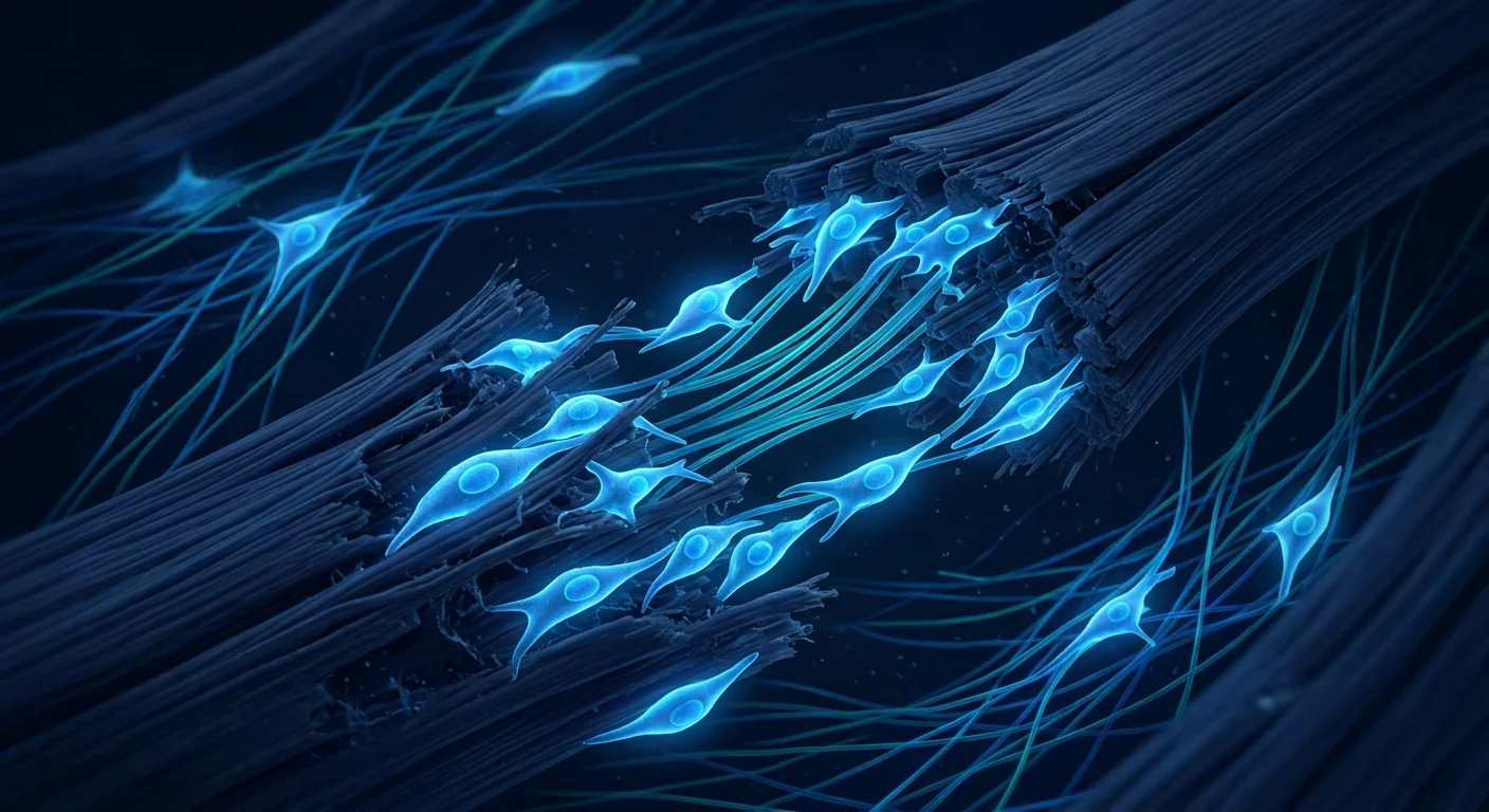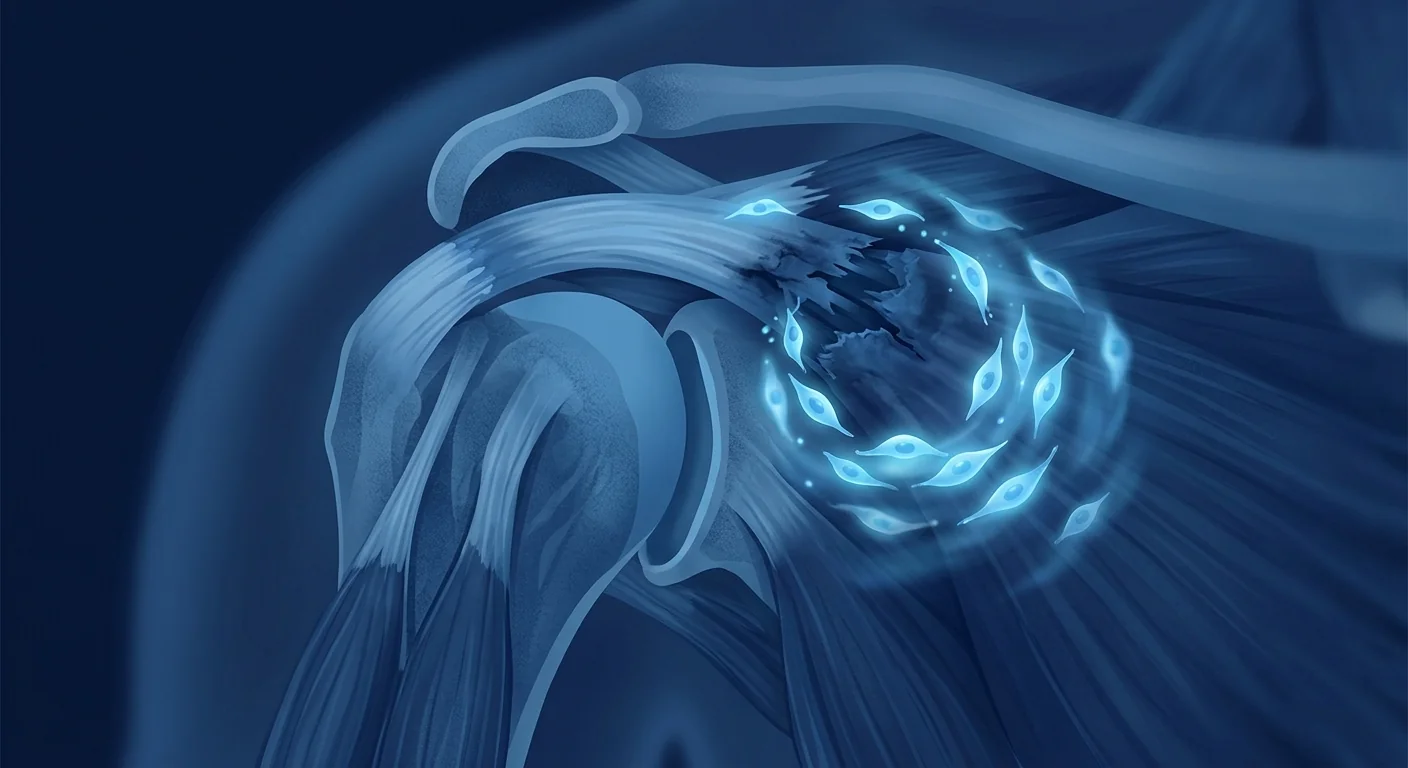
Rotator cuff injuries are among the most common — and most stubborn — musculoskeletal conditions in orthopaedic medicine. The rotator cuff is a group of four tendons that stabilise the shoulder joint and enable the full range of arm motion. When one or more of these tendons is torn or degenerated, the result is pain, weakness, and a shoulder that simply does not work the way it should. [1]
Where conventional treatment falls short. Standard care for rotator cuff tears follows a predictable ladder: rest, physiotherapy, corticosteroid injections, and — for full-thickness tears — surgical repair. While surgery can reattach the torn tendon to bone, the re-tear rate is sobering. Studies report failure rates of 20–94% depending on tear size, patient age, and tissue quality. The fundamental problem is biological: the tendon-to-bone insertion site (the enthesis) does not regenerate its original graded structure after repair. Instead, it heals with disorganised scar tissue that is mechanically weaker and prone to re-injury. [2][3]
The tissue-level challenge. The rotator cuff enthesis is a marvel of biological engineering — a continuous transition from flexible tendon to stiff bone across just a few hundred micrometres. This gradient is not reproduced by surgical repair or natural scarring. The result is a stress concentration at the repair site that predisposes to failure. Beyond the mechanical problem, rotator cuff tendons are intrinsically hypovascular, especially in the "critical zone" near the supraspinatus insertion, where most tears occur. This poor blood supply limits the delivery of the body's own repair cells and growth factors. [4]
MSC therapy targets the root cause. Mesenchymal Stem Cell therapy is being investigated not as a replacement for surgical repair when it is clearly indicated, but as a biological adjunct that may improve the quality of tendon-to-bone healing. By delivering a concentrated population of regenerative cells to the repair site, MSC therapy aims to shift the healing response from disorganised scar formation toward organised tissue regeneration — addressing the biological shortfall that makes rotator cuff repair so challenging. [5]
What is the rotator cuff and why does it tear?
The rotator cuff comprises four muscles and their tendons: the supraspinatus (abduction), infraspinatus and teres minor (external rotation), and subscapularis (internal rotation). Together, they form a cuff that centres the humeral head in the glenoid socket during arm movement. The supraspinatus tendon is the most frequently injured, accounting for the majority of tears, because it passes through a narrow subacromial space and bears significant mechanical load.
Tears are classified as partial-thickness (the tendon is frayed but not completely severed) or full-thickness (a complete discontinuity). Partial tears are further divided into articular-sided (on the joint side), bursal-sided (on the outer surface), and intrasubstance (within the tendon). Full-thickness tears are measured by the degree of tendon retraction — small (<1 cm), medium (1–3 cm), large (3–5 cm), and massive (>5 cm or involving two or more tendons). [6]
Risk factors include age (tears are present in >50% of individuals over 60), repetitive overhead activity (swimmers, throwers, painters), acute trauma (falls, heavy lifting), and smoking — which impairs tendon vascularity. Degenerative tears accumulate slowly over years as the tendon undergoes mucoid degeneration, fatty infiltration, and loss of organised collagen structure.
How MSC therapy supports rotator cuff healing
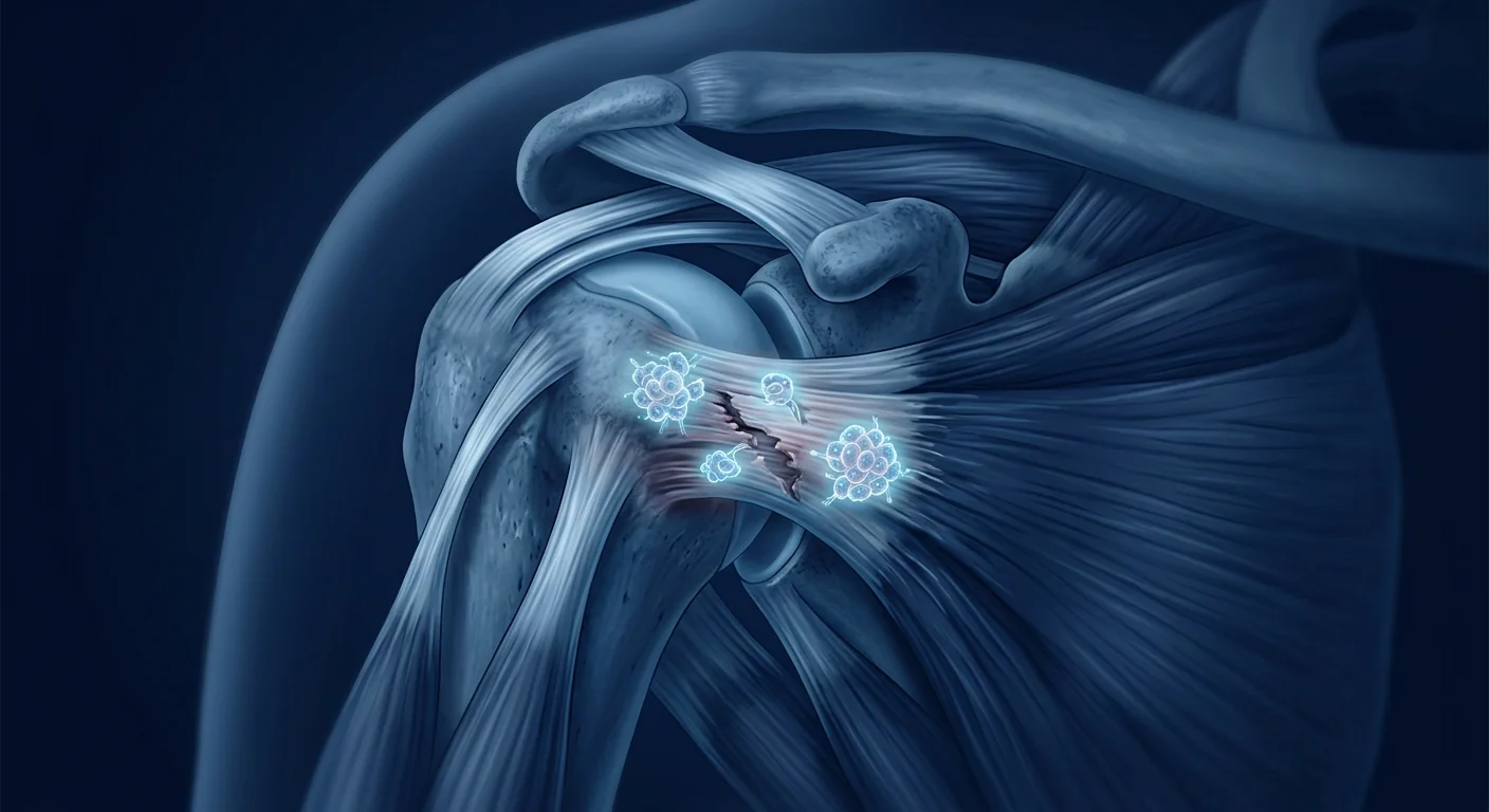
When Mesenchymal Stem Cells are delivered to the site of a rotator cuff tear — whether by direct injection under ultrasound guidance, or as an adjunct applied during arthroscopic repair — they engage the local environment through several coordinated mechanisms that are directly relevant to the biology of tendon-to-bone healing:
1. Modulation of the inflammatory environment
After a rotator cuff tear, the injured tendon and surrounding bursa enter a chronic inflammatory state characterised by elevated levels of IL-1β, IL-6, and TNF-α. While acute inflammation is necessary for the initial healing cascade, persistent inflammation drives matrix breakdown, inhibits tenocyte function, and promotes fatty infiltration of the muscle belly — a process that is essentially irreversible once established. MSCs respond to this high-cytokine environment by secreting anti-inflammatory mediators — TSG-6, PGE2, and IDO — that help shift the injury site from catabolic breakdown toward anabolic repair. [7]
2. Promotion of enthesis regeneration
The holy grail of rotator cuff repair is regeneration of the native enthesis — the four-zone transition from tendon (Zone I: collagen fibres) to fibrocartilage (Zone II) to mineralised fibrocartilage (Zone III) to bone (Zone IV). MSCs secrete a rich cocktail of growth factors — TGF-β3, BMP-2, and FGF-2 — that are known to be involved in enthesis development during embryogenesis. Preclinical studies in rat and rabbit rotator cuff models have shown that MSC-augmented repair results in a more organised fibrocartilage transition zone, with improved collagen fibre alignment and higher failure loads compared to repair alone. [8][9]
3. Reduction of post-surgical fibrosis and adhesion
One of the most clinically relevant complications of rotator cuff repair is post-operative stiffness caused by excessive scar tissue and subacromial adhesions. MSCs appear to influence the balance between regeneration and fibrosis through their effects on TGF-β1 signalling. By modulating the ratio of TGF-β1 (pro-fibrotic) to TGF-β3 (pro-regenerative), MSCs may help reduce the formation of disorganised scar and the adhesions that limit post-operative range of motion. [10]
4. Support for angiogenesis in the critical zone
The "critical zone" of the supraspinatus tendon — approximately 1 cm proximal to its insertion — is notoriously hypovascular, which is both a contributing factor to tear formation and a barrier to healing. MSCs secrete VEGF and other angiogenic factors that support the formation of new microvessels, improving oxygen and nutrient delivery to the healing tissue. This is particularly relevant in the early post-operative period when the repair site is dependent on diffusion from surrounding tissues.
What MSC therapy does NOT do for rotator cuff tears
It is essential to set honest expectations. MSC therapy does not close a retracted full-thickness tear — a tendon that has pulled away from its insertion by 3 cm requires surgical reapproximation. It does not replace the need for a structured rehabilitation programme — physiotherapy remains the cornerstone of recovery. It does not guarantee a return to pre-injury function, particularly in long-standing tears with significant muscle atrophy and fatty infiltration. What the evidence suggests it may do is improve the quality of healing at the cellular level, potentially reducing re-tear rates and improving functional outcomes — particularly in patients with poor native healing capacity.
Clinical evidence for MSC therapy in rotator cuff repair
The clinical evidence base for MSC therapy in rotator cuff disease is growing but still early-stage. Most published studies are small (n = 10–50), with follow-up periods of 1–3 years. The majority have investigated MSC augmentation of arthroscopic repair rather than stand-alone injection therapy. Here is a summary of the key findings:
MSC-augmented arthroscopic repair
Several prospective studies have compared arthroscopic rotator cuff repair with and without MSC augmentation. A 2020 randomised controlled trial of 30 patients with full-thickness supraspinatus tears reported that the MSC-augmented group had significantly lower re-tear rates at 12 months on MRI (13% vs. 40% in the control group, p < 0.05) and superior functional scores (ASES, Constant). [11]
A 2022 systematic review and meta-analysis of 7 studies (n = 327 patients) concluded that MSC augmentation was associated with a statistically significant reduction in re-tear rate (odds ratio 0.21, 95% CI 0.09–0.48) and improvement in the Constant-Murley score (mean difference 4.9 points, 95% CI 1.2–8.6) compared to repair alone. The authors noted that the quality of evidence was moderate and that larger, multicentre trials are needed. [12]
Injectable MSC therapy for partial tears
For partial-thickness tears — where the tendon is not completely detached and surgery may not be indicated — ultrasound-guided MSC injection is being explored as a less invasive option. A 2021 pilot study of 20 patients with high-grade partial-thickness supraspinatus tears (≥50% thickness) treated with a single ultrasound-guided injection of Wharton's jelly–derived MSCs reported significant improvements in VAS pain scores (from 7.2 to 2.1, p < 0.001) and ASES scores (from 48 to 82, p < 0.001) at 12 months. MRI demonstrated a reduction in tear size in 14 of 20 patients (70%). [13]
Key takeaway from the evidence
The current evidence suggests that MSC therapy is most promising in two clinical scenarios: (1) as an adjunct to arthroscopic repair of full-thickness tears, where the biological boost may reduce re-tear rates, and (2) as a standalone injection for partial-thickness tears in patients who wish to avoid or delay surgery. The evidence is not yet sufficient to recommend MSC therapy as a replacement for surgical repair of large or retracted tears.
Who is a candidate for MSC rotator cuff therapy?
Not every patient with a rotator cuff tear is a suitable candidate for MSC therapy. Clinical decision-making is individualised and based on several factors:
Patients who may benefit most
- Partial-thickness tears (grade 2–3). These are the sweet spot for injectable MSC therapy — the tendon is damaged but not completely detached, and the biological environment is still capable of supporting repair.
- Small to medium full-thickness tears undergoing surgical repair. MSC augmentation at the time of surgery may improve the biological quality of healing, particularly in patients with risk factors for poor healing (age >55, smoker, diabetes).
- Patients who have failed conservative management. Those who have completed 3–6 months of physiotherapy without satisfactory improvement.
- Patients wishing to avoid or delay surgery. Particularly relevant for partial tears and older patients with degenerative tears, where surgery carries higher risks.
Patients who are less suitable
- Massive, retracted tears (>5 cm) with significant muscle atrophy. Once the muscle belly has undergone fatty infiltration (Goutallier grade 3–4), the window for biological repair may have closed. Surgery is usually indicated, and even then, outcomes are guarded.
- Acute traumatic tears in young athletes. A 25-year-old with a full-thickness tear from a fall should generally undergo early surgical repair — the native healing capacity is strong, and the goal is anatomic restoration.
- Irreparable tears with cuff tear arthropathy. When the shoulder joint has developed arthritis secondary to chronic cuff deficiency, the primary problem is mechanical, not biological.
Recovery timeline: what to expect after MSC therapy
Recovery after MSC therapy for a rotator cuff injury follows a structured, phased approach. The timeline depends on whether the MSCs are delivered as a standalone injection or as an adjunct to surgery:
Week 1–2
Inflammatory phase. The shoulder is rested in a sling. Mild post-injection soreness is normal. For surgical patients, this is the standard post-operative immobilisation period. The MSCs are responding to the local cytokine environment and beginning to secrete paracrine factors.
Week 2–6
Proliferative phase. Passive range-of-motion exercises begin under physiotherapist guidance. The cellular repair response is actively underway — growth factor signalling is at its peak, and the first new collagen is being laid down. For injection-only patients, daily activities can gradually resume, but overhead movements and lifting remain restricted.
Week 6–12
Remodelling phase. Active-assisted range of motion begins. Strengthening exercises are introduced gradually. The repair tissue is maturing, and collagen fibres are being organised along lines of mechanical stress. This is the phase where the quality of biological healing — versus simple scar formation — becomes apparent.
Month 3–6
Strengthening phase. Progressive resistance training. Return to sport-specific drills for athletes. For surgical patients, this is the phase where the biological advantage of MSC augmentation may translate into a more durable repair — but the tissue is not yet at full strength.
Month 6–12
Return to full activity. Full return to sport, overhead work, and recreational activities. MRI at this stage is used to assess the structural integrity of the repair. The re-tear rate — the key outcome measure — is typically assessed at 12 months.
Comparing MSC sources for rotator cuff therapy
Two main categories of MSCs are used in clinical practice for rotator cuff pathology:
Wharton's jelly–derived MSCs (allogeneic)
These are mesenchymal stem cells extracted from the Wharton's jelly of donated umbilical cord tissue. They are expanded under GMP conditions, characterised for identity and purity, and administered as an off-the-shelf product. Advantages for rotator cuff applications include: consistent potency (not dependent on patient age or health), strong immunomodulatory and anti-inflammatory properties, high proliferative capacity, and the avoidance of a second harvesting procedure. At a clinical-grade facility, these cells are validated to ≥95% expression of MSC surface markers (CD73, CD90, CD105) and >95% viability at delivery. [14]
Bone marrow–derived MSCs (autologous)
Harvested from the patient's own iliac crest, these cells have the advantage of being autologous — eliminating any theoretical risk of immune rejection. The disadvantages include: variable cell quality (declining with patient age — many rotator cuff patients are over 55), the need for an additional bone marrow aspiration procedure, and a processing delay between harvest and administration. Some surgeons favour combining bone marrow concentrate (which contains MSCs along with platelets and growth factors) with the repair construct at the time of surgery.
The choice between allogeneic and autologous MSCs should be made on a case-by-case basis, considering the patient's age, tear characteristics, and preference. In the available clinical literature, both approaches have shown promising results, and no head-to-head comparison exists specifically for rotator cuff repair.
Limitations and honest perspective
It is important to state plainly what MSC therapy for rotator cuff tears is — and is not:
- MSC therapy is still investigational for rotator cuff pathology. While the evidence base is growing, large-scale multicentre randomised controlled trials are lacking. The current literature is dominated by small studies with methodological limitations.
- MSC therapy cannot reverse established muscle atrophy. Once the rotator cuff muscle belly has undergone significant fatty infiltration (Goutallier grade 3–4), the muscle has been replaced by fat. No amount of biological stimulation can regenerate muscle tissue that no longer exists. This is why early intervention is critical.
- Rehabilitation remains non-negotiable. MSC therapy is an adjunct to — not a replacement for — a structured, progressive physiotherapy programme. The cells provide the biological substrate; the mechanical loading provided by rehabilitation drives the organisation of that substrate into functional tissue.
- Not all tears are biological problems. Some rotator cuff tears are primarily mechanical — particularly acute traumatic tears in young patients, and tears associated with subacromial impingement from bony spurs. In these cases, addressing the mechanical problem (surgical repair, subacromial decompression) is the priority, and the added value of MSC augmentation is uncertain.
- Results vary significantly between patients. Age, tear chronicity, tear size, muscle quality, smoking status, diabetes, and compliance with rehabilitation all influence outcomes. MSC therapy is not a level playing field — it works best in patients with the best biological substrate.
Frequently asked questions
Can MSC therapy heal a full-thickness rotator cuff tear without surgery?
For a small, minimally retracted full-thickness tear in a patient who is not a surgical candidate, MSC injection may support biological healing and symptom improvement. However, for a retracted tear — where the tendon has pulled away from the bone — the gap must be closed mechanically. MSCs cannot bridge a physical gap. In these cases, MSC therapy is best considered as an adjunct at the time of surgical repair.
How soon after a rotator cuff tear can MSC therapy be administered?
There is no single answer. For acute traumatic tears in young patients, early surgical repair is generally recommended. For degenerative tears that have been present for weeks or months, MSC therapy can be considered at any point — though earlier intervention (before significant muscle atrophy develops) is likely to yield better results. Many clinicians recommend completing a 6–12 week trial of conservative management (physiotherapy, activity modification) before proceeding to MSC therapy, as a proportion of partial tears will improve without intervention.
Is MSC therapy covered by insurance?
In most jurisdictions, MSC therapy for rotator cuff pathology is considered elective and investigational, and is not covered by standard health insurance. Patients should expect to pay out of pocket. Costs vary significantly between clinics and countries. At VELAR Center, a detailed cost breakdown is provided during the consultation, and no patient proceeds to treatment without a clear understanding of the financial commitment.
What is the re-tear rate after MSC-augmented rotator cuff repair?
The available evidence suggests that MSC augmentation may reduce re-tear rates compared to repair alone. In the meta-analysis cited above, the pooled re-tear rate was approximately 13% in the MSC-augmented group versus 40% in the control group at 12 months. However, these figures come from small studies with selected patient populations and should be interpreted cautiously. Re-tear rates depend heavily on tear size, patient age, and rehabilitation quality — MSC therapy is one factor among many.
References
- Yamamoto A, Takagishi K, Osawa T, et al. Prevalence and risk factors of a rotator cuff tear in the general population. Journal of Shoulder and Elbow Surgery. 2010;19(1):116-120. doi:10.1016/j.jse.2009.04.006 ↩
- Galatz LM, Ball CM, Teefey SA, et al. The outcome and repair integrity of completely arthroscopically repaired large and massive rotator cuff tears. Journal of Bone and Joint Surgery. 2004;86(2):219-224. doi:10.2106/00004623-200402000-00002 ↩
- Le BTN, Wu XL, Lam PH, Murrell GAC. Factors predicting rotator cuff retears: an analysis of 1000 consecutive rotator cuff repairs. American Journal of Sports Medicine. 2014;42(5):1134-1142. doi:10.1177/0363546514525336 ↩
- Benjamin M, McGonagle D. Entheses: tendon and ligament attachment sites. Scandinavian Journal of Medicine & Science in Sports. 2009;19(4):520-527. doi:10.1111/j.1600-0838.2009.00906.x ↩
- Hernigou P, Flouzat Lachaniette CH, Delambre J, et al. Biologic augmentation of rotator cuff repair with mesenchymal stem cells during arthroscopy improves healing and prevents further tears: a case-controlled study. International Orthopaedics. 2014;38(9):1811-1818. doi:10.1007/s00264-014-2391-1 ↩
- Tashjian RZ. Epidemiology, natural history, and indications for treatment of rotator cuff tears. Clinics in Sports Medicine. 2012;31(4):589-604. doi:10.1016/j.csm.2012.07.001 ↩
- Proffen BL, Shakir S, Bifani J, et al. The effect of mesenchymal stromal cell sheets on the inflammatory stage of flexor tendon healing. Stem Cell Research & Therapy. 2016;7:144. doi:10.1186/s13287-016-0401-2 ↩
- Gulotta LV, Kovacevic D, Packer JD, et al. Bone marrow-derived mesenchymal stem cells transduced with scleraxis improve rotator cuff healing in a rat model. American Journal of Sports Medicine. 2011;39(6):1282-1289. doi:10.1177/0363546510395485 ↩
- Chen P, Cui L, Chen GC, et al. The application of BMP-12-overexpressing mesenchymal stem cells loaded on a collagen scaffold for rotator cuff repair in a rabbit model. Journal of Shoulder and Elbow Surgery. 2020;29(3):e90-e103. doi:10.1016/j.jse.2019.06.022 ↩
- Shapiro E, Grande D, Drakos M. Biologics in Achilles tendon healing and repair: a review. Current Reviews in Musculoskeletal Medicine. 2015;8(1):9-17. doi:10.1007/s12178-015-9257-z ↩
- Kim YS, Sung CH, Chung SH, et al. Does an injection of adipose-derived mesenchymal stem cells loaded in fibrin glue influence rotator cuff repair outcomes? A clinical and magnetic resonance imaging study. American Journal of Sports Medicine. 2020;48(8):1889-1898. doi:10.1177/0363546520917963 ↩
- Liu Y, Li Z, Li J, et al. Mesenchymal stem cell therapy for rotator cuff repair: a systematic review and meta-analysis. Orthopaedic Journal of Sports Medicine. 2022;10(4):23259671221087358. doi:10.1177/23259671221087358 ↩
- Jo CH, Chai JW, Jeong EC, et al. Intratendinous injection of mesenchymal stem cells for the treatment of rotator cuff disease: a 2-year follow-up study. Arthroscopy. 2021;37(3):858-868. doi:10.1016/j.arthro.2020.10.036 ↩
- Dominici M, Le Blanc K, Mueller I, et al. Minimal criteria for defining multipotent mesenchymal stromal cells. The International Society for Cellular Therapy position statement. Cytotherapy. 2006;8(4):315-317. doi:10.1080/14653240600855905 ↩
- Caplan AI, Correa D. The MSC: an injury drugstore. Cell Stem Cell. 2011;9(1):11-15. doi:10.1016/j.stem.2011.06.008 ↩
肩袖损伤是骨科医学中最常见——也是最顽固——的肌肉骨骼疾病之一。肩袖由四条肌腱组成,负责稳定肩关节并实现手臂的全方位运动。当其中一条或多条肌腱撕裂或退化时,就会出现疼痛、无力,以及肩关节功能受限。 [1]
常规治疗为何力不从心。 肩袖撕裂的标准治疗遵循可预测的阶梯路径:休息、物理治疗、皮质类固醇注射,以及——对于全层撕裂——手术修复。虽然手术可以将撕裂的肌腱重新附着到骨骼上,但再撕裂率令人担忧。研究表明,根据撕裂大小、患者年龄和组织质量的不同,失败率在20%至94%之间。根本问题在于生物学层面:肌腱-骨附着点(enthesis)在修复后无法再生其原始的梯度结构,而是以力学强度较弱的无序瘢痕组织愈合,容易再次受伤。 [2][3]
组织层面的挑战。 肩袖附着点是生物工程的奇迹——在短短几百微米内,从柔韧的肌腱连续过渡到坚硬的骨骼。这种梯度是手术修复或自然瘢痕无法重现的。结果是修复部位出现应力集中,易导致失败。除了力学问题,肩袖肌腱本身是低血管化的,尤其是在冈上肌附着点附近的"关键区域",这是大多数撕裂发生的部位。这种血供不足限制了身体自身修复细胞和生长因子的输送。 [4]
间充质干细胞疗法针对根本原因。 间充质干细胞疗法正在被研究作为手术修复的生物学辅助手段,而非替代品。通过将浓缩的再生细胞群体递送至修复部位,MSC疗法旨在将愈合反应从无序瘢痕形成转向有序组织再生——解决使肩袖修复如此具有挑战性的生物学短板。 [5]
什么是肩袖?为什么会撕裂?
肩袖由四块肌肉及其肌腱组成:冈上肌(外展)、冈下肌和小圆肌(外旋)、肩胛下肌(内旋)。它们共同形成一个袖套,在手臂运动时将肱骨头稳定在关节盂内。冈上肌腱是最常受伤的,占大多数撕裂,因为它穿过狭窄的肩峰下间隙并承受显著机械负荷。
撕裂分为部分厚度(肌腱磨损但未完全断裂)或全层(完全中断)。部分撕裂进一步分为关节侧(关节面)、滑囊侧(外表面)和腱内(肌腱内部)。全层撕裂按肌腱回缩程度衡量:小(<1 cm)、中(1-3 cm)、大(3-5 cm)和巨大(>5 cm或涉及两条或以上肌腱)。 [6]
风险因素包括年龄(60岁以上人群中超过50%存在撕裂)、重复性过顶活动(游泳者、投掷运动员、油漆工)、急性创伤(跌倒、重物提举)和吸烟——吸烟会损害肌腱血管。退行性撕裂在多年间缓慢累积,随着肌腱发生黏液样变性、脂肪浸润和有组织的胶原结构丧失而发展。
MSC疗法如何支持肩袖愈合
当间充质干细胞被递送至肩袖撕裂部位时——无论是通过超声引导下直接注射,还是作为关节镜修复时的辅助手段——它们通过多种协调机制与局部环境相互作用,这些机制与肌腱-骨愈合的生物学直接相关:
1. 调节炎症环境
肩袖撕裂后,受伤的肌腱和周围滑囊进入慢性炎症状态,以IL-1β、IL-6和TNF-α水平升高为特征。虽然急性炎症对初始愈合级联反应是必需的,但持续炎症会驱动基质分解、抑制肌腱细胞功能,并促进肌肉腹部的脂肪浸润——这一过程一旦形成基本上是不可逆的。MSC通过分泌抗炎介质——TSG-6、PGE2和IDO——来响应这种高细胞因子环境,帮助将损伤部位从分解代谢转向合成代谢修复。 [7]
2. 促进附着点再生
肩袖修复的圣杯是再生原始附着点——从肌腱(I区:胶原纤维)到纤维软骨(II区)到矿化纤维软骨(III区)到骨骼(IV区)的四区过渡。MSC分泌丰富的生长因子——TGF-β3、BMP-2和FGF-2——已知这些因子参与胚胎发育过程中的附着点形成。大鼠和兔肩袖模型的临床前研究表明,与单独修复相比,MSC增强修复产生更有序的纤维软骨过渡区,胶原纤维排列更好,失败负荷更高。 [8][9]
3. 减少术后纤维化和粘连
肩袖修复最具临床相关性的并发症之一是术后僵硬,由过度瘢痕组织和肩峰下粘连引起。MSC通过其对TGF-β1信号传导的影响,似乎能够影响再生与纤维化之间的平衡。通过调节TGF-β1(促纤维化)与TGF-β3(促再生)的比例,MSC可能有助于减少无序瘢痕的形成和限制术后活动范围的粘连。 [10]
4. 支持关键区域的血管生成
冈上肌腱的"关键区域"——大约在其附着点近端1 cm处——以低血管化著称,这既是撕裂形成的促成因素,也是愈合的障碍。MSC分泌VEGF和其他血管生成因子,支持新微血管的形成,改善愈合组织的氧气和营养输送。这在术后早期尤为相关,此时修复部位依赖于周围组织的扩散。
MSC疗法对肩袖撕裂不能做什么
设定诚实的期望至关重要。MSC疗法不能闭合回缩的全层撕裂——一条已经从附着点拉开3 cm的肌腱需要手术重新对合。它不能替代结构化的康复计划——物理治疗仍然是恢复的基石。它不能保证恢复到受伤前的功能水平,特别是对于长期存在的撕裂,伴有显著的肌肉萎缩和脂肪浸润。证据表明它可能改善的是细胞水平的愈合质量,可能降低再撕裂率并改善功能结果——特别是在天然愈合能力较差的患者中。
MSC治疗肩袖修复的临床证据
MSC疗法在肩袖疾病中的临床证据基础正在增长,但仍处于早期阶段。大多数已发表的研究规模较小(n=10-50),随访期为1-3年。大多数研究调查了MSC增强关节镜修复,而非单独注射疗法。以下是关键发现的总结:
MSC增强关节镜修复
几项前瞻性研究比较了有无MSC增强的关节镜肩袖修复。一项2020年针对30例全层冈上肌撕裂患者的随机对照试验报告,MSC增强组在12个月时MRI再撕裂率显著降低(13% vs 对照组40%,p<0.05),功能评分(ASES、Constant)更优。 [11]
一项2022年包含7项研究(n=327例患者)的系统评价和荟萃分析得出结论,MSC增强与再撕裂率显著降低(比值比0.21,95% CI 0.09-0.48)和Constant-Murley评分改善(平均差异4.9分,95% CI 1.2-8.6)相关。作者指出证据质量为中等,需要更大规模的多中心试验。 [12]
注射MSC疗法治疗部分撕裂
对于部分厚度撕裂——肌腱未完全分离,可能不需要手术——超声引导下MSC注射正在被探索作为一种创伤较小的选择。一项2021年针对20例高级别部分厚度冈上肌撕裂(≥50%厚度)患者的初步研究,使用单次超声引导下Wharton's jelly来源MSC注射,报告12个月时VAS疼痛评分(从7.2降至2.1,p<0.001)和ASES评分(从48升至82,p<0.001)显著改善。MRI显示20例患者中14例(70%)撕裂尺寸减小。 [13]
证据的关键要点
当前证据表明,MSC疗法在两种临床场景中最有前景:(1)作为全层撕裂关节镜修复的辅助手段,生物学增强可能降低再撕裂率;(2)作为部分厚度撕裂的独立注射,适用于希望避免或推迟手术的患者。证据尚不足以推荐MSC疗法替代大面积或回缩性撕裂的手术修复。
谁是MSC肩袖治疗的候选者?
并非每位肩袖撕裂患者都适合MSC疗法。临床决策是个体化的,基于以下因素:
可能获益最大的患者
- 部分厚度撕裂(2-3级)。 这是注射MSC疗法的最佳适应症——肌腱受损但未完全分离,生物环境仍能支持修复。
- 接受手术修复的中小型全层撕裂。 手术时MSC增强可能改善愈合的生物学质量,特别是在具有愈合不良风险因素的患者(年龄>55岁、吸烟者、糖尿病)中。
- 保守治疗失败的患者。 已完成3-6个月物理治疗但未获得满意改善的患者。
- 希望避免或推迟手术的患者。 特别适用于部分撕裂和退行性撕裂的老年患者,手术风险较高。
不太适合的患者
- 巨大回缩性撕裂(>5 cm)伴有显著肌肉萎缩。 一旦肌肉腹部发生脂肪浸润(Goutallier 3-4级),生物修复的窗口可能已经关闭。通常需要手术,即便如此,结果也不确定。
- 年轻运动员的急性创伤性撕裂。 25岁因跌倒导致全层撕裂的患者通常应进行早期手术修复——天然愈合能力强,目标是解剖恢复。
- 不可修复的撕裂伴肩袖撕裂关节病。 当肩关节因慢性肩袖缺损继发关节炎时,主要问题是机械性的,而非生物学性的。
恢复时间线:MSC治疗后的预期
肩袖损伤MSC治疗后的恢复遵循结构化、分阶段的方案。时间线取决于MSC是作为独立注射还是手术辅助手段递送:
第1-2周
炎症期。 肩部用吊带休息。轻微的注射后酸痛是正常的。对于手术患者,这是标准的术后固定期。MSC正在响应局部细胞因子环境,开始分泌旁分泌因子。
第2-6周
增殖期。 在物理治疗师指导下开始被动关节活动度练习。细胞修复反应正在积极进行——生长因子信号达到峰值,第一批新胶原蛋白正在沉积。对于仅注射的患者,日常活动可以逐渐恢复,但过顶运动和提举仍然受限。
第6-12周
重塑期。 开始主动辅助关节活动度练习。逐渐引入力量训练。修复组织正在成熟,胶原纤维正在沿机械应力方向排列。这是生物愈合质量——而非简单瘢痕形成——变得明显的阶段。
第3-6个月
力量训练期。 进行性阻力训练。运动员恢复运动专项训练。对于手术患者,MSC增强的生物学优势可能在此阶段转化为更耐久的修复——但组织尚未达到完全强度。
第6-12个月
恢复完全活动。 完全恢复运动、过顶工作和休闲活动。此阶段使用MRI评估修复的结构完整性。再撕裂率——关键结果指标——通常在12个月时评估。
肩袖治疗MSC来源的比较
在临床实践中,两类MSC用于肩袖病理:
Wharton's jelly来源MSC(异体)
这些是从捐赠脐带组织的Wharton's jelly中提取的间充质干细胞。它们在GMP条件下扩增,进行身份和纯度鉴定,作为即用型产品施用。肩袖应用的优点包括:效力一致(不依赖于患者年龄或健康状况)、强大的免疫调节和抗炎特性、高增殖能力、避免第二次采集手术。在临床级设施中,这些细胞经过验证,MSC表面标志物(CD73、CD90、CD105)表达≥95%,交付时活力>95%。 [14]
骨髓来源MSC(自体)
从患者自身髂嵴采集,这些细胞的优点是自体——消除了任何免疫排斥的理论风险。缺点包括:细胞质量不稳定(随患者年龄下降——许多肩袖患者超过55岁)、需要额外的骨髓穿刺手术、采集和施用之间的处理延迟。一些外科医生倾向于在手术时将骨髓浓缩物(含MSC及血小板和生长因子)与修复结构结合使用。
异体和自体MSC之间的选择应根据具体情况做出,考虑患者年龄、撕裂特征和偏好。在现有临床文献中,两种方法都显示出有希望的结果,且尚无专门针对肩袖修复的头对头比较。
局限性与诚实视角
明确陈述MSC疗法对肩袖撕裂是什么——以及不是什么,这一点很重要:
- MSC疗法对肩袖病理仍处于研究阶段。 虽然证据基础正在增长,但缺乏大规模多中心随机对照试验。当前文献以小规模研究为主,存在方法学局限性。
- MSC疗法不能逆转已形成的肌肉萎缩。 一旦肩袖肌肉腹部发生显著脂肪浸润(Goutallier 3-4级),肌肉已被脂肪替代。任何生物刺激都无法再生不再存在的肌肉组织。这就是为什么早期干预至关重要。
- 康复仍然不可或缺。 MSC疗法是结构化、渐进式物理治疗计划的辅助手段,而非替代品。细胞提供生物基质;康复提供的机械负荷驱动该基质组织成功能性组织。
- 并非所有撕裂都是生物学问题。 一些肩袖撕裂主要是机械性的——特别是年轻患者的急性创伤性撕裂,以及与骨刺引起的肩峰下撞击相关的撕裂。在这些情况下,解决机械问题(手术修复、肩峰下减压)是优先事项,MSC增强的附加价值不确定。
- 结果在患者之间差异显著。 年龄、撕裂慢性程度、撕裂大小、肌肉质量、吸烟状态、糖尿病和康复依从性都会影响结果。MSC疗法并非公平的竞争环境——它在具有最佳生物基质的患者中效果最好。
常见问题
MSC疗法能否在不手术的情况下治愈全层肩袖撕裂?
对于小型、轻微回缩的全层撕裂,在不适合手术的患者中,MSC注射可能支持生物愈合和症状改善。然而,对于回缩性撕裂——肌腱已从骨骼上拉开——必须通过机械方式闭合间隙。MSC无法桥接物理间隙。在这些情况下,MSC疗法最好在手术修复时作为辅助手段考虑。
肩袖撕裂后多久可以进行MSC治疗?
没有统一的答案。对于年轻患者的急性创伤性撕裂,通常建议早期手术修复。对于已存在数周或数月的退行性撕裂,MSC疗法可以在任何时间点考虑——尽管早期干预(在显著肌肉萎缩发生之前)可能产生更好的结果。许多临床医生建议在MSC治疗前完成6-12周的保守治疗试验(物理治疗、活动调整),因为部分部分撕裂将无需干预即可改善。
MSC疗法是否由保险覆盖?
在大多数司法管辖区,MSC疗法对肩袖病理被视为选择性和研究性的,不被标准健康保险覆盖。患者应预期自费支付。费用因诊所和国家而异。在VELAR Center,详细费用明细在咨询期间提供,没有患者在清楚了解财务承诺之前进行治疗。
MSC增强肩袖修复后的再撕裂率是多少?
现有证据表明,与单独修复相比,MSC增强可能降低再撕裂率。在上述荟萃分析中,MSC增强组的汇总再撕裂率约为13%,对照组为40%(12个月)。然而,这些数据来自小规模研究,患者群体经过选择,应谨慎解读。再撕裂率严重依赖于撕裂大小、患者年龄和康复质量——MSC疗法是众多因素之一。
参考文献
- Yamamoto A, Takagishi K, Osawa T, et al. Prevalence and risk factors of a rotator cuff tear in the general population. Journal of Shoulder and Elbow Surgery. 2010;19(1):116-120. doi:10.1016/j.jse.2009.04.006 ↩
- Galatz LM, Ball CM, Teefey SA, et al. The outcome and repair integrity of completely arthroscopically repaired large and massive rotator cuff tears. Journal of Bone and Joint Surgery. 2004;86(2):219-224. doi:10.2106/00004623-200402000-00002 ↩
- Le BTN, Wu XL, Lam PH, Murrell GAC. Factors predicting rotator cuff retears: an analysis of 1000 consecutive rotator cuff repairs. American Journal of Sports Medicine. 2014;42(5):1134-1142. doi:10.1177/0363546514525336 ↩
- Benjamin M, McGonagle D. Entheses: tendon and ligament attachment sites. Scandinavian Journal of Medicine & Science in Sports. 2009;19(4):520-527. doi:10.1111/j.1600-0838.2009.00906.x ↩
- Hernigou P, Flouzat Lachaniette CH, Delambre J, et al. Biologic augmentation of rotator cuff repair with mesenchymal stem cells during arthroscopy improves healing and prevents further tears: a case-controlled study. International Orthopaedics. 2014;38(9):1811-1818. doi:10.1007/s00264-014-2391-1 ↩
- Tashjian RZ. Epidemiology, natural history, and indications for treatment of rotator cuff tears. Clinics in Sports Medicine. 2012;31(4):589-604. doi:10.1016/j.csm.2012.07.001 ↩
- Proffen BL, Shakir S, Bifani J, et al. The effect of mesenchymal stromal cell sheets on the inflammatory stage of flexor tendon healing. Stem Cell Research & Therapy. 2016;7:144. doi:10.1186/s13287-016-0401-2 ↩
- Gulotta LV, Kovacevic D, Packer JD, et al. Bone marrow-derived mesenchymal stem cells transduced with scleraxis improve rotator cuff healing in a rat model. American Journal of Sports Medicine. 2011;39(6):1282-1289. doi:10.1177/0363546510395485 ↩
- Chen P, Cui L, Chen GC, et al. The application of BMP-12-overexpressing mesenchymal stem cells loaded on a collagen scaffold for rotator cuff repair in a rabbit model. Journal of Shoulder and Elbow Surgery. 2020;29(3):e90-e103. doi:10.1016/j.jse.2019.06.022 ↩
- Shapiro E, Grande D, Drakos M. Biologics in Achilles tendon healing and repair: a review. Current Reviews in Musculoskeletal Medicine. 2015;8(1):9-17. doi:10.1007/s12178-015-9257-z ↩
- Kim YS, Sung CH, Chung SH, et al. Does an injection of adipose-derived mesenchymal stem cells loaded in fibrin glue influence rotator cuff repair outcomes? A clinical and magnetic resonance imaging study. American Journal of Sports Medicine. 2020;48(8):1889-1898. doi:10.1177/0363546520917963 ↩
- Liu Y, Li Z, Li J, et al. Mesenchymal stem cell therapy for rotator cuff repair: a systematic review and meta-analysis. Orthopaedic Journal of Sports Medicine. 2022;10(4):23259671221087358. doi:10.1177/23259671221087358 ↩
- Jo CH, Chai JW, Jeong EC, et al. Intratendinous injection of mesenchymal stem cells for the treatment of rotator cuff disease: a 2-year follow-up study. Arthroscopy. 2021;37(3):858-868. doi:10.1016/j.arthro.2020.10.036 ↩
- Dominici M, Le Blanc K, Mueller I, et al. Minimal criteria for defining multipotent mesenchymal stromal cells. The International Society for Cellular Therapy position statement. Cytotherapy. 2006;8(4):315-317. doi:10.1080/14653240600855905 ↩
- Caplan AI, Correa D. The MSC: an injury drugstore. Cell Stem Cell. 2011;9(1):11-15. doi:10.1016/j.stem.2011.06.008 ↩
تُعد إصابات الكفة المدورة من أكثر الحالات العضلية الهيكلية شيوعًا — وأكثرها عنادًا — في طب العظام. الكفة المدورة هي مجموعة من أربعة أوتار تعمل على تثبيت مفصل الكتف وتمكين النطاق الكامل لحركة الذراع. عندما يتمزق واحد أو أكثر من هذه الأوتار أو يتدهور، تكون النتيجة ألمًا وضعفًا وكتفًا لا يعمل كما ينبغي. [1]
أوجه قصور العلاج التقليدي. تتبع الرعاية القياسية لتمزقات الكفة المدورة سلمًا متوقعًا: الراحة، والعلاج الطبيعي، وحقن الكورتيكوستيرويد، و — للتمزقات كاملة السماكة — الإصلاح الجراحي. بينما يمكن للجراحة إعادة ربط الوتر الممزق بالعظم، فإن معدل إعادة التمزق مثير للقلق. تشير الدراسات إلى معدلات فشل تتراوح بين 20% و94% اعتمادًا على حجم التمزق وعمر المريض وجودة الأنسجة. المشكلة الأساسية بيولوجية: موقع ارتباط الوتر بالعظم لا يستعيد بنيته المتدرجة الأصلية بعد الإصلاح، بل يلتئم بنسيج ندبي غير منظم أضعف ميكانيكيًا ومعرض للإصابة مرة أخرى. [2][3]
التحدي على مستوى الأنسجة. يُعد ارتباط الكفة المدورة بالعظم معجزة من معجزات الهندسة البيولوجية — انتقال مستمر من وتر مرن إلى عظم صلب عبر بضع مئات من الميكرومترات فقط. هذا التدرج لا يمكن إعادة إنتاجه بالإصلاح الجراحي أو التندب الطبيعي. النتيجة هي تركيز إجهاد في موقع الإصلاح مما يهيئ للفشل. بالإضافة إلى المشكلة الميكانيكية، فإن أوتار الكفة المدورة قليلة التوعية الدموية بطبيعتها، خاصة في "المنطقة الحرجة" بالقرب من ارتباط الوتر فوق الشوكي، حيث تحدث معظم التمزقات. هذا الإمداد الدموي الضعيف يحد من وصول خلايا الإصلاح وعوامل النمو الخاصة بالجسم. [4]
يستهدف علاج MSC السبب الجذري. يتم دراسة علاج الخلايا الجذعية الوسيطة ليس كبديل للإصلاح الجراحي عندما يكون ضروريًا بوضوح، ولكن كمساعد بيولوجي قد يحسن جودة التئام الوتر بالعظم. من خلال توصيل مجموعة مركزة من الخلايا التجديدية إلى موقع الإصلاح، يهدف علاج MSC إلى تحويل استجابة الالتئام من تكوين ندبة غير منظمة نحو تجديد الأنسجة المنظمة — معالجة النقص البيولوجي الذي يجعل إصلاح الكفة المدورة تحديًا كبيرًا. [5]
ما هي الكفة المدورة ولماذا تتمزق؟
تتكون الكفة المدورة من أربع عضلات وأوتارها: فوق الشوكي (الرفع الجانبي)، وتحت الشوكي والمدورة الصغيرة (الدوران الخارجي)، وتحت الكتف (الدوران الداخلي). تشكل معًا كفة تثبت رأس العضد في تجويف الحق أثناء حركة الذراع. وتر فوق الشوكي هو الأكثر إصابة، حيث يمثل غالبية التمزقات، لأنه يمر عبر مساحة تحت الأخرم الضيقة ويتحمل حملًا ميكانيكيًا كبيرًا.
تصنف التمزقات على أنها جزئية السماكة (الوتر متآكل ولكنه غير مقطوع بالكامل) أو كاملة السماكة (انقطاع كامل). تنقسم التمزقات الجزئية أيضًا إلى: على الجانب المفصلي، وعلى الجانب الجرابي، وداخل الوتر. تقاس التمزقات كاملة السماكة بدرجة تراجع الوتر: صغيرة (<1 سم)، ومتوسطة (1-3 سم)، وكبيرة (3-5 سم)، وضخمة (>5 سم أو تشمل وترين أو أكثر). [6]
تشمل عوامل الخطر العمر (التمزقات موجودة في >50% من الأفراد فوق 60 عامًا)، والنشاط المتكرر فوق الرأس (السباحون، والرماة، والدهانون)، والإصابات الحادة (السقوط، ورفع الأحمال الثقيلة)، والتدخين — الذي يضعف توعية الأوتار الدموية. تتراكم التمزقات التنكسية ببطء على مر السنين مع خضوع الوتر للتنكس المخاطي، والارتشاح الدهني، وفقدان بنية الكولاجين المنظمة.
كيف يدعم علاج MSC التئام الكفة المدورة
عندما يتم توصيل الخلايا الجذعية الوسيطة إلى موقع تمزق الكفة المدورة — سواء عن طريق الحقن المباشر تحت توجيه الموجات فوق الصوتية، أو كمساعد يُطبق أثناء الإصلاح بالمنظار — فإنها تتفاعل مع البيئة المحلية من خلال آليات منسقة متعددة ذات صلة مباشرة ببيولوجيا التئام الوتر بالعظم:
1. تعديل البيئة الالتهابية
بعد تمزق الكفة المدورة، يدخل الوتر المصاب والجراب المحيط به في حالة التهابية مزمنة تتميز بمستويات مرتفعة من IL-1β وIL-6 وTNF-α. بينما الالتهاب الحاد ضروري لسلسلة الالتئام الأولية، فإن الالتهاب المستمر يدفع تحلل المصفوفة، ويثبط وظيفة الخلايا الوترية، ويعزز الارتشاح الدهني لبطن العضلة — وهي عملية لا رجعة فيها أساسًا بمجرد تشكلها. تستجيب MSCs لهذه البيئة عالية السيتوكينات بإفراز وسطاء مضادين للالتهاب — TSG-6 وPGE2 وIDO — التي تساعد في تحويل موقع الإصابة من التحلل التقويضي نحو الإصلاح البنائي. [7]
2. تعزيز تجدد منطقة الارتباط
الكأس المقدسة لإصلاح الكفة المدورة هي تجديد منطقة الارتباط الأصلية — الانتقال رباعي المناطق من الوتر (المنطقة الأولى: ألياف الكولاجين) إلى الغضروف الليفي (المنطقة الثانية) إلى الغضروف الليفي المتكلس (المنطقة الثالثة) إلى العظم (المنطقة الرابعة). تفرز MSCs مزيجًا غنيًا من عوامل النمو — TGF-β3 وBMP-2 وFGF-2 — المعروف أنها تشارك في تطور منطقة الارتباط أثناء التكون الجنيني. أظهرت الدراسات قبل السريرية في نماذج الكفة المدورة للفئران والأرانب أن الإصلاح المعزز بـ MSC يؤدي إلى منطقة انتقالية غضروفية ليفية أكثر تنظيمًا، مع محاذاة أفضل لألياف الكولاجين وأحمال فشل أعلى مقارنة بالإصلاح وحده. [8][9]
3. تقليل التليف والالتصاقات بعد الجراحة
أحد أكثر مضاعفات إصلاح الكفة المدورة أهمية سريريًا هو التيبس بعد الجراحة الناجم عن النسيج الندبي المفرط والالتصاقات تحت الأخرم. يبدو أن MSCs تؤثر على التوازن بين التجدد والتليف من خلال تأثيراتها على إشارات TGF-β1. من خلال تعديل نسبة TGF-β1 (محفز للتليف) إلى TGF-β3 (محفز للتجدد)، قد تساعد MSCs في تقليل تكوين الندبة غير المنظمة والالتصاقات التي تحد من نطاق الحركة بعد الجراحة. [10]
4. دعم تكوين الأوعية الدموية في المنطقة الحرجة
"المنطقة الحرجة" لوتر فوق الشوكي — حوالي 1 سم قرب ارتباطه — معروفة بقلة توعيتها الدموية، وهو عامل مساهم في تكوين التمزق وعائق أمام الالتئام. تفرز MSCs عامل نمو بطانة الأوعية الدموية (VEGF) وعوامل مولدة للأوعية الأخرى التي تدعم تكوين أوعية دقيقة جديدة، مما يحسن توصيل الأكسجين والمغذيات إلى الأنسجة الملتئمة. هذا مهم بشكل خاص في الفترة المبكرة بعد الجراحة عندما يعتمد موقع الإصلاح على الانتشار من الأنسجة المحيطة.
ما لا يفعله علاج MSC لتمزقات الكفة المدورة
من الضروري وضع توقعات صادقة. علاج MSC لا يغلق تمزقًا كامل السماكة متراجعًا — الوتر الذي انسحب بعيدًا عن ارتباطه بمقدار 3 سم يتطلب إعادة تقريب جراحية. إنه لا يحل محل الحاجة إلى برنامج إعادة تأهيل منظم — يبقى العلاج الطبيعي حجر الزاوية للتعافي. إنه لا يضمن العودة إلى مستويات الوظيفة قبل الإصابة، خاصة في التمزقات طويلة الأمد مع ضمور عضلي كبير وارتشاح دهني. ما تشير إليه الأدلة هو أنه قد يحسن جودة الالتئام على المستوى الخلوي، مما قد يقلل معدلات إعادة التمزق ويحسن النتائج الوظيفية — خاصة في المرضى ذوي قدرة الالتئام الطبيعية الضعيفة.
الأدلة السريرية لعلاج MSC في إصلاح الكفة المدورة
قاعدة الأدلة السريرية لعلاج MSC في أمراض الكفة المدورة تنمو ولكنها لا تزال في مرحلة مبكرة. معظم الدراسات المنشورة صغيرة (n=10-50)، مع فترات متابعة 1-3 سنوات. بحثت الغالبية في تعزيز MSC للإصلاح بالمنظار بدلاً من العلاج بالحقن المستقل. فيما يلي ملخص للنتائج الرئيسية:
الإصلاح بالمنظار المعزز بـ MSC
قارنت عدة دراسات استباقية بين إصلاح الكفة المدورة بالمنظار مع وبدون تعزيز MSC. أبلغت تجربة عشوائية محكومة عام 2020 شملت 30 مريضًا يعانون من تمزقات فوق الشوكي كاملة السماكة أن مجموعة MSC المعززة كان لديها معدلات إعادة تمزق أقل بشكل ملحوظ في 12 شهرًا على الرنين المغناطيسي (13% مقابل 40% في مجموعة التحكم، p<0.05) ودرجات وظيفية متفوقة (ASES، Constant). [11]
خلصت مراجعة منهجية وتحليل تلوي عام 2022 لـ 7 دراسات (n=327 مريضًا) إلى أن تعزيز MSC ارتبط بانخفاض ذي دلالة إحصائية في معدل إعادة التمزق (نسبة الأرجحية 0.21، فاصل ثقة 95% 0.09-0.48) وتحسن في درجة Constant-Murley (فرق المتوسط 4.9 نقطة، فاصل ثقة 95% 1.2-8.6) مقارنة بالإصلاح وحده. لاحظ المؤلفون أن جودة الأدلة متوسطة وأن هناك حاجة لتجارب أكبر متعددة المراكز. [12]
علاج MSC بالحقن للتمزقات الجزئية
بالنسبة للتمزقات جزئية السماكة — حيث الوتر ليس منفصلاً تمامًا وقد لا تكون الجراحة ضرورية — يتم استكشاف حقن MSC تحت توجيه الموجات فوق الصوتية كخيار أقل توغلاً. أبلغت دراسة استطلاعية عام 2021 شملت 20 مريضًا يعانون من تمزقات فوق الشوكي جزئية السماكة عالية الدرجة (≥50% سماكة) عولجوا بحقنة واحدة تحت توجيه الموجات فوق الصوتية من MSCs مشتقة من هلام وارتون عن تحسنات كبيرة في درجات ألم VAS (من 7.2 إلى 2.1، p<0.001) ودرجات ASES (من 48 إلى 82، p<0.001) في 12 شهرًا. أظهر الرنين المغناطيسي انخفاضًا في حجم التمزق في 14 من 20 مريضًا (70%). [13]
الخلاصة الرئيسية من الأدلة
تشير الأدلة الحالية إلى أن علاج MSC أكثر وعدًا في سيناريوهين سريريين: (1) كمساعد للإصلاح بالمنظار للتمزقات كاملة السماكة، حيث قد يقلل التعزيز البيولوجي من معدلات إعادة التمزق، و(2) كحقن مستقل للتمزقات جزئية السماكة في المرضى الذين يرغبون في تجنب أو تأخير الجراحة. الأدلة ليست كافية بعد للتوصية بعلاج MSC كبديل للإصلاح الجراحي للتمزقات الكبيرة أو المتراجعة.
من هو المرشح لعلاج MSC للكفة المدورة؟
ليس كل مريض بتمزق الكفة المدورة مرشحًا مناسبًا لعلاج MSC. اتخاذ القرار السريري فردي ويستند إلى عدة عوامل:
المرضى الذين قد يستفيدون أكثر
- التمزقات جزئية السماكة (الدرجة 2-3). هذه هي النقطة المثالية لعلاج MSC بالحقن — الوتر تالف ولكنه غير منفصل تمامًا، والبيئة البيولوجية لا تزال قادرة على دعم الإصلاح.
- التمزقات كاملة السماكة الصغيرة إلى المتوسطة التي تخضع للإصلاح الجراحي. قد يحسن تعزيز MSC في وقت الجراحة الجودة البيولوجية للالتئام، خاصة في المرضى الذين لديهم عوامل خطر لضعف الالتئام (العمر >55، مدخن، سكري).
- المرضى الذين فشلوا في العلاج التحفظي. أولئك الذين أكملوا 3-6 أشهر من العلاج الطبيعي دون تحسن مرضٍ.
- المرضى الذين يرغبون في تجنب أو تأخير الجراحة. مهم بشكل خاص للتمزقات الجزئية والمرضى الأكبر سنًا ذوي التمزقات التنكسية، حيث تحمل الجراحة مخاطر أعلى.
المرضى الأقل ملاءمة
- التمزقات الضخمة المتراجعة (>5 سم) مع ضمور عضلي كبير. بمجرد أن يخضع بطن العضلة للارتشاح الدهني (Goutallier درجة 3-4)، قد تكون نافذة الإصلاح البيولوجي قد أغلقت. الجراحة ضرورية عادة، وحتى مع ذلك، النتائج غير مؤكدة.
- التمزقات الرضية الحادة في الرياضيين الشباب. يجب أن يخضع شخص عمره 25 عامًا بتمزق كامل السماكة من السقوط بشكل عام للإصلاح الجراحي المبكر — قدرة الالتئام الطبيعية قوية، والهدف هو الاستعادة التشريحية.
- التمزقات غير القابلة للإصلاح مع اعتلال مفصلي الكفة المدورة. عندما يكون مفصل الكتف قد أصيب بالتهاب المفاصل الثانوي لنقص الكفة المزمن، تكون المشكلة الأساسية ميكانيكية وليست بيولوجية.
الجدول الزمني للتعافي: ما يمكن توقعه بعد علاج MSC
يتبع التعافي بعد علاج MSC لإصابة الكفة المدورة نهجًا منظمًا ومتدرجًا. يعتمد الجدول الزمني على ما إذا تم توصيل MSCs كحقن مستقل أو كمساعد للجراحة:
الأسبوع 1-2
مرحلة الالتهاب. يتم إراحة الكتف في حمالة. الألم الخفيف بعد الحقن طبيعي. بالنسبة للمرضى الجراحيين، هذه هي فترة التثبيت القياسية بعد الجراحة. تستجيب MSCs لبيئة السيتوكينات المحلية وتبدأ في إفراز عوامل نظير صماوي.
الأسبوع 2-6
مرحلة التكاثر. تبدأ تمارين نطاق الحركة السلبي تحت إشراف أخصائي العلاج الطبيعي. استجابة الإصلاح الخلوي جارية بنشاط — إشارات عامل النمو في ذروتها، ويتم ترسيب أول كولاجين جديد. لمرضى الحقن فقط، يمكن استئناف الأنشطة اليومية تدريجيًا، لكن حركات فوق الرأس والرفع تبقى مقيدة.
الأسبوع 6-12
مرحلة إعادة التشكيل. يبدأ نطاق الحركة النشط المساعد. يتم إدخال تمارين التقوية تدريجيًا. الأنسجة المصلحة تنضج، ويتم تنظيم ألياف الكولاجين على طول خطوط الإجهاد الميكانيكي. هذه هي المرحلة التي تصبح فيها جودة الالتئام البيولوجي — مقابل تكوين الندبة البسيط — واضحة.
الشهر 3-6
مرحلة التقوية. تدريب المقاومة التقدمي. العودة إلى التدريبات الخاصة بالرياضة للرياضيين. بالنسبة للمرضى الجراحيين، هذه هي المرحلة التي قد تترجم فيها الميزة البيولوجية لتعزيز MSC إلى إصلاح أكثر متانة — لكن الأنسجة ليست بعد في قوتها الكاملة.
الشهر 6-12
العودة إلى النشاط الكامل. العودة الكاملة للرياضة، والعمل فوق الرأس، والأنشطة الترفيهية. يُستخدم الرنين المغناطيسي في هذه المرحلة لتقييم السلامة الهيكلية للإصلاح. معدل إعادة التمزق — مقياس النتيجة الرئيسي — يتم تقييمه عادة في 12 شهرًا.
مقارنة مصادر MSC لعلاج الكفة المدورة
تُستخدم فئتان رئيسيتان من MSCs في الممارسة السريرية لأمراض الكفة المدورة:
MSCs المشتقة من هلام وارتون (خيفي)
هذه خلايا جذعية وسيطة مستخرجة من هلام وارتون لأنسجة الحبل السري المتبرع بها. يتم توسيعها تحت ظروف GMP، وتوصيفها للهوية والنقاء، وإعطاؤها كمنتج جاهز للاستخدام. تشمل المزايا لتطبيقات الكفة المدورة: فعالية متسقة (لا تعتمد على عمر المريض أو صحته)، وخصائص قوية معدلة للمناعة ومضادة للالتهاب، وقدرة تكاثرية عالية، وتجنب إجراء حصاد ثانٍ. في منشأة من الدرجة السريرية، يتم التحقق من هذه الخلايا بتعبير ≥95% من علامات سطح MSC (CD73، CD90، CD105) وحيوية >95% عند التسليم. [14]
MSCs المشتقة من نخاع العظم (ذاتي)
يتم حصادها من العرف الحرقفي للمريض نفسه، وتتميز هذه الخلايا بكونها ذاتية — مما يلغي أي خطر نظري للرفض المناعي. تشمل العيوب: جودة خلايا متغيرة (تتناقص مع عمر المريض — العديد من مرضى الكفة المدورة فوق 55 عامًا)، والحاجة إلى إجراء شفط نخاع عظم إضافي، وتأخير المعالجة بين الحصاد والإعطاء. يفضل بعض الجراحين دمج مركز نخاع العظم (الذي يحتوي على MSCs مع الصفائح الدموية وعوامل النمو) مع بنية الإصلاح في وقت الجراحة.
يجب اتخاذ القرار بين MSCs الخيفية والذاتية على أساس كل حالة على حدة، مع مراعاة عمر المريض، وخصائص التمزق، والتفضيل. في الأدبيات السريرية المتاحة، أظهر كلا النهجين نتائج واعدة، ولا توجد مقارنة مباشرة خاصة بإصلاح الكفة المدورة.
القيود ومنظور صادق
من المهم أن نذكر بوضوح ما هو علاج MSC لتمزقات الكفة المدورة — وما ليس هو:
- لا يزال علاج MSC قيد البحث لأمراض الكفة المدورة. بينما قاعدة الأدلة تنمو، تفتقر التجارب العشوائية المحكومة واسعة النطاق متعددة المراكز. الأدبيات الحالية تهيمن عليها دراسات صغيرة ذات قيود منهجية.
- لا يمكن لعلاج MSC عكس الضمور العضلي المترسخ. بمجرد أن يخضع بطن عضلة الكفة المدورة لارتشاح دهني كبير (Goutallier درجة 3-4)، تم استبدال العضلة بالدهون. لا يمكن لأي قدر من التحفيز البيولوجي تجديد الأنسجة العضلية التي لم تعد موجودة. لهذا السبب التدخل المبكر أمر بالغ الأهمية.
- إعادة التأهيل تبقى غير قابلة للتفاوض. علاج MSC هو مساعد — وليس بديلاً — لبرنامج علاج طبيعي منظم وتقدمي. توفر الخلايا الركيزة البيولوجية؛ التحميل الميكانيكي الذي يوفره إعادة التأهيل يدفع تنظيم تلك الركيزة إلى نسيج وظيفي.
- ليست كل التمزقات مشاكل بيولوجية. بعض تمزقات الكفة المدورة ميكانيكية في المقام الأول — خاصة التمزقات الرضية الحادة في المرضى الشباب، والتمزقات المرتبطة بالانحشار تحت الأخرم من النتوءات العظمية. في هذه الحالات، معالجة المشكلة الميكانيكية (الإصلاح الجراحي، تخفيف الضغط تحت الأخرم) هي الأولوية، والقيمة المضافة لتعزيز MSC غير مؤكدة.
- تختلف النتائج بشكل كبير بين المرضى. العمر، ومزمنية التمزق، وحجم التمزق، وجودة العضلات، وحالة التدخين، والسكري، والامتثال لإعادة التأهيل كلها تؤثر على النتائج. علاج MSC ليس ساحة لعب متكافئة — يعمل بشكل أفضل في المرضى ذوي الركيزة البيولوجية الأفضل.
أسئلة شائعة
هل يمكن لعلاج MSC شفاء تمزق الكفة المدورة كامل السماكة بدون جراحة؟
بالنسبة لتمزق كامل السماكة صغير متراجع بشكل طفيف في مريض ليس مرشحًا للجراحة، قد يدعم حقن MSC الالتئام البيولوجي وتحسين الأعراض. ومع ذلك، بالنسبة للتمزق المتراجع — حيث انسحب الوتر بعيدًا عن العظم — يجب إغلاق الفجوة ميكانيكيًا. لا يمكن لـ MSCs سد فجوة فيزيائية. في هذه الحالات، من الأفضل اعتبار علاج MSC كمساعد في وقت الإصلاح الجراحي.
متى يمكن إعطاء علاج MSC بعد تمزق الكفة المدورة؟
لا توجد إجابة واحدة. للتمزقات الرضية الحادة في المرضى الشباب، يوصى عمومًا بالإصلاح الجراحي المبكر. للتمزقات التنكسية التي كانت موجودة لأسابيع أو أشهر، يمكن النظر في علاج MSC في أي وقت — على الرغم من أن التدخل المبكر (قبل تطور ضمور عضلي كبير) من المرجح أن يعطي نتائج أفضل. يوصي العديد من الأطباء بإكمال تجربة علاج تحفظي لمدة 6-12 أسبوعًا (علاج طبيعي، تعديل النشاط) قبل المضي قدمًا في علاج MSC، حيث أن نسبة من التمزقات الجزئية ستتحسن بدون تدخل.
هل يغطي التأمين علاج MSC؟
في معظم الولايات القضائية، يعتبر علاج MSC لأمراض الكفة المدورة اختياريًا وتجريبيًا، ولا يغطيه التأمين الصحي القياسي. يجب أن يتوقع المرضى الدفع من جيوبهم الخاصة. تختلف التكاليف بشكل كبير بين العيادات والبلدان. في مركز VELAR، يتم تقديم تفصيل للتكاليف أثناء الاستشارة، ولا يمضي أي مريض قدمًا في العلاج دون فهم واضح للالتزام المالي.
ما هو معدل إعادة التمزق بعد إصلاح الكفة المدورة المعزز بـ MSC؟
تشير الأدلة المتاحة إلى أن تعزيز MSC قد يقلل معدلات إعادة التمزق مقارنة بالإصلاح وحده. في التحليل التلوي المذكور أعلاه، كان معدل إعادة التمزق المجمع حوالي 13% في مجموعة MSC المعززة مقابل 40% في مجموعة التحكم في 12 شهرًا. ومع ذلك، تأتي هذه الأرقام من دراسات صغيرة مع مجموعات مرضى مختارة ويجب تفسيرها بحذر. تعتمد معدلات إعادة التمزق بشكل كبير على حجم التمزق، وعمر المريض، وجودة إعادة التأهيل — علاج MSC هو عامل واحد من بين العديد.
المراجع
- Yamamoto A, Takagishi K, Osawa T, et al. Prevalence and risk factors of a rotator cuff tear in the general population. Journal of Shoulder and Elbow Surgery. 2010;19(1):116-120. doi:10.1016/j.jse.2009.04.006 ↩
- Galatz LM, Ball CM, Teefey SA, et al. The outcome and repair integrity of completely arthroscopically repaired large and massive rotator cuff tears. Journal of Bone and Joint Surgery. 2004;86(2):219-224. doi:10.2106/00004623-200402000-00002 ↩
- Le BTN, Wu XL, Lam PH, Murrell GAC. Factors predicting rotator cuff retears: an analysis of 1000 consecutive rotator cuff repairs. American Journal of Sports Medicine. 2014;42(5):1134-1142. doi:10.1177/0363546514525336 ↩
- Benjamin M, McGonagle D. Entheses: tendon and ligament attachment sites. Scandinavian Journal of Medicine & Science in Sports. 2009;19(4):520-527. doi:10.1111/j.1600-0838.2009.00906.x ↩
- Hernigou P, Flouzat Lachaniette CH, Delambre J, et al. Biologic augmentation of rotator cuff repair with mesenchymal stem cells during arthroscopy improves healing and prevents further tears: a case-controlled study. International Orthopaedics. 2014;38(9):1811-1818. doi:10.1007/s00264-014-2391-1 ↩
- Tashjian RZ. Epidemiology, natural history, and indications for treatment of rotator cuff tears. Clinics in Sports Medicine. 2012;31(4):589-604. doi:10.1016/j.csm.2012.07.001 ↩
- Proffen BL, Shakir S, Bifani J, et al. The effect of mesenchymal stromal cell sheets on the inflammatory stage of flexor tendon healing. Stem Cell Research & Therapy. 2016;7:144. doi:10.1186/s13287-016-0401-2 ↩
- Gulotta LV, Kovacevic D, Packer JD, et al. Bone marrow-derived mesenchymal stem cells transduced with scleraxis improve rotator cuff healing in a rat model. American Journal of Sports Medicine. 2011;39(6):1282-1289. doi:10.1177/0363546510395485 ↩
- Chen P, Cui L, Chen GC, et al. The application of BMP-12-overexpressing mesenchymal stem cells loaded on a collagen scaffold for rotator cuff repair in a rabbit model. Journal of Shoulder and Elbow Surgery. 2020;29(3):e90-e103. doi:10.1016/j.jse.2019.06.022 ↩
- Shapiro E, Grande D, Drakos M. Biologics in Achilles tendon healing and repair: a review. Current Reviews in Musculoskeletal Medicine. 2015;8(1):9-17. doi:10.1007/s12178-015-9257-z ↩
- Kim YS, Sung CH, Chung SH, et al. Does an injection of adipose-derived mesenchymal stem cells loaded in fibrin glue influence rotator cuff repair outcomes? A clinical and magnetic resonance imaging study. American Journal of Sports Medicine. 2020;48(8):1889-1898. doi:10.1177/0363546520917963 ↩
- Liu Y, Li Z, Li J, et al. Mesenchymal stem cell therapy for rotator cuff repair: a systematic review and meta-analysis. Orthopaedic Journal of Sports Medicine. 2022;10(4):23259671221087358. doi:10.1177/23259671221087358 ↩
- Jo CH, Chai JW, Jeong EC, et al. Intratendinous injection of mesenchymal stem cells for the treatment of rotator cuff disease: a 2-year follow-up study. Arthroscopy. 2021;37(3):858-868. doi:10.1016/j.arthro.2020.10.036 ↩
- Dominici M, Le Blanc K, Mueller I, et al. Minimal criteria for defining multipotent mesenchymal stromal cells. The International Society for Cellular Therapy position statement. Cytotherapy. 2006;8(4):315-317. doi:10.1080/14653240600855905 ↩
- Caplan AI, Correa D. The MSC: an injury drugstore. Cell Stem Cell. 2011;9(1):11-15. doi:10.1016/j.stem.2011.06.008 ↩