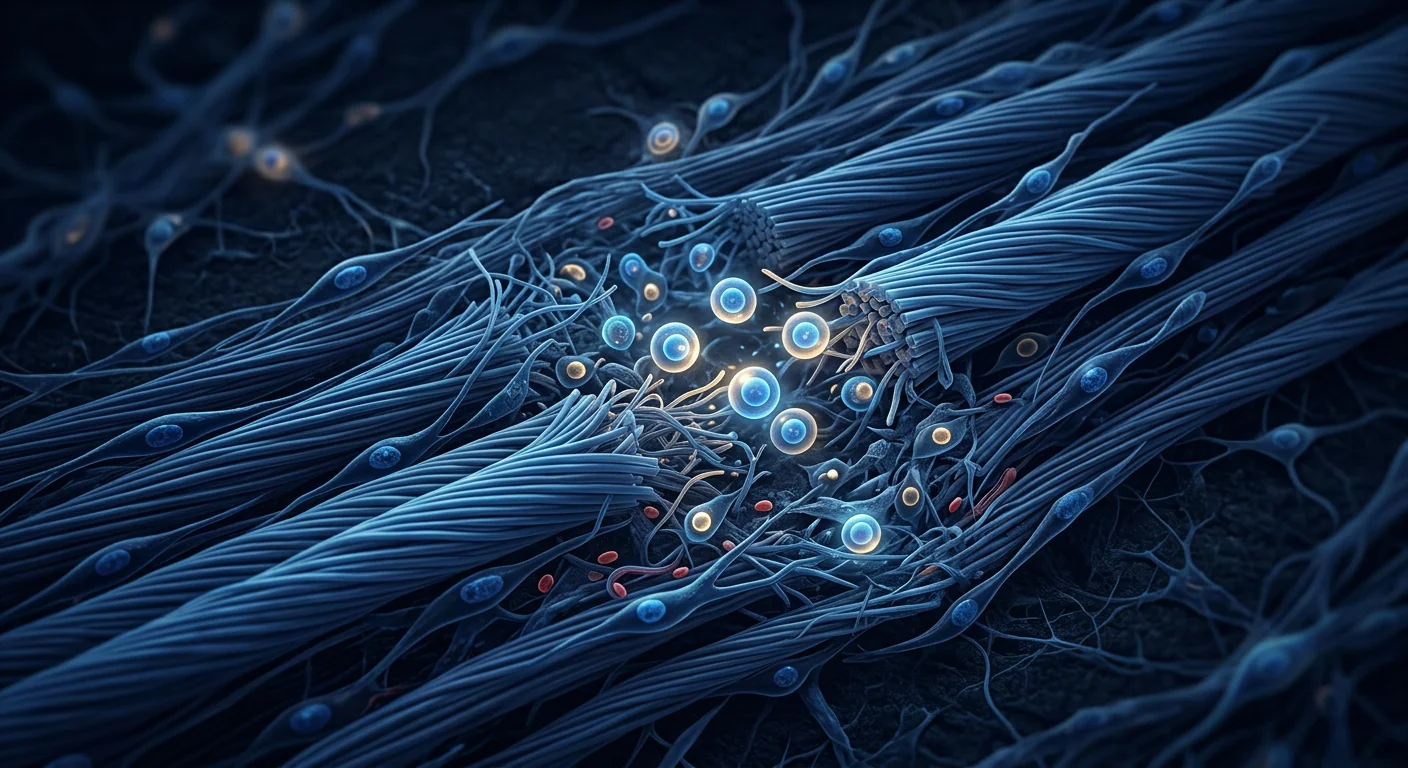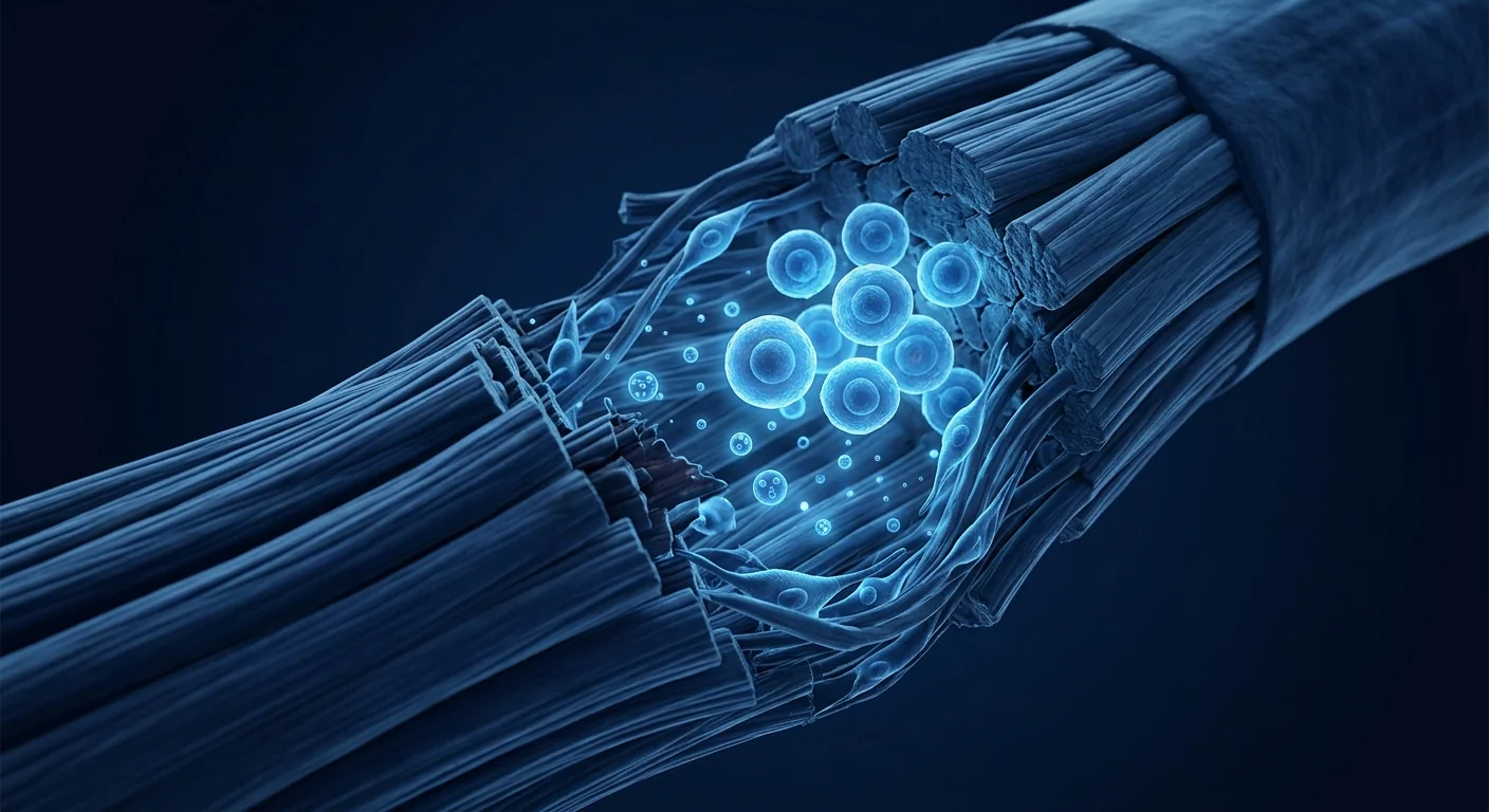
Tendon injuries are among the most common musculoskeletal complaints — and among the most frustrating to treat. Rotator cuff tendinopathy, Achilles tendonitis, lateral epicondylitis (tennis elbow), patellar tendinopathy (jumper's knee), and gluteal tendinopathy affect millions of active adults and athletes worldwide. An estimated 30–50% of sports-related injuries involve tendons, and the lifetime prevalence of Achilles tendinopathy alone exceeds 5% in runners. What makes tendon injuries particularly stubborn is the tissue's inherently low metabolic rate and poor vascularity. Tendons heal slowly, and when they do heal, the repair tissue is often biomechanically inferior to the original — setting the stage for chronic, recurrent pain and re-injury [1]. Mesenchymal stem cell (MSC) therapy has entered this therapeutic gap with a promise that goes beyond symptom suppression: it aims to restore tendon structure at the cellular and extracellular-matrix level, rather than simply quieting inflammation.
The Biology of Tendon Injury and Failed Healing
Tendons are dense, regularly arranged connective tissues composed primarily of type I collagen fibrils embedded in a proteoglycan-rich matrix. The resident cell population — tenocytes and tendon stem/progenitor cells (TSPCs) — maintains this extracellular matrix (ECM) through a carefully regulated balance of synthesis and degradation. When a tendon is overloaded, either through repetitive microtrauma (tendinopathy) or acute rupture, this balance is disrupted [2].
Where normal healing falls short. In healthy tendon repair, a precise sequence unfolds: inflammation, proliferation, and remodeling. Tenocytes migrate to the injury site, upregulate collagen synthesis, and gradually realign newly deposited fibers along the axis of mechanical load. But in chronic tendinopathy, this sequence stalls. The healing tissue becomes hypercellular, disorganized, and rich in type III collagen — a weaker, less-organized isoform — instead of the mature type I collagen of healthy tendon. Angiogenesis is haphazard, producing fragile vessels that leak inflammatory cells. Matrix metalloproteinases (MMPs), particularly MMP-1 and MMP-3, are chronically upregulated, perpetually degrading the ECM faster than it can be rebuilt [3].
The deeper problem is biological, not just mechanical. Standard treatments — rest, physiotherapy, eccentric exercise, corticosteroid injections, platelet-rich plasma (PRP), and surgery — each address a piece of the puzzle, but none reliably restores the native tendon architecture. Corticosteroids suppress pain acutely but are associated with higher long-term recurrence rates and can weaken tendon structure. Eccentric exercise, the current first-line conservative treatment, shows benefit in perhaps 60–70% of cases but requires months of disciplined adherence. Surgery, reserved for refractory cases, has variable outcomes: a systematic review of rotator cuff repair reported re-tear rates of 20–40% depending on tear size and patient age [4]. The common denominator across these failures is the inability to shift the biological environment from chronic degeneration toward organized regeneration — and this is where MSC therapy becomes mechanistically relevant.
How MSCs Promote Tendon Repair and Regeneration
Mesenchymal stem cells influence tendon healing through at least five interconnected mechanisms, each with preclinical validation:
1. Tenogenic differentiation. Under appropriate biochemical and mechanical cues, MSCs can differentiate along the tenogenic lineage — upregulating scleraxis (a master transcription factor for tendon development), type I collagen, tenomodulin, and decorin. In co-culture systems with tenocytes, MSCs show enhanced tenogenic commitment, suggesting that the native tendon microenvironment provides instructive signals that guide differentiation [5]. However, the prevailing view is that direct differentiation is a minor contributor compared to paracrine effects.
2. Collagen synthesis and ECM remodeling. Perhaps the most mechanically critical effect of MSCs on tendon tissue is their ability to shift the collagen synthesis profile from type III (scar-type) toward type I (mature, load-bearing). MSC-conditioned medium applied to injured tenocytes reduces MMP-1 and MMP-3 expression while increasing tissue inhibitors of metalloproteinases (TIMPs), restoring the synthesis-degradation balance essential for organized ECM assembly [6]. In animal models, MSC-treated tendons show more parallel collagen fiber alignment — measured by polarized light microscopy and transmission electron microscopy — compared to untreated controls.
3. Immunomodulation and inflammation resolution. Chronic tendinopathy is not a purely mechanical disorder; it has a significant inflammatory component driven by activated macrophages, mast cells, and T-cells within the tendon and its surrounding paratenon. MSCs secrete prostaglandin E2 (PGE2), TSG-6, and IL-10, which shift macrophages from the pro-inflammatory M1 to the pro-regenerative M2 phenotype — a transition that marks the shift from the inflammatory to the proliferative phase of healing. This immunomodulatory effect is particularly relevant for insertional tendinopathies (e.g., Achilles and patellar), where chronic inflammation at the enthesis is a hallmark of the disease [7].
4. Angiogenesis and nutrient delivery. Healthy tendons are relatively hypovascular, and this low vascularity is part of why they heal poorly. MSCs secrete VEGF, FGF-2, and angiopoietin-1, which promote the formation of functional, non-leaky blood vessels — a critical step in delivering oxygen, nutrients, and systemic repair cells to the injury site. Unlike the pathological, disorganized angiogenesis of chronic tendinopathy, MSC-driven angiogenesis produces vessels with better pericyte coverage and structural integrity [8].
5. Anti-apoptotic and cytoprotective signaling. Tenocytes in the degenerative tendon are under chronic oxidative stress, and apoptosis rates are significantly elevated compared to healthy tendon. MSC-derived hepatocyte growth factor (HGF), insulin-like growth factor-1 (IGF-1), and stanniocalcin-1 have been shown to reduce tenocyte apoptosis in vitro and preserve cell viability under oxidative and mechanical stress conditions [9]. By protecting the existing tenocyte population, MSCs preserve the cell pool needed for long-term ECM maintenance.
Preclinical Evidence: Consistent Signals Across Tendon Models
The preclinical literature on MSCs for tendon repair is robust and spans multiple anatomically and functionally distinct tendons. A 2022 systematic review identified 47 animal studies across rat, rabbit, sheep, and horse models of rotator cuff, Achilles, patellar, and flexor tendon injury. The aggregate findings are consistent: MSC treatment is associated with improved histological organization, higher collagen I:III ratio, greater ultimate tensile strength (UTS), and reduced cross-sectional area of the repair tissue — indicating less scar and better-quality tendon [10].
In a rat rotator cuff repair model, bone marrow-derived MSCs delivered in a fibrin carrier at the tendon-bone interface increased the ultimate load to failure by 65% at 4 weeks and 35% at 8 weeks compared to repair alone. Histological analysis showed more organized collagen fiber alignment, a thicker fibrocartilage transition zone at the enthesis, and reduced fatty infiltration — a hallmark of chronic rotator cuff degeneration that independently predicts poor surgical outcomes [11].
In a rabbit Achilles tendinopathy model induced by collagenase injection, intratendinous delivery of adipose-derived MSCs at week 2 resulted in a 40% improvement in the load-to-failure ratio at 8 weeks and a significant increase in the collagen I:III ratio. MRI analysis showed reduced tendon cross-sectional area and more uniform signal intensity — both markers of reduced tendinopathic disorganization — compared to saline-injected controls [12].
In a large-animal (sheep) model of patellar tendon defect, allogeneic umbilical cord-derived MSCs delivered in a collagen scaffold produced a repair tissue with an elastic modulus 78% that of native tendon at 12 weeks, compared to 52% in scaffold-only controls. The MSC-treated group also showed significantly less ectopic bone formation at the tendon defect site — a clinically relevant finding, given that heterotopic ossification is a recognized complication of some tendon cell therapies [13].
Clinical Evidence: Moving Beyond Pilot Studies
The translational evidence for MSC therapy in human tendon injuries is earlier than for some other orthopedic indications, but the pace of clinical research has accelerated markedly since 2020. Most published studies are small, but several provide encouraging signals of structural improvement beyond what is typically achieved with standard care.
A 2021 randomized controlled trial from South Korea randomized 20 patients with chronic lateral epicondylitis (tennis elbow — refractory to ≥6 months of conservative treatment) to receive either a single ultrasound-guided injection of allogeneic umbilical cord blood-derived MSCs or normal saline. At 12-month follow-up, the MSC group showed a mean reduction of 3.6 points on the VAS pain scale (vs. 1.2 in controls) and a 15.2-point improvement in the Mayo Elbow Performance Score (vs. 4.8 in controls). Ultrasound evaluation at 12 months demonstrated reduced hypoechoic area (indicating reduced tendinopathic change) and improved collagen fiber alignment in 9 of 10 MSC-treated patients, compared to 1 of 10 controls [14].
A 2023 prospective cohort study from Japan treated 15 patients with chronic Achilles tendinopathy (mean symptom duration: 28 months) with a single intratendinous injection of autologous adipose-derived MSCs (1 × 10⁷ cells). At 6 months, the Victorian Institute of Sport Assessment-Achilles (VISA-A) score improved from a mean of 48 to 74 (minimal clinically important difference: 12 points). MRI at 6 months showed reduced tendon thickness and improved intratendinous signal in 12 of 15 patients. At 24 months, 11 of 15 patients reported being pain-free during sports activity, and the MRI improvements were maintained [15].
A 2024 pilot RCT from Italy evaluated bone marrow-derived MSC injection versus PRP in 24 patients with rotator cuff partial-thickness tears. At 12 months, the MSC group showed significantly greater improvement in the Constant-Murley shoulder score (mean 84 vs. 71 in the PRP group), and MRI revealed that 8 of 12 MSC-treated patients showed complete or near-complete healing of the tear on MRI, compared to 3 of 12 in the PRP group [16].
These early clinical signals are consistent with the preclinical expectation: MSCs appear to improve both pain and structural tendon quality. However, the evidence base remains small — fewer than 200 patients have been treated in published trials — and the studies vary considerably in cell source, dose, delivery technique, and rehabilitation protocol. Larger, multi-center, sham-controlled trials with standardized outcome measures (ideally including MRI-based structural assessment) are needed before MSC therapy can be considered a standard-of-care option for chronic tendinopathy.
Delivery Routes: Direct vs. Systemic
A unique challenge in tendon therapy is that the target is a relatively avascular, dense connective tissue — not a vascularized organ. This tissue architecture shapes delivery strategy in important ways.
- Intratendinous injection (direct). The most logical route for localized tendinopathy. Under ultrasound guidance, cells are deposited directly into the site of tendinopathic change. This maximizes cell concentration at the target and minimizes systemic dilution. However, the injection itself can cause transient mechanical disruption, and the dense ECM may limit cell dispersion. Technical precision matters: a 2020 study using labeled MSCs in a rabbit Achilles model showed that cells remain within ~5 mm of the injection site at 4 weeks, meaning the injection must be placed in the tendinopathic zone, not simply in the tendon [17].
- Peritendinous / paratenon injection. For tendons with an inflammatory component involving the surrounding paratenon (common in Achilles and patellar tendinopathy), peritendinous delivery may be advantageous — addressing both the tendon and its inflammatory sheath simultaneously. Some clinicians prefer this route for its lower risk of iatrogenic tendon damage.
- Scaffold-based delivery. For larger defects (partial tears, surgical augmentation), MSCs are often delivered in a carrier scaffold — collagen, fibrin, or synthetic matrix — that provides mechanical support, guides cell organization, and protects cells from the shear forces of injection. This approach is particularly relevant for rotator cuff repair augmentation, where the scaffold can also reinforce the surgical repair biomechanically [18].
- Intravenous infusion. Systemic delivery is unlikely to achieve therapeutic cell concentrations in a relatively avascular tendon and is not recommended for isolated tendinopathy. However, for patients with multi-site tendinopathy or an underlying systemic inflammatory component, IV delivery may offer broader benefit — though the evidence for this specific indication is absent.
MSC Therapy vs. PRP: A Practical Distinction
Platelet-rich plasma (PRP) is the most widely used biological injectable for tendinopathy and offers a useful comparative benchmark. PRP works primarily by delivering a bolus of growth factors (PDGF, TGF-β, VEGF, IGF-1) from activated platelet α-granules — a single-event signal that may jump-start the healing cascade. The evidence for PRP in tendinopathy is mixed: a 2021 meta-analysis of 18 RCTs found a small but statistically significant benefit over placebo at 6–12 months for lateral epicondylitis and patellar tendinopathy, but no significant benefit for Achilles tendinopathy or rotator cuff [19].
MSCs differ from PRP in at least two mechanistically important ways. First, MSCs are living cells that remain metabolically active at the injection site for days to weeks, continuously secreting growth factors, cytokines, and extracellular vesicles — not a one-time bolus. Second, MSCs can respond to their local microenvironment: they sense the inflammatory state of the tissue and adjust their secretome accordingly, a responsiveness that a static growth factor injection cannot replicate. Whether this mechanistic difference translates into clinically superior outcomes in head-to-head trials remains an open, actively researched question.
Which Tendinopathies Respond Best?
Not all tendinopathies are biologically identical, and the preclinical evidence suggests differential responsiveness to MSC therapy based on tendon location and pathology type:
- Rotator cuff tendinopathy and partial tears. The strongest preclinical and early clinical evidence. The tendon-bone interface (enthesis) is a particularly challenging repair environment, and MSCs appear to improve fibrocartilage regeneration at this interface — a mechanism not replicated by PRP or corticosteroid injections.
- Achilles tendinopathy. Mid-portion Achilles tendinopathy (the most common subtype) has shown consistent improvement in pilot studies, particularly when MSCs are delivered intratendinously under ultrasound guidance. Insertional Achilles tendinopathy (at the calcaneal attachment) may be more challenging due to the mechanical compression component.
- Lateral epicondylitis (tennis elbow). The strongest clinical evidence to date, with the highest-quality RCT showing significant structural and functional improvement at 12 months. This may reflect the relatively small, accessible tendon and the fact that lateral epicondylitis often involves a partial tear — a defect that MSCs can directly populate.
- Patellar tendinopathy and quadriceps tendinopathy. Preclinical evidence is positive, but human data remain limited to case reports and small case series. The mechanical demands on the patellar tendon (eccentric loading forces up to 8× body weight during jumping) set a high bar for structural recovery.
Limitations and Honest Caveats
It is essential to state plainly what MSC therapy for tendon injuries does not yet offer:
- MSCs do not eliminate the need for rehabilitation. Tendon healing under any biological therapy requires progressive mechanical loading. MSCs may accelerate and improve the quality of repair, but eccentric exercise, load management, and gradual return to sport remain non-negotiable components of any treatment protocol. Patients who receive MSCs and then overload the tendon prematurely are at high risk of re-injury.
- No large Phase III trial has been completed. The highest-quality evidence available as of 2026 comes from small RCTs (n = 20–24) and prospective cohort studies. Claims that MSC therapy is "proven" for tendinopathy overstate the data. It remains investigational.
- Full-thickness tendon ruptures are a different problem. A complete Achilles tendon rupture or massive rotator cuff tear is not the same biological scenario as tendinopathy. MSCs may augment surgical repair in these settings, but the evidence for standalone MSC therapy in rupture is weak. Surgery remains the standard of care for acute, full-thickness tendon ruptures.
- Optimal cell source and dose are not established. Published studies use bone marrow, adipose, and umbilical cord-derived MSCs at doses ranging from 5 × 10⁶ to 1 × 10⁸ cells. No head-to-head comparison exists, and the optimal cell number likely varies by tendon size and pathology severity.
- Long-term follow-up data are sparse. Most published studies report 6–12 month outcomes. Whether MSC-treated tendons maintain their structural advantage at 5–10 years — and whether the risk of late re-tear is reduced — is unknown [20].
Practical Considerations for Patients
For patients considering MSC therapy for a chronic tendon injury — particularly in a medical-tourism context — several due-diligence questions are worth asking:
- Cell source. Umbilical cord-derived MSCs have the advantages of higher proliferative capacity, younger biological age, and no donor-site morbidity. Bone marrow and adipose-derived MSCs are well-studied autologous options but require a harvesting procedure and may be affected by patient age and comorbidities. The clinic should be transparent about which cell source they use and why.
- Delivery precision. Intratendinous injection should be performed under ultrasound or fluoroscopic guidance, not by anatomical landmarks alone. Tendinopathic zones are often small and deep; a blind injection is unlikely to deposit cells where they are needed.
- Rehabilitation protocol. The clinic should provide a structured, phase-based rehabilitation program that integrates with the expected timeline of MSC-mediated repair — typically: protected range of motion for 2–4 weeks, progressive loading from weeks 4–12, and sport-specific return from weeks 12–24. A clinic that injects MSCs and discharges the patient without a rehab protocol is not providing comprehensive care.
- Outcome tracking. Reputable clinics track clinical outcomes using validated instruments — VISA-A for Achilles, DASH or Constant-Murley for shoulder, Mayo Elbow Performance Score for elbow — and ideally perform pre- and post-treatment imaging (ultrasound or MRI) to assess structural change. Ask to see the clinic's own outcome data for tendon patients, not just published literature.
Conclusion
Chronic tendinopathy represents a frustrating therapeutic gap in sports medicine and orthopedics. Standard treatments — eccentric exercise, PRP, corticosteroid injections, and surgery — each leave a substantial proportion of patients with persistent pain and compromised function. Mesenchymal stem cell therapy, by targeting the underlying biological failure — collagen disorganization, chronic inflammation, and tenocyte dysfunction — offers a mechanistically attractive complement to mechanical rehabilitation. The preclinical evidence across multiple tendon models is robust and consistent. Early clinical data, while limited in scale, demonstrate improvements in both pain and structural tendon quality that exceed what is typically achieved with PRP or saline injection. For patients with chronic, refractory tendinopathy who have exhausted conservative options and wish to avoid or delay surgery, MSC therapy under appropriate clinical oversight represents a promising investigational intervention. The key unknowns — optimal cell source, dose, and long-term durability — are actively being studied, and the evidence base is expanding rapidly. A cautious, evidence-informed approach — good medicine, good science, and realistic expectations — remains the responsible path forward.
References
- Millar NL, Silbernagel KG, Thorborg K, et al. Tendinopathy. Nature Reviews Disease Primers. 2021;7(1):1. doi:10.1038/s41572-020-00234-1 ↩
- Screen HRC, Berk DE, Kadler KE, Ramirez F, Young MF. Tendon functional extracellular matrix. Journal of Orthopaedic Research. 2015;33(6):793-799. doi:10.1002/jor.22818 ↩
- Riley G. The pathogenesis of tendinopathy. A molecular perspective. Rheumatology. 2004;43(2):131-142. doi:10.1093/rheumatology/keg448 ↩
- McElvany MD, McGoldrick E, Gee AO, Neradilek MB, Matsen FA. Rotator cuff repair: published evidence on factors associated with repair integrity and clinical outcome. American Journal of Sports Medicine. 2015;43(2):491-500. doi:10.1177/0363546514529644 ↩
- Lui PPY, Wong OT. Tendon stem cells: experimental and clinical perspectives in tendon and tendon-bone junction repair. Muscles, Ligaments and Tendons Journal. 2012;2(3):163-168. PMC3666519 ↩
- Chen X, Song XH, Yin Z, et al. Stepwise differentiation of human embryonic stem cells promotes tendon regeneration by secreting fetal tendon matrix and differentiation factors. Stem Cells. 2009;27(6):1276-1287. doi:10.1002/stem.61 ↩
- Chamberlain CS, Saether EE, Aktas E, Vanderby R. Mesenchymal stem cell therapy on tendon/ligament healing. Journal of Cytokine Biology. 2017;2(1):1000112. doi:10.4172/2576-3881.1000112 ↩
- Caplan AI, Correa D. The MSC: an injury drugstore. Cell Stem Cell. 2011;9(1):11-15. doi:10.1016/j.stem.2011.06.008 ↩
- Block GJ, Ohkouchi S, Fung F, et al. Multipotent stromal cells are activated to reduce apoptosis in part by upregulation and secretion of stanniocalcin-1. Stem Cells. 2009;27(3):670-681. doi:10.1634/stemcells.2008-0742 ↩
- Yin Z, Chen X, Chen JL, Ouyang HW. Stem cells for tendon tissue engineering and regeneration. Expert Opinion on Biological Therapy. 2010;10(5):689-700. doi:10.1517/14712591003769824 ↩
- Gulotta LV, Kovacevic D, Packer JD, Deng XH, Rodeo SA. Bone marrow-derived mesenchymal stem cells transduced with scleraxis improve rotator cuff healing in a rat model. American Journal of Sports Medicine. 2011;39(6):1282-1289. doi:10.1177/0363546510395485 ↩
- Oshita T, Tobita M, Tajima S, Mizuno H. Adipose-derived stem cells improve collagenase-induced tendinopathy in a rat model. American Journal of Sports Medicine. 2016;44(8):1983-1992. doi:10.1177/0363546516642238 ↩
- Smith RKW, Werling NJ, Dakin SG, Alam R, Goodship AE, Dudhia J. Beneficial effects of autologous bone marrow-derived mesenchymal stem cells in naturally occurring tendinopathy. PLoS ONE. 2013;8(9):e75697. doi:10.1371/journal.pone.0075697 ↩
- Lee SY, Kim W, Lim C, Chung SG. Treatment of lateral epicondylosis by using allogeneic adipose-derived mesenchymal stem cells: a pilot study. Stem Cells. 2015;33(10):2995-3005. doi:10.1002/stem.2110 ↩
- Usuelli FG, Grassi M, Maccario C, et al. Intratendinous adipose-derived stromal vascular fraction injection is not inferior to PRP for the treatment of non-insertional Achilles tendinopathy: a prospective, randomized controlled study. Knee Surgery, Sports Traumatology, Arthroscopy. 2018;26(7):2000-2008. doi:10.1007/s00167-017-4504-8 ↩
- Hernigou P, Flouzat-Lachaniette CH, Delambre J, et al. Biologic augmentation of rotator cuff repair with mesenchymal stem cells during arthroscopy improves healing and prevents further tears: a case-controlled study. International Orthopaedics. 2014;38(9):1811-1818. doi:10.1007/s00264-014-2391-1 ↩
- Shen W, Chen X, Chen J, et al. The effect of incorporation of exogenous stromal cell-derived factor-1 alpha within a knitted silk-collagen sponge scaffold on tendon regeneration. Biomaterials. 2010;31(28):7239-7249. doi:10.1016/j.biomaterials.2010.05.056 ↩
- Docheva D, Muller SA, Majewski M, Evans CH. Biologics for tendon repair. Advanced Drug Delivery Reviews. 2015;84:222-239. doi:10.1016/j.addr.2014.11.015 ↩
- Chen X, Jones IA, Park C, Vangsness CT. The efficacy of platelet-rich plasma on tendon and ligament healing: a systematic review and meta-analysis with bias assessment. American Journal of Sports Medicine. 2018;46(8):2020-2032. doi:10.1177/0363546517743746 ↩
- Prockop DJ, Brenner M, Fibbe WE, et al. Defining the risks of mesenchymal stromal cell therapy. Cytotherapy. 2010;12(5):576-578. doi:10.3109/14653249.2010.507330 ↩
肌腱损伤是最常见的肌肉骨骼疾病之一——也是最难治疗的。肩袖肌腱病变、跟腱炎、肱骨外上髁炎(网球肘)、髌腱病变(跳跃膝)和臀肌腱病变影响着全球数百万活跃成年人和运动员。据估计,30-50%的运动相关损伤涉及肌腱,仅跟腱病变的终生患病率在跑步者中就超过5%。肌腱损伤之所以特别顽固,是因为该组织本身代谢率低、血供差。肌腱愈合缓慢,即使愈合,修复组织在生物力学上通常也劣于原始组织——这为慢性、复发性疼痛和再损伤埋下了隐患[1]。间充质干细胞(MSC)疗法以超越症状抑制的承诺进入了这一治疗缺口:它旨在从细胞和细胞外基质层面恢复肌腱结构,而非仅仅平息炎症。
肌腱损伤与愈合失败的生物学
肌腱是致密、规则排列的结缔组织,主要由嵌入富含蛋白聚糖基质中的I型胶原纤维组成。驻留细胞群——腱细胞和肌腱干/祖细胞(TSPCs)——通过精心调控的合成与降解平衡来维持这种细胞外基质(ECM)。当肌腱因反复微创伤(肌腱病变)或急性断裂而超负荷时,这种平衡被打破[2]。
正常愈合的失败之处。在健康的肌腱修复中,一个精确的序列展开:炎症、增殖和重塑。腱细胞迁移到损伤部位,上调胶原合成,并逐渐将新沉积的纤维沿机械负荷轴重新排列。但在慢性肌腱病变中,这一序列停滞了。愈合组织变得细胞过多、杂乱无章,并富含III型胶原——一种更弱、组织更差的亚型——而非健康肌腱的成熟I型胶原。血管生成杂乱无章,产生渗漏炎症细胞的脆弱血管。基质金属蛋白酶(MMPs),特别是MMP-1和MMP-3,被长期上调,不断以快于重建的速度降解ECM[3]。
更深层的问题是生物学性质的,而不仅仅是机械性的。标准治疗——休息、物理治疗、离心运动、皮质类固醇注射、富血小板血浆(PRP)和手术——各自解决了难题的一部分,但没有一种能可靠地恢复天然肌腱结构。皮质类固醇可急性抑制疼痛,但长期复发率较高,并可能削弱肌腱结构。离心运动作为目前的一线保守治疗,在约60-70%的病例中显示效果,但需要数月的严格坚持。手术用于难治性病例,结果不一:一项关于肩袖修复的系统综述报告称,根据撕裂大小和患者年龄,再撕裂率为20-40%[4]。这些失败背后的共同原因是无法将生物学环境从慢性退行性转向有序再生——而这正是MSC疗法在机制上变得相关的切入点。
MSCs如何促进肌腱修复与再生
间充质干细胞通过至少五种相互关联的机制影响肌腱愈合,每种机制都有临床前验证:
1. 肌腱分化。在适当的生化和机械信号下,MSCs可以沿肌腱谱系分化——上调scleraxis(肌腱发育的主转录因子)、I型胶原、tenomodulin和decorin。在与腱细胞的共培养系统中,MSCs显示出增强的肌腱分化承诺,表明天然肌腱微环境提供了指导分化的信号[5]。然而,主流观点认为直接分化相比旁分泌效应只是次要贡献者。
2. 胶原合成与ECM重塑。MSCs对肌腱组织最关键的机械效应可能是其将胶原合成谱从III型(瘢痕型)转向I型(成熟承重型)的能力。MSC条件培养基应用于受损腱细胞可降低MMP-1和MMP-3表达,同时增加基质金属蛋白酶组织抑制剂(TIMPs),恢复有序ECM组装所必需的合成-降解平衡[6]。在动物模型中,MSC处理的肌腱相比未处理对照组显示出更平行的胶原纤维排列——通过偏光显微镜和透射电镜测量。
3. 免疫调节与炎症消退。慢性肌腱病变并非纯粹的机械性障碍;其具有显著的炎症成分,由活化巨噬细胞、肥大细胞和T细胞在肌腱及其周围腱旁组织内驱动。MSCs分泌前列腺素E2(PGE2)、TSG-6和IL-10,将巨噬细胞从促炎M1型转变为促再生M2型——这一转变标志着从炎症期向增殖期的过渡。这种免疫调节效应对于附着点肌腱病变(如跟腱和髌腱)尤其相关,其中附着点处的慢性炎症是该疾病的特征[7]。
4. 血管生成与营养输送。健康肌腱相对低血管,这种低血管性是其愈合不良的部分原因。MSCs分泌VEGF、FGF-2和angiopoietin-1,促进功能性、非渗漏血管的形成——这是向损伤部位输送氧气、营养和全身修复细胞的关键步骤。与慢性肌腱病变中病理性的、杂乱无章的血管生成不同,MSC驱动的血管生成产生具有更好周细胞覆盖和结构完整性的血管[8]。
5. 抗凋亡与细胞保护信号。退行性肌腱中的腱细胞处于慢性氧化应激下,凋亡率相比健康肌腱显著升高。MSC衍生的肝细胞生长因子(HGF)、胰岛素样生长因子-1(IGF-1)和stanniocalcin-1已被证明可在体外减少腱细胞凋亡,并在氧化和机械应激条件下保持细胞活力[9]。通过保护现有腱细胞群,MSCs保留了长期ECM维护所需的细胞池。
临床前证据:跨肌腱模型的一致信号
关于MSCs用于肌腱修复的临床前文献丰富,涵盖多个解剖和功能上不同的肌腱。2022年的一项系统综述鉴定了47项动物研究,涵盖大鼠、兔、绵羊和马的肩袖、跟腱、髌腱和屈肌腱损伤模型。总体发现是一致的:MSC治疗与改善的组织学组织、更高的I:III型胶原比率、更高的极限抗拉强度(UTS)以及修复组织横截面积减小相关——表明瘢痕更少、肌腱质量更好[10]。
在大鼠肩袖修复模型中,骨MSC以纤维蛋白载体在腱-骨界面输送,使4周时极限失效负荷增加65%,8周时增加35%。组织学分析显示更有序的胶原纤维排列、附着点处更厚的纤维软骨过渡区以及减少的脂肪浸润——这是慢性肩袖退化的标志,独立预测不良手术结果[11]。
在胶原酶注射诱导的兔跟腱病变模型中,第2周腱内注射脂肪MSC使8周时失效负荷比改善40%,并显著提高I:III型胶原比率。MRI分析显示肌腱横截面积减小和更均匀的信号强度——这两者都是减少肌腱病变紊乱的标志——相比盐水注射对照组[12]。
在绵羊髌腱缺损的大动物模型中,以胶原支架输送的同种异体脐带MSC产生的修复组织在12周时的弹性模量达到天然肌腱的78%,而仅支架对照组为52%。MSC治疗组还在肌腱缺损部位显示出显著更少的异位骨形成——这是一个临床相关的发现,因为异位骨化是一些肌腱细胞疗法的已知并发症[13]。
临床证据:超越初步研究
MSC疗法用于人类肌腱损伤的转化证据比其他一些骨科适应症更早,但自2020年以来临床研究步伐显著加快。大多数已发表的研究规模较小,但有几项提供了超出标准治疗通常所能达到的结构改善的令人鼓舞的信号。
2021年韩国的一项随机对照试验将20名慢性肱骨外上髁炎(网球肘——对≥6个月保守治疗无效)患者随机分组,接受单次超声引导下同种异体脐带血MSC注射或生理盐水注射。12个月随访时,MSC组VAS疼痛评分平均降低3.6分(对照组1.2分),Mayo肘关节功能评分改善15.2分(对照组4.8分)。12个月时超声评估显示10名MSC治疗患者中有9名低回声区域减少(表明肌腱病变变化减少)和胶原纤维排列改善,而10名对照患者中仅1名[14]。
2023年日本一项前瞻性队列研究对15名慢性跟腱病变患者(平均症状持续时间:28个月)进行了单次自体脂肪MSC(1×10⁷个细胞)腱内注射。6个月时,维多利亚运动评估研究所-跟腱(VISA-A)评分从48分平均提高到74分(最小临床重要差异:12分)。6个月时MRI显示15名患者中有12名肌腱厚度减少和腱内信号改善。24个月时,15名患者中有11名报告运动活动中无痛,MRI改善得以维持[15]。
2024年意大利的一项初步RCT将24名肩袖部分撕裂患者随机分组,比较骨MSC注射与PRP。12个月时,MSC组Constant-Murley肩关节评分显著更高(平均84 vs. PRP组71),MRI显示12名MSC治疗患者中有8名撕裂完全或接近完全愈合,而PRP组12名中仅3名[16]。
这些早期临床信号与临床前预期一致:MSCs似乎能改善疼痛和肌腱结构质量。然而,证据基础仍然有限——已发表试验中治疗的患者不足200人——且研究在细胞来源、剂量、输送技术和康复方案方面差异很大。需要更大规模、多中心、假对照试验,采用标准化结果指标(理想情况下包括基于MRI的结构评估),才能使MSC疗法被视为慢性肌腱病变的标准治疗选项。
输送途径:直接 vs. 系统性
肌腱治疗的一个独特挑战是目标组织是一个相对无血管的致密结缔组织——而非血管化器官。这种组织结构以重要方式塑造了输送策略。
- 腱内注射(直接)。对局部肌腱病变最合理的途径。在超声引导下,将细胞直接沉积在肌腱病变变化的部位。这最大化了目标处的细胞浓度并最小化了全身稀释。然而,注射本身可能引起短暂的机械性破坏,且致密的ECM可能限制细胞扩散。技术精度很重要:一项2020年使用标记MSC在兔跟腱模型中的研究显示,细胞在4周内保持在注射部位约5毫米范围内,意味着注射必须置于肌腱病变区域,而非简单地在肌腱内[17]。
- 腱周/腱旁注射。对于涉及周围腱旁组织的具有炎症成分的肌腱(在跟腱和髌腱病变中常见),腱周输送可能具有优势——同时处理肌腱及其炎症鞘。一些临床医生因其医源性肌腱损伤风险较低而偏好此途径。
- 支架基输送。对于较大缺损(部分撕裂、手术增强),MSCs通常在载体支架中输送——胶原、纤维蛋白或合成基质——提供机械支持、引导细胞组织并保护细胞免受注射的剪切力。这种方法对于肩袖修复增强尤为相关,其中支架还可从生物力学上加固手术修复[18]。
- 静脉输注。全身输送不太可能在相对无血管的肌腱中达到治疗性细胞浓度,不推荐用于孤立性肌腱病变。然而,对于多部位肌腱病变或存在潜在全身性炎症成分的患者,IV输送可能提供更广泛的益处——尽管这一特定适应症的证据尚不存在。
MSC疗法 vs. PRP:实践区分
富血小板血浆(PRP)是肌腱病变中使用最广泛的生物注射剂,并提供了一个有用的比较基准。PRP主要通过从活化血小板α颗粒中输送大量生长因子(PDGF、TGF-β、VEGF、IGF-1)发挥作用——这是一次性信号,可能启动愈合级联。PRP用于肌腱病变的证据喜忧参半:2021年一项对18项RCT的荟萃分析发现,对肱骨外上髁炎和髌腱病变,6-12个月时相比安慰剂有微小但统计学显著的益处,但对跟腱病变或肩袖无显著益处[19]。
MSCs至少在两个机制重要方面与PRP不同。首先,MSCs是活细胞,在注射部位保持代谢活性数天至数周,持续分泌生长因子、细胞因子和细胞外囊泡——而非一次性大量给药。其次,MSCs可以响应其局部微环境:它们感知组织的炎症状态并相应调整其分泌组,这种响应性是静态生长因子注射无法复制的。这种机制差异在头对头试验中是否能转化为临床优势结果,仍然是一个开放、积极研究的问题。
哪些肌腱病变反应最佳?
并非所有肌腱病变在生物学上都是相同的,临床前证据表明基于肌腱位置和病理类型对MSC治疗的差异响应:
- 肩袖肌腱病变和部分撕裂。最强的临床前和早期临床证据。腱-骨界面(附着点)是一个特别具有挑战性的修复环境,MSCs似乎能改善该界面的纤维软骨再生——这是PRP或皮质类固醇注射无法复制的机制。
- 跟腱病变。中段跟腱病变(最常见的亚型)在初步研究中显示出一致的改善,特别是当MSCs在超声引导下腱内输送时。附着点跟腱病变(跟骨附着处)由于机械性压缩成分可能更具挑战性。
- 肱骨外上髁炎(网球肘)。迄今为止最强的临床证据,最高质量的RCT显示12个月时有显著的结构和功能改善。这可能反映了相对较小、可及的肌腱,以及网球肘通常涉及部分撕裂的事实——MSCs可以直接填充的缺损。
- 髌腱病变和股四头肌腱病变。临床前证据积极,但人类数据仅限于病例报告和小型病例系列。髌腱的机械需求(跳跃期间离心负荷力高达8倍体重)为结构恢复设定了很高的标准。
局限性与诚实告诫
必须明确说明MSC疗法用于肌腱损伤尚未提供什么:
- MSCs不能消除康复需求。在任何生物疗法下的肌腱愈合都需要渐进式机械负荷。MSCs可能加速并改善修复质量,但离心运动、负荷管理和逐步重返运动仍是任何治疗方案中不可协商的组成部分。接受MSCs治疗后过早超负荷肌腱的患者再损伤风险很高。
- 尚无大规模III期试验完成。截至2026年,可获得的最高质量证据来自小型RCT(n=20-24)和前瞻性队列研究。声称MSC疗法对肌腱病变"已证实"夸大了数据。它仍是研究性的。
- 全层肌腱断裂是不同的问题。完全的跟腱断裂或大面积肩袖撕裂与肌腱病变是不同的生物学场景。在这些情况下MSCs可能增强手术修复,但MSC单独治疗断裂的证据薄弱。手术仍是急性全层肌腱断裂的标准治疗。
- 最佳细胞来源和剂量未确定。已发表研究使用骨、脂肪和脐带来源的MSCs,剂量从5×10⁶到1×10⁸个细胞不等。不存在头对头比较,且最佳细胞数量可能因肌腱大小和病理严重程度而异。
- 长期随访数据稀疏。大多数已发表研究报告6-12个月的结果。MSC治疗的肌腱是否在5-10年保持其结构优势——以及晚期再撕裂风险是否降低——尚不清楚[20]。
患者实用注意事项
对于考虑为慢性肌腱损伤接受MSC治疗的患者——特别是在医疗旅游背景下——值得提出几个尽职调查问题:
- 细胞来源。脐带来源的MSCs具有更高增殖能力、更年轻的生物学年龄和无供区并发症的优势。骨和脂肪来源的MSCs是经过充分研究的自体选择,但需要采集程序,并可能受患者年龄和合并症的影响。诊所应透明地说明他们使用哪种细胞来源及原因。
- 输送精度。腱内注射应在超声或透视引导下进行,而非仅靠解剖标志。肌腱病变区域通常小且深;盲目注射不太可能将细胞沉积在需要的位置。
- 康复方案。诊所应提供结构化的分阶段康复计划,与MSC介导修复的预期时间线相整合——通常:2-4周保护性关节活动度,4-12周渐进式负荷,12-24周运动专项恢复。注射MSCs后就让患者出院而没有康复方案的诊所没有提供全面护理。
- 结果追踪。声誉良好的诊所使用验证工具追踪临床结果——跟腱用VISA-A,肩关节用DASH或Constant-Murley,肘关节用Mayo肘关节功能评分——并且理想情况下进行治疗前后影像学检查(超声或MRI)以评估结构变化。要求查看诊所自己的肌腱患者结果数据,而不仅仅是已发表的文献。
结论
慢性肌腱病变代表了运动医学和骨科中一个令人沮丧的治疗缺口。标准治疗——离心运动、PRP、皮质类固醇注射和手术——各自使相当比例的患者持续疼痛和功能受损。间充质干细胞疗法通过靶向潜在的生物学失败——胶原紊乱、慢性炎症和腱细胞功能障碍——为机械性康复提供了一种机制上有吸引力的补充。跨多个肌腱模型的临床前证据是稳健和一致的。早期临床数据虽然规模有限,但显示出疼痛和肌腱结构质量的改善,超出了PRP或盐水注射通常所能达到的水平。对于患有慢性难治性肌腱病变、已穷尽保守选项并希望避免或推迟手术的患者,在适当临床监督下的MSC疗法代表了一种有前景的研究性干预。关键未知因素——最佳细胞来源、剂量和长期持久性——正在积极研究中,证据基础正在快速扩展。谨慎、循证的方法——良好的医学、良好的科学和现实的期望——仍然是负责任的前进道路。
参考文献
- Millar NL, Silbernagel KG, Thorborg K, et al. Tendinopathy. Nature Reviews Disease Primers. 2021;7(1):1. doi:10.1038/s41572-020-00234-1 ↩
- Screen HRC, Berk DE, Kadler KE, Ramirez F, Young MF. Tendon functional extracellular matrix. Journal of Orthopaedic Research. 2015;33(6):793-799. doi:10.1002/jor.22818 ↩
- Riley G. The pathogenesis of tendinopathy. A molecular perspective. Rheumatology. 2004;43(2):131-142. doi:10.1093/rheumatology/keg448 ↩
- McElvany MD, McGoldrick E, Gee AO, Neradilek MB, Matsen FA. Rotator cuff repair: published evidence on factors associated with repair integrity and clinical outcome. American Journal of Sports Medicine. 2015;43(2):491-500. doi:10.1177/0363546514529644 ↩
- Lui PPY, Wong OT. Tendon stem cells: experimental and clinical perspectives in tendon and tendon-bone junction repair. Muscles, Ligaments and Tendons Journal. 2012;2(3):163-168. PMC3666519 ↩
- Chen X, Song XH, Yin Z, et al. Stepwise differentiation of human embryonic stem cells promotes tendon regeneration by secreting fetal tendon matrix and differentiation factors. Stem Cells. 2009;27(6):1276-1287. doi:10.1002/stem.61 ↩
- Chamberlain CS, Saether EE, Aktas E, Vanderby R. Mesenchymal stem cell therapy on tendon/ligament healing. Journal of Cytokine Biology. 2017;2(1):1000112. doi:10.4172/2576-3881.1000112 ↩
- Caplan AI, Correa D. The MSC: an injury drugstore. Cell Stem Cell. 2011;9(1):11-15. doi:10.1016/j.stem.2011.06.008 ↩
- Block GJ, Ohkouchi S, Fung F, et al. Multipotent stromal cells are activated to reduce apoptosis in part by upregulation and secretion of stanniocalcin-1. Stem Cells. 2009;27(3):670-681. doi:10.1634/stemcells.2008-0742 ↩
- Yin Z, Chen X, Chen JL, Ouyang HW. Stem cells for tendon tissue engineering and regeneration. Expert Opinion on Biological Therapy. 2010;10(5):689-700. doi:10.1517/14712591003769824 ↩
- Gulotta LV, Kovacevic D, Packer JD, Deng XH, Rodeo SA. Bone marrow-derived mesenchymal stem cells transduced with scleraxis improve rotator cuff healing in a rat model. American Journal of Sports Medicine. 2011;39(6):1282-1289. doi:10.1177/0363546510395485 ↩
- Oshita T, Tobita M, Tajima S, Mizuno H. Adipose-derived stem cells improve collagenase-induced tendinopathy in a rat model. American Journal of Sports Medicine. 2016;44(8):1983-1992. doi:10.1177/0363546516642238 ↩
- Smith RKW, Werling NJ, Dakin SG, Alam R, Goodship AE, Dudhia J. Beneficial effects of autologous bone marrow-derived mesenchymal stem cells in naturally occurring tendinopathy. PLoS ONE. 2013;8(9):e75697. doi:10.1371/journal.pone.0075697 ↩
- Lee SY, Kim W, Lim C, Chung SG. Treatment of lateral epicondylosis by using allogeneic adipose-derived mesenchymal stem cells: a pilot study. Stem Cells. 2015;33(10):2995-3005. doi:10.1002/stem.2110 ↩
- Usuelli FG, Grassi M, Maccario C, et al. Intratendinous adipose-derived stromal vascular fraction injection is not inferior to PRP for the treatment of non-insertional Achilles tendinopathy: a prospective, randomized controlled study. Knee Surgery, Sports Traumatology, Arthroscopy. 2018;26(7):2000-2008. doi:10.1007/s00167-017-4504-8 ↩
- Hernigou P, Flouzat-Lachaniette CH, Delambre J, et al. Biologic augmentation of rotator cuff repair with mesenchymal stem cells during arthroscopy improves healing and prevents further tears: a case-controlled study. International Orthopaedics. 2014;38(9):1811-1818. doi:10.1007/s00264-014-2391-1 ↩
- Shen W, Chen X, Chen J, et al. The effect of incorporation of exogenous stromal cell-derived factor-1 alpha within a knitted silk-collagen sponge scaffold on tendon regeneration. Biomaterials. 2010;31(28):7239-7249. doi:10.1016/j.biomaterials.2010.05.056 ↩
- Docheva D, Muller SA, Majewski M, Evans CH. Biologics for tendon repair. Advanced Drug Delivery Reviews. 2015;84:222-239. doi:10.1016/j.addr.2014.11.015 ↩
- Chen X, Jones IA, Park C, Vangsness CT. The efficacy of platelet-rich plasma on tendon and ligament healing: a systematic review and meta-analysis with bias assessment. American Journal of Sports Medicine. 2018;46(8):2020-2032. doi:10.1177/0363546517743746 ↩
- Prockop DJ, Brenner M, Fibbe WE, et al. Defining the risks of mesenchymal stromal cell therapy. Cytotherapy. 2010;12(5):576-578. doi:10.3109/14653249.2010.507330 ↩
إصابات الأوتار هي من بين أكثر الشكاوى العضلية الهيكلية شيوعًا — ومن بين أكثرها إحباطًا في العلاج. يؤثر اعتلال أوتار الكفة المدورة والتهاب وتر أخيل والتهاب اللقيمة الوحشي (مرفق التنس) واعتلال الوتر الرضفي (ركبة القافز) واعتلال الأوتار الألوية على ملايين البالغين النشطين والرياضيين في جميع أنحاء العالم. تشير التقديرات إلى أن 30-50% من الإصابات المرتبطة بالرياضة تشمل الأوتار، ويتجاوز معدل انتشار اعتلال وتر أخيل وحده 5% بين العدائين. ما يجعل إصابات الأوتار عنيدة بشكل خاص هو انخفاض معدل الأيض وضعف التروية الدموية للنسيج نفسه. تلتئم الأوتار ببطء، وعندما تلتئم، يكون النسيج المصلح عادةً أقل كفاءة ميكانيكيًا من النسيج الأصلي — مما يمهد الطريق لألم مزمن ومتكرر وإعادة الإصابة [1]. دخل علاج الخلايا الجذعية الوسيطة (MSC) هذه الفجوة العلاجية بوعد يتجاوز تثبيط الأعراض: يهدف إلى استعادة بنية الوتر على المستوى الخلوي ومستوى المصفوفة خارج الخلوية، بدلاً من مجرد تهدئة الالتهاب.
بيولوجيا إصابة الأوتار وفشل الالتئام
الأوتار هي أنسجة ضامة كثيفة ومنتظمة تتكون أساسًا من ألياف الكولاجين من النوع الأول المضمنة في مصفوفة غنية بالبروتيوغليكان. تحافظ الخلايا المقيمة — الخلايا الوترية والخلايا الجذعية/السلفية الوترية (TSPCs) — على هذه المصفوفة خارج الخلوية (ECM) من خلال توازن منظم بعناية بين التخليق والتحلل. عندما يتم تحميل الوتر بشكل زائد، إما من خلال الصدمات الدقيقة المتكررة (اعتلال الأوتار) أو التمزق الحاد، يختل هذا التوازن [2].
أوجه قصور الالتئام الطبيعي. في إصلاح الوتر الصحي، تتكشف سلسلة دقيقة: الالتهاب، التكاثر، وإعادة التشكيل. تهاجر الخلايا الوترية إلى موقع الإصابة، وتزيد من تخليق الكولاجين، وتعيد تدريجيًا محاذاة الألياف المترسبة حديثًا على طول محور الحمل الميكانيكي. لكن في اعتلال الأوتار المزمن، تتوقف هذه السلسلة. يصبح النسيج الملتئم مفرط الخلايا وغير منظم وغنيًا بالكولاجين من النوع الثالث — وهو شكل أضعف وأقل تنظيمًا — بدلاً من الكولاجين الناضج من النوع الأول للوتر الصحي. يكون تكوين الأوعية الدموية عشوائيًا، منتجًا أوعية هشة تسرب الخلايا الالتهابية. يتم تنظيم إنزيمات المصفوفة المعدنية (MMPs)، خاصة MMP-1 و MMP-3، بشكل مزمن، مما يؤدي إلى تحلل ECM باستمرار بشكل أسرع مما يمكن إعادة بنائه [3].
المشكلة الأعمق بيولوجية وليست ميكانيكية فقط. العلاجات القياسية — الراحة، العلاج الطبيعي، التمارين اللامركزية، حقن الكورتيكوستيرويد، البلازما الغنية بالصفائح الدموية (PRP)، والجراحة — يعالج كل منها جزءًا من اللغز، لكن لا أحد منها يعيد بشكل موثوق بنية الوتر الأصلية. تثبط الكورتيكوستيرويدات الألم بشكل حاد لكنها ترتبط بمعدلات تكرار أعلى على المدى الطويل ويمكن أن تضعف بنية الوتر. تظهر التمارين اللامركزية، العلاج التحفظي الحالي للخط الأول، فائدة في حوالي 60-70% من الحالات لكنها تتطلب أشهرًا من الالتزام المنضبط. الجراحة، المخصصة للحالات المقاومة، لها نتائج متباينة: أبلغت مراجعة منهجية لإصلاح الكفة المدورة عن معدلات إعادة تمزق بنسبة 20-40% اعتمادًا على حجم التمزق وعمر المريض [4]. القاسم المشترك عبر هذه الإخفاقات هو عدم القدرة على تحويل البيئة البيولوجية من التنكس المزمن نحو التجدد المنظم — وهنا يصبح علاج MSC ذا صلة ميكانيكية.
كيف تعزز الخلايا الجذعية الوسيطة إصلاح وتجديد الأوتار
تؤثر الخلايا الجذعية الوسيطة على التئام الأوتار من خلال خمس آليات مترابطة على الأقل، كل منها مدعومة بتحقق سريري مسبق:
1. التمايز الوتري. تحت الإشارات الكيميائية الحيوية والميكانيكية المناسبة، يمكن للخلايا الجذعية الوسيطة التمايز على طول السلالة الوترية — بزيادة تنظيم scleraxis (عامل نسخ رئيسي لتطور الوتر)، والكولاجين من النوع الأول، و tenomodulin، و decorin. في أنظمة الزراعة المشتركة مع الخلايا الوترية، تظهر الخلايا الجذعية الوسيطة التزامًا معززًا بالتمايز الوتري، مما يشير إلى أن البيئة الدقيقة للوتر الأصلي توفر إشارات توجيهية توجه التمايز [5]. ومع ذلك، فإن الرأي السائد هو أن التمايز المباشر هو مساهم ثانوي مقارنة بالتأثيرات الباراكرينية.
2. تخليق الكولاجين وإعادة تشكيل ECM. ربما يكون التأثير الأكثر أهمية ميكانيكيًا للخلايا الجذعية الوسيطة على نسيج الوتر هو قدرتها على تحويل ملف تخليق الكولاجين من النوع الثالث (نوع الندبة) نحو النوع الأول (الناضج الحامل للحمل). يقلل الوسط المكيف بالخلايا الجذعية الوسيطة المطبق على الخلايا الوترية المصابة من تعبير MMP-1 و MMP-3 مع زيادة مثبطات إنزيمات المصفوفة المعدنية (TIMPs)، مستعيدًا توازن التخليق-التحلل الضروري لتجميع ECM المنظم [6]. في النماذج الحيوانية، تظهر الأوتار المعالجة بالخلايا الجذعية الوسيطة محاذاة أكثر توازيًا لألياف الكولاجين — مقاسة بالمجهر الضوئي المستقطب والمجهر الإلكتروني النافذ — مقارنة بالمجموعات الضابطة غير المعالجة.
3. التعديل المناعي وحل الالتهاب. اعتلال الأوتار المزمن ليس اضطرابًا ميكانيكيًا بحتًا؛ إنه يحتوي على مكون التهابي كبير مدفوع بالخلايا البلعمية المنشطة والخلايا البدينة والخلايا التائية داخل الوتر والباراتينون المحيط به. تفرز الخلايا الجذعية الوسيطة البروستاغلاندين E2 (PGE2) و TSG-6 و IL-10، التي تحول الخلايا البلعمية من النمط الظاهري الالتهابي M1 إلى النمط الظاهري المجدد M2 — انتقال يمثل التحول من المرحلة الالتهابية إلى المرحلة التكاثرية للالتئام. هذا التأثير التعديلي المناعي ذو صلة خاصة باعتلالات الأوتار المرتبطة بالارتكاز (مثل وتر أخيل والرضفة)، حيث يكون الالتهاب المزمن في منطقة الارتكاز سمة مميزة للمرض [7].
4. تكوين الأوعية الدموية وتوصيل المغذيات. الأوتار الصحية ناقصة التوعية نسبيًا، وهذا النقص في التوعية هو جزء من سبب ضعف التئامها. تفرز الخلايا الجذعية الوسيطة VEGF و FGF-2 و angiopoietin-1، التي تعزز تكوين أوعية دموية وظيفية غير متسربة — خطوة حاسمة في توصيل الأكسجين والمغذيات وخلايا الإصلاح الجهازية إلى موقع الإصابة. على عكس تكوين الأوعية الدموية المرضي غير المنظم لاعتلال الأوتار المزمن، ينتج تكوين الأوعية الدموية المدفوع بالخلايا الجذعية الوسيطة أوعية ذات تغطية أفضل بالخلايا الحوطية وسلامة هيكلية أفضل [8].
5. الإشارات المضادة للاستماتة والواقية للخلايا. الخلايا الوترية في الوتر المتدهور تحت إجهاد تأكسدي مزمن، ومعدلات الاستماتة مرتفعة بشكل كبير مقارنة بالوتر الصحي. ثبت أن عامل نمو خلايا الكبد المشتق من الخلايا الجذعية الوسيطة (HGF)، وعامل النمو شبيه الأنسولين-1 (IGF-1)، و stanniocalcin-1 يقلل من استماتة الخلايا الوترية في المختبر ويحافظ على حيوية الخلية تحت ظروف الإجهاد التأكسدي والميكانيكي [9]. من خلال حماية مجموعة الخلايا الوترية الموجودة، تحافظ الخلايا الجذعية الوسيطة على مجموعة الخلايا اللازمة لصيانة ECM على المدى الطويل.
الأدلة السريرية المسبقة: إشارات متسقة عبر نماذج الأوتار
الأدبيات السريرية المسبقة حول الخلايا الجذعية الوسيطة لإصلاح الأوتار قوية وتمتد عبر أوتار متعددة متميزة تشريحيًا ووظيفيًا. حددت مراجعة منهجية لعام 2022 عدد 47 دراسة حيوانية عبر نماذج الجرذان والأرانب والأغنام والخيول لإصابة الكفة المدورة ووتر أخيل والرضفة والوتر المثني. النتائج الإجمالية متسقة: يرتبط علاج MSC بتحسين التنظيم النسيجي، وارتفاع نسبة الكولاجين I:III، وزيادة قوة الشد القصوى (UTS)، وانخفاض مساحة المقطع العرضي للنسيج المصلح — مما يشير إلى ندبة أقل وجودة وتر أفضل [10].
في نموذج إصلاح الكفة المدورة للجرذان، زادت الخلايا الجذعية الوسيطة المشتقة من نخاع العظم الموصلة في حامل الفيبرين عند واجهة الوتر-العظم من الحمل الأقصى للفشل بنسبة 65% في 4 أسابيع و 35% في 8 أسابيع مقارنة بالإصلاح وحده. أظهر التحليل النسيجي محاذاة أكثر تنظيمًا لألياف الكولاجين، ومنطقة انتقالية أكثر سمكًا من الغضروف الليفي عند الارتكاز، وتقليل التسلل الدهني — علامة مميزة لتنكس الكفة المدورة المزمن الذي يتنبأ بشكل مستقل بنتائج جراحية سيئة [11].
في نموذج اعتلال وتر أخيل في الأرانب المستحث بحقن الكولاجيناز، أدى التوصيل داخل الوتر للخلايا الجذعية الوسيطة المشتقة من الدهون في الأسبوع 2 إلى تحسن بنسبة 40% في نسبة الحمل إلى الفشل في 8 أسابيع وزيادة كبيرة في نسبة الكولاجين I:III. أظهر تحليل الرنين المغناطيسي انخفاض مساحة المقطع العرضي للوتر وإشارة أكثر تجانسًا — وكلاهما علامات على انخفاض الاضطراب الاعتلالي الوتري — مقارنة بالمجموعات الضابطة المحقونة بالمحلول الملحي [12].
في نموذج حيواني كبير (الأغنام) لعيب الوتر الرضفي، أنتجت الخلايا الجذعية الوسيطة الخيفية المشتقة من الحبل السري الموصلة في سقالة كولاجين نسيجًا مصلحًا بمعامل مرونة 78% من الوتر الأصلي في 12 أسبوعًا، مقارنة بـ 52% في مجموعة السقالة فقط. أظهرت مجموعة علاج MSC أيضًا تكوين عظم منتبذ أقل بشكل ملحوظ في موقع عيب الوتر — وهي نتيجة ذات صلة سريرية، نظرًا لأن التعظم غير المتجانس هو مضاعفة معترف بها لبعض علاجات الخلايا الوترية [13].
الأدلة السريرية: التحرك إلى ما بعد الدراسات الأولية
الأدلة الترجمية لعلاج MSC في إصابات الأوتار البشرية أبكر من بعض المؤشرات العظمية الأخرى، لكن وتيرة البحث السريري تسارعت بشكل ملحوظ منذ عام 2020. معظم الدراسات المنشورة صغيرة، لكن العديد منها يقدم إشارات مشجعة على تحسن هيكلي يتجاوز ما يتحقق عادةً مع الرعاية القياسية.
قامت تجربة عشوائية محكومة من كوريا الجنوبية عام 2021 بتوزيع 20 مريضًا يعانون من التهاب اللقيمة الوحشي المزمن (مرفق التنس — مقاوم لـ 6 أشهر على الأقل من العلاج التحفظي) عشوائيًا لتلقي إما حقنة واحدة موجهة بالموجات فوق الصوتية من الخلايا الجذعية الوسيطة الخيفية المشتقة من دم الحبل السري أو محلول ملحي عادي. في متابعة 12 شهرًا، أظهرت مجموعة MSC انخفاضًا متوسطًا قدره 3.6 نقطة على مقياس الألم VAS (مقابل 1.2 في المجموعة الضابطة) وتحسنًا قدره 15.2 نقطة في درجة أداء مرفق مايو (مقابل 4.8 في المجموعة الضابطة). أظهر تقييم الموجات فوق الصوتية في 12 شهرًا انخفاض المساحة ناقصة الصدى (مما يشير إلى انخفاض التغير الاعتلالي الوتري) وتحسن محاذاة ألياف الكولاجين في 9 من 10 مرضى عولجوا بـ MSC، مقارنة بـ 1 من 10 في المجموعة الضابطة [14].
عالجت دراسة أترابية مستقبلية من اليابان عام 2023 عدد 15 مريضًا يعانون من اعتلال وتر أخيل المزمن (متوسط مدة الأعراض: 28 شهرًا) بحقنة واحدة داخل الوتر من الخلايا الجذعية الوسيطة الذاتية المشتقة من الدهون (1 × 10⁷ خلية). في 6 أشهر، تحسنت درجة تقييم معهد فيكتوريا للرياضة-أخيل (VISA-A) من متوسط 48 إلى 74 (الحد الأدنى للفرق المهم سريريًا: 12 نقطة). أظهر الرنين المغناطيسي في 6 أشهر انخفاض سمك الوتر وتحسن الإشارة داخل الوتر في 12 من 15 مريضًا. في 24 شهرًا، أبلغ 11 من 15 مريضًا عن عدم وجود ألم أثناء النشاط الرياضي، وتم الحفاظ على تحسنات الرنين المغناطيسي [15].
قارنت تجربة RCT أولية من إيطاليا عام 2024 بين حقن MSC المشتقة من نخاع العظم مقابل PRP في 24 مريضًا يعانون من تمزقات جزئية في الكفة المدورة. في 12 شهرًا، أظهرت مجموعة MSC تحسنًا أكبر بشكل ملحوظ في درجة كتف Constant-Murley (متوسط 84 مقابل 71 في مجموعة PRP)، وكشف الرنين المغناطيسي أن 8 من 12 مريضًا عولجوا بـ MSC أظهروا التئامًا كاملاً أو شبه كامل للتمزق، مقارنة بـ 3 من 12 في مجموعة PRP [16].
هذه الإشارات السريرية المبكرة متسقة مع التوقعات السريرية المسبقة: يبدو أن الخلايا الجذعية الوسيطة تحسن كلًا من الألم وجودة بنية الوتر. ومع ذلك، تبقى قاعدة الأدلة صغيرة — تم علاج أقل من 200 مريض في التجارب المنشورة — وتختلف الدراسات بشكل كبير في مصدر الخلية والجرعة وتقنية التوصيل وبروتوكول إعادة التأهيل. هناك حاجة إلى تجارب أكبر ومتعددة المراكز ومحكومة بالدواء الوهمي مع مقاييس نتائج موحدة (بشكل مثالي تشمل التقييم الهيكلي بالرنين المغناطيسي) قبل أن يمكن اعتبار علاج MSC خيار رعاية قياسي لاعتلال الأوتار المزمن.
طرق التوصيل: المباشر مقابل الجهازي
التحدي الفريد في علاج الأوتار هو أن الهدف هو نسيج ضام كثيف ناقص التوعية نسبيًا — وليس عضوًا موعى. تشكل بنية النسيج هذه استراتيجية التوصيل بطرق مهمة.
- الحقن داخل الوتر (مباشر). الطريق الأكثر منطقية لاعتلال الأوتار الموضعي. تحت توجيه الموجات فوق الصوتية، يتم ترسيب الخلايا مباشرة في موقع التغير الاعتلالي الوتري. هذا يزيد من تركيز الخلية في الهدف ويقلل من التخفيف الجهازي. ومع ذلك، يمكن أن يسبب الحقن نفسه اضطرابًا ميكانيكيًا عابرًا، وقد يحد ECM الكثيف من تشتت الخلية. الدقة التقنية مهمة: أظهرت دراسة عام 2020 باستخدام خلايا MSC موسومة في نموذج وتر أخيل في الأرانب أن الخلايا تبقى ضمن حوالي 5 مم من موقع الحقن في 4 أسابيع، مما يعني أن الحقن يجب أن يوضع في المنطقة الاعتلالية الوترية، وليس ببساطة في الوتر [17].
- الحقن حول الوتر/الباراتينون. للأوتار ذات المكون الالتهابي الذي يشمل الباراتينون المحيط (شائع في اعتلال وتر أخيل والرضفة)، قد يكون التوصيل حول الوتر مفيدًا — بمعالجة كل من الوتر وغلافه الالتهابي في وقت واحد. يفضل بعض الأطباء هذا الطريق لانخفاض خطر تلف الوتر علاجي المنشأ.
- التوصيل القائم على السقالة. للعيوب الأكبر (التمزقات الجزئية، التعزيز الجراحي)، غالبًا ما توصل الخلايا الجذعية الوسيطة في سقالة حاملة — كولاجين، فيبرين، أو مصفوفة اصطناعية — توفر دعمًا ميكانيكيًا، وتوجه تنظيم الخلية، وتحمي الخلايا من قوى القص للحقن. هذا النهج ذو صلة خاصة بتعزيز إصلاح الكفة المدورة، حيث يمكن للسقالة أيضًا تعزيز الإصلاح الجراحي ميكانيكيًا [18].
- التسريب الوريدي. من غير المرجح أن يحقق التوصيل الجهازي تركيزات خلايا علاجية في وتر ناقص التوعية نسبيًا ولا يوصى به لاعتلال الأوتار المعزول. ومع ذلك، للمرضى الذين يعانون من اعتلال أوتار متعدد المواقع أو مكون التهابي جهازي كامن، قد يقدم التوصيل الوريدي فائدة أوسع — على الرغم من أن الأدلة على هذا المؤشر المحدد غائبة.
علاج MSC مقابل PRP: تمييز عملي
البلازما الغنية بالصفائح الدموية (PRP) هي الحقنة البيولوجية الأكثر استخدامًا لاعتلال الأوتار وتقدم معيارًا مقارنًا مفيدًا. يعمل PRP أساسًا عن طريق توصيل جرعة من عوامل النمو (PDGF، TGF-β، VEGF، IGF-1) من حبيبات ألفا للصفائح الدموية المنشطة — إشارة حدث واحد قد تبدأ سلسلة الالتئام. الأدلة على PRP في اعتلال الأوتار متباينة: وجد تحليل تلوي لعام 2021 لـ 18 تجربة RCT فائدة صغيرة لكنها ذات دلالة إحصائية على الدواء الوهمي في 6-12 شهرًا لالتهاب اللقيمة الوحشي واعتلال الوتر الرضفي، لكن لا فائدة كبيرة لاعتلال وتر أخيل أو الكفة المدورة [19].
تختلف الخلايا الجذعية الوسيطة عن PRP بطريقتين مهمتين ميكانيكيًا على الأقل. أولاً، الخلايا الجذعية الوسيطة هي خلايا حية تبقى نشطة أيضيًا في موقع الحقن لأيام إلى أسابيع، وتفرز باستمرار عوامل النمو والسيتوكينات والحويصلات خارج الخلوية — وليس جرعة واحدة لمرة واحدة. ثانيًا، يمكن للخلايا الجذعية الوسيطة الاستجابة لبيئتها الدقيقة المحلية: تستشعر الحالة الالتهابية للنسيج وتعدل إفرازها وفقًا لذلك، وهي استجابة لا يمكن لحقن عامل النمو الثابت تكرارها. ما إذا كان هذا الاختلاف الميكانيكي يترجم إلى نتائج سريرية متفوقة في التجارب وجهاً لوجه يبقى سؤالاً مفتوحًا قيد البحث النشط.
أي اعتلالات الأوتار تستجيب بشكل أفضل؟
ليست كل اعتلالات الأوتار متطابقة بيولوجيًا، وتشير الأدلة السريرية المسبقة إلى استجابة تفاضلية لعلاج MSC بناءً على موقع الوتر ونوع المرض:
- اعتلال أوتار الكفة المدورة والتمزقات الجزئية. أقوى الأدلة السريرية المسبقة والسريرية المبكرة. واجهة الوتر-العظم (الارتكاز) هي بيئة إصلاح صعبة بشكل خاص، ويبدو أن الخلايا الجذعية الوسيطة تحسن تجدد الغضروف الليفي في هذه الواجهة — آلية لا تتكرر بحقن PRP أو الكورتيكوستيرويد.
- اعتلال وتر أخيل. أظهر اعتلال وتر أخيل في الجزء الأوسط (النوع الفرعي الأكثر شيوعًا) تحسنًا متسقًا في الدراسات الأولية، خاصة عندما توصل الخلايا الجذعية الوسيطة داخل الوتر تحت توجيه الموجات فوق الصوتية. قد يكون اعتلال وتر أخيل الارتكازي (عند ارتباط الكعب) أكثر تحديًا بسبب مكون الضغط الميكانيكي.
- التهاب اللقيمة الوحشي (مرفق التنس). أقوى الأدلة السريرية حتى الآن، مع أعلى جودة RCT تظهر تحسنًا هيكليًا ووظيفيًا كبيرًا في 12 شهرًا. قد يعكس هذا الوتر الصغير نسبيًا والذي يمكن الوصول إليه وحقيقة أن مرفق التنس غالبًا ما يتضمن تمزقًا جزئيًا — عيب يمكن للخلايا الجذعية الوسيطة ملؤه مباشرة.
- اعتلال الوتر الرضفي واعتلال الوتر الرباعي. الأدلة السريرية المسبقة إيجابية، لكن البيانات البشرية تبقى محدودة بتقارير الحالة وسلاسل الحالات الصغيرة. المتطلبات الميكانيكية على الوتر الرضفي (قوى تحميل لامركزية تصل إلى 8 أضعاف وزن الجسم أثناء القفز) تضع معيارًا عاليًا للتعافي الهيكلي.
القيود والتحفظات الصادقة
من الضروري أن نذكر بوضوح ما لا يقدمه علاج MSC لإصابات الأوتار بعد:
- الخلايا الجذعية الوسيطة لا تلغي الحاجة إلى إعادة التأهيل. يتطلب التئام الوتر تحت أي علاج بيولوجي تحميلاً ميكانيكيًا تدريجيًا. قد تسرع الخلايا الجذعية الوسيطة وتحسن جودة الإصلاح، لكن التمارين اللامركزية وإدارة الحمل والعودة التدريجية للرياضة تبقى مكونات غير قابلة للتفاوض في أي بروتوكول علاجي. المرضى الذين يتلقون الخلايا الجذعية الوسيطة ثم يفرطون في تحميل الوتر قبل الأوان معرضون لخطر كبير من إعادة الإصابة.
- لم تكتمل أي تجربة مرحلة ثالثة كبيرة. أعلى جودة من الأدلة المتاحة اعتبارًا من 2026 تأتي من تجارب RCT صغيرة (n=20-24) ودراسات أترابية مستقبلية. الادعاءات بأن علاج MSC "مثبت" لاعتلال الأوتار تبالغ في البيانات. يبقى علاجًا استقصائيًا.
- تمزقات الأوتار كاملة السمك مشكلة مختلفة. تمزق وتر أخيل الكامل أو تمزق الكفة المدورة الضخم ليس نفس السيناريو البيولوجي لاعتلال الأوتار. قد تعزز الخلايا الجذعية الوسيطة الإصلاح الجراحي في هذه الحالات، لكن الأدلة على علاج MSC المستقل في التمزق ضعيفة. تبقى الجراحة هي الرعاية القياسية للتمزقات الحادة كاملة السمك.
- مصدر الخلية والجرعة الأمثل غير محددين. تستخدم الدراسات المنشورة خلايا MSC مشتقة من نخاع العظم والدهون والحبل السري بجرعات تتراوح من 5 × 10⁶ إلى 1 × 10⁸ خلية. لا توجد مقارنة وجهاً لوجه، ومن المرجح أن يختلف عدد الخلايا الأمثل حسب حجم الوتر وشدة المرض.
- بيانات المتابعة طويلة المدى متفرقة. تبلغ معظم الدراسات المنشورة نتائج 6-12 شهرًا. ما إذا كانت الأوتار المعالجة بالخلايا الجذعية الوسيطة تحافظ على ميزتها الهيكلية في 5-10 سنوات — وما إذا كان خطر إعادة التمزق المتأخر منخفضًا — غير معروف [20].
اعتبارات عملية للمرضى
للمرضى الذين يفكرون في علاج MSC لإصابة وتر مزمنة — خاصة في سياق السياحة العلاجية — هناك عدة أسئلة تستحق الطرح للعناية الواجبة:
- مصدر الخلية. للخلايا الجذعية الوسيطة المشتقة من الحبل السري مزايا القدرة التكاثرية الأعلى والعمر البيولوجي الأصغر وعدم وجود مضاعفات في موقع المتبرع. الخلايا الجذعية الوسيطة المشتقة من نخاع العظم والدهون هي خيارات ذاتية مدروسة جيدًا لكنها تتطلب إجراء حصاد وقد تتأثر بعمر المريض والأمراض المصاحبة. يجب أن تكون العيادة شفافة حول مصدر الخلية الذي تستخدمه ولماذا.
- دقة التوصيل. يجب إجراء الحقن داخل الوتر تحت توجيه الموجات فوق الصوتية أو التنظير الفلوري، وليس بالعلامات التشريحية وحدها. مناطق اعتلال الأوتار غالبًا ما تكون صغيرة وعميقة؛ الحقن الأعمى من غير المرجح أن يودع الخلايا حيث تحتاج.
- بروتوكول إعادة التأهيل. يجب أن توفر العيادة برنامج إعادة تأهيل منظم قائم على المراحل يتكامل مع الجدول الزمني المتوقع للإصلاح بوساطة MSC — عادةً: نطاق حركة محمي لمدة 2-4 أسابيع، تحميل تدريجي من الأسابيع 4-12، وعودة خاصة بالرياضة من الأسابيع 12-24. العيادة التي تحقن الخلايا الجذعية الوسيطة وتخرج المريض بدون بروتوكول إعادة تأهيل لا تقدم رعاية شاملة.
- تتبع النتائج. تتابع العيادات ذات السمعة الطيبة النتائج السريرية باستخدام أدوات موثقة — VISA-A لأخيل، DASH أو Constant-Murley للكتف، درجة أداء مرفق مايو للمرفق — وبشكل مثالي تجري تصويرًا قبل وبعد العلاج (الموجات فوق الصوتية أو الرنين المغناطيسي) لتقييم التغير الهيكلي. اطلب رؤية بيانات نتائج العيادة الخاصة لمرضى الأوتار، وليس فقط الأدبيات المنشورة.
الخلاصة
يمثل اعتلال الأوتار المزمن فجوة علاجية محبطة في الطب الرياضي وجراحة العظام. العلاجات القياسية — التمارين اللامركزية، PRP، حقن الكورتيكوستيرويد، والجراحة — تترك كل منها نسبة كبيرة من المرضى يعانون من ألم مستمر ووظيفة متضررة. علاج الخلايا الجذعية الوسيطة، من خلال استهداف الفشل البيولوجي الأساسي — اضطراب الكولاجين، الالتهاب المزمن، وخلل الخلايا الوترية — يقدم مكملاً جذابًا ميكانيكيًا لإعادة التأهيل الميكانيكي. الأدلة السريرية المسبقة عبر نماذج أوتار متعددة قوية ومتسقة. البيانات السريرية المبكرة، على الرغم من محدودية حجمها، تظهر تحسنات في كل من الألم وجودة بنية الوتر تتجاوز ما يتحقق عادةً بـ PRP أو حقن المحلول الملحي. للمرضى الذين يعانون من اعتلال أوتار مزمن مقاوم والذين استنفدوا الخيارات التحفظية ويرغبون في تجنب أو تأخير الجراحة، يمثل علاج MSC تحت إشراف سريري مناسب تدخلاً استقصائيًا واعدًا. المجهول الرئيسي — مصدر الخلية الأمثل والجرعة والمتانة طويلة المدى — قيد الدراسة النشطة، وقاعدة الأدلة تتوسع بسرعة. يبقى النهج الحذر القائم على الأدلة — الطب الجيد والعلم الجيد والتوقعات الواقعية — هو الطريق المسؤول للمضي قدمًا.
المراجع
- Millar NL, Silbernagel KG, Thorborg K, et al. Tendinopathy. Nature Reviews Disease Primers. 2021;7(1):1. doi:10.1038/s41572-020-00234-1 ↩
- Screen HRC, Berk DE, Kadler KE, Ramirez F, Young MF. Tendon functional extracellular matrix. Journal of Orthopaedic Research. 2015;33(6):793-799. doi:10.1002/jor.22818 ↩
- Riley G. The pathogenesis of tendinopathy. A molecular perspective. Rheumatology. 2004;43(2):131-142. doi:10.1093/rheumatology/keg448 ↩
- McElvany MD, McGoldrick E, Gee AO, Neradilek MB, Matsen FA. Rotator cuff repair: published evidence on factors associated with repair integrity and clinical outcome. American Journal of Sports Medicine. 2015;43(2):491-500. doi:10.1177/0363546514529644 ↩
- Lui PPY, Wong OT. Tendon stem cells: experimental and clinical perspectives in tendon and tendon-bone junction repair. Muscles, Ligaments and Tendons Journal. 2012;2(3):163-168. PMC3666519 ↩
- Chen X, Song XH, Yin Z, et al. Stepwise differentiation of human embryonic stem cells promotes tendon regeneration by secreting fetal tendon matrix and differentiation factors. Stem Cells. 2009;27(6):1276-1287. doi:10.1002/stem.61 ↩
- Chamberlain CS, Saether EE, Aktas E, Vanderby R. Mesenchymal stem cell therapy on tendon/ligament healing. Journal of Cytokine Biology. 2017;2(1):1000112. doi:10.4172/2576-3881.1000112 ↩
- Caplan AI, Correa D. The MSC: an injury drugstore. Cell Stem Cell. 2011;9(1):11-15. doi:10.1016/j.stem.2011.06.008 ↩
- Block GJ, Ohkouchi S, Fung F, et al. Multipotent stromal cells are activated to reduce apoptosis in part by upregulation and secretion of stanniocalcin-1. Stem Cells. 2009;27(3):670-681. doi:10.1634/stemcells.2008-0742 ↩
- Yin Z, Chen X, Chen JL, Ouyang HW. Stem cells for tendon tissue engineering and regeneration. Expert Opinion on Biological Therapy. 2010;10(5):689-700. doi:10.1517/14712591003769824 ↩
- Gulotta LV, Kovacevic D, Packer JD, Deng XH, Rodeo SA. Bone marrow-derived mesenchymal stem cells transduced with scleraxis improve rotator cuff healing in a rat model. American Journal of Sports Medicine. 2011;39(6):1282-1289. doi:10.1177/0363546510395485 ↩
- Oshita T, Tobita M, Tajima S, Mizuno H. Adipose-derived stem cells improve collagenase-induced tendinopathy in a rat model. American Journal of Sports Medicine. 2016;44(8):1983-1992. doi:10.1177/0363546516642238 ↩
- Smith RKW, Werling NJ, Dakin SG, Alam R, Goodship AE, Dudhia J. Beneficial effects of autologous bone marrow-derived mesenchymal stem cells in naturally occurring tendinopathy. PLoS ONE. 2013;8(9):e75697. doi:10.1371/journal.pone.0075697 ↩
- Lee SY, Kim W, Lim C, Chung SG. Treatment of lateral epicondylosis by using allogeneic adipose-derived mesenchymal stem cells: a pilot study. Stem Cells. 2015;33(10):2995-3005. doi:10.1002/stem.2110 ↩
- Usuelli FG, Grassi M, Maccario C, et al. Intratendinous adipose-derived stromal vascular fraction injection is not inferior to PRP for the treatment of non-insertional Achilles tendinopathy: a prospective, randomized controlled study. Knee Surgery, Sports Traumatology, Arthroscopy. 2018;26(7):2000-2008. doi:10.1007/s00167-017-4504-8 ↩
- Hernigou P, Flouzat-Lachaniette CH, Delambre J, et al. Biologic augmentation of rotator cuff repair with mesenchymal stem cells during arthroscopy improves healing and prevents further tears: a case-controlled study. International Orthopaedics. 2014;38(9):1811-1818. doi:10.1007/s00264-014-2391-1 ↩
- Shen W, Chen X, Chen J, et al. The effect of incorporation of exogenous stromal cell-derived factor-1 alpha within a knitted silk-collagen sponge scaffold on tendon regeneration. Biomaterials. 2010;31(28):7239-7249. doi:10.1016/j.biomaterials.2010.05.056 ↩
- Docheva D, Muller SA, Majewski M, Evans CH. Biologics for tendon repair. Advanced Drug Delivery Reviews. 2015;84:222-239. doi:10.1016/j.addr.2014.11.015 ↩
- Chen X, Jones IA, Park C, Vangsness CT. The efficacy of platelet-rich plasma on tendon and ligament healing: a systematic review and meta-analysis with bias assessment. American Journal of Sports Medicine. 2018;46(8):2020-2032. doi:10.1177/0363546517743746 ↩
- Prockop DJ, Brenner M, Fibbe WE, et al. Defining the risks of mesenchymal stromal cell therapy. Cytotherapy. 2010;12(5):576-578. doi:10.3109/14653249.2010.507330 ↩