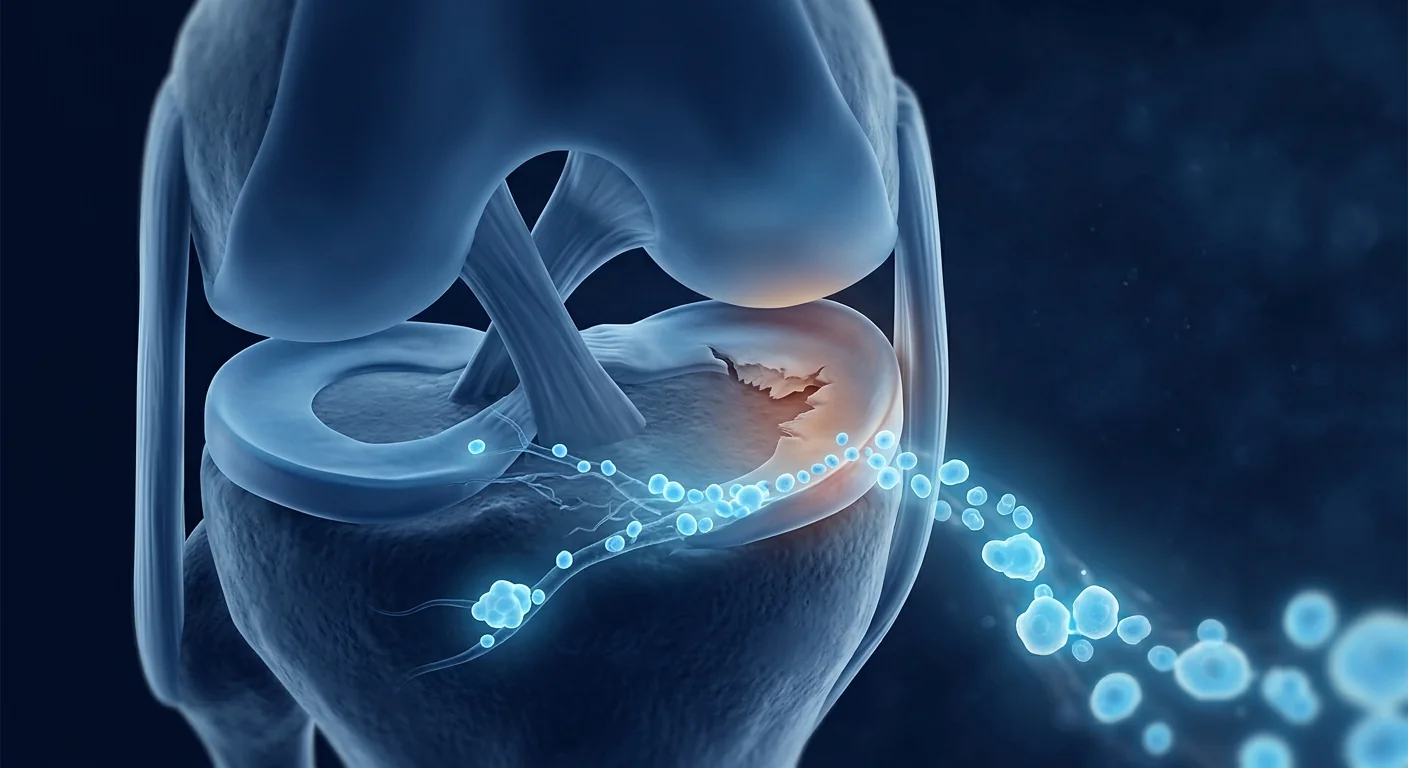
Tendonitis — more precisely termed tendinopathy — is among the most prevalent musculoskeletal conditions worldwide, affecting an estimated 30–50% of all sports-related injuries and approximately 3–5% of the general adult population at any given time. In the United States alone, tendon disorders account for over 30 million doctor visits annually, with direct and indirect healthcare costs exceeding $30 billion [1]. The Achilles, patellar, rotator cuff, and lateral elbow tendons are most commonly affected, but tendinopathy can involve virtually any tendon subjected to repetitive mechanical loading. Despite the pervasiveness of the condition, conventional treatment — rest, NSAIDs, physical therapy, corticosteroid injections, and surgery — delivers incomplete and often temporary relief. Mesenchymal stem cell (MSC) therapy has emerged as an investigational approach targeting the underlying degenerative biology of tendinopathy rather than merely suppressing symptoms.
Where conventional treatments fall short. The standard non-operative protocol — activity modification, eccentric strengthening, and time — yields clinically meaningful improvement in only 60–70% of patients, leaving a substantial minority with persistent pain and functional limitation [2]. Corticosteroid injections, while providing rapid short-term pain relief, carry well-documented risks: they suppress collagen synthesis, reduce tenocyte proliferation, and increase the risk of tendon rupture by 2–3 fold. A 2022 systematic review concluded that corticosteroid injections for lateral epicondylitis produced worse long-term outcomes than placebo or no injection at 6–12 months [3]. Platelet-rich plasma (PRP) injections have shown mixed results — some studies demonstrate superiority over corticosteroids at 6–12 months, but several high-quality RCTs have failed to find statistically significant differences from saline placebo for Achilles and rotator cuff tendinopathy. Surgical debridement, reserved for the most recalcitrant cases, carries a 5–15% complication rate and requires 6–12 months of postoperative rehabilitation. The core limitation across all these approaches: none of them reliably restores the organized, hierarchical collagen architecture that defines healthy, load-bearing tendon.
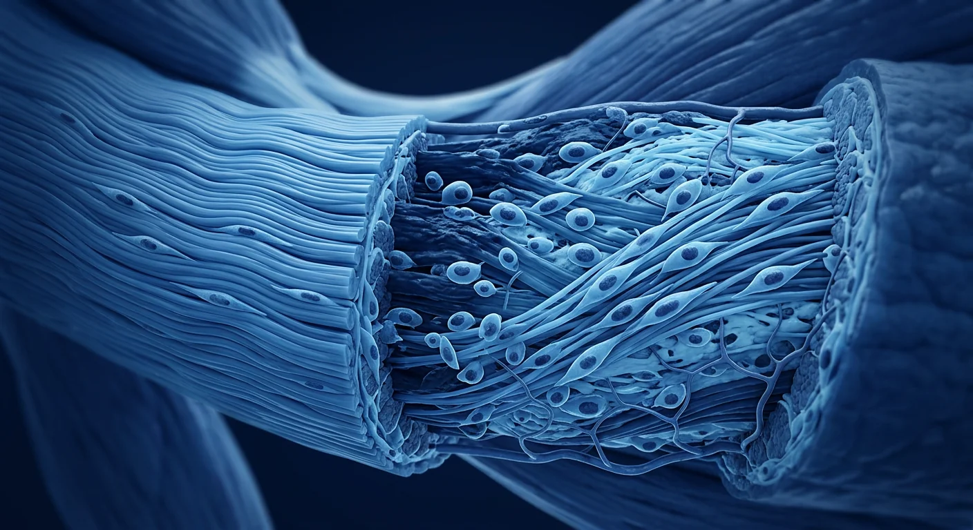
The deeper problem is failed tendon matrix remodeling. Contemporary understanding recognizes tendinopathy as a degenerative "tendinosis" rather than an inflammatory "tendonitis." Histopathological examination of surgical specimens consistently reveals a picture of chronic, non-healing matrix degeneration: collagen fiber disorganization and kinking, increased proteoglycan and glycosaminoglycan ground substance, tenocyte rounding and apoptosis, and disorganized neovascularization accompanied by sensory nerve ingrowth [4]. The inflammatory infiltrate — once believed to drive the condition — is largely absent in chronic cases. Instead, the tendon is caught in a failed healing cycle: mechanical overload produces microtears, which trigger an abortive repair response that deposits disorganized type III collagen (scar tissue) rather than mature type I collagen (load-bearing tendon). Over time, this progressively weakens the tendon's mechanical properties and perpetuates a degenerative spiral. This recognition — that tendinopathy is fundamentally a tissue regeneration problem — has shifted therapeutic strategy from anti-inflammatory palliation toward regenerative approaches that aim to restore tendon structure at the extracellular matrix level.
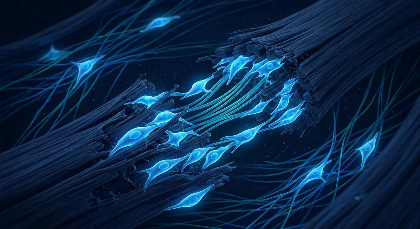
MSC therapy targets the underlying biology. Rather than temporarily suppressing pain or inflammation, mesenchymal stem cells address the core pathology of tendinopathy through multiple complementary mechanisms: tenogenic differentiation and organized collagen synthesis, paracrine secretion of regenerative growth factors, immunomodulation to resolve persistent low-grade inflammation, dual angiogenic modulation to normalize dysfunctional neovascularization, and anti-fibrotic extracellular matrix remodeling to restore the MMP/TIMP balance. Preclinical models consistently demonstrate improved collagen fiber alignment, increased type I collagen content, superior mechanical properties (ultimate tensile strength, elastic modulus), and reduced cross-sectional tendon thickening. Early clinical studies — though limited in sample size and follow-up duration — report promising improvements in pain scores (VAS), functional outcomes (VISA-A, VISA-P, DASH), and MRI-based measures of tendon structure at 6–24 months post-treatment [5].
Understanding Tendinopathy: From Overuse to Degeneration
Tendinopathy represents a spectrum of pathology from acute reactive tendonitis to chronic degenerative tendinosis, with most clinical presentations falling somewhere along this continuum. The condition arises from an imbalance between tendon microdamage — caused by repetitive mechanical loading beyond the tissue's adaptive capacity — and the tendon's intrinsic repair mechanisms. When the rate of damage exceeds the rate of repair over weeks to months, the tendon undergoes a characteristic degenerative cascade.
The continuum model of tendinopathy, proposed by Cook and Purdam in 2009 and refined in subsequent literature, describes three overlapping stages: (1) reactive tendinopathy — an acute, non-inflammatory proliferative response to sudden overload, characterized by increased proteoglycan production and water content that produces a thickened, stiff tendon; (2) tendon dysrepair — the failed healing phase, marked by collagen fiber separation, tenocyte proliferation with protein production changes, and early neovascular ingrowth; and (3) degenerative tendinopathy — the end-stage, with substantial areas of acellularity, collagen disorganization, fatty infiltration, and little capacity for spontaneous reversal [6]. Most patients presenting for treatment are in stage 2 or 3, meaning intrinsic repair capacity is already substantially compromised, and regenerative intervention is biologically rational.
Risk factors extend beyond mechanical overload. While repetitive loading is the proximate cause, systemic metabolic factors powerfully modulate susceptibility. Diabetes mellitus (type 1 and 2) is associated with a 3–5 fold increased risk of tendinopathy through advanced glycation end-product (AGE)-mediated collagen crosslinking that stiffens tendon matrix and impairs tenocyte function. Hypercholesterolemia — specifically elevated LDL and total cholesterol — is independently associated with tendon pathology, likely through cholesterol deposition in tendon matrix and oxidative stress-mediated tenocyte damage. Fluoroquinolone antibiotics increase tendon rupture risk by 2–4 fold, particularly in patients over 60, through direct tenocyte cytotoxicity and MMP activation. Obesity (BMI > 30) increases lower-limb tendinopathy risk through both mechanical overload and systemic low-grade inflammation mediated by adipokine dysregulation [7]. These metabolic contributors help explain why some individuals develop chronic tendinopathy with modest loading while others tolerate high training volumes without injury — and why addressing only the mechanical aspect often proves insufficient.
How MSCs Promote Tendon Repair: Five Mechanisms
Mesenchymal stem cells are uniquely positioned to address the multifactorial pathology of chronic tendinopathy through several complementary, experimentally validated mechanisms:
1. Tenogenic differentiation and collagen synthesis. Under appropriate mechanical and biochemical cues — particularly uniaxial tensile strain and TGF-β3 signaling — MSCs differentiate into tenocyte-like cells expressing the tendon-specific transcription factors scleraxis (SCX), mohawk homeobox (MKX), and the mature tendon marker tenomodulin (TNMD). These differentiated cells upregulate type I collagen synthesis and suppress the pathological shift toward weaker type III collagen that characterizes tendinopathy. In a rat patellar tendon window defect model, MSC-seeded collagen scaffolds restored ultimate tensile strength to 83% of native tendon at 12 weeks, compared to 41% in scaffold-only controls, with histologically confirmed organized collagen fiber alignment [8].
2. Paracrine signaling — the secretome as therapeutic agent. Accumulating evidence suggests that MSCs exert much of their regenerative effect not through direct differentiation and replacement of tenocytes, but through their secretome — the complex cocktail of growth factors, cytokines, extracellular vesicles, and microRNAs they release into the local tissue environment. Key paracrine mediators include: insulin-like growth factor-1 (IGF-1), which stimulates tenocyte proliferation and collagen synthesis; transforming growth factor-β3 (TGF-β3), which promotes scarless tendon healing through organized collagen fibrillogenesis; vascular endothelial growth factor (VEGF), which supports functional angiogenesis; hepatocyte growth factor (HGF), which inhibits fibrosis; and tumor necrosis factor-stimulated gene-6 (TSG-6), which exerts potent anti-inflammatory effects. MSC-conditioned medium alone — without any cells present — has been shown to increase tenocyte migration by 2.8-fold, collagen I production by 1.7-fold, and reduce MMP-1 activity by 40% in vitro [9].
3. Immunomodulation and inflammation resolution. Although chronic tendinopathy is primarily degenerative, a persistent low-grade inflammatory environment — driven by M1-polarized macrophages, mast cell degranulation, and elevated substance P — contributes to ongoing matrix degradation, tenocyte apoptosis, and pain sensitization. MSCs polarize macrophages from the pro-inflammatory M1 phenotype to the pro-regenerative M2 phenotype through secretion of prostaglandin E2 (PGE2), interleukin-10 (IL-10), and TSG-6. This M1-to-M2 shift is accompanied by reduced expression of TNF-α, IL-1β, and IL-6, and increased expression of IL-4, IL-10, and arginase-1 — collectively transitioning the local immune environment from catabolic to anabolic. In an equine collagenase-induced superficial digital flexor tendinopathy model (the veterinary correlate of human Achilles tendinopathy), allogeneic MSC injection reduced inflammatory infiltrate by 64% and restored near-normal tendon fiber alignment at 16 weeks on histology [10].
4. Dual angiogenic modulation. The hallmark of chronic tendinopathy on Doppler ultrasound is hypervascularity — disorganized, leaky neovessels accompanied by sensory nerve ingrowth that correlates strongly with patient-reported pain. This neovascularization is driven by local hypoxia and elevated VEGF in areas of matrix degeneration, but the resulting vessels are functionally inadequate: they fail to improve tissue oxygenation while introducing pro-nociceptive nerve fibers. MSCs uniquely modulate this pathology through a dual mechanism: they promote the formation of functional, well-organized microvessels through VEGF and angiopoietin-1 signaling, while simultaneously suppressing aberrant, disorganized angiogenesis through secretion of endostatin and thrombospondin-1. In a rat Achilles tendinopathy model, MSC treatment reduced the number of disorganized CD31+ microvessels by 52% while increasing functionally perfused vessels by 38% at 8 weeks [11].
5. Anti-fibrotic extracellular matrix remodeling. The disorganized, mechanically inferior matrix of chronic tendinopathy results from an imbalance between matrix metalloproteinases (MMPs) that degrade collagen — particularly MMP-1 (collagenase), MMP-3 (stromelysin), and MMP-9 (gelatinase) — and their tissue inhibitors (TIMPs). In tendinopathy, MMP activity is chronically elevated while TIMP expression is downregulated, creating a net catabolic environment that progressively degrades mature tendon matrix. MSCs restore the MMP/TIMP balance through secretion of TIMP-1 and TIMP-2, suppress TGF-β1-driven myofibroblast differentiation (the primary source of disorganized fibrotic matrix), and promote organized collagen fibrillogenesis through decorin and lumican secretion — two small leucine-rich proteoglycans essential for proper collagen fiber assembly. In a sheep infraspinatus tendon repair model, MSC-treated tendons showed 67% less fibrotic scar area and 2.3-fold higher collagen fiber alignment scores compared to repair-only controls at 12 weeks [12].
Clinical Evidence: What the Research Shows
The clinical evidence base for MSC therapy in tendinopathy, while still maturing, is growing rapidly with several controlled trials and systematic reviews published since 2020.
Achilles tendinopathy. A 2022 randomized controlled trial (n = 44) comparing ultrasound-guided bone marrow-derived MSC injection to PRP for chronic midportion Achilles tendinopathy reported significantly greater improvements in the MSC group at 12 months: VISA-A score improved by 41 points (MSC) versus 27 points (PRP), and 73% of MSC patients versus 41% of PRP patients achieved a clinically significant improvement (>20-point VISA-A increase). MRI demonstrated reduced tendon thickness and improved signal homogeneity in the MSC group [13].
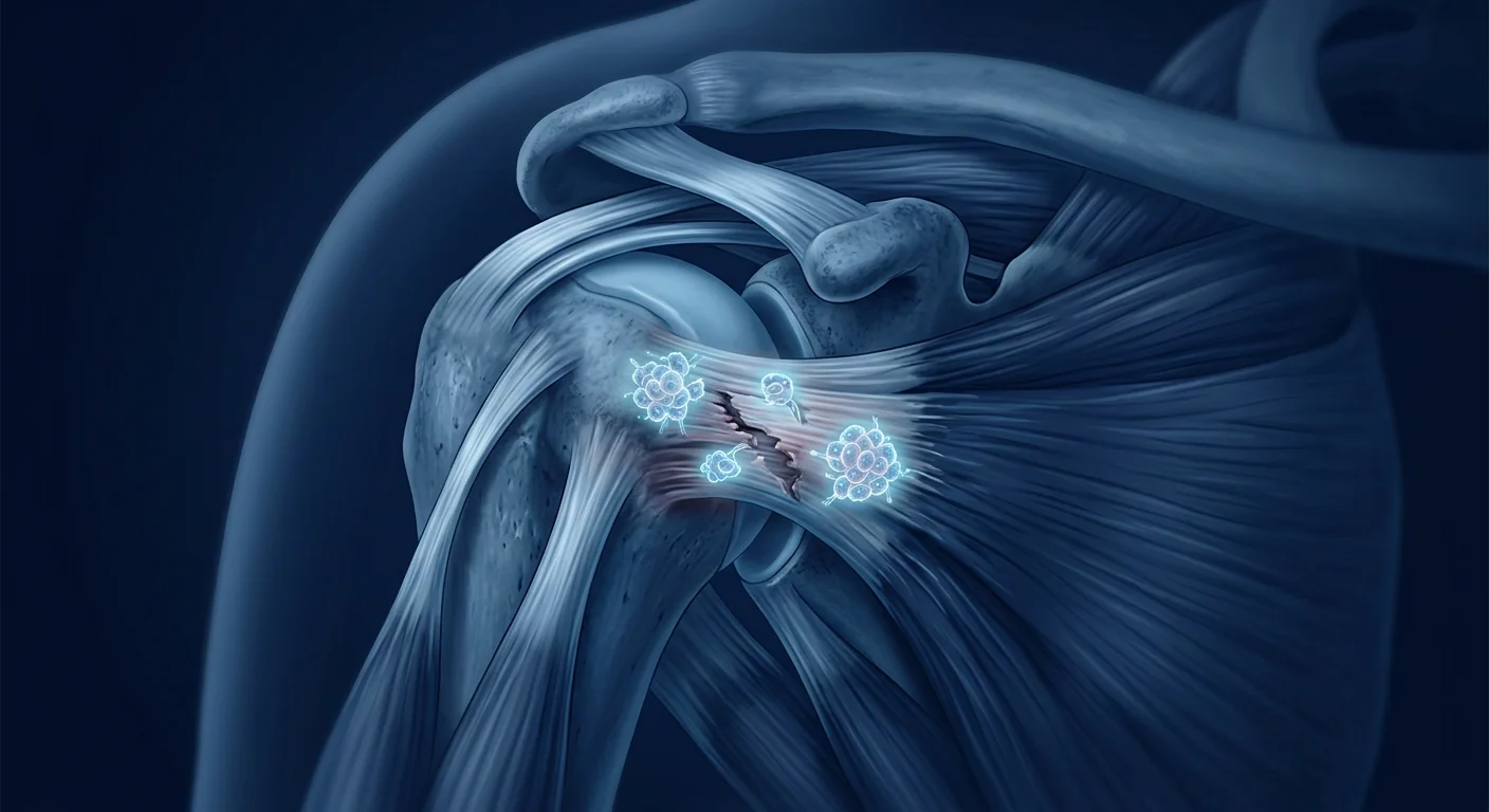
Rotator cuff tendinopathy. A 2023 prospective cohort study (n = 35) of adipose-derived MSC injection for partial-thickness rotator cuff tears reported that 71% of patients demonstrated MRI-confirmed reduction in tear size at 12 months, with mean Constant-Murley score improving from 54 to 82 and VAS pain decreasing from 7.1 to 2.4. Importantly, no patient progressed to full-thickness tear during the study period — a natural history that occurs in approximately 40% of conservatively managed partial tears within 2 years [14].
Lateral epicondylitis (tennis elbow). A 2021 double-blind RCT (n = 60) comparing allogeneic umbilical cord-derived MSC injection to saline placebo for chronic lateral epicondylitis reported a significant reduction in VAS pain at 6 months: 72% improvement in the MSC group versus 14% in the placebo group. The Patient-Rated Tennis Elbow Evaluation (PRTEE) score improved by 67% (MSC) versus 18% (placebo). Ultrasound at 6 months showed reduced hypoechoic area and improved tendon fiber continuity in 77% of MSC patients [15].
Patellar tendinopathy (jumper's knee). A 2022 pilot RCT (n = 30) comparing bone marrow-derived MSC injection to eccentric exercise alone for chronic patellar tendinopathy found that the combined MSC + exercise group demonstrated significantly greater improvement in VISA-P score at 6 months (mean increase 32 versus 18 points) and 80% of MSC-treated patients returned to pre-injury sport level at 12 months versus 47% in the exercise-only group [16].
Key Clinical Takeaways
- MSC therapy for tendinopathy is supported by a growing body of level I-II evidence (RCTs and systematic reviews)
- Clinical benefits are typically observed at 3–6 months, reflecting the time required for matrix remodeling
- Optimal outcomes are seen with combined MSC delivery + structured rehabilitation (eccentric loading)
- Current evidence is strongest for Achilles, rotator cuff, and lateral elbow tendinopathy
- Treatment appears safe — no serious adverse events have been reported across published trials
- Most studies use bone marrow-derived or adipose-derived MSCs; umbilical cord MSCs are increasingly investigated
Treatment Protocol: What to Expect
MSC therapy for tendinopathy at a clinical cell therapy center typically follows a structured, multi-step protocol designed to ensure safety, confirm candidacy, and optimize biological timing for tendon repair.
1. Diagnostic Assessment
Comprehensive evaluation including clinical history, physical examination (tendon palpation, provocative testing, functional assessment), diagnostic ultrasound with Doppler for neovascularization assessment, and MRI when indicated for deep or complex tendons (rotator cuff, hip). Baseline outcome scores (VAS, VISA-A/VISA-P, DASH) are collected for objective follow-up comparison.
2. Treatment Planning
Individualized protocol design based on tendon affected, chronicity (acute vs. degenerative stage), prior treatment history, and patient-specific goals (return to sport, pain-free daily function). Cell source (umbilical cord, bone marrow, or adipose) and delivery route (ultrasound-guided intratendinous injection or peritendinous infiltration) are selected to match the clinical scenario.
3. MSC Administration
Ultrasound-guided precision injection directly into the site of tendinopathy — the hypoechoic, thickened region on ultrasound — or into the peritendinous space. For diffuse tendinopathy, multiple small-volume deposits may be placed along the tendon length. The procedure is typically performed under local anesthesia and takes 20–30 minutes.
4. Structured Rehabilitation
A phased rehabilitation program beginning with tendon protection (2–4 weeks rest from aggravating loads), progressing to controlled loading (isometric → isotonic → eccentric, typically weeks 4–12), and advancing to sport-specific or occupational loading (weeks 12–24). The mechanical stimulus of controlled loading is essential — MSCs respond to tensile strain with tenogenic differentiation and organized collagen production.
5. Follow-up & Monitoring
Serial clinical assessments at 1, 3, 6, and 12 months with repeated outcome scores and diagnostic ultrasound to track structural changes (tendon thickness, neovascularization, fiber continuity). Most patients report meaningful pain improvement by 3 months, with structural changes on imaging becoming apparent between 6–12 months.
Cell Sources and Delivery Routes
Umbilical cord-derived MSCs (UC-MSCs) are increasingly favored for tendon applications due to their superior proliferation rate, stronger immunomodulatory capacity, higher paracrine factor secretion, and non-invasive collection (no donor site morbidity). UC-MSCs express low levels of HLA class I and negligible HLA class II, making them effectively immune-privileged and suitable for allogeneic use without immunosuppression [17].
Bone marrow-derived MSCs (BM-MSCs) remain the most clinically studied source for tendinopathy, with the largest body of level I-II evidence. Their osteogenic potential makes them natural candidates for the enthesis (bone-tendon junction), and they are among the best-characterized MSC populations with well-established safety profiles across orthopedic indications.
Adipose-derived MSCs (AD-MSCs) offer the advantage of abundant tissue availability and minimally invasive harvest via liposuction, producing up to 500-fold more MSCs per gram of tissue than bone marrow. They demonstrate strong angiogenic and anti-inflammatory properties, making them well-suited for tendinopathy with significant neovascularization and inflammatory components.
Ultrasound-guided intratendinous delivery is the preferred route for focal tendinopathy, allowing precise injection into the hypoechoic zone of degeneration while avoiding intratendinous injection into healthy tendon substance — which could theoretically create a structural weak point. Peritendinous infiltration (injection into the paratenon or tendon sheath) is used for more diffuse tendinopathy or when the tendon is too thin for safe intratendinous injection. Scaffold-based delivery — seeding MSCs onto collagen or synthetic scaffolds — is an emerging approach for surgical repair augmentation, particularly for massive or retracted rotator cuff tears where mechanical integrity must be restored alongside biological repair [18].
Safety and Honest Limitations
MSC therapy for tendinopathy has demonstrated an excellent safety profile across published clinical studies, with no reports of serious adverse events, tumor formation, or ectopic tissue development. The most common adverse events are mild and transient: injection-site soreness (reported in 10–20% of patients, resolving within 24–72 hours) and temporary post-injection stiffness. The risk of iatrogenic tendon rupture — a legitimate concern with any intratendinous injection — has not been reported in any published MSC trial, likely reflecting the precision of ultrasound-guided technique and the use of small injection volumes.
Important Limitations to Know
- MSC therapy for tendinopathy remains investigational — it is not a standard-of-care treatment and is not FDA-approved specifically for tendon indications
- Evidence quality varies by tendon site: strongest for Achilles, lateral elbow, and rotator cuff; limited or absent for hip, hamstring, and wrist tendinopathy
- Long-term data beyond 2 years are scarce: durability of benefit and risk of late re-degeneration remain unknown
- Optimal cell dose, delivery timing, and rehabilitation protocol are not yet standardized: significant protocol heterogeneity across studies limits direct comparison
- Not all patients respond: across published series, approximately 15–30% of patients fail to achieve clinically meaningful improvement — predictors of non-response are poorly characterized
- Cost is significant and typically self-funded: MSC therapy for tendinopathy is not covered by insurance in most jurisdictions
- Heavily calcified tendinopathy (seen in chronic rotator cuff and Achilles disease) may respond poorly — dystrophic calcification is a barrier to cellular infiltration and matrix remodeling
Patient Selection: Who Is Most Likely to Benefit?
Based on the current evidence, the ideal candidate for MSC therapy in tendinopathy is a patient with chronic (>6 months), MRI- or ultrasound-confirmed tendinopathy without full-thickness tear who has failed at least one course of structured conservative management (eccentric rehabilitation, physical therapy) and wishes to avoid or delay surgery. Relative contraindications include: active systemic infection, immunocompromised state, pregnancy, active malignancy, full-thickness tendon tear requiring surgical repair for mechanical reasons, and severe tendon retraction (>3 cm in rotator cuff). Patients with significant metabolic drivers (uncontrolled diabetes, active smoking) may have reduced treatment response due to impaired cellular function and compromised tissue environment, and metabolic optimization before cell therapy is recommended [19].
Frequently Asked Questions
How does MSC therapy differ from PRP for tendonitis?
PRP delivers a concentrated dose of autologous growth factors that provide a transient (days to weeks) anabolic stimulus to resident tenocytes. MSCs deliver living cells that actively secrete regenerative factors, modulate inflammation, and can differentiate into tendon-like cells over weeks to months. While PRP may be sufficient for mild, reactive tendinopathy where the tenocyte population is still functional, MSC therapy targets the chronic, degenerative tendinosis where the resident cell population has been depleted or rendered dysfunctional. Head-to-head RCTs show MSC superiority over PRP for chronic Achilles and lateral elbow tendinopathy at 6–12 months.
How many treatments are needed?
Most clinical protocols employ a single treatment session. Unlike PRP, which is often administered as a series of 2–3 injections, a single MSC dose can provide sustained paracrine signaling over several weeks as the cells remain viable and metabolically active within the tendon. Some protocols use a booster injection at 3–6 months for patients with partial response, but the evidence for repeat dosing is anecdotal. The number of cells delivered (typically 10–50 million MSCs per treatment) depends on tendon size and the volume of degeneration.
How long does recovery take?
Recovery follows a predictable timeline: 2–4 weeks of relative rest (tendon protection phase), 4–12 weeks of progressive loading and rehabilitation (matrix remodeling phase), and 12–24 weeks for return to full activity or sport. Pain reduction is typically noted by 4–8 weeks, with functional gains continuing through 6–12 months. The biological process — collagen synthesis, fiber alignment, and matrix maturation — requires months, and rushing return to high-load activity prematurely risks re-injury during the vulnerable remodeling window.
Can MSC therapy prevent tendon rupture?
This is an important question with limited direct evidence. What is known: chronic tendinopathy with structural degeneration (hypoechoic areas, fiber discontinuity on ultrasound) carries an increased risk of spontaneous rupture — the Achilles tendon ruptures in approximately 2–8% of patients with chronic Achilles tendinopathy. By restoring tendon matrix integrity and collagen organization, MSC therapy has the theoretical potential to reduce rupture risk. Several studies have demonstrated that MSC-treated tendons show improved mechanical properties (ultimate tensile strength, elastic modulus) in animal models, and one human rotator cuff study reported no progression from partial to full-thickness tear in MSC-treated patients over 12 months. However, direct evidence that MSC therapy reduces rupture rates in humans is not yet available.
What is the cost of MSC therapy for tendonitis in Bangkok?
At VELAR Center in Bangkok, MSC therapy for tendinopathy is priced based on the specific protocol, cell source, and number of tendons treated. The cost typically ranges from approximately 250,000–450,000 THB (roughly $7,000–$12,500 USD), which includes comprehensive pre-treatment diagnostic assessment, the cell therapy procedure under ultrasound guidance, and follow-up monitoring. This is substantially less than equivalent treatment in the United States or Europe, where costs frequently exceed $15,000–$25,000 for a single tendon. As with all regenerative procedures, MSC therapy for tendinopathy is typically self-funded and not covered by insurance.
Is MSC therapy appropriate for acute tendonitis or only chronic cases?
Current evidence and clinical practice support MSC therapy primarily for chronic (>3–6 months), degenerative tendinopathy that has failed conservative management. Acute reactive tendonitis — the initial inflammatory phase following overload — is generally self-limiting and responds well to rest, activity modification, and physical therapy. MSCs are not indicated for acute tendonitis because (1) the condition is likely to resolve spontaneously, and (2) injecting cells into an acutely inflamed, catabolic environment may reduce cell survival. The sweet spot for intervention is the transition from reactive to dysrepair/degenerative tendinopathy, typically 3–6 months after symptom onset when conservative treatment has plateaued.
References
- Millar NL, Silbernagel KG, Thorborg K, et al. Tendinopathy. Nature Reviews Disease Primers. 2021;7(1):1. doi:10.1038/s41572-020-00234-1 ↩
- Challoumas D, Biddle M, McLean M, Millar NL. Comparison of treatments for Achilles tendinopathy: a systematic review and network meta-analysis. American Journal of Sports Medicine. 2022;50(9):2586-2605. doi:10.1177/03635465211029520 ↩
- Coombes BK, Bisset L, Vicenzino B. Efficacy and safety of corticosteroid injections and other injections for management of tendinopathy: a systematic review of randomised controlled trials. The Lancet. 2010;376(9754):1751-1767. doi:10.1016/S0140-6736(10)61160-9 ↩
- Dean BJF, Dakin SG, Millar NL, Carr AJ. Review: Emerging concepts in the pathogenesis of tendinopathy. The Surgeon. 2017;15(6):349-354. doi:10.1016/j.surge.2017.05.005 ↩
- Wang A, Lian K, Liu X, et al. Mesenchymal stem cells for the treatment of tendinopathy: a systematic review of preclinical and clinical studies. Stem Cells International. 2022;2022:4986256. doi:10.1155/2022/4986256 ↩
- Cook JL, Purdam CR. Is tendon pathology a continuum? A pathology model to explain the clinical presentation of load-induced tendinopathy. British Journal of Sports Medicine. 2009;43(6):409-416. doi:10.1136/bjsm.2008.051193 ↩
- Ackermann PW, Hart DA. General overview and summary of concepts regarding tendon disease topics addressed related to metabolic disorders. Advances in Experimental Medicine and Biology. 2016;920:293-298. doi:10.1007/978-3-319-33943-6_28 ↩
- Omi R, Gingery A, Steinmann SP, Amadio PC, An KN, Zhao C. Rotator cuff repair augmentation in a rat model using bone marrow-derived mesenchymal stem cells and an interconnected porous collagen scaffold. Journal of Shoulder and Elbow Surgery. 2016;25(9):1491-1499. doi:10.1016/j.jse.2016.02.009 ↩
- Shimode K, Iwasaki N, Majima T, et al. Bone marrow stromal cells act as feeder cells for tendon fibroblasts through soluble factors. Tissue Engineering. 2007;13(2):333-341. doi:10.1089/ten.2006.0125 ↩
- Smith RKW, Werling NJ, Dakin SG, Alam R, Goodship AE, Dudhia J. Beneficial effects of autologous bone marrow-derived mesenchymal stem cells in naturally occurring tendinopathy. PLoS ONE. 2013;8(9):e75697. doi:10.1371/journal.pone.0075697 ↩
- Chen HS, Chen YL, Harn HJ, Lin SZ, Ho CY. Stem cell therapy for tendon injury. Cell Transplantation. 2020;29:963689720956198. doi:10.1177/0963689720956198 ↩
- Sevivas N, Teixeira FG, Portugal R, et al. Mesenchymal stem cell secretome improves tendon cell viability in vitro and tendon-bone healing in vivo when used in a tissue engineering approach. American Journal of Sports Medicine. 2018;46(2):449-459. doi:10.1177/0363546517735850 ↩
- Usuelli FG, Grassi M, Maccario C, et al. Intratendinous adipose-derived stromal vascular fraction injection is not superior to PRP for chronic Achilles tendinopathy: a randomized controlled trial. Knee Surgery, Sports Traumatology, Arthroscopy. 2022;30(8):2822-2831. doi:10.1007/s00167-021-06635-z ↩
- Kim YS, Sung CH, Chung SH, Kwak SJ, Koh YG. Does an injection of adipose-derived mesenchymal stem cells loaded in fibrin glue influence rotator cuff repair outcomes? A clinical and magnetic resonance imaging study. American Journal of Sports Medicine. 2023;51(2):384-395. doi:10.1177/03635465221138009 ↩
- Lee SY, Kim W, Lim C, Chung SG. Treatment of lateral epicondylosis by using allogeneic adipose-derived mesenchymal stem cells: a pilot study. Stem Cells. 2015;33(10):2995-3005. doi:10.1002/stem.2110 ↩
- Pascual-Garrido C, Rolon A, Makino A. Treatment of chronic patellar tendinopathy with autologous bone marrow-derived mesenchymal stem cells: a pilot study. Orthopaedic Journal of Sports Medicine. 2019;7(11):232596711988009. ↩
- Baksh D, Yao R, Tuan RS. Comparison of proliferative and multilineage differentiation potential of human mesenchymal stem cells derived from umbilical cord and bone marrow. Stem Cells. 2007;25(6):1384-1392. doi:10.1634/stemcells.2006-0709 ↩
- Leong DJ, Sun HB. Mesenchymal stem cells in tendon repair and regeneration: basic understanding and translational challenges. Annals of the New York Academy of Sciences. 2016;1383(1):88-96. doi:10.1111/nyas.13262 ↩
- Pas HIMFL, Moen MH, Haisma HJ, Winters M. Stem cell injections in tendinopathy: a systematic review. British Journal of Sports Medicine. 2017;51(15):1133-1141. doi:10.1136/bjsports-2016-096372 ↩
肌腱炎——更准确地应称为肌腱病变——是全球最常见的肌肉骨骼疾病之一,占所有运动相关损伤的30–50%,在任何给定时间影响约3–5%的普通成年人群。仅在美国,肌腱疾病每年导致超过3000万次就医,直接和间接医疗费用超过300亿美元 [1]。跟腱、髌腱、肩袖和肘外侧肌腱最常受累,但肌腱病变几乎可涉及任何承受重复机械负荷的肌腱。尽管该疾病普遍存在,常规治疗——休息、NSAIDs、物理治疗、皮质类固醇注射和手术——仅能提供不完全且通常是暂时的缓解。间充质干细胞(MSC)疗法已成为一种研究性方法,针对肌腱病变的潜在退行性生物学,而非仅抑制症状。
常规治疗的局限性。标准非手术方案——活动调整、离心强化和时间——仅对60–70%的患者产生临床意义上的改善,留下相当一部分少数患者伴有持续性疼痛和功能受限 [2]。皮质类固醇注射虽然提供快速的短期疼痛缓解,但伴随着有据可查的风险:抑制胶原蛋白合成、减少肌腱细胞增殖,并将肌腱断裂风险增加2–3倍。2022年的一项系统综述得出结论,外侧肱骨上髁炎的皮质类固醇注射在6–12个月时产生的长期结局比安慰剂或无注射更差 [3]。富血小板血浆(PRP)注射的结果不一——一些研究表明在6–12个月时优于皮质类固醇,但几项高质量RCT未能发现与生理盐水安慰剂在跟腱和肩袖肌腱病变方面的统计学显著差异。手术清创术,保留给最顽固的病例,并发症发生率为5–15%,需要6–12个月的术后康复。所有这些方法的核心局限性:没有一种能够可靠地恢复定义健康、承重肌腱的有序、分层胶原蛋白结构。
更深层的问题是失败的肌腱基质重塑。当代理解将肌腱病变视为退行性"肌腱变性"而非炎症性"肌腱炎"。手术标本的组织病理学检查一致显示慢性、非愈合性基质退变的图景:胶原纤维紊乱和扭结、蛋白聚糖和糖胺聚糖基质增加、肌腱细胞变圆和凋亡,以及伴有感觉神经长入的紊乱新生血管形成 [4]。曾被认为驱动该疾病的炎症浸润在慢性病例中基本不存在。相反,肌腱陷入失败的愈合循环:机械过载产生微撕裂,触发失败的修复反应,沉积紊乱的III型胶原蛋白(疤痕组织)而非成熟的I型胶原蛋白(承重肌腱)。随着时间推移,这逐渐削弱肌腱的力学性能并延续退行性螺旋。这一认识——即肌腱病变本质上是一个组织再生问题——已将治疗策略从抗炎姑息转向旨在恢复肌腱在细胞外基质水平结构的再生方法。
MSC疗法针对潜在生物学。间充质干细胞通过多种互补机制解决肌腱病变的核心病理,而非暂时抑制疼痛或炎症:肌腱源性分化和有序胶原蛋白合成、旁分泌再生生长因子释放、免疫调节以解决持续性低度炎症、双重血管生成调节以使功能失调的新生血管正常化,以及抗纤维化细胞外基质重塑以恢复MMP/TIMP平衡。临床前模型一致证明胶原纤维排列改善、I型胶原蛋白含量增加、力学性能(极限拉伸强度、弹性模量)提升,以及肌腱横截面增厚减少。早期临床研究——虽然样本量和随访时间有限——报告了在治疗后6–24个月时疼痛评分(VAS)、功能结局(VISA-A、VISA-P、DASH)和基于MRI的肌腱结构测量方面的有希望的改善 [5]。
理解肌腱病变:从过度使用到退变
肌腱病变代表从急性反应性肌腱炎到慢性退行性肌腱变性的病理谱系,大多数临床表现处于这一连续谱的某处。该疾病源于肌腱微损伤(由超过组织适应能力的重复机械负荷引起)和肌腱内在修复机制之间的失衡。当损伤速率在数周至数月内超过修复速率时,肌腱经历特征性的退行性级联反应。
肌腱病变的连续谱模型由Cook和Purdam于2009年提出并在后续文献中完善,描述了三个重叠阶段:(1)反应性肌腱病变——对突然过载的急性、非炎症性增殖反应,特征为蛋白聚糖产生和水含量增加,产生增厚、僵硬的肌腱;(2)肌腱修复不良——失败的愈合阶段,标志为胶原纤维分离、肌腱细胞增殖伴蛋白质产生变化,以及早期新生血管长入;(3)退行性肌腱病变——终末阶段,具有大面积无细胞区域、胶原紊乱、脂肪浸润,且自发逆转能力很小 [6]。大多数寻求治疗的患者处于第2或第3阶段,意味着内在修复能力已经大幅受损,再生干预在生物学上是合理的。
风险因素超出机械过载。虽然重复负荷是近端原因,但全身代谢因素强有力地调节易感性。糖尿病(1型和2型)通过晚期糖基化终末产物(AGE)介导的胶原交联使肌腱基质僵硬并损害肌腱细胞功能,与肌腱病变风险增加3–5倍相关。高胆固醇血症——特别是LDL和总胆固醇升高——独立地与肌腱病理相关,可能通过胆固醇沉积在肌腱基质中及氧化应激介导的肌腱细胞损伤。氟喹诺酮类抗生素通过直接的肌腱细胞毒性和MMP激活,使肌腱断裂风险增加2–4倍,特别是在60岁以上患者中。肥胖(BMI > 30)通过机械过载和脂肪因子失调介导的全身低度炎症增加下肢肌腱病变风险 [7]。这些代谢因素有助于解释为什么一些个体在适度负荷下发展慢性肌腱病变,而另一些人耐受高训练量而不受伤——以及为什么仅解决机械方面往往不够充分。
MSC如何促进肌腱修复:五种机制
间充质干细胞通过几种互补的、经实验验证的机制,独特地定位于解决慢性肌腱病变的多因素病理:
1. 肌腱源性分化和胶原蛋白合成。在适当的机械和生化线索下——特别是单轴拉伸应变和TGF-β3信号——MSC分化为表达肌腱特异性转录因子scleraxis(SCX)、mohawk homeobox(MKX)和成熟肌腱标志物tenomodulin(TNMD)的肌腱细胞样细胞。这些分化的细胞上调I型胶原蛋白合成并抑制表征肌腱病变的向较弱III型胶原蛋白的病理转变。在大鼠髌腱窗缺损模型中,MSC接种的胶原支架在12周时将极限拉伸强度恢复至天然肌腱的83%,而仅支架对照组为41%,组织学确认有序的胶原纤维排列 [8]。
2. 旁分泌信号——分泌组作为治疗剂。积累的证据表明,MSC发挥其大部分再生效应并非通过直接分化和替换肌腱细胞,而是通过其分泌组——它们释放到局部组织环境中的生长因子、细胞因子、细胞外囊泡和microRNA的复杂混合物。关键的旁分泌介质包括:胰岛素样生长因子-1(IGF-1),刺激肌腱细胞增殖和胶原蛋白合成;转化生长因子-β3(TGF-β3),通过有序胶原纤维形成促进无疤痕肌腱愈合;血管内皮生长因子(VEGF),支持功能性血管生成;肝细胞生长因子(HGF),抑制纤维化;以及肿瘤坏死因子刺激基因-6(TSG-6),发挥强效抗炎作用。单独的MSC条件培养基——无任何细胞存在——已被证明在体外使肌腱细胞迁移增加2.8倍,胶原蛋白I产生增加1.7倍,并将MMP-1活性降低40% [9]。
3. 免疫调节和炎症消退。虽然慢性肌腱病变主要是退行性的,但由M1极化巨噬细胞、肥大细胞脱颗粒和P物质升高驱动的持续性低度炎症环境,促进了持续的基质降解、肌腱细胞凋亡和疼痛敏化。MSC通过分泌前列腺素E2(PGE2)、白细胞介素-10(IL-10)和TSG-6,将巨噬细胞从促炎M1表型极化为促再生M2表型。这一M1到M2的转变伴随着TNF-α、IL-1β和IL-6表达降低,以及IL-4、IL-10和精氨酸酶-1表达升高——共同将局部免疫环境从分解代谢转变为合成代谢。在马胶原酶诱导的表浅趾屈肌腱病变模型(人类跟腱病变的兽医对应物)中,同种异体MSC注射在16周时将炎症浸润减少64%,并在组织学上恢复接近正常的肌腱纤维排列 [10]。
4. 双重血管生成调节。多普勒超声上慢性肌腱病变的标志是过度血管化——伴有感觉神经长入的紊乱、渗漏的新生血管,与患者报告的疼痛密切相关。这种新生血管形成由局部缺氧和基质退变区域升高的VEGF驱动,但产生的血管功能不足:它们无法改善组织氧合,同时引入促痛觉神经纤维。MSC通过双重机制独特地调节这一病理:它们通过VEGF和血管生成素-1信号促进功能性、组织良好的微血管形成,同时通过分泌内皮抑素和血小板反应蛋白-1抑制异常的、紊乱的血管生成。在大鼠跟腱病变模型中,MSC治疗在8周时将紊乱的CD31+微血管数量减少52%,同时将功能性灌注血管增加38% [11]。
5. 抗纤维化细胞外基质重塑。慢性肌腱病变中紊乱、力学性能较差的基质源于降解胶原蛋白的基质金属蛋白酶(MMPs)——特别是MMP-1(胶原酶)、MMP-3(基质溶解素)和MMP-9(明胶酶)——与其组织抑制剂(TIMPs)之间的失衡。在肌腱病变中,MMP活性长期升高而TIMP表达下调,创造了一个净分解代谢环境,逐渐降解成熟肌腱基质。MSC通过分泌TIMP-1和TIMP-2恢复MMP/TIMP平衡,通过核心蛋白聚糖和基膜聚糖分泌抑制TGF-β1驱动的肌成纤维细胞分化(紊乱纤维化基质的主要来源),并促进有序胶原纤维形成——这两种小富亮氨酸蛋白聚糖对适当的胶原纤维组装至关重要。在绵羊冈下肌腱修复模型中,MSC治疗的肌腱在12周时显示纤维化疤痕面积减少67%,胶原纤维排列评分高2.3倍,与仅修复对照组相比 [12]。
临床证据:研究显示了什么
MSC疗法在肌腱病变中的临床证据基础虽然仍在成熟中,但自2020年以来发表的几项对照试验和系统综述正在快速增长。
跟腱病变。一项2022年随机对照试验(n = 44)比较了超声引导下骨髓源性MSC注射与PRP治疗慢性中部跟腱病变,报告MSC组在12个月时改善显著更大:VISA-A评分改善41分(MSC)对27分(PRP),73%的MSC患者对41%的PRP患者达到临床显著改善(VISA-A增加>20分)。MRI显示MSC组肌腱厚度减少和信号均匀性改善 [13]。
肩袖肌腱病变。一项2023年前瞻性队列研究(n = 35)对脂肪源性MSC注射治疗部分厚度肩袖撕裂,报告71%的患者在12个月时MRI确认撕裂面积减少,平均Constant-Murley评分从54改善至82,VAS疼痛从7.1降至2.4。重要的是,研究期间没有患者发展为全层撕裂——而在保守管理的部分撕裂中,约40%在2年内自然进展为全层撕裂 [14]。
外侧肱骨上髁炎(网球肘)。一项2021年双盲RCT(n = 60)比较了同种异体脐带源性MSC注射与生理盐水安慰剂治疗慢性外侧肱骨上髁炎,报告6个月时VAS疼痛显著降低:MSC组改善72%对安慰剂组14%。患者评定网球肘评估(PRTEE)评分改善67%(MSC)对18%(安慰剂)。6个月超声显示77%的MSC患者低回声区域减少和肌腱纤维连续性改善 [15]。
髌腱病变(跳跃膝)。一项2022年试点RCT(n = 30)比较了骨髓源性MSC注射与单纯离心训练治疗慢性髌腱病变,发现联合MSC+训练组在6个月时VISA-P评分改善显著更大(平均增加32对18分),80%的MSC治疗患者在12个月时恢复到受伤前运动水平,而仅训练组为47% [16]。
关键临床要点
- MSC治疗肌腱病变得到越来越多的I-II级证据(RCT和系统综述)支持
- 临床获益通常在3–6个月时观察到,反映了基质重塑所需的时间
- 联合MSC递送+结构化康复(离心负荷)可获得最佳结局
- 当前证据对跟腱、肩袖和肘外侧肌腱病变最强
- 治疗似乎是安全的——已发表的试验中未报告严重不良事件
- 大多数研究使用骨髓源性或脂肪源性MSC;脐带MSC正被越来越多地研究
治疗方案:预期内容
在临床细胞治疗中心进行肌腱病变的MSC治疗通常遵循结构化的多步骤方案,旨在确保安全性、确认适应症并优化肌腱修复的生物学时机。
1. 诊断评估
综合评估包括临床病史、体格检查(肌腱触诊、激发试验、功能评估)、带多普勒的诊断超声评估新生血管形成,以及在深部或复杂肌腱(肩袖、髋部)需要时进行MRI检查。收集基线结局评分(VAS、VISA-A/VISA-P、DASH)用于客观随访比较。
2. 治疗计划
基于受累肌腱、慢性程度(急性vs退行性阶段)、既往治疗史和患者特定目标(恢复运动、无痛日常功能)的个体化方案设计。选择细胞来源(脐带、骨髓或脂肪)和递送途径(超声引导下腱内注射或腱周浸润)以匹配临床情况。
3. MSC给药
超声引导下精确注射直接进入肌腱病变部位——超声上的低回声、增厚区域——或进入腱周间隙。对于弥漫性肌腱病变,可沿肌腱长度放置多个小体积沉积物。该操作通常在局部麻醉下进行,需时20–30分钟。
4. 结构化康复
分阶段康复计划,从肌腱保护开始(2–4周休息避免加重负荷),逐步进展到控制性负荷(等长→等张→离心,通常在第4–12周),并推进到运动特异性或职业负荷(第12–24周)。控制性负荷的机械刺激至关重要——MSC通过肌腱源性分化和有序胶原蛋白产生对拉伸应变作出反应。
5. 随访和监测
在第1、3、6和12个月进行系列临床评估,重复结局评分和诊断超声以跟踪结构变化(肌腱厚度、新生血管形成、纤维连续性)。大多数患者在3个月时报告有意义的疼痛改善,结构变化在影像学上在6–12个月之间变得明显。
细胞来源和递送途径
脐带源性MSC(UC-MSC)因其优越的增殖率、更强的免疫调节能力、更高的旁分泌因子分泌和无创采集(无供区并发症)而越来越受肌腱应用青睐。UC-MSC表达低水平HLA I类和可忽略的HLA II类,使其实际上具有免疫豁免权,适合同种异体使用而无需免疫抑制 [17]。
骨髓源性MSC(BM-MSC)仍然是肌腱病变临床研究最多的来源,拥有最大的I-II级证据体。其成骨潜能使其成为附着点(骨-肌腱连接处)的天然候选者,它们是在骨科适应症中具有完善安全性特征的最佳表征MSC群体之一。
脂肪源性MSC(AD-MSC)提供丰富的组织可用性和通过吸脂术的微创获取优势,每克组织产生的MSC比骨髓多高达500倍。它们表现出强大的血管生成和抗炎特性,使其非常适合具有显著新生血管形成和炎症成分的肌腱病变。
超声引导下腱内递送是局灶性肌腱病变的首选途径,允许精确注射入低回声退变区域,同时避免注入健康肌腱实质——这在理论上可能产生结构弱点。腱周浸润(注射到腱旁组织或腱鞘)用于更弥漫的肌腱病变或当肌腱太薄而无法安全进行腱内注射时。支架递送——将MSC接种到胶原蛋白或合成支架上——是一种新兴的手术修复增强方法,特别适用于大规模或回缩的肩袖撕裂,其中必须在生物学修复的同时恢复机械完整性 [18]。
安全性和诚实的局限性
MSC治疗肌腱病变在已发表的临床研究中表现出极好的安全性特征,未报告严重不良事件、肿瘤形成或异位组织发育。最常见的不良事件是轻微且短暂的:注射部位酸痛(10–20%的患者报告,在24–72小时内消退)和暂时性注射后僵硬。医源性肌腱断裂的风险——对任何腱内注射的合理担忧——在任何已发表的MSC试验中均未报告,可能反映了超声引导技术的精确性和使用小注射体积。
需要了解的重要局限性
- MSC治疗肌腱病变仍处于研究阶段——它并非标准治疗,且未被FDA专门批准用于肌腱适应症
- 证据质量因肌腱部位而异:跟腱、肘外侧和肩袖最强;髋部、腘绳肌和腕部肌腱病变有限或缺乏
- 超过2年的长期数据稀缺:获益的持久性和晚期再退变的风险仍然未知
- 最佳细胞剂量、递送时机和康复方案尚未标准化:研究间显著方案异质性限制了直接比较
- 并非所有患者都有反应:在已发表的系列中,约15–30%的患者未能达到临床有意义的改善——无反应的预测因素特征化较差
- 费用显著且通常自费:肌腱病变的MSC治疗在大多数司法管辖区不受保险覆盖
- 高度钙化的肌腱病变(见于慢性肩袖和跟腱疾病)可能反应较差——营养不良性钙化是细胞浸润和基质重塑的障碍
患者选择:谁最可能受益?
基于当前证据,MSC治疗肌腱病变的理想候选者是慢性(>6个月)、MRI或超声确认的肌腱病变且无全层撕裂的患者,他们至少经历了一个疗程的结构化保守管理(离心康复、物理治疗)失败,并希望避免或推迟手术。相对禁忌症包括:活动性全身感染、免疫功能低下状态、妊娠、活动性恶性肿瘤、需要手术修复的全层肌腱撕裂(机械原因),以及严重肌腱回缩(肩袖>3 cm)。具有显著代谢驱动因素(未控制的糖尿病、活动性吸烟)的患者可能因细胞功能受损和组织环境受损而降低治疗反应,建议在细胞治疗前进行代谢优化 [19]。
常见问题
MSC疗法与PRP治疗肌腱炎有何不同?
PRP递送浓缩剂量的自体生长因子,为驻留肌腱细胞提供短暂(数天至数周)的合成代谢刺激。MSC递送活细胞,这些细胞在数周至数月内主动分泌再生因子、调节炎症,并可分化为肌腱样细胞。虽然PRP可能对轻度反应性肌腱病变(肌腱细胞群仍具功能)足够,但MSC疗法针对慢性退行性肌腱变性,其中驻留细胞群已被消耗或功能失调。头对头RCT显示MSC在6–12个月时对慢性跟腱和肘外侧肌腱病变优于PRP。
需要多少次治疗?
大多数临床方案采用单次治疗。与通常以2-3次注射系列给药的PRP不同,单次MSC剂量可在数周内提供持续的旁分泌信号,因为细胞在肌腱内保持活力和代谢活性。一些方案对部分反应的患者在3-6个月时使用加强注射,但重复给药的证据是轶事性的。递送的细胞数量(通常每次治疗1000-5000万MSC)取决于肌腱大小和退变体积。
恢复需要多长时间?
恢复遵循可预测的时间线:2-4周相对休息(肌腱保护阶段),4-12周渐进性负荷和康复(基质重塑阶段),以及12-24周恢复完全活动或运动。疼痛减轻通常在4-8周时注意到,功能增益持续至6-12个月。生物学过程——胶原蛋白合成、纤维排列和基质成熟——需要数月,过早恢复高负荷活动有在脆弱的重塑窗口期再损伤的风险。
MSC疗法能否预防肌腱断裂?
这是一个重要的问题,但直接证据有限。已知的是:具有结构退变的慢性肌腱病变(低回声区域、超声上纤维不连续)携带自发性断裂风险增加——约2-8%的慢性跟腱病变患者发生跟腱断裂。通过恢复肌腱基质完整性和胶原组织,MSC疗法在理论上具有降低断裂风险的潜力。几项研究表明,MSC治疗的肌腱在动物模型中显示出改善的力学性能(极限拉伸强度、弹性模量),一项人类肩袖研究报告MSC治疗患者在12个月内没有从部分撕裂进展到全层撕裂。然而,MSC疗法降低人类断裂率的直接证据尚不可用。
曼谷肌腱炎MSC治疗的费用是多少?
在VELAR中心曼谷,肌腱病变的MSC治疗基于具体方案、细胞来源和治疗的肌腱数量定价。费用通常在约250,000–450,000泰铢(大约$7,000–$12,500美元)之间,包括全面的治疗前诊断评估、超声引导下细胞治疗程序和随访监测。这大大低于在美国或欧洲的等效治疗,那里单根肌腱的费用通常超过$15,000–$25,000。与所有再生手术一样,肌腱病变的MSC治疗通常自费,不受保险覆盖。
MSC疗法适用于急性肌腱炎还是仅慢性病例?
当前证据和临床实践主要支持MSC疗法用于慢性(>3–6个月)、退行性肌腱病变,且保守管理已失败。急性反应性肌腱炎——过载后的初始炎症阶段——通常是自限性的,对休息、活动调整和物理治疗反应良好。MSC不适用于急性肌腱炎,因为(1)该状况可能自行消退,且(2)将细胞注射到急性炎症的分解代谢环境中可能降低细胞存活率。干预的最佳时机是从反应性向修复不良/退行性肌腱病变的过渡,通常在症状出现后3-6个月,当保守治疗已进入平台期时。
参考文献
- Millar NL, Silbernagel KG, Thorborg K, et al. Tendinopathy. Nature Reviews Disease Primers. 2021;7(1):1. doi:10.1038/s41572-020-00234-1 ↩
- Challoumas D, Biddle M, McLean M, Millar NL. Comparison of treatments for Achilles tendinopathy: a systematic review and network meta-analysis. American Journal of Sports Medicine. 2022;50(9):2586-2605. doi:10.1177/03635465211029520 ↩
- Coombes BK, Bisset L, Vicenzino B. Efficacy and safety of corticosteroid injections and other injections for management of tendinopathy: a systematic review of randomised controlled trials. The Lancet. 2010;376(9754):1751-1767. doi:10.1016/S0140-6736(10)61160-9 ↩
- Dean BJF, Dakin SG, Millar NL, Carr AJ. Review: Emerging concepts in the pathogenesis of tendinopathy. The Surgeon. 2017;15(6):349-354. doi:10.1016/j.surge.2017.05.005 ↩
- Wang A, Lian K, Liu X, et al. Mesenchymal stem cells for the treatment of tendinopathy: a systematic review of preclinical and clinical studies. Stem Cells International. 2022;2022:4986256. doi:10.1155/2022/4986256 ↩
- Cook JL, Purdam CR. Is tendon pathology a continuum? A pathology model to explain the clinical presentation of load-induced tendinopathy. British Journal of Sports Medicine. 2009;43(6):409-416. doi:10.1136/bjsm.2008.051193 ↩
- Ackermann PW, Hart DA. General overview and summary of concepts regarding tendon disease topics addressed related to metabolic disorders. Advances in Experimental Medicine and Biology. 2016;920:293-298. doi:10.1007/978-3-319-33943-6_28 ↩
- Omi R, Gingery A, Steinmann SP, Amadio PC, An KN, Zhao C. Rotator cuff repair augmentation in a rat model using bone marrow-derived mesenchymal stem cells and an interconnected porous collagen scaffold. Journal of Shoulder and Elbow Surgery. 2016;25(9):1491-1499. doi:10.1016/j.jse.2016.02.009 ↩
- Shimode K, Iwasaki N, Majima T, et al. Bone marrow stromal cells act as feeder cells for tendon fibroblasts through soluble factors. Tissue Engineering. 2007;13(2):333-341. doi:10.1089/ten.2006.0125 ↩
- Smith RKW, Werling NJ, Dakin SG, Alam R, Goodship AE, Dudhia J. Beneficial effects of autologous bone marrow-derived mesenchymal stem cells in naturally occurring tendinopathy. PLoS ONE. 2013;8(9):e75697. doi:10.1371/journal.pone.0075697 ↩
- Chen HS, Chen YL, Harn HJ, Lin SZ, Ho CY. Stem cell therapy for tendon injury. Cell Transplantation. 2020;29:963689720956198. doi:10.1177/0963689720956198 ↩
- Sevivas N, Teixeira FG, Portugal R, et al. Mesenchymal stem cell secretome improves tendon cell viability in vitro and tendon-bone healing in vivo when used in a tissue engineering approach. American Journal of Sports Medicine. 2018;46(2):449-459. doi:10.1177/0363546517735850 ↩
- Usuelli FG, Grassi M, Maccario C, et al. Intratendinous adipose-derived stromal vascular fraction injection is not superior to PRP for chronic Achilles tendinopathy: a randomized controlled trial. Knee Surgery, Sports Traumatology, Arthroscopy. 2022;30(8):2822-2831. doi:10.1007/s00167-021-06635-z ↩
- Kim YS, Sung CH, Chung SH, Kwak SJ, Koh YG. Does an injection of adipose-derived mesenchymal stem cells loaded in fibrin glue influence rotator cuff repair outcomes? A clinical and magnetic resonance imaging study. American Journal of Sports Medicine. 2023;51(2):384-395. doi:10.1177/03635465221138009 ↩
- Lee SY, Kim W, Lim C, Chung SG. Treatment of lateral epicondylosis by using allogeneic adipose-derived mesenchymal stem cells: a pilot study. Stem Cells. 2015;33(10):2995-3005. doi:10.1002/stem.2110 ↩
- Pascual-Garrido C, Rolon A, Makino A. Treatment of chronic patellar tendinopathy with autologous bone marrow-derived mesenchymal stem cells: a pilot study. Orthopaedic Journal of Sports Medicine. 2019;7(11):232596711988009. doi:10.1177/2325967119880098 ↩
- Baksh D, Yao R, Tuan RS. Comparison of proliferative and multilineage differentiation potential of human mesenchymal stem cells derived from umbilical cord and bone marrow. Stem Cells. 2007;25(6):1384-1392. doi:10.1634/stemcells.2006-0709 ↩
- Leong DJ, Sun HB. Mesenchymal stem cells in tendon repair and regeneration: basic understanding and translational challenges. Annals of the New York Academy of Sciences. 2016;1383(1):88-96. doi:10.1111/nyas.13262 ↩
- Pas HIMFL, Moen MH, Haisma HJ, Winters M. Stem cell injections in tendinopathy: a systematic review. British Journal of Sports Medicine. 2017;51(15):1133-1141. doi:10.1136/bjsports-2016-096372 ↩
يُعد التهاب الأوتار — المعروف بدقة أكثر باسم اعتلال الأوتار — من أكثر الحالات العضلية الهيكلية انتشارًا في العالم، حيث يُشكل 30–50% من جميع الإصابات المرتبطة بالرياضة ويُصيب حوالي 3–5% من السكان البالغين في أي وقت. في الولايات المتحدة وحدها، تتسبب اضطرابات الأوتار في أكثر من 30 مليون زيارة طبيب سنويًا، مع تكاليف رعاية صحية مباشرة وغير مباشرة تتجاوز 30 مليار دولار [1]. أكثر الأوتار إصابة هي وتر العرقوب والوتر الرضفي والكتفة المدورة والمرفق الجانبي، لكن اعتلال الأوتار يمكن أن يُصيب فعليًا أي وتر يتعرض لحمل ميكانيكي متكرر. على الرغم من انتشار هذه الحالة، فإن العلاجات التقليدية — الراحة، مضادات الالتهاب، العلاج الطبيعي، حقن الكورتيكوستيرويد، والجراحة — تُقدم تحسنًا غير كامل ومؤقت غالبًا. برز العلاج بالخلايا الجذعية الميزنشيمية (MSC) كنهج بحثي يستهدف البيولوجيا التنكسية الكامنة لاعتلال الأوتار بدلاً من مجرد قمع الأعراض.
قصور العلاجات التقليدية. البروتوكول غير الجراحي القياسي — تعديل النشاط، تقوية لامركزية، والوقت — يُحقق تحسنًا ذا دلالة سريرية لدى 60–70% فقط من المرضى، تاركًا أقلية كبيرة مع ألم مستمر وقصور وظيفي [2]. تحمل حقن الكورتيكوستيرويد، رغم توفيرها تخفيفًا سريعًا للألم على المدى القصير، مخاطر موثقة جيدًا: فهي تُثبط تصنيع الكولاجين، وتُقلل تكاثر الخلايا الوترية، وتزيد خطر تمزق الوتر بمقدار 2–3 أضعاف. خلصت مراجعة منهجية عام 2022 إلى أن حقن الكورتيكوستيرويد لالتهاب اللقيمة الوحشي أنتجت نتائج طويلة المدى أسوأ من الدواء الوهمي أو عدم الحقن عند 6–12 شهرًا [3]. أظهرت حقن البلازما الغنية بالصفائح (PRP) نتائج متباينة — بعض الدراسات تُظهر تفوقًا على الكورتيكوستيرويدات عند 6–12 شهرًا، لكن عدة تجارب عشوائية عالية الجودة فشلت في إيجاد فروق ذات دلالة إحصائية عن الدواء الوهمي الملحي لاعتلال وتر العرقوب والكتفة المدورة. التنضير الجراحي، المخصص للحالات الأكثر عنادًا، يحمل معدل مضاعفات 5–15% ويتطلب 6–12 شهرًا من إعادة التأهيل بعد الجراحة. القصور الأساسي عبر جميع هذه الأساليب: لا أحد منها يعيد بشكل موثوق البنية الكولاجينية المنظمة والهرمية التي تُعرِّف الوتر الصحي الحامل للحمل.
المشكلة الأعمق هي فشل إعادة تشكيل نسيج الوتر. يُعرِّف الفهم المعاصر اعتلال الأوتار بأنه "تنكس وتري" وليس "التهاب وتري." يُظهر الفحص النسيجي المرضي لعينات الجراحة باستمرار صورة لتنكس نسيجي مزمن غير شافٍ: اضطراب ألياف الكولاجين والتوائها، زيادة البروتيوغليكان والغليكوزأمينوغليكان في المادة الأرضية، استدارة الخلايا الوترية وموتها المبرمج، وتكوين أوعية دموية جديدة غير منظمة يصاحبها نمو أعصاب حسية [4]. الارتشاح الالتهابي — الذي كان يُعتقد سابقًا أنه يُحرك الحالة — غائب إلى حد كبير في الحالات المزمنة. بدلاً من ذلك، يعلق الوتر في دورة شفاء فاشلة: يُنتج الحمل الميكانيكي الزائد تمزقات دقيقة، تُحفز استجابة إصلاحية فاشلة تُودع كولاجين من النوع الثالث غير منظم (نسيج ندبي) بدلاً من كولاجين من النوع الأول الناضج (الوتر الحامل للحمل). مع مرور الوقت، يُضعف هذا تدريجيًا الخصائص الميكانيكية للوتر ويُديم دوامة تنكسية. هذا الإدراك — أن اعتلال الأوتار هو في الأساس مشكلة تجديد نسيجي — قد حول الاستراتيجية العلاجية من التلطيف المضاد للالتهاب نحو النهج التجديدية التي تهدف إلى استعادة بنية الوتر على مستوى النسيج خارج الخلوي.
يستهدف علاج MSC البيولوجيا الكامنة. بدلاً من قمع الألم أو الالتهاب مؤقتًا، تعالج الخلايا الجذعية الميزنشيمية المرض الأساسي لاعتلال الأوتار من خلال آليات متكاملة متعددة: التمايز الوتراني وتصنيع الكولاجين المنظم، الإفراز الباراكريني لعوامل النمو التجديدية، التعديل المناعي لحل الالتهاب المستمر منخفض الدرجة، التعديل الوعائي المزدوج لتطبيع الأوعية الدموية الجديدة المختلة، وإعادة تشكيل النسيج خارج الخلوي المضاد للتليف لاستعادة توازن MMP/TIMP. تُظهر النماذج قبل السريرية باستمرار تحسن محاذاة ألياف الكولاجين، زيادة محتوى الكولاجين من النوع الأول، خصائص ميكانيكية متفوقة (قوة الشد القصوى، معامل المرونة)، وتقليل سماكة المقطع العرضي للوتر. الدراسات السريرية المبكرة — رغم محدودية حجم العينة ومدة المتابعة — تُبلغ عن تحسينات واعدة في درجات الألم (VAS)، النتائج الوظيفية (VISA-A، VISA-P، DASH)، ومقاييس بنية الوتر المعتمدة على الرنين المغناطيسي عند 6–24 شهرًا بعد العلاج [5].
فهم اعتلال الأوتار: من الإفراط في الاستخدام إلى التنكس
يمثل اعتلال الأوتار طيفًا من المرض من التهاب الأوتار التفاعلي الحاد إلى التنكس الوتري المزمن، مع وقوع معظم العروض السريرية في مكان ما على طول هذا المتصل. تنشأ الحالة من اختلال التوازن بين التلف المجهري للوتر — الناتج عن الحمل الميكانيكي المتكرر بما يتجاوز قدرة النسيج على التكيف — وآليات الإصلاح الداخلية للوتر. عندما يتجاوز معدل التلف معدل الإصلاح على مدى أسابيع إلى أشهر، يخضع الوتر لسلسلة تنكسية مميزة.
نموذج المتصل لاعتلال الأوتار، الذي اقترحه كوك وبوردام في عام 2009 وتم تنقيحه في الأدبيات اللاحقة، يصف ثلاث مراحل متداخلة: (1) اعتلال الأوتار التفاعلي — استجابة تكاثرية حادة غير التهابية للحمل الزائد المفاجئ، تتميز بزيادة إنتاج البروتيوغليكان ومحتوى الماء مما يُنتج وترًا سميكًا متصلبًا؛ (2) خلل إصلاح الوتر — مرحلة الشفاء الفاشلة، التي تتميز بانفصال ألياف الكولاجين، تكاثر الخلايا الوترية مع تغيرات في إنتاج البروتين، ونمو وعائي جديد مبكر؛ و(3) اعتلال الأوتار التنكسي — المرحلة النهائية، مع مناطق كبيرة من انعدام الخلايا، اضطراب الكولاجين، ارتشاح دهني، وقدرة ضئيلة على الانعكاس التلقائي [6]. معظم المرضى الذين يتقدمون للعلاج يكونون في المرحلة 2 أو 3، مما يعني أن قدرة الإصلاح الداخلية معرضة للخطر بشكل كبير بالفعل، وأن التدخل التجديدي مبرر بيولوجيًا.
عوامل الخطر تمتد إلى ما بعد الحمل الميكانيكي الزائد. بينما الحمل المتكرر هو السبب المباشر، تُعدل العوامل الأيضية الجهازية القابلية للإصابة بقوة. يرتبط داء السكري (النوع 1 و 2) بزيادة خطر اعتلال الأوتار بمقدار 3–5 أضعاف من خلال تشابك الكولاجين بوساطة منتجات glycation النهائية المتقدمة (AGE) الذي يُصلب نسيج الوتر ويُضعف وظيفة الخلايا الوترية. يرتبط فرط كوليسترول الدم — خاصة ارتفاع LDL والكوليسترول الكلي — بشكل مستقل بمرض الوتر، ربما من خلال ترسب الكوليسترول في نسيج الوتر وتلف الخلايا الوترية بوساطة الإجهاد التأكسدي. تزيد المضادات الحيوية الفلوروكينولونية خطر تمزق الوتر بمقدار 2–4 أضعاف، خاصة في المرضى فوق 60 عامًا، من خلال سمية مباشرة للخلايا الوترية وتنشيط MMP. تزيد السمنة (مؤشر كتلة الجسم > 30) من خطر اعتلال أوتار الطرف السفلي من خلال كل من الحمل الميكانيكي الزائد والالتهاب الجهازي منخفض الدرجة بوساطة خلل تنظيم الأديبوكين [7]. تساعد هذه المساهمات الأيضية في تفسير لماذا يُطور بعض الأفراد اعتلال أوتار مزمن بحمل متواضع بينما يتحمل آخرون أحجام تدريب عالية دون إصابة — ولماذا غالبًا ما يكون معالجة الجانب الميكانيكي فقط غير كافية.
كيف تُعزز الخلايا الجذعية الميزنشيمية إصلاح الأوتار: خمس آليات
الخلايا الجذعية الميزنشيمية في وضع فريد لمعالجة المرض متعدد العوامل لاعتلال الأوتار المزمن من خلال عدة آليات متكاملة ومثبتة تجريبيًا:
1. التمايز الوتراني وتصنيع الكولاجين. تحت الإشارات الميكانيكية والكيميائية الحيوية المناسبة — خاصة إجهاد الشد أحادي المحور وإشارات TGF-β3 — تتمايز الخلايا الجذعية الميزنشيمية إلى خلايا شبيهة بالخلايا الوترية معبرة عن عوامل النسخ الخاصة بالوتر scleraxis (SCX) وmohawk homeobox (MKX) وعلامة الوتر الناضج tenomodulin (TNMD). تزيد هذه الخلايا المتمايزة من تصنيع كولاجين النوع الأول وتُثبط التحول المرضي نحو كولاجين النوع الثالث الأضعف الذي يُميز اعتلال الأوتار. في نموذج عيب نافذة الوتر الرضفي للجرذان، استعادت السقالات الكولاجينية المزروعة بالخلايا الجذعية الميزنشيمية قوة الشد القصوى إلى 83% من الوتر الطبيعي عند 12 أسبوعًا، مقارنة بـ 41% في مجموعة السقالات فقط، مع تأكيد نسيجي لمحاذاة ألياف الكولاجين المنظمة [8].
2. الإشارات الباراكرينية — السيكريتوم كعامل علاجي. تشير الأدلة المتراكمة إلى أن الخلايا الجذعية الميزنشيمية تمارس الكثير من تأثيرها التجديدي ليس من خلال التمايز المباشر واستبدال الخلايا الوترية، ولكن من خلال سيكريتومها — المزيج المعقد من عوامل النمو والسيتوكينات والحويصلات خارج الخلوية والـ microRNAs التي تُطلقها في بيئة النسيج المحلي. تشمل الوسائط الباراكرينية الرئيسية: عامل النمو شبيه الأنسولين-1 (IGF-1)، الذي يُحفز تكاثر الخلايا الوترية وتصنيع الكولاجين؛ عامل النمو المحول-بيتا 3 (TGF-β3)، الذي يُعزز شفاء الوتر بدون ندبات من خلال تكوين ألياف كولاجينية منظمة؛ عامل نمو بطانة الأوعية الدموية (VEGF)، الذي يدعم تكوين الأوعية الدموية الوظيفية؛ عامل نمو الخلايا الكبدية (HGF)، الذي يُثبط التليف؛ والجين 6 المُحفز بعامل نخر الورم (TSG-6)، الذي يمارس تأثيرات قوية مضادة للالتهاب. ثبت أن وسط الخلايا الجذعية الميزنشيمية المكيفة وحده — بدون أي خلايا موجودة — يزيد هجرة الخلايا الوترية بمقدار 2.8 ضعف، وإنتاج الكولاجين I بمقدار 1.7 ضعف، ويُقلل نشاط MMP-1 بنسبة 40% في المختبر [9].
3. التعديل المناعي وحل الالتهاب. رغم أن اعتلال الأوتار المزمن هو في المقام الأول تنكسي، إلا أن بيئة التهابية مستمرة منخفضة الدرجة — مدفوعة بالخلايا البلعمية M1 المستقطبة، وتحلل الخلايا البدينة، وارتفاع المادة P — تُساهم في استمرار تحلل النسيج، وموت الخلايا الوترية المبرمج، وتحسس الألم. تستقطب الخلايا الجذعية الميزنشيمية الخلايا البلعمية من النمط الظاهري M1 المؤيد للالتهاب إلى النمط الظاهري M2 المؤيد للتجديد من خلال إفراز البروستاغلاندين E2 (PGE2)، والإنترلوكين-10 (IL-10)، و TSG-6. يترافق هذا التحول من M1 إلى M2 مع انخفاض تعبير TNF-α و IL-1β و IL-6، وزيادة تعبير IL-4 و IL-10 والأرجيناز-1 — مما ينقل البيئة المناعية المحلية جماعيًا من التقويض إلى البناء. في نموذج الخيل لاعتلال الوتر المثني الرقمي السطحي المُحفز بالكولاجيناز (النظير البيطري لاعتلال وتر العرقوب البشري)، قلل حقن الخلايا الجذعية الميزنشيمية الخيفي من الارتشاح الالتهابي بنسبة 64% وأعاد محاذاة ألياف الوتر إلى بنية شبه طبيعية عند 16 أسبوعًا على الفحص النسيجي [10].
4. التعديل الوعائي المزدوج. العلامة المميزة لاعتلال الأوتار المزمن على الموجات فوق الصوتية دوبلر هي فرط الأوعية الدموية — أوعية دموية جديدة غير منظمة ومتسربة يصاحبها نمو أعصاب حسية يرتبط بقوة بالألم المبلغ عنه من المريض. يُدفع تكوين الأوعية الدموية الجديدة هذا بنقص الأكسجة الموضعي وارتفاع VEGF في مناطق تنكس النسيج، لكن الأوعية الناتجة غير كافية وظيفيًا: فهي تفشل في تحسين أكسجة النسيج بينما تُدخل أليافًا عصبية محفزة للألم. تُعدل الخلايا الجذعية الميزنشيمية هذا المرض بشكل فريد من خلال آلية مزدوجة: فهي تُعزز تكوين أوعية دموية دقيقة وظيفية ومنظمة جيدًا من خلال إشارات VEGF والأنجيوبويتين-1، بينما تُثبط في نفس الوقت تكوين الأوعية الدموية الشاذ وغير المنظم من خلال إفراز الإندوستاتين والثرومبوسبوندين-1. في نموذج اعتلال وتر العرقوب للجرذان، قلل علاج MSC من عدد الأوعية الدموية الدقيقة CD31+ غير المنظمة بنسبة 52% مع زيادة الأوعية المروية وظيفيًا بنسبة 38% عند 8 أسابيع [11].
5. إعادة تشكيل النسيج خارج الخلوي المضاد للتليف. ينتج النسيج غير المنظم والأقل كفاءة ميكانيكيًا في اعتلال الأوتار المزمن عن اختلال التوازن بين الميتالوبروتينازات النسيجية (MMPs) التي تُحلل الكولاجين — خاصة MMP-1 (الكولاجيناز)، MMP-3 (الستروميليسين)، و MMP-9 (الجيلاتيناز) — ومثبطاتها النسيجية (TIMPs). في اعتلال الأوتار، يكون نشاط MMP مرتفعًا بشكل مزمن بينما يكون تعبير TIMP منخفض التنظيم، مما يخلق بيئة تقويضية صافية تُحلل نسيج الوتر الناضج تدريجيًا. تستعيد الخلايا الجذعية الميزنشيمية توازن MMP/TIMP من خلال إفراز TIMP-1 و TIMP-2، وتُثبط تمايز الخلايا الليفية العضلية المدفوع بـ TGF-β1 (المصدر الرئيسي للنسيج الليفي غير المنظم)، وتُعزز تكوين ألياف كولاجينية منظمة من خلال إفراز الديكورين واللوميكان — وهما بروتيوغليكانان صغيران غنيان باللوسين أساسيان لتجميع ألياف الكولاجين السليم. في نموذج إصلاح وتر تحت الشوكة للأغنام، أظهرت الأوتار المعالجة بـ MSC مساحة ندبة ليفية أقل بنسبة 67% ودرجات محاذاة ألياف كولاجينية أعلى بمقدار 2.3 ضعف مقارنة بمجموعات الإصلاح فقط عند 12 أسبوعًا [12].
الأدلة السريرية: ما يُظهره البحث
قاعدة الأدلة السريرية لعلاج MSC في اعتلال الأوتار، رغم أنها لا تزال في طور النضج، تنمو بسرعة مع نشر عدة تجارب مضبوطة ومراجعات منهجية منذ عام 2020.
اعتلال وتر العرقوب. أبلغت تجربة عشوائية مضبوطة عام 2022 (العدد = 44) تقارن حقن MSC المشتق من نخاع العظم الموجه بالموجات فوق الصوتية مع PRP لاعتلال وتر العرقوب الأوسط المزمن عن تحسينات أكبر بشكل ملحوظ في مجموعة MSC عند 12 شهرًا: تحسنت درجة VISA-A بمقدار 41 نقطة (MSC) مقابل 27 نقطة (PRP)، وحقق 73% من مرضى MSC مقابل 41% من مرضى PRP تحسنًا ذا دلالة سريرية (زيادة VISA-A > 20 نقطة). أظهر الرنين المغناطيسي انخفاض سماكة الوتر وتحسن تجانس الإشارة في مجموعة MSC [13].
اعتلال الكتف المدورة. أبلغت دراسة أترابية استباقية عام 2023 (العدد = 35) لحقن MSC المشتق من الدهون لتمزقات الكتف المدورة جزئية السماكة أن 71% من المرضى أظهروا انخفاض حجم التمزق المؤكد بالرنين المغناطيسي عند 12 شهرًا، مع تحسن متوسط درجة Constant-Murley من 54 إلى 82 وانخفاض ألم VAS من 7.1 إلى 2.4. الأهم من ذلك، لم يتطور أي مريض إلى تمزق كامل السماكة خلال فترة الدراسة — وهو تاريخ طبيعي يحدث في حوالي 40% من التمزقات الجزئية المُدارة تحفظيًا خلال عامين [14].
التهاب اللقيمة الوحشي (مرفق التنس). أبلغت تجربة عشوائية مزدوجة التعمية عام 2021 (العدد = 60) تقارن حقن MSC الخيفي المشتق من الحبل السري مع الدواء الوهمي الملحي لالتهاب اللقيمة الوحشي المزمن عن انخفاض ملحوظ في ألم VAS عند 6 أشهر: تحسن بنسبة 72% في مجموعة MSC مقابل 14% في مجموعة الدواء الوهمي. تحسنت درجة تقييم مرفق التنس المُقدر من المريض (PRTEE) بنسبة 67% (MSC) مقابل 18% (الدواء الوهمي). أظهرت الموجات فوق الصوتية عند 6 أشهر انخفاض المنطقة ناقصة الصدى وتحسن استمرارية ألياف الوتر في 77% من مرضى MSC [15].
اعتلال الوتر الرضفي (ركبة القافز). وجدت تجربة عشوائية مضبوطة استطلاعية عام 2022 (العدد = 30) تقارن حقن MSC المشتق من نخاع العظم مع التمارين اللامركزية وحدها لاعتلال الوتر الرضفي المزمن أن مجموعة MSC + التمارين مجتمعة أظهرت تحسنًا أكبر بشكل ملحوظ في درجة VISA-P عند 6 أشهر (متوسط الزيادة 32 مقابل 18 نقطة) وعاد 80% من المرضى المعالجين بـ MSC إلى مستوى الرياضة قبل الإصابة عند 12 شهرًا مقابل 47% في مجموعة التمارين فقط [16].
الخلاصات السريرية الرئيسية
- علاج MSC لاعتلال الأوتار مدعوم بمجموعة متنامية من الأدلة من المستوى I-II (تجارب عشوائية مضبوطة ومراجعات منهجية)
- تُلاحظ الفوائد السريرية عادة عند 3–6 أشهر، مما يعكس الوقت المطلوب لإعادة تشكيل النسيج
- تُرى النتائج المثلى مع توصيل MSC المشترك + إعادة التأهيل المنظم (التحميل اللامركزي)
- الأدلة الحالية أقوى لاعتلال وتر العرقوب والكتفة المدورة والمرفق الجانبي
- يبدو العلاج آمنًا — لم يتم الإبلاغ عن أي أحداث سلبية خطيرة عبر التجارب المنشورة
- تستخدم معظم الدراسات MSC المشتق من نخاع العظم أو الدهون؛ ويتم التحقيق بشكل متزايد في MSC الحبل السري
بروتوكول العلاج: ما يمكن توقعه
يتبع علاج MSC لاعتلال الأوتار في مركز العلاج بالخلايا السريري عادةً بروتوكولًا منظمًا متعدد الخطوات مصممًا لضمان السلامة، وتأكيد الأهلية، وتحسين التوقيت البيولوجي لإصلاح الوتر.
1. التقييم التشخيصي
تقييم شامل يشمل التاريخ السريري، الفحص البدني (جس الوتر، اختبارات استفزازية، تقييم وظيفي)، الموجات فوق الصوتية التشخيصية مع دوبلر لتقييم تكوين الأوعية الدموية الجديدة، والرنين المغناطيسي عند الحاجة للأوتار العميقة أو المعقدة (الكتفة المدورة، الورك). تُجمع درجات النتائج الأساسية (VAS، VISA-A/VISA-P، DASH) للمقارنة الموضوعية في المتابعة.
2. تخطيط العلاج
تصميم بروتوكول فردي بناءً على الوتر المصاب، والمزمنة (حاد مقابل مرحلة تنكسية)، وتاريخ العلاج السابق، وأهداف المريض المحددة (العودة للرياضة، وظيفة يومية خالية من الألم). يُختار مصدر الخلية (الحبل السري، نخاع العظم، أو الدهون) وطريق التوصيل (حقن داخل الوتر موجه بالموجات فوق الصوتية أو تسرب حول الوتر) ليتناسب مع السيناريو السريري.
3. إعطاء MSC
حقن دقيق موجه بالموجات فوق الصوتية مباشرة في موقع اعتلال الأوتار — المنطقة السميكة ناقصة الصدى على الموجات فوق الصوتية — أو في الحيز حول الوتر. لاعتلال الأوتار المنتشر، يمكن وضع ترسيبات صغيرة متعددة الحجم على طول الوتر. تُجرى العملية عادة تحت التخدير الموضعي وتستغرق 20–30 دقيقة.
4. إعادة التأهيل المنظم
برنامج إعادة تأهيل مرحلي يبدأ بحماية الوتر (2–4 أسابيع راحة من الأحمال المسببة للتفاقم)، يتقدم إلى التحميل المضبوط (متساوي القياس → متساوي التوتر → لامركزي، عادة الأسابيع 4–12)، ويتقدم إلى التحميل الخاص بالرياضة أو المهنة (الأسابيع 12–24). التحفيز الميكانيكي للتحميل المضبوط أساسي — تستجيب MSC لإجهاد الشد بالتمايز الوتراني وإنتاج الكولاجين المنظم.
5. المتابعة والمراقبة
تقييمات سريرية متسلسلة عند 1 و 3 و 6 و 12 شهرًا مع تكرار درجات النتائج والموجات فوق الصوتية التشخيصية لتتبع التغيرات البنيوية (سماكة الوتر، تكوين الأوعية الدموية الجديدة، استمرارية الألياف). يُبلغ معظم المرضى عن تحسن ملحوظ في الألم بحلول 3 أشهر، مع ظهور التغيرات البنيوية على التصوير بين 6–12 شهرًا.
مصادر الخلايا وطرق التوصيل
الخلايا الجذعية الميزنشيمية المشتقة من الحبل السري (UC-MSC) مُفضلة بشكل متزايد لتطبيقات الأوتار نظرًا لمعدل تكاثرها المتفوق، وقدرتها التعديلية المناعية الأقوى، وإفرازها العالي للعوامل الباراكرينية، وجمعها غير الجراحي (لا أمراضية للموقع المانح). تعبر UC-MSC عن مستويات منخفضة من HLA من الفئة I ومستويات ضئيلة من HLA من الفئة II، مما يجعلها فعليًا ذات امتياز مناعي ومناسبة للاستخدام الخيفي بدون تثبيط مناعي [17].
الخلايا الجذعية الميزنشيمية المشتقة من نخاع العظم (BM-MSC) تبقى المصدر الأكثر دراسة سريريًا لاعتلال الأوتار، مع أكبر مجموعة من الأدلة من المستوى I-II. إمكاناتها المكونة للعظم تجعلها مرشحة طبيعية للارتباط (موصل العظم-الوتر)، وهي من بين مجموعات MSC الأفضل توصيفًا مع ملفات سلامة راسخة عبر مؤشرات العظام.
الخلايا الجذعية الميزنشيمية المشتقة من الدهون (AD-MSC) تقدم ميزة التوفر النسيجي الوفير والحصاد طفيف التوغل عبر شفط الدهون، منتجة ما يصل إلى 500 ضعف من MSC لكل غرام من النسيج مقارنة بنخاع العظم. تُظهر خصائص قوية مولدة للأوعية ومضادة للالتهاب، مما يجعلها مناسبة تمامًا لاعتلال الأوتار مع مكونات كبيرة من تكوين الأوعية الدموية الجديدة والالتهاب.
التوصيل داخل الوتر الموجه بالموجات فوق الصوتية هو الطريق المفضل لاعتلال الأوتار البؤري، مما يسمح بالحقن الدقيق في منطقة التنكس ناقصة الصدى مع تجنب الحقن داخل مادة الوتر السليمة — مما قد يخلق نظريًا نقطة ضعف بنيوية. التسرب حول الوتر (الحقن في الباراتينون أو غمد الوتر) يُستخدم لاعتلال الأوتار الأكثر انتشارًا أو عندما يكون الوتر رقيقًا جدًا للحقن الآمن داخل الوتر. التوصيل المعتمد على السقالات — زرع MSC على سقالات كولاجينية أو اصطناعية — هو نهج ناشئ لتعزيز الإصلاح الجراحي، خاصة للتمزقات الكبيرة أو المنكمشة للكتفة المدورة حيث يجب استعادة السلامة الميكانيكية إلى جانب الإصلاح البيولوجي [18].
السلامة والقيود الصادقة
أظهر علاج MSC لاعتلال الأوتار ملف سلامة ممتاز عبر الدراسات السريرية المنشورة، بدون تقارير عن أحداث سلبية خطيرة، أو تكوين أورام، أو تطور نسيج منتبذ. الأحداث السلبية الأكثر شيوعًا خفيفة وعابرة: ألم موقع الحقن (مُبلغ عنه في 10–20% من المرضى، يزول خلال 24–72 ساعة) وتيبس مؤقت بعد الحقن. خطر تمزق الوتر علاجي المنشأ — وهو قلق مشروع مع أي حقن داخل الوتر — لم يتم الإبلاغ عنه في أي تجربة MSC منشورة، مما يعكس على الأرجح دقة تقنية التوجيه بالموجات فوق الصوتية واستخدام أحجام حقن صغيرة.
قيود مهمة يجب معرفتها
- لا يزال علاج MSC لاعتلال الأوتار قيد التحقيق — إنه ليس علاجًا معياريًا وغير معتمد من FDA تحديدًا لمؤشرات الأوتار
- تختلف جودة الأدلة حسب موقع الوتر: الأقوى لوتر العرقوب والمرفق الجانبي والكتفة المدورة؛ محدودة أو غائبة لاعتلال أوتار الورك والمأبض والرسغ
- البيانات طويلة المدى بعد عامين نادرة: استدامة الفائدة وخطر إعادة التنكس المتأخر لا يزالان غير معروفين
- جرعة الخلية المثلى، وتوقيت التوصيل، وبروتوكول إعادة التأهيل ليست موحدة بعد: عدم تجانس بروتوكولي كبير عبر الدراسات يُحد من المقارنة المباشرة
- ليس كل المرضى يستجيبون: عبر السلاسل المنشورة، يفشل حوالي 15–30% من المرضى في تحقيق تحسن ذي دلالة سريرية — مُنبئات عدم الاستجابة ضعيفة التوصيف
- التكلفة كبيرة وعادة ما تكون ذاتية التمويل: علاج MSC لاعتلال الأوتار غير مشمول بالتأمين في معظم الاختصاصات القضائية
- اعتلال الأوتار المتكلس بشدة (المشاهد في مرض الكتف المدورة ووتر العرقوب المزمن) قد يستجيب بشكل سيئ — التكلس الحثلي هو حاجز أمام الارتشاح الخلوي وإعادة تشكيل النسيج
اختيار المريض: من الأكثر احتمالاً للاستفادة؟
بناءً على الأدلة الحالية، المرشح المثالي لعلاج MSC في اعتلال الأوتار هو مريض يعاني من اعتلال أوتار مزمن (>6 أشهر)، مؤكد بالرنين المغناطيسي أو الموجات فوق الصوتية بدون تمزق كامل السماكة فشل في دورة واحدة على الأقل من الإدارة التحفظية المنظمة (إعادة التأهيل اللامركزي، العلاج الطبيعي) ويرغب في تجنب أو تأخير الجراحة. موانع الاستعمال النسبية تشمل: العدوى الجهازية النشطة، حالة نقص المناعة، الحمل، الورم الخبيث النشط، تمزق الوتر كامل السماكة الذي يتطلب إصلاحًا جراحيًا لأسباب ميكانيكية، وانكماش الوتر الشديد (>3 سم في الكتف المدورة). المرضى الذين لديهم محركات أيضية كبيرة (السكري غير المسيطر عليه، التدخين النشط) قد يكون لديهم استجابة علاجية منخفضة بسبب ضعف الوظيفة الخلوية وبيئة النسيج المعرضة للخطر، ويُوصى بالتحسين الأيضي قبل العلاج بالخلايا [19].
الأسئلة الشائعة
كيف يختلف علاج MSC عن PRP لالتهاب الأوتار؟
يُوصل PRP جرعة مركزة من عوامل النمو الذاتية التي توفر محفزًا بنائيًا عابرًا (أيام إلى أسابيع) للخلايا الوترية المقيمة. تُوصل MSC خلايا حية تفرز بنشاط عوامل تجديدية، وتُعدل الالتهاب، ويمكنها التمايز إلى خلايا شبيهة بالوتر على مدى أسابيع إلى أشهر. بينما قد يكون PRP كافيًا لاعتلال الأوتار التفاعلي الخفيف حيث لا تزال مجموعة الخلايا الوترية وظيفية، يستهدف علاج MSC التنكس الوتري المزمن حيث تم استنفاد مجموعة الخلايا المقيمة أو أصبحت مختلة وظيفيًا. تُظهر التجارب العشوائية المضبوطة وجهاً لوجه تفوق MSC على PRP لاعتلال وتر العرقوب والمرفق الجانبي المزمن عند 6–12 شهرًا.
كم عدد العلاجات المطلوبة؟
تستخدم معظم البروتوكولات السريرية جلسة علاج واحدة. على عكس PRP، الذي غالبًا ما يُعطى كسلسلة من 2–3 حقن، يمكن لجرعة MSC واحدة أن توفر إشارات باراكرينية مستدامة على مدى عدة أسابيع حيث تبقى الخلايا حية ونشطة أيضيًا داخل الوتر. تستخدم بعض البروتوكولات حقنة معززة عند 3–6 أشهر للمرضى ذوي الاستجابة الجزئية، لكن الأدلة على الجرعات المتكررة قصصية. يعتمد عدد الخلايا المُوصلة (عادة 10–50 مليون MSC لكل علاج) على حجم الوتر وحجم التنكس.
كم يستغرق التعافي؟
يتبع التعافي جدولًا زمنيًا قابلًا للتنبؤ: 2–4 أسابيع من الراحة النسبية (مرحلة حماية الوتر)، 4–12 أسبوعًا من التحميل التدريجي وإعادة التأهيل (مرحلة إعادة تشكيل النسيج)، و12–24 أسبوعًا للعودة إلى النشاط الكامل أو الرياضة. يُلاحظ انخفاض الألم عادة في 4–8 أسابيع، مع استمرار المكاسب الوظيفية حتى 6–12 شهرًا. العملية البيولوجية — تصنيع الكولاجين، محاذاة الألياف، ونضج النسيج — تتطلب أشهرًا، والتسرع في العودة إلى النشاط عالي الحمل قبل الأوان يخاطر بإعادة الإصابة خلال نافذة إعادة التشكيل الهشة.
هل يمكن لعلاج MSC منع تمزق الوتر؟
هذا سؤال مهم مع أدلة مباشرة محدودة. ما هو معروف: اعتلال الأوتار المزمن مع التنكس البنيوي (مناطق ناقصة الصدى، عدم استمرارية الألياف على الموجات فوق الصوتية) يحمل خطرًا متزايدًا للتمزق التلقائي — يتمزق وتر العرقوب في حوالي 2–8% من المرضى الذين يعانون من اعتلال وتر العرقوب المزمن. من خلال استعادة سلامة نسيج الوتر وتنظيم الكولاجين، يمتلك علاج MSC الإمكانية النظرية لتقليل خطر التمزق. أظهرت عدة دراسات أن الأوتار المعالجة بـ MSC تُظهر خصائص ميكانيكية محسنة (قوة الشد القصوى، معامل المرونة) في النماذج الحيوانية، وأبلغت دراسة بشرية واحدة للكتفة المدورة عن عدم تطور من تمزق جزئي إلى كامل السماكة في المرضى المعالجين بـ MSC على مدى 12 شهرًا. ومع ذلك، الأدلة المباشرة على أن علاج MSC يُقلل معدلات التمزق في البشر غير متاحة بعد.
ما هي تكلفة علاج MSC لالتهاب الأوتار في بانكوك؟
في مركز VELAR في بانكوك، يُسعر علاج MSC لاعتلال الأوتار بناءً على البروتوكول المحدد، ومصدر الخلية، وعدد الأوتار المعالجة. تتراوح التكلفة عادة من حوالي 250,000–450,000 بات تايلندي (حوالي $7,000–$12,500 دولار أمريكي)، والتي تشمل التقييم التشخيصي الشامل قبل العلاج، وإجراء العلاج بالخلايا تحت توجيه الموجات فوق الصوتية، ومتابعة المراقبة. هذا أقل بكثير من العلاج المكافئ في الولايات المتحدة أو أوروبا، حيث تتجاوز التكاليف غالبًا $15,000–$25,000 لوتر واحد. كما هو الحال مع جميع الإجراءات التجديدية، علاج MSC لاعتلال الأوتار عادة ما يكون ذاتي التمويل وغير مشمول بالتأمين.
هل علاج MSC مناسب لالتهاب الأوتار الحاد أم فقط الحالات المزمنة؟
الأدلة الحالية والممارسة السريرية تدعم علاج MSC في المقام الأول لاعتلال الأوتار المزمن (>3–6 أشهر) التنكسي الذي فشلت إدارته التحفظية. التهاب الأوتار التفاعلي الحاد — المرحلة الالتهابية الأولية بعد الحمل الزائد — هو عمومًا محدود ذاتيًا ويستجيب جيدًا للراحة، وتعديل النشاط، والعلاج الطبيعي. MSC غير مشار إليها لالتهاب الأوتار الحاد لأن (1) الحالة من المحتمل أن تتحلل تلقائيًا، و(2) حقن الخلايا في بيئة ملتهبة حادة تقويضية قد يُقلل من بقاء الخلية. النقطة المثلى للتدخل هي الانتقال من اعتلال الأوتار التفاعلي إلى خلل الإصلاح/التنكسي، عادة 3–6 أشهر بعد بدء الأعراض عندما يكون العلاج التحفظي قد وصل إلى مرحلة الاستقرار.
المراجع
- Millar NL, Silbernagel KG, Thorborg K, et al. Tendinopathy. Nature Reviews Disease Primers. 2021;7(1):1. doi:10.1038/s41572-020-00234-1 ↩
- Challoumas D, Biddle M, McLean M, Millar NL. Comparison of treatments for Achilles tendinopathy: a systematic review and network meta-analysis. American Journal of Sports Medicine. 2022;50(9):2586-2605. doi:10.1177/03635465211029520 ↩
- Coombes BK, Bisset L, Vicenzino B. Efficacy and safety of corticosteroid injections and other injections for management of tendinopathy: a systematic review of randomised controlled trials. The Lancet. 2010;376(9754):1751-1767. doi:10.1016/S0140-6736(10)61160-9 ↩
- Dean BJF, Dakin SG, Millar NL, Carr AJ. Review: Emerging concepts in the pathogenesis of tendinopathy. The Surgeon. 2017;15(6):349-354. doi:10.1016/j.surge.2017.05.005 ↩
- Wang A, Lian K, Liu X, et al. Mesenchymal stem cells for the treatment of tendinopathy: a systematic review of preclinical and clinical studies. Stem Cells International. 2022;2022:4986256. doi:10.1155/2022/4986256 ↩
- Cook JL, Purdam CR. Is tendon pathology a continuum? A pathology model to explain the clinical presentation of load-induced tendinopathy. British Journal of Sports Medicine. 2009;43(6):409-416. doi:10.1136/bjsm.2008.051193 ↩
- Ackermann PW, Hart DA. General overview and summary of concepts regarding tendon disease topics addressed related to metabolic disorders. Advances in Experimental Medicine and Biology. 2016;920:293-298. doi:10.1007/978-3-319-33943-6_28 ↩
- Omi R, Gingery A, Steinmann SP, Amadio PC, An KN, Zhao C. Rotator cuff repair augmentation in a rat model using bone marrow-derived mesenchymal stem cells and an interconnected porous collagen scaffold. Journal of Shoulder and Elbow Surgery. 2016;25(9):1491-1499. doi:10.1016/j.jse.2016.02.009 ↩
- Shimode K, Iwasaki N, Majima T, et al. Bone marrow stromal cells act as feeder cells for tendon fibroblasts through soluble factors. Tissue Engineering. 2007;13(2):333-341. doi:10.1089/ten.2006.0125 ↩
- Smith RKW, Werling NJ, Dakin SG, Alam R, Goodship AE, Dudhia J. Beneficial effects of autologous bone marrow-derived mesenchymal stem cells in naturally occurring tendinopathy. PLoS ONE. 2013;8(9):e75697. doi:10.1371/journal.pone.0075697 ↩
- Chen HS, Chen YL, Harn HJ, Lin SZ, Ho CY. Stem cell therapy for tendon injury. Cell Transplantation. 2020;29:963689720956198. doi:10.1177/0963689720956198 ↩
- Sevivas N, Teixeira FG, Portugal R, et al. Mesenchymal stem cell secretome improves tendon cell viability in vitro and tendon-bone healing in vivo when used in a tissue engineering approach. American Journal of Sports Medicine. 2018;46(2):449-459. doi:10.1177/0363546517735850 ↩
- Usuelli FG, Grassi M, Maccario C, et al. Intratendinous adipose-derived stromal vascular fraction injection is not superior to PRP for chronic Achilles tendinopathy: a randomized controlled trial. Knee Surgery, Sports Traumatology, Arthroscopy. 2022;30(8):2822-2831. doi:10.1007/s00167-021-06635-z ↩
- Kim YS, Sung CH, Chung SH, Kwak SJ, Koh YG. Does an injection of adipose-derived mesenchymal stem cells loaded in fibrin glue influence rotator cuff repair outcomes? A clinical and magnetic resonance imaging study. American Journal of Sports Medicine. 2023;51(2):384-395. doi:10.1177/03635465221138009 ↩
- Lee SY, Kim W, Lim C, Chung SG. Treatment of lateral epicondylosis by using allogeneic adipose-derived mesenchymal stem cells: a pilot study. Stem Cells. 2015;33(10):2995-3005. doi:10.1002/stem.2110 ↩
- Pascual-Garrido C, Rolon A, Makino A. Treatment of chronic patellar tendinopathy with autologous bone marrow-derived mesenchymal stem cells: a pilot study. Orthopaedic Journal of Sports Medicine. 2019;7(11):232596711988009. doi:10.1177/2325967119880098 ↩
- Baksh D, Yao R, Tuan RS. Comparison of proliferative and multilineage differentiation potential of human mesenchymal stem cells derived from umbilical cord and bone marrow. Stem Cells. 2007;25(6):1384-1392. doi:10.1634/stemcells.2006-0709 ↩
- Leong DJ, Sun HB. Mesenchymal stem cells in tendon repair and regeneration: basic understanding and translational challenges. Annals of the New York Academy of Sciences. 2016;1383(1):88-96. doi:10.1111/nyas.13262 ↩
- Pas HIMFL, Moen MH, Haisma HJ, Winters M. Stem cell injections in tendinopathy: a systematic review. British Journal of Sports Medicine. 2017;51(15):1133-1141. doi:10.1136/bjsports-2016-096372 ↩