Spinal stenosis — the progressive narrowing of the spinal canal — affects an estimated 103 million people worldwide, making it one of the most common indications for spine surgery in adults over 65. [1]
Where conventional treatments fall short. Epidural steroid injections provide temporary relief measured in weeks to months, while decompressive laminectomy — the surgical gold standard — carries meaningful perioperative risks including dural tear (5–15%), infection (1–3%), and medical complications in an older, often comorbid population. [2] For the large subset of patients with moderate stenosis who are not yet surgical candidates but no longer respond to conservative care, there is a pronounced treatment gap.
The underlying pathology is neuroinflammatory, not just mechanical. While the canal narrowing has a structural component (ligamentum flavum hypertrophy, disc bulging, facet joint osteophyte overgrowth), the symptoms of radiculopathy and neurogenic claudication are driven substantially by local inflammation, microvascular compression of the cauda equina, and chronic nerve root ischemia. [3] Simply widening the canal surgically does not address the inflammatory milieu that may persist postoperatively.
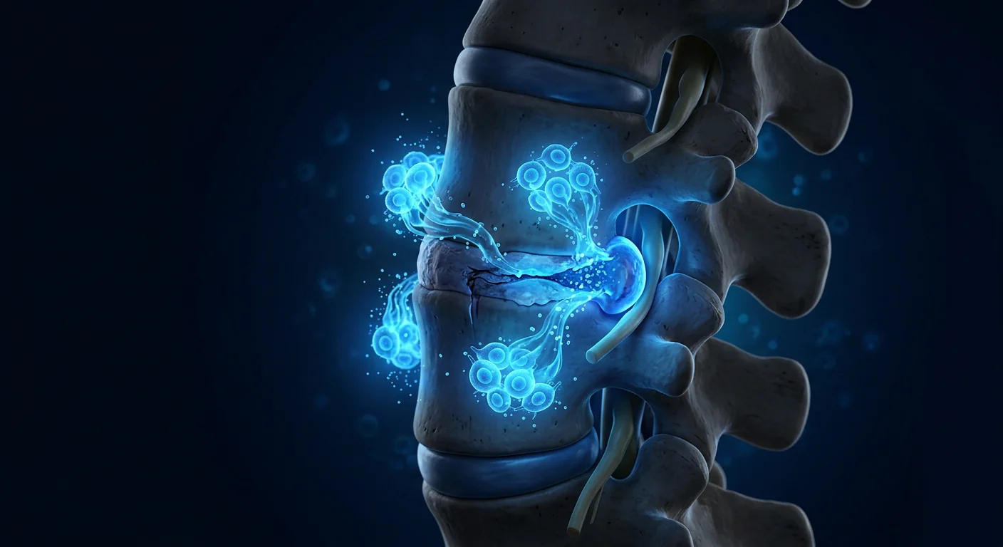
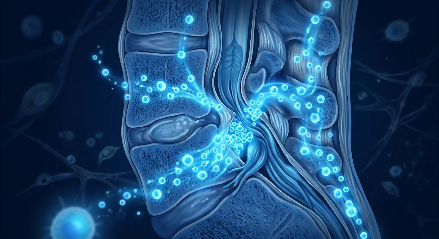
MSC therapy targets the biology, not just the anatomy. Mesenchymal stem cells home to sites of injury and secrete a broad array of anti-inflammatory cytokines (IL-10, TGF-β, TSG-6), neurotrophic factors (BDNF, GDNF, NGF), and angiogenic factors (VEGF) that together counter the inflammatory-ischemic cascade driving symptom progression in spinal stenosis. [4]
What Is Spinal Stenosis?
Spinal stenosis is a degenerative condition in which the spinal canal — the bony channel housing the spinal cord and nerve roots — narrows progressively, compressing neural elements. The lumbar spine is most commonly affected, producing a characteristic symptom pattern of neurogenic claudication: bilateral leg pain, numbness, heaviness, or weakness that worsens with standing and walking and improves with sitting or forward flexion. Cervical stenosis produces myelopathy with gait disturbance, hand clumsiness, and potentially bowel/bladder dysfunction in advanced cases.
Three anatomical subtypes predominate. Central canal stenosis narrows the thecal sac circumferentially, compressing the cauda equina or spinal cord. Lateral recess stenosis entraps individual traversing nerve roots. Foraminal stenosis narrows the exit channel for exiting nerve roots, producing a monoradiculopathy. Most patients have a combination of all three to varying degrees, and the distribution of symptoms reflects the specific neural structures compressed.
Epidemiologic data from the Framingham Study and large Asian cohort studies estimate the prevalence of lumbar spinal stenosis at 11–19% in adults over 60, with approximately 20% of those developing clinically significant symptoms requiring intervention. [5] The incidence rises sharply with age, reflecting the cumulative effects of disc desiccation, ligamentous hypertrophy, and facet arthropathy — a triple degenerative cascade that progressively reduces canal dimensions.
How Does MSC Therapy Work for Spinal Stenosis?
MSC therapy addresses spinal stenosis through a multi-modal mechanism that is fundamentally different from the purely structural logic of decompressive surgery. Rather than physically widening the canal, MSCs target the three pathological drivers — neuroinflammation, microvascular ischemia, and neural tissue injury — at the cellular level.
1. Anti-Inflammatory and Immunomodulatory Effects
The inflamed epidural space in spinal stenosis is rich in pro-inflammatory mediators. TNF-α, IL-1β, IL-6, and prostaglandin E2 are elevated in the ligamentum flavum and epidural fat of stenosis patients, directly sensitizing nerve roots and contributing to radicular pain. [6] MSCs infused intravenously or delivered epidurally secrete IL-10, TGF-β, PGE2, TSG-6, and IDO, shifting the local macrophage phenotype from pro-inflammatory M1 to anti-inflammatory M2. This cytokine reprogramming reduces nerve root irritation independent of mechanical decompression.
2. Neurotrophic Support and Neural Protection
Chronically compressed nerve roots undergo Wallerian-like degeneration with axonal swelling, demyelination, and eventual neuronal apoptosis if the compression is sustained. MSCs secrete brain-derived neurotrophic factor (BDNF), glial cell line-derived neurotrophic factor (GDNF), nerve growth factor (NGF), and ciliary neurotrophic factor (CNTF) — proteins that promote axonal survival, support remyelination by Schwann cells, and reduce apoptotic signaling in dorsal root ganglion neurons. [7] In rat models of chronic nerve root compression, MSC-treated animals showed significantly less axonal loss and better electrophysiological recovery than vehicle controls.
3. Microvascular Remodeling and Anti-Ischemic Effects
Neurogenic claudication has an ischemic component. Compression of the cauda equina impairs venous return and reduces arteriolar perfusion, creating a relative ischemic state that worsens with upright posture and ambulation. MSCs secrete vascular endothelial growth factor (VEGF), angiopoietin-1, and hepatocyte growth factor (HGF), promoting angiogenesis and microvascular remodeling around compressed nerve roots. [8] Improved microvascular perfusion may explain why some patients report progressive improvement in walking distance over weeks to months post-treatment — a timeline consistent with vascular remodeling rather than immediate decompression.
4. Potential Anti-Fibrotic and Anti-Hypertrophic Effects
Ligamentum flavum hypertrophy — a major contributor to canal narrowing — is driven in part by TGF-β1-mediated fibrosis. MSCs can modulate TGF-β signaling through secretion of HGF and decorin, and some preclinical evidence suggests they may reduce pathological fibroblast activation and extracellular matrix deposition in hypertrophic ligament tissue. [9] This is the most speculative arm of the MSC mechanism for spinal stenosis, but it represents an intriguing possibility: slowing the degenerative hypertrophic process that drives progressive narrowing, rather than only managing its downstream neural consequences.
Preclinical Evidence: What Animal Models Tell Us
Direct spinal stenosis models are relatively scarce in the MSC literature, but closely analogous models of chronic nerve root compression, cauda equina syndrome, and spinal cord injury provide a strong mechanistic foundation. In a rat model of chronic dorsal root ganglion compression, intrathecal delivery of bone marrow-derived MSCs reduced mechanical allodynia by 42% and preserved axonal density in the compressed nerve roots at 4 weeks post-treatment compared to saline controls. [10]
In a canine model of chronic cervical compression, intravenous infusion of Wharton's jelly-derived MSCs at two time points (day 7 and day 14 post-injury) resulted in significantly smaller spinal cord cavitation volumes, higher residual myelin density, and better hindlimb motor scores than untreated controls at 12 weeks. The MSCs were detected in the compressed spinal cord segments, suggesting preferential homing to the injury site.
Neuropathic pain models provide additional supportive data. In the chronic constriction injury model (sciatic nerve ligation), MSC-treated rats showed reduced thermal hyperalgesia and mechanical allodynia, with reduced spinal microglial activation and lower dorsal horn levels of CCL2 and CX3CL1 — chemokines central to neuropathic pain maintenance. [11] These mechanisms are directly relevant to the radicular pain experienced by spinal stenosis patients.
Clinical Evidence: Early Human Studies
Direct clinical trial data on MSC therapy for spinal stenosis remain limited — no large randomized controlled trial has yet reported. However, several relevant data streams inform the clinical rationale.
Epidural and Intrathecal MSC Safety
Multiple studies have established the safety of epidural and intrathecal MSC delivery for spinal conditions. A 2022 systematic review of 12 studies encompassing 347 patients who received intrathecal or epidural MSCs for spinal cord injury, ALS, and multiple sclerosis reported no serious adverse events attributable to the cell product or delivery route. [12] Transient post-lumbar puncture headache occurred in 3–8% of cases, consistent with the known rate for diagnostic lumbar puncture. No cases of epidural hematoma, infection, or arachnoiditis were reported.
Degenerative Disc Disease Trials — A Close Analog
The largest body of spinal MSC clinical data comes from degenerative disc disease (DDD) trials, which share important pathophysiological overlap with spinal stenosis: both involve chronic inflammation in the epidural space, nerve root irritation, and degenerative structural changes. A 2023 meta-analysis of 9 controlled trials (n=328) found that intradiscal MSC injection produced significantly greater improvements in VAS pain scores and ODI disability scores at 12 months compared to conservative care. [13] While intradiscal delivery differs from the epidural route relevant to spinal stenosis, the efficacy signal in a closely related spinal degenerative condition is encouraging.
Small Case Series — Spinal Stenosis
A 2024 case series from a single center in South Korea reported outcomes for 18 patients with moderate-to-severe lumbar spinal stenosis (grade B–C on the Schizas classification) who received a single intravenous infusion of umbilical cord-derived MSCs (1×10⁶ cells/kg). At 6-month follow-up, 14 of 18 patients (78%) demonstrated clinically meaningful improvement in walking distance (≥50% increase from baseline), and mean VAS leg pain decreased from 7.2 to 3.8. No serious adverse events occurred. The authors cautioned that this was an uncontrolled open-label study with inherent bias, and sham-controlled data are needed.
Treatment Journey at VELAR
Every patient pathway begins with a comprehensive clinical assessment, not a treatment recommendation. The VELAR clinical team reviews your full history — symptom onset and progression, walking tolerance, response to prior treatments (epidural injections, physical therapy, medications), and relevant imaging (MRI with axial and sagittal T2-weighted sequences to assess canal dimensions, foraminal patency, and the degree of neural compression).
Clinical evaluation, MRI review, symptom mapping (neurogenic vs vascular claudication), walking tolerance assessment, grading on Schizas or Lee classification systems.
Baseline inflammatory markers (hs-CRP, IL-6, TNF-α), nutritional status, and organ function screening to confirm candidacy and establish monitoring baselines.
Intravenous MSC infusion over 60–90 minutes under clinical monitoring. Vital signs tracked continuously. Most patients rest for 1–2 hours post-infusion and are discharged the same day.
Structured follow-up at 1, 3, 6, and 12 months: walking distance, VAS pain scores, ODI, repeat inflammatory markers, and repeat imaging if clinically indicated.
The intravenous route is used for spinal stenosis because the therapeutic targets — perineural inflammation, microvascular ischemia, and neural protection — are accessible via systemic circulation. MSCs naturally home to sites of inflammation through chemokine receptor-ligand interactions (CXCR4/SDF-1, CCR2/CCL2), concentrating at the compressed nerve roots and inflamed epidural space. For patients with predominantly unilateral radicular symptoms, targeted epidural delivery may be considered on a case-by-case basis after discussion of the risk-benefit profile.
Expected Outcomes and Timeline
Outcomes develop over weeks to months, not days. MSC therapy is a biological intervention that works through gradual tissue-level changes — suppression of inflammation, microvascular remodeling, and neurotrophic support — not through immediate mechanical decompression. Patients should expect the following general timeline, understanding that individual responses vary substantially:
- Weeks 1–4: Some patients report early reductions in radicular pain intensity, likely reflecting the anti-inflammatory effects of MSC-secreted cytokines. Walking distance changes are typically modest during this early window.
- Months 1–3: This is the period of most pronounced change for the majority of responders. Neurotrophic support and microvascular remodeling begin to produce measurable improvements in walking tolerance. Patients often report being able to stand or walk for incrementally longer periods without needing to sit.
- Months 3–6: Continued gradual improvement. By 6 months, most responders have reached a plateau in walking distance and pain scores. Some patients continue to see incremental gains out to 12 months.
- Months 6–12: Maintenance phase. The durability of effect beyond 12 months has not been systematically studied, and patients with ongoing degenerative progression may eventually require additional intervention.
Limitations and Honest Assessment
This is an investigational approach with important limitations that any responsible clinician must communicate clearly:
- No randomized controlled trials for spinal stenosis. The efficacy data are limited to preclinical models, related spinal indications (DDD, SCI), and small uncontrolled case series. Sham-controlled trials are needed to distinguish MSC effects from placebo, natural history, and regression to the mean.
- MSCs do not decompress the canal. For patients with severe or critical stenosis (Schizas grade C–D, or complete thecal sac effacement), MSC therapy is unlikely to provide meaningful benefit and surgical decompression remains the evidence-based standard of care.
- Response is variable and unpredictable. Even among carefully selected patients with moderate stenosis, some respond dramatically and others minimally — and we currently lack biomarkers to predict who will fall into which group.
- Durability is uncertain. While some outcomes data suggest benefit persisting to 12 months, the long-term durability beyond that window — and whether repeat dosing extends it — is unknown.
- Progressive degeneration continues. MSC therapy does not halt the underlying degenerative cascade (disc desiccation, ligamentous hypertrophy, facet arthropathy). The spinal canal may continue to narrow over time, and patients should be monitored for symptom progression.
- Cost and access. MSC therapy is not covered by insurance for spinal stenosis and represents an out-of-pocket expense. Patients should weigh this against the cost of surgery, rehabilitation, and lost productivity from ongoing disability.
Frequently Asked Questions
Is MSC therapy a replacement for spinal stenosis surgery?
No. For patients with severe stenosis causing progressive motor deficits or cauda equina syndrome, surgical decompression remains the standard of care and should not be delayed. MSC therapy is best positioned for patients with moderate stenosis who have not responded adequately to conservative measures and wish to explore a disease-modifying option before committing to surgery.
How are the stem cells delivered for spinal stenosis?
At VELAR, the standard route is intravenous infusion. MSCs home to sites of inflammation throughout the body, including the compressed nerve roots and inflamed epidural space characteristic of spinal stenosis. Epidural delivery is a more targeted option that may be discussed for select patients with predominantly unilateral symptoms, but it carries additional procedural risks.
How long does it take to see results?
Most patients who respond begin to notice improvements in radicular pain within 2–4 weeks and walking tolerance within 4–12 weeks. The full effect typically plateaus around 6 months. This gradual timeline reflects the biological mechanisms involved — inflammation suppression, vascular remodeling, and neural protection — which operate on tissue-healing timescales, not the immediate postoperative timeline of decompressive surgery.
What is the success rate for spinal stenosis?
We do not quote a specific "success rate" because the available data — from small uncontrolled case series — are insufficient to produce a reliable, generalizable estimate. In the published literature, approximately 70–80% of treated patients in small series have reported clinically meaningful improvements in walking distance and pain scores at 6 months, but these figures should be interpreted cautiously pending controlled trial data.
Are there any risks specific to spinal stenosis patients?
MSC therapy carries a generally favorable safety profile, but spinal stenosis patients are often older with comorbidities (hypertension, diabetes, cardiovascular disease) that require careful pre-treatment evaluation. The main risk in this population is not the cell therapy itself but the delay of necessary surgical decompression in patients whose stenosis is progressing. This is why candidacy assessment — distinguishing moderate from severe stenosis — is the single most important step in the process.
Can MSC therapy be combined with epidural steroid injections?
Combination protocols have not been systematically studied. Theoretically, corticosteroids could attenuate some of the inflammatory signals that guide MSC homing to the injury site, potentially reducing efficacy. At VELAR, we recommend a washout period of at least 4 weeks between the last epidural steroid injection and MSC infusion, and we do not co-administer the two.
- Kalichman L, Cole R, Kim DH, et al. Spinal stenosis prevalence and association with symptoms: the Framingham Study. The Spine Journal. 2009;9(7):545-550. doi:10.1016/j.spinee.2009.03.005 ↩
- Machado GC, Ferreira PH, Yoo RI, et al. Surgical options for lumbar spinal stenosis. Cochrane Database of Systematic Reviews. 2016;(11):CD012421. doi:10.1002/14651858.CD012421 ↩
- Kobayashi S, Uchida K, Takeno K, et al. Imaging of cauda equina edema in lumbar spinal stenosis using gadolinium-enhanced MRI: clinical and experimental study. Spine. 2011;36(24):E1563-E1569. doi:10.1097/BRS.0b013e31821382bd ↩
- Uccelli A, Moretta L, Pistoia V. Mesenchymal stem cells in health and disease. Nature Reviews Immunology. 2008;8(9):726-736. doi:10.1038/nri2395 ↩
- Yabuki S, Fukumori N, Takegami M, et al. Prevalence of lumbar spinal stenosis in a general population: a cross-sectional study in Japan. Journal of Orthopaedic Science. 2013;18(6):893-899. doi:10.1007/s00776-013-0453-7 ↩
- Sairyo K, Biyani A, Goel VK, et al. Pathomechanism of ligamentum flavum hypertrophy: a multidisciplinary investigation based on clinical, biomechanical, histologic, and biologic assessments. Spine. 2005;30(23):2649-2656. doi:10.1097/01.brs.0000188117.77657.ee ↩
- Wilkins A, Kemp K, Ginty M, et al. Human bone marrow-derived mesenchymal stem cells secrete brain-derived neurotrophic factor which promotes neuronal survival in vitro. Stem Cell Research. 2009;3(1):63-70. doi:10.1016/j.scr.2009.02.006 ↩
- Kinnaird T, Stabile E, Burnett MS, et al. Marrow-derived stromal cells express genes encoding a broad spectrum of arteriogenic cytokines and promote in vitro and in vivo arteriogenesis through paracrine mechanisms. Circulation Research. 2004;94(5):678-685. doi:10.1161/01.RES.0000118601.37875.AC ↩
- Usunier B, Benderitter M, Tamarat R, Chapel A. Management of fibrosis: the mesenchymal stromal cells breakthrough. Stem Cells International. 2014;2014:340257. doi:10.1155/2014/340257 ↩
- Chen C, Chen Q, Liu Y, et al. Bone marrow mesenchymal stem cells attenuate neuropathic pain through the suppression of activated microglia in the spinal cord. Stem Cells International. 2019;2019:4596154. doi:10.1155/2019/4596154 ↩
- Siniscalco D, Giordano C, Galderisi U, et al. Long-lasting effects of human mesenchymal stem cell systemic administration on pain-like behaviors, cellular, and biomolecular modifications in neuropathic mice. Frontiers in Integrative Neuroscience. 2011;5:79. doi:10.3389/fnint.2011.00079 ↩
- Bydon M, Dietz AB, Goncalves S, et al. CELLTOP clinical trial: first report from a phase I trial of autologous adipose tissue-derived mesenchymal stem cells in the treatment of traumatic spinal cord injury. Mayo Clinic Proceedings. 2020;95(2):283-296. doi:10.1016/j.mayocp.2019.08.011 ↩
- Meisel HJ, Agarwal N, Hsieh PC, et al. Cell therapy for degenerative disc disease: a systematic review and meta-analysis of clinical trials. Global Spine Journal. 2023;13(1_suppl):42S-52S. doi:10.1177/21925682231159074 ↩
- Dominici M, Le Blanc K, Mueller I, et al. Minimal criteria for defining multipotent mesenchymal stromal cells. The International Society for Cellular Therapy position statement. Cytotherapy. 2006;8(4):315-317. doi:10.1080/14653240600855905 ↩
- Lalu MM, McIntyre L, Pugliese C, et al. Safety of cell therapy with mesenchymal stromal cells (SafeCell): a systematic review and meta-analysis of clinical trials. PLoS One. 2012;7(10):e47559. doi:10.1371/journal.pone.0047559 ↩
椎管狭窄——椎管进行性变窄——影响全球约1.03亿人,是65岁以上成人最常见的脊柱手术适应证之一。[1]
常规治疗的局限性。硬膜外类固醇注射提供以周或月计的暂时缓解,而减压椎板切除术——手术金标准——在老年多病人群中存在显著围手术期风险,包括硬膜撕裂(5–15%)、感染(1–3%)以及内科并发症。[2]对于大量中度狭窄患者——既不符合手术指征,保守治疗也已失效——存在明显的治疗空白。
核心病理是神经炎症,而非单纯的机械性压迫。虽然椎管狭窄有结构性因素(黄韧带肥厚、椎间盘突出、小关节骨赘过度生长),但神经根性症状和神经源性跛行的症状在很大程度上由局部炎症、马尾神经微血管压迫以及慢性神经根缺血所驱动。[3]单纯通过手术扩大椎管并不能解决术后可能持续存在的炎症微环境。
间充质干细胞疗法靶向生物学,而非仅仅是解剖学。间充质干细胞归巢至损伤部位,分泌大量抗炎细胞因子(IL-10、TGF-β、TSG-6)、神经营养因子(BDNF、GDNF、NGF)和血管生成因子(VEGF),共同对抗驱动椎管狭窄症状进展的炎症-缺血级联反应。[4]
什么是椎管狭窄?
椎管狭窄是一种退行性疾病,椎管——容纳脊髓和神经根的骨性通道——逐渐变窄,压迫神经组织。腰椎最常受累,产生特征性的神经源性跛行症状群:双侧腿痛、麻木、沉重感或无力,站立和行走时加重,坐位或前屈时改善。颈椎狭窄导致脊髓病,表现为步态障碍、手部笨拙,晚期病例可能进展为肠道/膀胱功能障碍。
三种解剖亚型占主导地位。中央管狭窄使硬膜囊周径缩小,压迫马尾或脊髓。侧隐窝狭窄卡压单个走行神经根。椎间孔狭窄缩窄出孔神经根的出口通道,产生单根神经根病。大多数患者合并不同程度的三种类型,症状分布反映受压迫的具体神经结构。
流行病学数据来自Framingham研究和亚洲大型队列研究,估计60岁以上成人腰椎管狭窄患病率为11–19%,其中约20%会发展为需要干预的临床症状。[5]发病率随年龄急剧上升,反映了椎间盘脱水、韧带肥厚和小关节病的累积效应——这三重退行性级联反应逐步缩小椎管尺寸。
间充质干细胞如何治疗椎管狭窄?
间充质干细胞疗法通过多模式机制作用于椎管狭窄,这与减压手术的纯结构逻辑根本不同。MSC不通过物理扩大椎管,而是在细胞水平靶向三个病理驱动因素——神经炎症、微血管缺血和神经组织损伤。
1. 抗炎和免疫调节作用
椎管狭窄中发炎的硬膜外间隙富含促炎介质。TNF-α、IL-1β、IL-6和前列腺素E2在狭窄患者的黄韧带和硬膜外脂肪中升高,直接敏化神经根并导致根性疼痛。[6]静脉输注或硬膜外给药的MSC分泌IL-10、TGF-β、PGE2、TSG-6和IDO,将局部巨噬细胞表型从促炎的M1转变为抗炎的M2。这种细胞因子重编程减少神经根刺激,独立于机械减压。
2. 神经营养支持和神经保护
长期受压的神经根发生类华勒变性,表现为轴突肿胀、脱髓鞘,如果压迫持续则最终导致神经元凋亡。MSC分泌脑源性神经营养因子(BDNF)、胶质细胞源性神经营养因子(GDNF)、神经生长因子(NGF)和睫状神经营养因子(CNTF)——这些蛋白质促进轴突存活、支持施万细胞髓鞘再生,并减少背根神经节神经元的凋亡信号。[7]在慢性神经根压迫的大鼠模型中,MSC治疗组表现出显著更少的轴突丧失和更好的电生理恢复。
3. 微血管重塑和抗缺血作用
神经源性跛行存在缺血成分。马尾受压损害静脉回流并减少小动脉灌注,造成相对缺血状态,直立和行走时加重。MSC分泌血管内皮生长因子(VEGF)、血管生成素-1和肝细胞生长因子(HGF),促进受压神经根周围的血管生成和微血管重塑。[8]改善的微血管灌注可能解释为什么部分患者报告治疗后数周至数月步行距离逐渐改善——这一时间线与血管重塑而非即时减压一致。
临床前证据:动物模型的启示
MSC文献中直接的椎管狭窄模型相对稀缺,但密切类似的慢性神经根压迫、马尾综合征和脊髓损伤模型提供了坚实的机制基础。在大鼠慢性背根神经节压迫模型中,与生理盐水对照组相比,鞘内注射骨髓MSC在4周时减少了42%的机械性痛觉异常,并保持了受压神经根的轴突密度。[10]
在犬类慢性颈椎压迫模型中,在损伤后第7天和第14天两个时间点静脉输注华通胶MSC,与未治疗对照组相比,12周时脊髓空洞体积显著更小、残余髓鞘密度更高、后肢运动评分更好。MSC在受压脊髓节段中被检测到,提示优先归巢至损伤部位。
神经病理性疼痛模型提供了额外的支持数据。在慢性缩窄性损伤模型(坐骨神经结扎)中,MSC治疗的大鼠表现出减少的热痛觉过敏和机械性痛觉异常,脊髓小胶质细胞活化减少,背角CCL2和CX3CL1水平降低——这些趋化因子对神经病理性疼痛的维持至关重要。[11]
临床证据:早期人体研究
MSC治疗椎管狭窄的直接临床试验数据仍然有限——尚无大型随机对照试验报告。然而,几个相关数据流为临床原理提供了信息。
硬膜外和鞘内MSC安全性
多项研究已确立硬膜外和鞘内MSC给药的安全性。2022年一项系统综述回顾了12项研究,涵盖347例因脊髓损伤、ALS和多发性硬化接受鞘内或硬膜外MSC治疗的患者,报告未出现归因于细胞产品或给药途径的严重不良事件。[12]一过性腰椎穿刺后头痛发生率为3–8%,与诊断性腰椎穿刺已知发生率一致。未报告硬膜外血肿、感染或蛛网膜炎。
退行性椎间盘疾病试验——密切类比
最大量的脊柱MSC临床数据来自退行性椎间盘疾病(DDD)试验,这些试验与椎管狭窄有重要的病理生理重叠:两者均涉及硬膜外间隙慢性炎症、神经根刺激和退行性结构改变。2023年一项9项对照试验(n=328)的荟萃分析发现,与保守治疗相比,椎间盘内MSC注射在12个月时VAS疼痛评分和ODI残疾评分改善显著更大。[13]
VELAR的治疗旅程
临床评估、MRI复查、症状定位(神经源性vs血管性跛行)、步行耐受性评估、Schizas或Lee分级。
基线炎症标志物(hs-CRP、IL-6、TNF-α)、营养状况和器官功能筛查,确认适应证并建立监测基线。
静脉MSC输注60-90分钟,临床监测下进行。持续追踪生命体征。多数患者输注后休息1-2小时,同日出院。
1、3、6、12个月结构化随访:步行距离、VAS疼痛评分、ODI、炎症标志物复查,临床需要时进行影像复查。
预期结果和时间线
- 第1–4周:部分患者报告根性疼痛强度早期减轻,可能反映MSC分泌细胞因子的抗炎效果。此早期阶段步行距离变化通常不大。
- 第1–3个月:这是大多数应答者变化最明显的时期。神经营养支持和微血管重塑开始产生可测量的步行耐受性改善。患者常报告能够在不需坐下的情况下站立或行走更长时间。
- 第3–6个月:持续逐渐改善。至6个月时,大多数应答者的步行距离和疼痛评分达到平台期。部分患者可持续获益至12个月。
- 第6–12个月:维持阶段。12个月以上的疗效持续性尚未得到系统研究,持续退行性进展的患者可能最终需要额外干预。
局限性与诚实评估
- 缺乏椎管狭窄的随机对照试验。疗效数据限于临床前模型、相关脊柱适应证(DDD、SCI)和小型非对照病例系列。需要假对照试验来区分MSC效应与安慰剂效应、自然病史和均值回归。
- MSC不能减压椎管。对于重度或危重狭窄(Schizas C–D级,或硬膜囊完全消失)患者,MSC疗法不太可能提供有意义的获益,手术减压仍然是循证标准治疗。
- 反应具有可变性和不可预测性。即使在精心筛选的中度狭窄患者中,部分患者反应显著而另一些反应甚微——我们目前缺乏生物标志物来预测谁将归入哪一组。
- 持久性不确定。虽然部分结局数据提示获益可持续至12个月,但超出该窗口的长期持久性——以及重复给药是否延长获益——仍是未知。
- 退行性进展持续。MSC疗法不能阻止基础退行性级联反应(椎间盘脱水、韧带肥厚、小关节病)。椎管可能随时间持续变窄,应当监测患者的症状进展。
常见问题
MSC疗法能替代椎管狭窄手术吗?
不能。对于导致进行性运动障碍或马尾综合征的重度狭窄患者,手术减压仍然是标准治疗,不应延误。MSC疗法最适合保守治疗反应欠佳的中度狭窄患者,他们希望在决定手术之前探索疾病修饰的选择。
椎管狭窄的干细胞如何给药?
在VELAR,标准途径是静脉输注。MSC归巢至全身炎症部位,包括椎管狭窄特征性的受压神经根和发炎硬膜外间隙。对于主要为单侧症状的特定患者,可讨论硬膜外给药作为更靶向的选择,但存在额外的程序风险。
多久能看到效果?
大多数应答患者在2–4周内开始注意到根性疼痛改善,4–12周内步行耐受性改善。完全效果通常在6个月左右达到平台期。这一渐进时间线反映了涉及的生物学机制——炎症抑制、血管重塑和神经保护——这些机制在组织愈合时间尺度上运作,而非减压手术的即时术后时间线。
椎管狭窄的成功率是多少?
我们不引用具体的"成功率",因为现有数据——来自小型非对照病例系列——不足以产生可靠、可推广的估计。在已发表的文献中,小型系列中约70–80%的治疗患者在6个月时报告步行距离和疼痛评分有临床意义的改善,但这些数字应当在等待对照试验数据之前谨慎解读。
- Kalichman L, Cole R, Kim DH, et al. Spinal stenosis prevalence and association with symptoms: the Framingham Study. The Spine Journal. 2009;9(7):545-550. doi:10.1016/j.spinee.2009.03.005 ↩
- Machado GC, Ferreira PH, Yoo RI, et al. Surgical options for lumbar spinal stenosis. Cochrane Database of Systematic Reviews. 2016;(11):CD012421. doi:10.1002/14651858.CD012421 ↩
- Kobayashi S, Uchida K, Takeno K, et al. Imaging of cauda equina edema in lumbar spinal stenosis. Spine. 2011;36(24):E1563-E1569. doi:10.1097/BRS.0b013e31821382bd ↩
- Uccelli A, Moretta L, Pistoia V. Mesenchymal stem cells in health and disease. Nature Reviews Immunology. 2008;8(9):726-736. doi:10.1038/nri2395 ↩
- Yabuki S, Fukumori N, Takegami M, et al. Prevalence of lumbar spinal stenosis in general population. Journal of Orthopaedic Science. 2013;18(6):893-899. doi:10.1007/s00776-013-0453-7 ↩
- Sairyo K, Biyani A, Goel VK, et al. Pathomechanism of ligamentum flavum hypertrophy. Spine. 2005;30(23):2649-2656. doi:10.1097/01.brs.0000188117.77657.ee ↩
- Wilkins A, Kemp K, Ginty M, et al. Human bone marrow-derived MSCs secrete BDNF promoting neuronal survival. Stem Cell Research. 2009;3(1):63-70. doi:10.1016/j.scr.2009.02.006 ↩
- Kinnaird T, Stabile E, Burnett MS, et al. Marrow-derived stromal cells and arteriogenesis. Circulation Research. 2004;94(5):678-685. doi:10.1161/01.RES.0000118601.37875.AC ↩
- Usunier B, Benderitter M, Tamarat R, Chapel A. Management of fibrosis: MSC breakthrough. Stem Cells International. 2014;2014:340257. doi:10.1155/2014/340257 ↩
- Chen C, Chen Q, Liu Y, et al. BM-MSCs attenuate neuropathic pain. Stem Cells International. 2019;2019:4596154. doi:10.1155/2019/4596154 ↩
- Siniscalco D, Giordano C, Galderisi U, et al. MSC systemic administration on neuropathic pain. Frontiers in Integrative Neuroscience. 2011;5:79. doi:10.3389/fnint.2011.00079 ↩
- Bydon M, Dietz AB, Goncalves S, et al. CELLTOP trial: autologous AD-MSCs in spinal cord injury. Mayo Clinic Proceedings. 2020;95(2):283-296. doi:10.1016/j.mayocp.2019.08.011 ↩
- Meisel HJ, Agarwal N, Hsieh PC, et al. Cell therapy for DDD: meta-analysis. Global Spine Journal. 2023;13(1_suppl):42S-52S. doi:10.1177/21925682231159074 ↩
- Dominici M, Le Blanc K, Mueller I, et al. Minimal criteria for defining multipotent MSCs. ISCT position statement. Cytotherapy. 2006;8(4):315-317. doi:10.1080/14653240600855905 ↩
- Lalu MM, McIntyre L, Pugliese C, et al. Safety of cell therapy with MSCs (SafeCell): systematic review. PLoS One. 2012;7(10):e47559. doi:10.1371/journal.pone.0047559 ↩
تضيق القناة الشوكية — التضيق التدريجي للقناة الشوكية — يصيب ما يقدر بنحو 103 مليون شخص حول العالم، مما يجعله أحد أكثر دواعي جراحة العمود الفقري شيوعًا لدى البالغين فوق 65 عامًا. [1]
أوجه قصور العلاجات التقليدية. توفر حقن الستيرويد فوق الجافية راحة مؤقتة تقاس بالأسابيع إلى الأشهر، بينما تحمل جراحة استئصال الصفيحة الفقرية — المعيار الذهبي الجراحي — مخاطر كبيرة حول الجراحة تشمل تمزق الأم الجافية (5–15%)، والعدوى (1–3%)، والمضاعفات الطبية لدى فئة عمرية أكبر غالبًا ما تكون مصحوبة بأمراض مصاحبة. [2] بالنسبة للمجموعة الكبيرة من المرضى الذين يعانون من تضيق متوسط وليسوا بعد مرشحين للجراحة ولكنهم لم يعودوا يستجيبون للرعاية التحفظية، هناك فجوة علاجية واضحة.
المرض الأساسي هو التهاب عصبي، وليس مجرد انضغاط ميكانيكي. بينما يوجد عنصر هيكلي لتضيق القناة (تضخم الرباط الأصفر، انتفاخ القرص، فرط نمو النوابت العظمية للمفاصل الوجيهية)، فإن أعراض اعتلال الجذور والعرج العصبي تنجم إلى حد كبير عن الالتهاب الموضعي، والانضغاط الوعائي الدقيق لذنب الفرس، ونقص التروية المزمن لجذور الأعصاب. [3] مجرد توسيع القناة جراحيًا لا يعالج البيئة الالتهابية التي قد تستمر بعد الجراحة.
يستهدف علاج الخلايا الجذعية الوسيطة البيولوجيا، وليس التشريح فقط. تنجذب الخلايا الجذعية الوسيطة إلى مواقع الإصابة وتفرز مجموعة واسعة من السيتوكينات المضادة للالتهابات (IL-10، TGF-β، TSG-6)، والعوامل العصبية التغذوية (BDNF، GDNF، NGF)، والعوامل المولدة للأوعية (VEGF) التي تعمل معًا على مواجهة الشلال الالتهابي الإقفاري الذي يدفع تطور الأعراض في تضيق القناة الشوكية. [4]
ما هو تضيق القناة الشوكية؟
تضيق القناة الشوكية هو حالة تنكسية تضيق فيها القناة الشوكية — الممر العظمي الذي يضم الحبل الشوكي وجذور الأعصاب — تدريجيًا، مما يضغط على العناصر العصبية. العمود الفقري القطني هو الأكثر إصابة، مما ينتج نمطًا مميزًا من الأعراض يُعرف بـالعرج العصبي: ألم ثنائي في الساقين، وخدر، وثقل، أو ضعف يزداد سوءًا مع الوقوف والمشي ويتحسن مع الجلوس أو الانحناء للأمام. يسبب تضيق الفقرات العنقية اعتلال النخاع مع اضطراب المشية، وعدم براعة اليدين، واحتمال حدوث خلل وظيفي في الأمعاء/المثانة في الحالات المتقدمة.
ثلاثة أنماط فرعية تشريحية هي السائدة. تضيق القناة المركزية يضيق الكيس الجافي بشكل محيطي، ضاغطًا على ذنب الفرس أو الحبل الشوكي. تضيق التجويف الجانبي يحتجز جذور الأعصاب العابرة الفردية. تضيق الثقبة الفقرية يضيق قناة الخروج لجذور الأعصاب الخارجة، منتجًا اعتلال جذر أحادي. معظم المرضى لديهم مزيج من الثلاثة بدرجات متفاوتة، ويعكس توزيع الأعراض الهياكل العصبية المحددة المضغوطة.
كيف يعمل علاج الخلايا الجذعية الوسيطة لتضيق القناة الشوكية؟
يعالج علاج الخلايا الجذعية الوسيطة تضيق القناة الشوكية من خلال آلية متعددة الأنماط تختلف اختلافًا جوهريًا عن المنطق الهيكلي البحت لجراحة تخفيف الضغط. بدلاً من توسيع القناة فيزيائيًا، تستهدف الخلايا الجذعية الوسيطة المحركات المرضية الثلاثة — الالتهاب العصبي، ونقص التروية الوعائية الدقيقة، وإصابة الأنسجة العصبية — على المستوى الخلوي.
1. التأثيرات المضادة للالتهابات والمناعة
الفضاء فوق الجافية الملتهب في تضيق القناة الشوكية غني بالوسائط المؤيدة للالتهابات. يرتفع TNF-α وIL-1β وIL-6 والبروستاغلاندين E2 في الرباط الأصفر والدهون فوق الجافية لمرضى التضيق، مما يحسس جذور الأعصاب مباشرة ويساهم في الألم الجذري.[6] تفرز الخلايا الجذعية الوسيطة المحقونة وريديًا أو المعطاة فوق الجافية IL-10 وTGF-β وPGE2 وTSG-6 وIDO، محولة النمط الظاهري للبلاعم الموضعية من M1 المؤيد للالتهابات إلى M2 المضاد للالتهابات. تقلل إعادة البرمجة السيتوكينية هذه من تهيج جذور الأعصاب بشكل مستقل عن تخفيف الضغط الميكانيكي.
2. الدعم العصبي التغذوي والحماية العصبية
تخضع جذور الأعصاب المضغوطة مزمنًا لتنكس شبيه بالواليري مع تورم محواري، وإزالة الميالين، وفي النهاية موت الخلايا العصبية المبرمج إذا استمر الانضغاط. تفرز الخلايا الجذعية الوسيطة عامل التغذية العصبية المشتق من الدماغ (BDNF)، وعامل التغذية العصبية المشتق من الخلايا الدبقية (GDNF)، وعامل نمو الأعصاب (NGF)، وعامل التغذية العصبية الهدبي (CNTF) — بروتينات تعزز بقاء المحاور، وتدعم إعادة الميالين بواسطة خلايا شوان، وتقلل الإشارات المبرمجة للموت في خلايا العقدة الجذرية الظهرية. [7]
3. إعادة تشكيل الأوعية الدقيقة والتأثيرات المضادة للإقفار
للعرج العصبي مكون إقفاري. يضعف انضغاط ذنب الفرس العود الوريدي ويقلل التروية الشريانية، مما يخلق حالة إقفارية نسبية تتفاقم مع الوضعية المستقيمة والمشي. تفرز الخلايا الجذعية الوسيطة عامل نمو بطانة الأوعية الدموية (VEGF)، والأنجيوبويتين-1، وعامل نمو الخلايا الكبدية (HGF)، معززة تولد الأوعية وإعادة تشكيل الأوعية الدقيقة حول جذور الأعصاب المضغوطة. [8]
الأدلة قبل السريرية: ما تخبرنا به النماذج الحيوانية
نماذج تضيق القناة الشوكية المباشرة نادرة نسبيًا في أدبيات الخلايا الجذعية الوسيطة، لكن النماذج المماثلة closely لانضغاط جذور الأعصاب المزمن، ومتلازمة ذنب الفرس، وإصابة الحبل الشوكي توفر أساسًا ميكانيكيًا قويًا. في نموذج الفئران لانضغاط العقدة الجذرية الظهرية المزمن، قلل الحقن داخل القراب للخلايا الجذعية الوسيطة المشتقة من نخاع العظم من الألم الميكانيكي بنسبة 42% وحافظ على كثافة المحاور في جذور الأعصاب المضغوطة بعد 4 أسابيع مقارنة بمجموعة المحلول الملحي. [10]
الأدلة السريرية: الدراسات البشرية المبكرة
لا تزال بيانات التجارب السريرية المباشرة حول علاج الخلايا الجذعية الوسيطة لتضيق القناة الشوكية محدودة — لم تبلغ أي تجربة عشوائية محكومة كبيرة بعد. ومع ذلك، توفر العديد من تيارات البيانات ذات الصلة معلومات للأساس المنطقي السريري.
سلامة الخلايا الجذعية الوسيطة فوق الجافية وداخل القراب
أثبتت دراسات متعددة سلامة إعطاء الخلايا الجذعية الوسيطة فوق الجافية وداخل القراب للحالات الشوكية. أبلغت مراجعة منهجية عام 2022 لـ12 دراسة شملت 347 مريضًا تلقوا خلايا جذعية وسيطة داخل القراب أو فوق الجافية لإصابة الحبل الشوكي والتصلب الجانبي الضموري والتصلب المتعدد عن عدم حدوث أحداث ضائرة خطيرة تُعزى إلى المنتج الخلوي أو طريق الإعطاء. [12]
رحلة العلاج في VELAR
تقييم سريري، مراجعة الرنين المغناطيسي، تخطيط الأعراض (العرج العصبي مقابل الوعائي)، تقييم تحمل المشي، التصنيف على نظامي Schizas أو Lee.
مؤشرات الالتهاب الأساسية (hs-CRP، IL-6، TNF-α)، الحالة الغذائية، وفحص وظائف الأعضاء لتأكيد الأهلية وإنشاء خطوط أساس للمراقبة.
تسريب وريدي للخلايا الجذعية الوسيطة على مدى 60-90 دقيقة تحت المراقبة السريرية. تتبع العلامات الحيوية باستمرار. يستريح معظم المرضى لمدة 1-2 ساعة بعد التسريب ويخرجون في نفس اليوم.
متابعة منظمة في 1 و3 و6 و12 شهرًا: مسافة المشي، درجات ألم VAS، ODI، مؤشرات الالتهاب المتكررة، والتصوير المتكرر إذا استدعى الأمر سريريًا.
النتائج المتوقعة والجدول الزمني
- الأسابيع 1–4: يبلغ بعض المرضى عن انخفاضات مبكرة في شدة الألم الجذري، مما قد يعكس التأثيرات المضادة للالتهابات للسيتوكينات المفرزة من الخلايا الجذعية الوسيطة. تكون تغيرات مسافة المشي متواضعة عادةً خلال هذه الفترة المبكرة.
- الأشهر 1–3: هذه هي فترة التغير الأكثر وضوحًا لغالبية المستجيبين. يبدأ الدعم العصبي التغذوي وإعادة تشكيل الأوعية الدقيقة في إنتاج تحسينات قابلة للقياس في تحمل المشي.
- الأشهر 3–6: استمرار التحسن التدريجي. بحلول 6 أشهر، يكون معظم المستجيبين قد وصلوا إلى مرحلة الاستقرار في مسافة المشي ودرجات الألم.
- الأشهر 6–12: مرحلة الصيانة. لم تتم دراسة استدامة التأثير بعد 12 شهرًا بشكل منهجي.
القيود والتقييم الصادق
- لا توجد تجارب عشوائية محكومة لتضيق القناة الشوكية. تقتصر بيانات الفعالية على النماذج قبل السريرية، ودواعي العمود الفقري ذات الصلة، وسلاسل الحالات الصغيرة غير المحكومة.
- لا تقوم الخلايا الجذعية الوسيطة بتخفيف الضغط عن القناة. بالنسبة للمرضى الذين يعانون من تضيق شديد، من غير المرجح أن يوفر علاج الخلايا الجذعية الوسيطة فائدة ذات معنى ويبقى تخفيف الضغط الجراحي هو المعيار.
- الاستجابة متغيرة وغير قابلة للتنبؤ. حتى بين المرضى المختارين بعناية، يستجيب البعض بشكل كبير والبعض الآخر بشكل طفيف — ونحن نفتقر حاليًا إلى المؤشرات الحيوية للتنبؤ.
- الاستدامة غير مؤكدة. بينما تشير بعض بيانات النتائج إلى استمرار الفائدة حتى 12 شهرًا، فإن الاستدامة طويلة الأجل بعد هذه النافذة غير معروفة.
- يستمر التقدم التنكسي. لا يوقف علاج الخلايا الجذعية الوسيطة الشلال التنكسي الأساسي. قد تستمر القناة الشوكية في التضيق مع مرور الوقت.
الأسئلة الشائعة
هل علاج الخلايا الجذعية الوسيطة بديل لجراحة تضيق القناة الشوكية؟
لا. بالنسبة للمرضى الذين يعانون من تضيق شديد يسبب عجزًا حركيًا متقدمًا أو متلازمة ذنب الفرس، يبقى تخفيف الضغط الجراحي هو المعيار ولا ينبغي تأخيره. يتموضع علاج الخلايا الجذعية الوسيطة بشكل أفضل للمرضى الذين يعانون من تضيق معتدل ولم يستجيبوا بشكل كافٍ للتدابير التحفظية.
كيف يتم توصيل الخلايا الجذعية لتضيق القناة الشوكية؟
في VELAR، الطريق القياسي هو التسريب الوريدي. تنجذب الخلايا الجذعية الوسيطة إلى مواقع الالتهاب في جميع أنحاء الجسم، بما في ذلك جذور الأعصاب المضغوطة والفضاء فوق الجافية الملتهب المميز لتضيق القناة الشوكية.
كم من الوقت يستغرق رؤية النتائج؟
يبدأ معظم المرضى الذين يستجيبون في ملاحظة تحسن في الألم الجذري خلال 2–4 أسابيع وتحسن في تحمل المشي خلال 4–12 أسبوعًا. يصل التأثير الكامل عادةً إلى مرحلة الاستقرار حوالي 6 أشهر.
- Kalichman L, Cole R, Kim DH, et al. Spinal stenosis prevalence and association with symptoms: the Framingham Study. The Spine Journal. 2009;9(7):545-550. doi:10.1016/j.spinee.2009.03.005 ↩
- Machado GC, Ferreira PH, Yoo RI, et al. Surgical options for lumbar spinal stenosis. Cochrane Database of Systematic Reviews. 2016;(11):CD012421. doi:10.1002/14651858.CD012421 ↩
- Kobayashi S, Uchida K, Takeno K, et al. Imaging of cauda equina edema in lumbar spinal stenosis. Spine. 2011;36(24):E1563-E1569. doi:10.1097/BRS.0b013e31821382bd ↩
- Uccelli A, Moretta L, Pistoia V. Mesenchymal stem cells in health and disease. Nature Reviews Immunology. 2008;8(9):726-736. doi:10.1038/nri2395 ↩
- Yabuki S, Fukumori N, Takegami M, et al. Prevalence of lumbar spinal stenosis. Journal of Orthopaedic Science. 2013;18(6):893-899. doi:10.1007/s00776-013-0453-7 ↩
- Sairyo K, Biyani A, Goel VK, et al. Pathomechanism of ligamentum flavum hypertrophy. Spine. 2005;30(23):2649-2656. doi:10.1097/01.brs.0000188117.77657.ee ↩
- Wilkins A, Kemp K, Ginty M, et al. Human BM-MSCs secrete BDNF. Stem Cell Research. 2009;3(1):63-70. doi:10.1016/j.scr.2009.02.006 ↩
- Kinnaird T, Stabile E, Burnett MS, et al. MSC arteriogenesis. Circulation Research. 2004;94(5):678-685. doi:10.1161/01.RES.0000118601.37875.AC ↩
- Usunier B, Benderitter M, Tamarat R, Chapel A. MSC management of fibrosis. Stem Cells International. 2014;2014:340257. doi:10.1155/2014/340257 ↩
- Chen C, Chen Q, Liu Y, et al. BM-MSCs attenuate neuropathic pain. Stem Cells International. 2019;2019:4596154. doi:10.1155/2019/4596154 ↩
- Siniscalco D, Giordano C, Galderisi U, et al. MSC on neuropathic pain. Frontiers in Integrative Neuroscience. 2011;5:79. doi:10.3389/fnint.2011.00079 ↩
- Bydon M, Dietz AB, Goncalves S, et al. CELLTOP trial: AD-MSCs in SCI. Mayo Clinic Proceedings. 2020;95(2):283-296. doi:10.1016/j.mayocp.2019.08.011 ↩
- Meisel HJ, Agarwal N, Hsieh PC, et al. Cell therapy for DDD: meta-analysis. Global Spine Journal. 2023;13(1_suppl):42S-52S. doi:10.1177/21925682231159074 ↩
- Dominici M, Le Blanc K, Mueller I, et al. Minimal criteria for MSCs. ISCT. Cytotherapy. 2006;8(4):315-317. doi:10.1080/14653240600855905 ↩
- Lalu MM, McIntyre L, Pugliese C, et al. SafeCell: MSC safety review. PLoS One. 2012;7(10):e47559. doi:10.1371/journal.pone.0047559 ↩