Osteoarthritis (OA) is the most common form of arthritis worldwide, affecting an estimated 500 million people — yet it is frequently misunderstood as simply "wear and tear." In reality, OA is a complex, whole-joint disease in which mechanical stress, low-grade inflammation, and failed repair processes converge to degrade articular cartilage, remodel subchondral bone, and inflame the synovial lining. Unlike rheumatoid arthritis, which is driven by systemic autoimmunity, OA arises from an imbalance between the joint's capacity to maintain and repair its tissues and the loads placed upon it. Age, obesity, previous injury, and genetic predisposition all tip that balance. For decades, treatment has been palliative — pain relief, physiotherapy, and eventually joint replacement. The question that mesenchymal stem cell (MSC) research is now asking is whether the biology of repair can be meaningfully supported before the joint reaches the surgical endpoint.
What actually happens inside an osteoarthritic joint
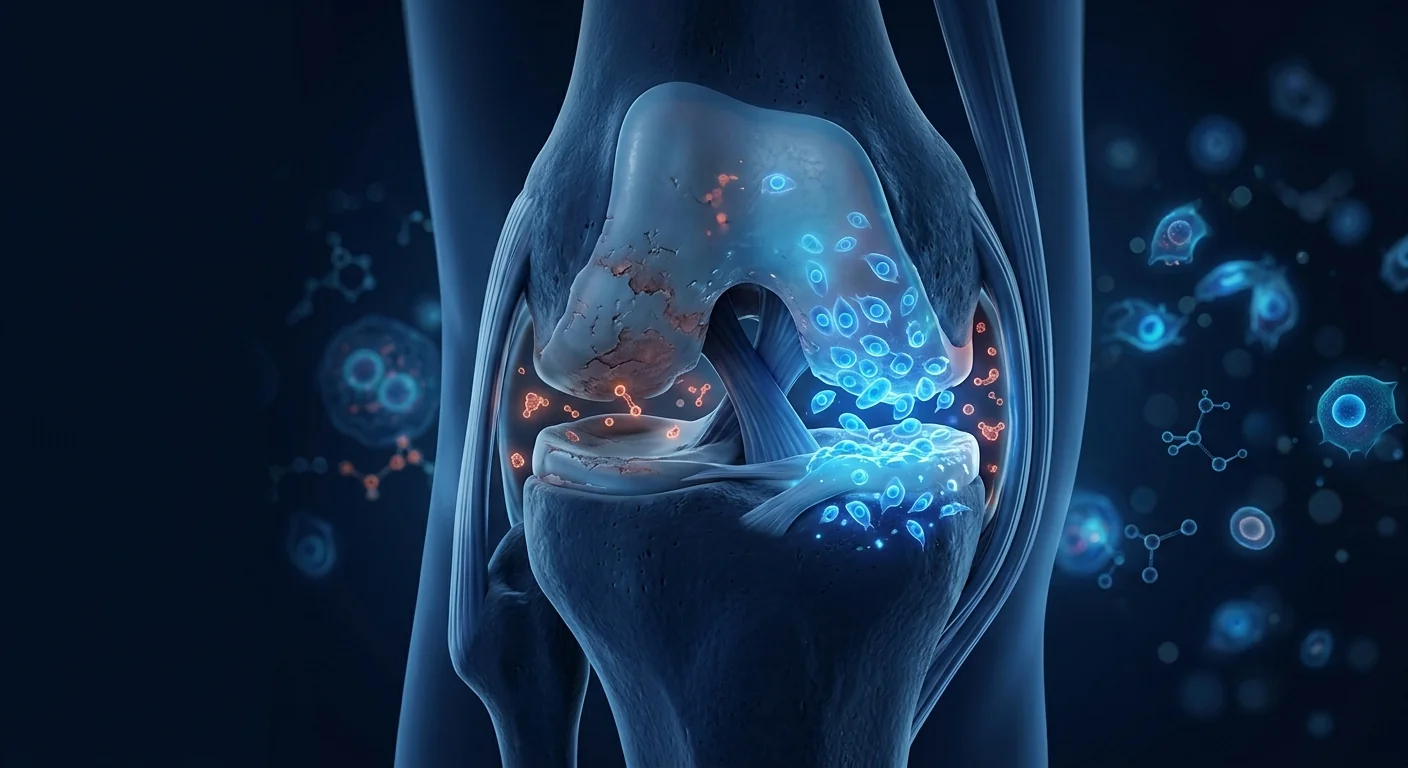
The healthy joint is an exquisitely tuned biomechanical system. Articular cartilage — a smooth, resilient tissue that covers the ends of bones — is maintained by chondrocytes, the resident cells that continuously synthesise and remodel the extracellular matrix of collagen and proteoglycans. In OA, this equilibrium breaks. Chondrocytes become senescent — they stop dividing and begin secreting pro-inflammatory cytokines, matrix-degrading enzymes (notably MMP-13 and ADAMTS-5), and reactive oxygen species that further damage the surrounding matrix. Cartilage loses its water-binding proteoglycans, its surface fibrillates, and fissures deepen until bone is exposed.
What was once considered a purely mechanical disease is now understood to have a significant inflammatory component. Synovial macrophages become activated, releasing IL-1β, TNF-α, and IL-6 into the joint space. These cytokines amplify chondrocyte catabolism and sensitise peripheral nociceptors, contributing to the pain that defines the patient's experience. The subchondral bone remodels abnormally, forming sclerotic plates and osteophytes. Critically, unlike in rheumatoid arthritis, this inflammation is low-grade and compartmentalised — it stays largely within the joint rather than becoming systemic. That distinction is important because it shapes what a cell therapy would need to achieve: not systemic immune suppression, but local modulation of a degenerative microenvironment.
Why mesenchymal stem cells are a candidate for osteoarthritis
The rationale for MSCs in OA rests on three overlapping capabilities that map directly onto the disease process. First, MSCs can exert chondroprotective and trophic effects — they secrete growth factors including TGF-β, IGF-1, and BMPs that support the survival and matrix-producing activity of the patient's own chondrocytes. In co-culture experiments, MSCs reduce chondrocyte apoptosis and slow the loss of proteoglycans from cartilage explants exposed to inflammatory stimuli. Second, they are anti-inflammatory — MSCs suppress the activation of synovial macrophages and shift them from a pro-inflammatory (M1) toward a tissue-remodelling (M2) phenotype, thereby lowering the intra-articular concentrations of IL-1β and TNF-α. Third, they provide direct matrix support — although MSCs are not expected to regenerate a pristine articular surface de novo, they can differentiate into chondrocyte-like cells and contribute to the repair tissue that fills cartilage defects, particularly when delivered on a scaffold or in combination with microfracture.
Importantly, MSC therapy for OA is almost always delivered intra-articularly — injected directly into the joint space — unlike the intravenous route used in many systemic autoimmune and inflammatory conditions. This local delivery concentrates the cells where they are needed, reduces systemic exposure, and has contributed to the favourable safety profile observed across dozens of clinical studies.
What the clinical evidence actually shows
The clinical literature on MSCs for knee OA is now substantial — more than for any other MSC indication — and it tells a story that is encouraging but incomplete. Multiple systematic reviews and meta-analyses have aggregated data from randomised controlled trials (RCTs) and concluded that intra-articular MSC injection is associated with statistically significant and clinically meaningful reductions in pain, as measured by the Visual Analogue Scale (VAS) and the WOMAC pain subscale, and with improvements in function that persist for 12 to 24 months in many studies [1][2].
Several individual trials merit attention for what they show — and for what they leave open. A 2015 RCT by Vega and colleagues compared intra-articular allogeneic bone-marrow MSCs against hyaluronic acid in 30 patients and reported significantly better pain and cartilage quality scores (measured by T2 mapping on MRI) in the MSC group at 12 months [3]. A larger 2016 phase I/II multicentre trial by Lamo-Espinosa et al. randomised patients to two different MSC doses or hyaluronic acid alone, finding that the higher dose (100 × 10⁶ cells) produced significant WOMAC and VAS improvements at 12 months compared to the control [4]. In 2019, Matas and colleagues reported a randomised comparison of single versus repeated umbilical-cord MSC injections against hyaluronic acid: the repeated-dose group showed superior pain and function outcomes at 12 months, and importantly, MRI analysis suggested a disease-modifying effect — less progression of cartilage loss in the MSC-treated knees [5].
The cell sources studied are diverse. Bone-marrow-derived MSCs have the longest track record, but umbilical-cord-derived MSCs — which are younger, more proliferative, and avoid the need for a bone-marrow harvest — have produced some of the strongest signals in recent trials [6]. Adipose-derived stromal vascular fraction (SVF) and culture-expanded adipose MSCs have also been widely studied, with meta-analyses suggesting they produce comparable pain relief to bone-marrow MSCs in the short to medium term, though comparative data remain limited [7][8].
The honest headline
As of mid-2026, no MSC product is approved by the FDA or EMA as a standard-of-care treatment for osteoarthritis. The evidence base is considerably stronger than for many other MSC indications — multiple RCTs with consistent pain and function signals — but it has not yet reached the level of regulatory approval. The responsible description is advanced investigational therapy with a growing evidence base, not a proven cure. Any clinic promising guaranteed cartilage regrowth or complete pain resolution is misrepresenting the data.
How OA outcomes are measured — and what MSCs actually move
To weigh the evidence seriously requires knowing what is being measured. In OA trials, the gold-standard patient-reported outcome is the WOMAC (Western Ontario and McMaster Universities Osteoarthritis Index), which captures pain, stiffness, and physical function across 24 items. The VAS pain score (0–100 mm) provides a simpler, single-dimension readout. The KOOS (Knee Injury and Osteoarthritis Outcome Score) extends WOMAC to include sports and quality-of-life domains. On the structural side, MRI with T2 mapping and delayed gadolinium-enhanced techniques (dGEMRIC) can quantify cartilage composition and volume, while plain radiographs track joint-space narrowing — though structural change lags symptom change by months to years.
Across the RCTs, the pattern is consistent: MSC-treated groups show greater pain reduction than controls (typically hyaluronic acid or saline) by 6–12 months, with mean VAS improvements in the range of 20–40 mm and WOMAC improvements of 15–25 points — both exceeding the thresholds considered clinically meaningful. Functional gains (stair climbing, walking distance) follow a similar trajectory. The structural data are more preliminary: several trials have reported slower cartilage volume loss or improved T2 relaxation times on MRI, but the effects are modest and not universal [9]. The strongest structural signals have come from trials using repeated dosing rather than a single injection [5].
What the evidence supports — and what it doesn't
A fair reading of the data supports several conclusions. Intra-articular MSC therapy for knee OA has a well-established short- and medium-term safety record, with adverse events generally limited to transient post-injection pain and swelling. It consistently reduces pain and improves function in controlled trials, with effect sizes that are clinically meaningful and appear durable for 12–24 months. There are preliminary structural signals suggesting possible disease modification, but these require confirmation in larger, longer trials with MRI-based primary endpoints.
What the evidence does NOT yet support is equally important. It does not establish that MSCs reverse established OA — grade 4 (bone-on-bone) joints with complete cartilage loss do not regenerate a new cartilage surface. It does not confirm durability beyond 2–3 years, as most trials follow patients for 12–24 months. It does not identify a single optimal protocol — cell source, dose, frequency, and patient selection criteria vary widely across studies. And it does not demonstrate that MSC therapy is superior to total knee arthroplasty for end-stage disease — surgery remains the definitive option when conservative measures have been exhausted.
- Grade matters. Patients with moderate OA (Kellgren-Lawrence grades 2–3) are the population in whom the strongest trial signals have been seen. Grade 4 joints have minimal cartilage remaining and are less likely to benefit from a biologic intervention aimed at supporting repair.
- Dosing and repetition are open questions. The most promising structural data come from repeated-dose protocols, but the optimal interval and total dose have not been defined. Single-injection regimens produce meaningful symptom relief but weaker structural signals.
- Combination approaches are under investigation. Several groups are studying MSCs co-delivered with platelet-rich plasma (PRP), hyaluronic acid scaffolds, or microfracture, with the hypothesis that the scaffold or marrow stimulation provides a substrate for MSC-mediated repair [10].
Osteoarthritis is not a single disease with a single time course — it is a spectrum from early metabolic disturbance to end-stage joint failure. The honest contribution of MSC therapy is likely to be in the middle of that spectrum: slowing progression and reducing symptoms in joints that still have cartilage worth preserving.
— VELAR Clinical Team
How to evaluate any offer responsibly
If you are considering MSC therapy for osteoarthritis, the diligence is straightforward and the same questions protect against over-promise in any medical domain. Ask what cell type and source are used — umbilical-cord MSCs, bone-marrow MSCs, and adipose SVF are biologically different products with different evidence bases. Ask whether the provider can cite published trial data for their specific protocol, not just general references to the field. Ask what outcome measures they use — a provider who tracks your WOMAC and VAS scores before and after treatment is operating with a level of rigour that is absent from clinics offering only testimonials. Ask about cell characterisation: are the cells tested for identity (ISCT criteria: CD73⁺, CD90⁺, CD105⁺, CD45⁻), viability (>95% at delivery), and sterility before release? Third-party quality data are a meaningful signal of laboratory standards. Be deeply sceptical of guarantees, of claims that grade-4 joints will regenerate cartilage, and of any suggestion that MSC therapy is an alternative to weight management, physiotherapy, and appropriate analgesia — it is an adjunct, not a replacement, for established conservative care.
The VELAR perspective
At VELAR Center, osteoarthritis is one of the conditions we treat most frequently — and precisely because it is common, we believe the standard of evidence required before making any claim is higher, not lower. Our approach is conservative by design: we use umbilical-cord-derived MSCs manufactured under cGMP conditions with ISCT-compliant characterisation and third-party sterility release, we track structured outcomes (WOMAC, VAS, and where indicated, follow-up imaging), and we position MSC therapy as a bridge — a biological intervention that may extend the functional life of a joint and defer arthroplasty — not as a miracle that undoes decades of degeneration. Every consultation begins with an honest conversation about what the evidence says, what it does not say, and whether, given your specific imaging and clinical status, a biologic approach is a rational next step or whether the data suggest you would be better served by other options. That is the standard we would want for our own families, and it is the only standard we offer.
References
- Jevotovsky DS, Alfonso AR, Einhorn TA, Chiu ES. Osteoarthritis and stem cell therapy in humans: a systematic review. Osteoarthritis and Cartilage. 2018;26(6):711-729. doi:10.1016/j.joca.2018.02.906 ↩
- Ma W, Liu C, Wang S, Xu H, Sun L. Efficacy and safety of mesenchymal stem cell therapy for knee osteoarthritis: a systematic review and meta-analysis of randomized controlled trials. Stem Cell Research & Therapy. 2021;12:187. doi:10.1186/s13287-021-02234-6 ↩
- Vega A, Martín-Ferrero MA, Del Canto F, et al. Treatment of knee osteoarthritis with allogeneic bone marrow mesenchymal stem cells: a randomized controlled trial. Transplantation. 2015;99(8):1681-1690. doi:10.1097/TP.0000000000000678 ↩
- Lamo-Espinosa JM, Mora G, Blanco JF, et al. Intra-articular injection of two different doses of autologous bone marrow mesenchymal stem cells versus hyaluronic acid in the treatment of knee osteoarthritis: multicenter randomized controlled clinical trial (phase I/II). Journal of Translational Medicine. 2016;14:246. doi:10.1186/s12967-016-0998-2 ↩
- Matas J, Orrego M, Amenabar D, et al. Umbilical cord-derived mesenchymal stromal cells (MSCs) for knee osteoarthritis: repeated MSC dosing is superior to a single MSC dose and to hyaluronic acid in a controlled randomized phase I/II trial. Stem Cells Translational Medicine. 2019;8(3):215-224. doi:10.1002/sctm.18-0053 ↩
- Song Y, Du H, Dai C, et al. Human adipose-derived mesenchymal stem cells for osteoarthritis: a pilot study with long-term follow-up and repeated injections. Regenerative Medicine. 2018;13(3):295-307. doi:10.2217/rme-2017-0152 ↩
- Freitag J, Bates D, Boyd R, et al. Mesenchymal stem cell therapy in the treatment of osteoarthritis: reparative pathways, safety and efficacy — a review. BMC Musculoskeletal Disorders. 2016;17:230. doi:10.1186/s12891-016-1085-9 ↩
- Jo CH, Lee YG, Shin WH, et al. Intra-articular injection of mesenchymal stem cells for the treatment of osteoarthritis of the knee: a proof-of-concept clinical trial. Stem Cells. 2014;32(5):1254-1266. doi:10.1002/stem.1634 ↩
- Kim YS, Choi YJ, Lee SW, et al. Assessment of clinical and MRI outcomes after mesenchymal stem cell implantation in patients with knee osteoarthritis: a prospective study. Osteoarthritis and Cartilage. 2016;24(2):237-245. doi:10.1016/j.joca.2015.08.009 ↩
- Bastos R, Mathias M, Andrade R, et al. Intra-articular injection of culture-expanded mesenchymal stem cells with or without addition of platelet-rich plasma is effective in decreasing pain and symptoms in knee osteoarthritis: a controlled, double-blind clinical trial. Knee Surgery, Sports Traumatology, Arthroscopy. 2020;28(7):1989-1999. doi:10.1007/s00167-019-05732-8 ↩
- Pers YM, Rackwitz L, Ferreira R, et al. Adipose mesenchymal stromal cell-based therapy for severe osteoarthritis of the knee: a phase I dose-escalation trial. Stem Cells Translational Medicine. 2016;5(7):847-856. doi:10.5966/sctm.2015-0245 ↩
- Gupta PK, Chullikana A, Rengasamy M, et al. Efficacy and safety of adult human bone marrow-derived, cultured, pooled, allogeneic mesenchymal stromal cells (Stempeucel®): preclinical and clinical trial in osteoarthritis of the knee joint. Arthritis Research & Therapy. 2016;18(1):301. doi:10.1186/s13075-016-1195-4 ↩
- Emadedin M, Labibzadeh N, Liastani MG, et al. Intra-articular implantation of autologous bone marrow-derived mesenchymal stromal cells to treat knee osteoarthritis: a randomized, triple-blind, placebo-controlled phase 1/2 clinical trial. Cytotherapy. 2018;20(10):1238-1246. doi:10.1016/j.jcyt.2018.08.005 ↩
- Wakitani S, Imoto K, Yamamoto T, Saito M, Murata N, Yoneda M. Human autologous culture expanded bone marrow mesenchymal cell transplantation for repair of cartilage defects in osteoarthritic knees. Osteoarthritis and Cartilage. 2002;10(3):199-206. doi:10.1053/joca.2001.0504 ↩
- Khalifeh Soltani S, Forogh B, Ahmadbeigi N, et al. Safety and efficacy of allogenic placental mesenchymal stem cells for treating knee osteoarthritis: a pilot study. Cytotherapy. 2019;21(1):54-63. doi:10.1016/j.jcyt.2018.11.003 ↩
骨关节炎(OA)是全球最常见的关节炎类型,估计影响约5亿人,但它常被误解为单纯的"磨损"。实际上,OA是一种复杂的全关节疾病,机械应力、低度炎症和修复失败过程共同作用,导致关节软骨退化、软骨下骨重塑和滑膜内层发炎。与由系统性自身免疫驱动的类风湿关节炎不同,OA源于关节维持和修复组织的能力与施加其上的负荷之间的失衡。年龄、肥胖、既往损伤和遗传易感性都会打破这一平衡。几十年来,治疗一直是对症性的——缓解疼痛、物理治疗,最终进行关节置换。间充质干细胞(MSC)研究现在提出的问题是:在关节到达手术终点之前,修复生物学能否得到有意义的支持。
骨关节炎关节内部究竟发生了什么
健康关节是一个精妙调谐的生物力学系统。关节软骨——覆盖骨端的光滑而有弹性的组织——由软骨细胞维持,这些驻留细胞持续合成和重塑胶原蛋白和蛋白聚糖的细胞外基质。在OA中,这种平衡被打破。软骨细胞变得衰老——它们停止分裂,开始分泌促炎细胞因子、基质降解酶(特别是MMP-13和ADAMTS-5)以及进一步损伤周围基质的活性氧。软骨失去其结合水分的蛋白聚糖,表面发生纤维化,裂缝加深直至骨暴露。
曾经被认为是纯粹机械性疾病的OA,现在被认为具有显著的炎症成分。滑膜巨噬细胞被激活,向关节腔释放IL-1β、TNF-α和IL-6。这些细胞因子放大软骨细胞分解代谢并使外周伤害感受器敏感化,导致定义患者体验的疼痛。软骨下骨异常重塑,形成硬化板和骨赘。关键的是,与类风湿关节炎不同,这种炎症是低度且局限的——它主要停留在关节内而非变为系统性。这一区别很重要,因为它决定了细胞疗法需要实现什么:不是系统性免疫抑制,而是对退行性微环境的局部调节。
为何间充质干细胞是骨关节炎的候选
MSC用于OA的理论基础基于三个相互重叠的能力,直接映射到疾病过程。第一,MSC可以发挥软骨保护与营养作用——它们分泌包括TGF-β、IGF-1和BMP在内的生长因子,支持患者自身软骨细胞的存活和基质产生活性。在共培养实验中,MSC减少软骨细胞凋亡,减缓暴露于炎症刺激的软骨外植体中蛋白聚糖的丢失。第二,它们具有抗炎作用——MSC抑制滑膜巨噬细胞的激活,并将其从促炎(M1)表型转向组织重塑(M2)表型,从而降低关节内IL-1β和TNF-α的浓度。第三,它们提供直接的基质支持——虽然不期望MSC从头再生出完美的关节表面,但它们可以分化为软骨细胞样细胞,并有助于填充软骨缺损的修复组织,特别是在与支架结合或联合微骨折时。
重要的是,OA的MSC治疗几乎总是通过关节内注射——直接注入关节腔——与许多系统性自身免疫和炎症性疾病中使用的静脉途径不同。这种局部给药将细胞集中在需要的地方,减少了全身暴露,并促成了在数十项临床研究中观察到的良好安全性特征。
临床证据实际显示了什么
关于MSC治疗膝骨关节炎的临床文献现在相当丰富——比任何其他MSC适应症都多——它讲述了一个令人鼓舞但不完整的故事。多项系统评价和荟萃分析汇总了随机对照试验(RCT)的数据,得出结论:关节内MSC注射与具有统计学意义和临床意义的疼痛减轻相关(通过视觉模拟量表VAS和WOMAC疼痛分量表测量),以及与功能改善相关,在许多研究中这些改善持续12至24个月 [1][2]。
几项个别试验值得关注,既因为它们显示了什么,也因为它们留下了什么未解。2015年Vega及其同事的RCT在30名患者中比较了关节内异体骨髓MSC与透明质酸,报告12个月时MSC组的疼痛和软骨质量评分(通过MRI T2映射测量)显著更好 [3]。2016年Lamo-Espinosa等人进行的一项更大的I/II期多中心试验将患者随机分配到两种不同MSC剂量或单独透明质酸,发现与对照组相比,较高剂量(100 × 10⁶细胞)在12个月时产生了显著的WOMAC和VAS改善 [4]。2019年,Matas及其同事报告了单次与重复脐带MSC注射对比透明质酸的随机比较:重复剂量组在12个月时显示出优越的疼痛和功能结果,重要的是,MRI分析提示了疾病修饰效应——MSC治疗的膝关节中软骨丢失进展较少 [5]。
所研究的细胞来源是多样的。骨髓来源的MSC拥有最长的追踪记录,但脐带来源的MSC——更年轻、增殖能力更强,且避免了骨髓采集的需要——在最近的试验中产生了一些最强的信号 [6]。脂肪来源的基质血管组分(SVF)和培养扩增的脂肪MSC也被广泛研究,荟萃分析提示它们在短期至中期产生与骨髓MSC相当的疼痛缓解,尽管比较数据仍然有限 [7][8]。
诚实的标题
截至2026年中,没有任何MSC产品被FDA或EMA批准作为骨关节炎的标准治疗。证据基础比许多其他MSC适应症要强得多——多项RCT具有一致的疼痛和功能信号——但尚未达到监管批准的水平。负责任的描述是具有不断增长证据基础的先进研究性疗法,而非已证实的治愈方法。任何承诺保证软骨再生或完全消除疼痛的诊所都在歪曲数据。
OA的结果如何衡量——以及MSC实际改变了什么
要认真评估证据,需要知道衡量的是什么。在OA试验中,金标准的患者报告结果是WOMAC(西安大略和麦克马斯特大学骨关节炎指数),涵盖24个项目的疼痛、僵硬和身体功能。VAS疼痛评分(0–100 mm)提供更简单、单一维度的读数。KOOS(膝关节损伤和骨关节炎结果评分)将WOMAC扩展到包括运动和生命质量领域。在结构方面,MRI配合T2映射和延迟钆增强技术(dGEMRIC)可以量化软骨成分和体积,而平片跟踪关节间隙狭窄——尽管结构变化滞后于症状变化数月到数年。
在各项RCT中,模式是一致的:MSC治疗组在6–12个月时显示比对照组(通常为透明质酸或生理盐水)更大的疼痛减轻,平均VAS改善范围为20–40 mm,WOMAC改善为15–25分——两者均超过被认为具有临床意义的阈值。功能增益(爬楼梯、步行距离)遵循类似的轨迹。结构数据更为初步:几项试验报告了MRI上软骨体积丢失较慢或T2弛豫时间改善,但效果温和且并非普遍 [9]。最强的结构信号来自使用重复给药而非单次注射的试验 [5]。
证据支持什么——又不支持什么
对数据的公允解读支持几个结论。膝骨关节炎的关节内MSC治疗拥有确立良好的短期和中期安全记录,不良事件通常限于短暂的注射后疼痛和肿胀。在对照试验中,它持续减轻疼痛并改善功能,效应量具有临床意义且似乎持续12–24个月。存在初步的结构信号提示可能的疾病修饰,但这些需要在以MRI为主要终点的更大、更长的试验中确认。
证据尚未支持的内容同样重要。它并未确立MSC逆转已确立的OA——完全丧失软骨的4级(骨对骨)关节不会再生出新的软骨表面。它并未确认超过2–3年的持久性,因为大多数试验随访患者12–24个月。它并未确定单一最佳方案——细胞来源、剂量、频率和患者选择标准在研究中差异很大。它并未证明MSC治疗对终末期疾病优于全膝关节置换术——当保守措施耗尽时,手术仍是确定性的选择。
- 分级很重要。中度OA(Kellgren-Lawrence 2–3级)患者是观察到最强试验信号的人群。4级关节几乎没有剩余软骨,不太可能从旨在支持修复的生物学干预中获益。
- 剂量和重复是开放问题。最有前景的结构数据来自重复剂量方案,但最佳间隔和总剂量尚未确定。单次注射方案产生有意义的症状缓解但结构信号较弱。
- 联合方法正在研究中。多个研究组正在研究MSC与富血小板血浆(PRP)、透明质酸支架或微骨折联合递送,假设支架或骨髓刺激为MSC介导的修复提供基质 [10]。
骨关节炎不是一种具有单一时间进程的单一疾病——它是从早期代谢紊乱到终末期关节衰竭的谱系。MSC治疗的诚实贡献可能在于该谱系的中间:减缓仍有软骨值得保留的关节的进展并减轻症状。
— VELAR 临床团队
如何负责任地评估任何提议
如果您正在考虑骨关节炎的MSC治疗,所需的审慎是直接的,同样的问题可以在任何医疗领域防范过度承诺。询问使用何种细胞类型和来源——脐带MSC、骨髓MSC和脂肪SVF是具有不同证据基础的不同生物学产品。询问提供者能否为其特定方案引用已发表的试验数据,而非仅对该领域的一般性引用。询问他们使用什么结果测量——一个在治疗前后追踪您的WOMAC和VAS评分的提供者,其操作严谨程度是仅提供患者感言的诊所所不具备的。询问细胞鉴定:细胞是否经过身份检测(ISCT标准:CD73⁺、CD90⁺、CD105⁺、CD45⁻)、活性检测(交付时>95%)和无菌检测后才放行?第三方质量数据是实验室标准的有意义信号。对保证、对4级关节将再生软骨的声称、以及对MSC治疗是体重管理、物理治疗和适当镇痛的替代品的任何暗示,都要深度怀疑——它是已确立的保守治疗的辅助手段,而非替代品。
VELAR 的观点
在VELAR中心,骨关节炎是我们最常治疗的病症之一——正因为它常见,我们相信在做出任何声称之前所需的证据标准更高而非更低。我们的方法刻意保守:我们使用在cGMP条件下制造、具有ISCT合规鉴定和第三方无菌放行的脐带来源MSC,我们追踪结构化结果(WOMAC、VAS,以及适当时的随访影像),我们将MSC治疗定位为桥梁——一种可能延长关节功能寿命并推迟关节置换术的生物学干预——而非消除数十年退化的奇迹。每次咨询都以关于证据说了什么、没说什么的诚实对话开始,以及根据您的具体影像和临床状态,生物学方法是否是合理的下一步,还是数据表明其他选择可能更适合您。这是我们希望为自家家人设定的标准,也是我们提供的唯一标准。
参考文献
- Jevotovsky DS, Alfonso AR, Einhorn TA, Chiu ES. Osteoarthritis and stem cell therapy in humans: a systematic review. Osteoarthritis and Cartilage. 2018;26(6):711-729. doi:10.1016/j.joca.2018.02.906 ↩
- Ma W, Liu C, Wang S, Xu H, Sun L. Efficacy and safety of mesenchymal stem cell therapy for knee osteoarthritis: a systematic review and meta-analysis of randomized controlled trials. Stem Cell Research & Therapy. 2021;12:187. doi:10.1186/s13287-021-02234-6 ↩
- Vega A, Martín-Ferrero MA, Del Canto F, et al. Treatment of knee osteoarthritis with allogeneic bone marrow mesenchymal stem cells: a randomized controlled trial. Transplantation. 2015;99(8):1681-1690. doi:10.1097/TP.0000000000000678 ↩
- Lamo-Espinosa JM, Mora G, Blanco JF, et al. Intra-articular injection of two different doses of autologous bone marrow mesenchymal stem cells versus hyaluronic acid in the treatment of knee osteoarthritis: multicenter randomized controlled clinical trial (phase I/II). Journal of Translational Medicine. 2016;14:246. doi:10.1186/s12967-016-0998-2 ↩
- Matas J, Orrego M, Amenabar D, et al. Umbilical cord-derived mesenchymal stromal cells (MSCs) for knee osteoarthritis: repeated MSC dosing is superior to a single MSC dose and to hyaluronic acid in a controlled randomized phase I/II trial. Stem Cells Translational Medicine. 2019;8(3):215-224. doi:10.1002/sctm.18-0053 ↩
- Song Y, Du H, Dai C, et al. Human adipose-derived mesenchymal stem cells for osteoarthritis: a pilot study with long-term follow-up and repeated injections. Regenerative Medicine. 2018;13(3):295-307. doi:10.2217/rme-2017-0152 ↩
- Freitag J, Bates D, Boyd R, et al. Mesenchymal stem cell therapy in the treatment of osteoarthritis: reparative pathways, safety and efficacy — a review. BMC Musculoskeletal Disorders. 2016;17:230. doi:10.1186/s12891-016-1085-9 ↩
- Jo CH, Lee YG, Shin WH, et al. Intra-articular injection of mesenchymal stem cells for the treatment of osteoarthritis of the knee: a proof-of-concept clinical trial. Stem Cells. 2014;32(5):1254-1266. doi:10.1002/stem.1634 ↩
- Kim YS, Choi YJ, Lee SW, et al. Assessment of clinical and MRI outcomes after mesenchymal stem cell implantation in patients with knee osteoarthritis: a prospective study. Osteoarthritis and Cartilage. 2016;24(2):237-245. doi:10.1016/j.joca.2015.08.009 ↩
- Bastos R, Mathias M, Andrade R, et al. Intra-articular injection of culture-expanded mesenchymal stem cells with or without addition of platelet-rich plasma is effective in decreasing pain and symptoms in knee osteoarthritis: a controlled, double-blind clinical trial. Knee Surgery, Sports Traumatology, Arthroscopy. 2020;28(7):1989-1999. doi:10.1007/s00167-019-05732-8 ↩
- Pers YM, Rackwitz L, Ferreira R, et al. Adipose mesenchymal stromal cell-based therapy for severe osteoarthritis of the knee: a phase I dose-escalation trial. Stem Cells Translational Medicine. 2016;5(7):847-856. doi:10.5966/sctm.2015-0245 ↩
- Gupta PK, Chullikana A, Rengasamy M, et al. Efficacy and safety of adult human bone marrow-derived, cultured, pooled, allogeneic mesenchymal stromal cells (Stempeucel®): preclinical and clinical trial in osteoarthritis of the knee joint. Arthritis Research & Therapy. 2016;18(1):301. doi:10.1186/s13075-016-1195-4 ↩
- Emadedin M, Labibzadeh N, Liastani MG, et al. Intra-articular implantation of autologous bone marrow-derived mesenchymal stromal cells to treat knee osteoarthritis: a randomized, triple-blind, placebo-controlled phase 1/2 clinical trial. Cytotherapy. 2018;20(10):1238-1246. doi:10.1016/j.jcyt.2018.08.005 ↩
- Wakitani S, Imoto K, Yamamoto T, Saito M, Murata N, Yoneda M. Human autologous culture expanded bone marrow mesenchymal cell transplantation for repair of cartilage defects in osteoarthritic knees. Osteoarthritis and Cartilage. 2002;10(3):199-206. doi:10.1053/joca.2001.0504 ↩
- Khalifeh Soltani S, Forogh B, Ahmadbeigi N, et al. Safety and efficacy of allogenic placental mesenchymal stem cells for treating knee osteoarthritis: a pilot study. Cytotherapy. 2019;21(1):54-63. doi:10.1016/j.jcyt.2018.11.003 ↩
الفصال العظمي (OA) هو أكثر أنواع التهاب المفاصل شيوعاً في العالم، إذ يصيب قرابة 500 مليون شخص — ومع ذلك كثيراً ما يُساء فهمه على أنه مجرد "تآكل وتمزق". في الواقع، OA مرض معقد يشمل المفصل بأكمله، تتقارب فيه الإجهادات الميكانيكية والالتهاب منخفض الدرجة وعمليات الإصلاح الفاشلة لتدمير الغضروف المفصلي وإعادة تشكيل العظم تحت الغضروفي والتهاب البطانة الزليلية. وبخلاف التهاب المفاصل الروماتويدي الذي يحركه مناعة ذاتية جهازية، ينشأ OA من اختلال التوازن بين قدرة المفصل على صيانة وإصلاح أنسجته والأحمال المفروضة عليه. والعمر والسمنة والإصابات السابقة والاستعداد الوراثي كلها عوامل ترجّح كفة هذا الاختلال. ولعقود، كان العلاج تلطيفياً — تخفيف الألم والعلاج الطبيعي ثم استبدال المفصل في النهاية. والسؤال الذي تطرحه أبحاث الخلايا الجذعية الوسيطة (MSC) الآن هو: هل يمكن دعم بيولوجيا الإصلاح بشكل ذي معنى قبل أن يصل المفصل إلى نقطة الجراحة النهائية.
ما الذي يحدث فعلياً داخل المفصل المصاب بالفصال العظمي
المفصل السليم نظام ميكانيكي حيوي مضبوط بدقة. الغضروف المفصلي — نسيج أملس مرن يغطي أطراف العظام — تحافظ عليه الخلايا الغضروفية، وهي الخلايا المقيمة التي تصنّع وتعيد تشكيل النسيج البين-خلوي من الكولاجين والبروتيوغليكان باستمرار. وفي OA، ينهار هذا التوازن. تصبح الخلايا الغضروفية شائخة — تتوقف عن الانقسام وتبدأ بإفراز سيتوكينات محرّضة للالتهاب وإنزيمات محلّلة للنسيج (أبرزها MMP-13 وADAMTS-5) وأنواع الأكسجين التفاعلية التي تزيد الضرر في النسيج المحيط. يفقد الغضروف بروتيوغليكاناته الرابطة للماء، ويتليف سطحه، وتتعمق الشقوق حتى ينكشف العظم.
وما كان يُعتبر يوماً مرضاً ميكانيكياً بحتاً صار يُفهم الآن على أن له مكوّناً التهابياً كبيراً. إذ تتنشط البلاعم الزليلية مطلقةً IL-1β وTNF-α وIL-6 في جوف المفصل. وتضخّم هذه السيتوكينات تقويض الخلايا الغضروفية وتُحسّس مستقبلات الألم الطرفية، مساهمةً في الألم الذي يحدد تجربة المريض. ويعيد العظم تحت الغضروفي تشكيل نفسه بشكل غير طبيعي، مكوّناً صفائح متصلبة ونوابت عظمية. والأهم، خلافاً لالتهاب المفاصل الروماتويدي، هذا الالتهاب منخفض الدرجة ومقصور على الحيّز — يبقى غالباً داخل المفصل بدلاً من أن يصبح جهازياً. وهذا التمييز مهم لأنه يحدد ما يحتاج العلاج الخلوي أن يحققه: ليس تثبيطاً مناعياً جهازياً، بل تعديلاً موضعياً لبيئة دقيقة تنكسية.
لماذا الخلايا الجذعية الوسيطة مرشحة للفصال العظمي
يقوم الأساس المنطقي لاستخدام MSCs في OA على ثلاث قدرات متداخلة تنطبق مباشرة على عملية المرض. أولاً، يمكن لـ MSCs أن تمارس تأثيرات واقية للغضروف وغذائية — إذ تفرز عوامل نمو تشمل TGF-β وIGF-1 وBMPs تدعم بقاء ونشاط إنتاج النسيج للخلايا الغضروفية الخاصة بالمريض. وفي تجارب الزرع المشترك، تقلّل MSCs استماتة الخلايا الغضروفية وتبطئ فقدان البروتيوغليكان من رقاقات الغضروف المعرّضة لمنبهات التهابية. ثانياً، هي مضادة للالتهاب — تثبّط MSCs تنشيط البلاعم الزليلية وتنقلها من نمط محرّض للالتهاب (M1) نحو نمط معيد لتشكيل النسيج (M2)، خافضةً بذلك تركيزات IL-1β وTNF-α داخل المفصل. ثالثاً، توفّر دعماً مباشراً للنسيج — ومع أن MSCs لا يُتوقع منها أن تجدّد سطحاً مفصلياً سليماً من العدم، فإنها تستطيع التمايز إلى خلايا شبيهة بالغضروفية والإسهام في نسيج الإصلاح الذي يملأ عيوب الغضروف، خصوصاً عند توصيلها على دعامة أو بالتزامن مع الكسور الدقيقة.
والأهم أن علاج OA بالـ MSCs يُعطى دوماً تقريباً داخل المفصل — حقناً مباشرةً في جوف المفصل — خلافاً للطريق الوريدي المستخدم في كثير من الحالات المناعية الذاتية والالتهابية الجهازية. وهذا التوصيل الموضعي يركّز الخلايا حيث الحاجة، ويقلّل التعرض الجهازي، وقد أسهم في ملمح الأمان المواتي الملاحظ عبر عشرات الدراسات السريرية.
ماذا تظهر الأدلة السريرية فعلياً
أصبحت الأدبيات السريرية حول MSCs لـ OA الركبة الآن كبيرة — أكثر من أي استطباب آخر لـ MSC — وهي تروي قصة مشجّعة لكنها ناقصة. وقد جمعت مراجعات منهجية واستعراضات تحليلية تلوية عديدة بيانات من تجارب معشّاة مضبوطة (RCTs) وخلصت إلى أن حقن MSC داخل المفصل يرتبط بانخفاض ذي دلالة إحصائية وأهمية سريرية في الألم، مقيساً بمقياس التمثيل البصري (VAS) ومقياس WOMAC الفرعي للألم، وبتحسّنات في الوظيفة تستمر من 12 إلى 24 شهراً في كثير من الدراسات [1][2].
وتستحق عدة تجارب فردية الانتباه لما تظهره — ولما تتركه مفتوحاً. قارنت تجربة معشاة مضبوطة أجراها Vega وزملاؤه عام 2015 بين MSCs خيفية من نخاع العظم داخل المفصل وحمض الهيالورونيك لدى 30 مريضاً، وأبلغت عن درجات ألم وجودة غضروف أفضل بكثير (مقيسةً بتخطيط T2 بالرنين المغناطيسي) في مجموعة MSC عند 12 شهراً [3]. وفي تجربة أكبر متعددة المراكز من المرحلة I/II أجراها Lamo-Espinosa وزملاؤه عام 2016، عُشّي المرضى على جرعتين مختلفتين من MSC أو حمض الهيالورونيك وحده، فوجدت أن الجرعة الأعلى (100 × 10⁶ خلية) أنتجت تحسّنات ذات دلالة في WOMAC وVAS عند 12 شهراً مقارنةً بالشاهد [4]. وفي 2019، أبلغ Matas وزملاؤه عن مقارنة معشّاة بين حقن MSC وحيدة من الحبل السري مقابل حقن متكررة مقابل حمض الهيالورونيك: أظهرت مجموعة الجرعات المتكررة نتائج ألم ووظيفة أفضل عند 12 شهراً، والأهم أن تحليل الرنين المغناطيسي أشار إلى تأثير معدّل للمرض — تقدّم أقل في فقدان الغضروف في الركَب المعالَجة بـ MSC [5].
ومصادر الخلايا المدروسة متنوعة. فلخلايا MSC مشتقة نخاع العظم أطول سجل، لكن خلايا MSC مشتقة الحبل السري — الأصغر سناً والأكثر تكاثراً والتي تتجنب الحاجة لبزل نخاع العظم — أنتجت بعض أقوى الإشارات في التجارب الحديثة [6]. كما دُرست على نطاق واسع الخلايا الجذعية مشتقة الشحم (SVF) وخلايا MSC الشحمية الموسَّعة بالزرع، وأشارت التحليلات التلوية إلى أنها تنتج تخفيفاً للألم مماثلاً لخلايا MSC النخاعية في المدى القصير إلى المتوسط، وإن بقيت البيانات المقارنة محدودة [7][8].
العنوان الصادق
حتى منتصف 2026، لا يوجد منتج MSC معتمد من FDA أو EMA كعلاج معياري للفصال العظمي. قاعدة الأدلة أقوى بكثير مما هي عليه لكثير من استطبابات MSC الأخرى — عدة تجارب RCTs ذات إشارات ألم ووظيفة متسقة — لكنها لم تصل بعد إلى مستوى الاعتماد التنظيمي. الوصف المسؤول هو علاج بحثي متقدم ذو قاعدة أدلة متنامية، لا علاج مثبت. أي عيادة تعد بتجدد غضروفي مضمون أو زوال كامل للألم تسيء تمثيل البيانات.
كيف تُقاس نتائج OA — وما الذي تحرّكه MSCs فعلاً
لوزن الأدلة بجدية يتطلب معرفة ما يُقاس. في تجارب OA، النتيجة المعيارية الذهبية المبلَّغ عنها من المريض هي WOMAC (مؤشر الفصال العظمي لجامعتي ويسترن أونتاريو وماكماستر)، الذي يلتقط الألم والتيبّس والوظيفة الجسدية عبر 24 بنداً. وتقدم درجة VAS للألم (0–100 مم) قراءة أبسط أحادية البعد. ويوسّع KOOS (درجة نتيجة إصابة الركبة والفصال العظمي) مقياس WOMAC ليشمل مجالات الرياضة وجودة الحياة. وعلى الجانب البنيوي، يمكن للرنين المغناطيسي مع تخطيط T2 وتقنيات التعزيز المتأخر بالغادولينيوم (dGEMRIC) أن يكمّم تركيب الغضروف وحجمه، بينما تتابع الصور الشعاعية البسيطة تضيّق الحيز المفصلي — وإن كان التغير البنيوي يتأخر عن تغير الأعراض بأشهر إلى سنوات.
وعبر التجارب RCTs، النمط متسق: تظهر المجموعات المعالَجة بـ MSC انخفاضاً أكبر في الألم من الشواهد (عادةً حمض الهيالورونيك أو المحلول الملحي) بحلول 6–12 شهراً، مع تحسّنات وسطية في VAS في حدود 20–40 مم وتحسّنات في WOMAC قدرها 15–25 نقطة — وكلاهما يتجاوزان العتبات المعتبرة ذات أهمية سريرية. وتتبع المكاسب الوظيفية (صعود الدرج، مسافة المشي) مساراً مشابهاً. والبيانات البنيوية أكثر أولية: أبلغت عدة تجارب عن فقدان أبطأ لحجم الغضروف أو تحسّن أزمنة استرخاء T2 بالرنين المغناطيسي، لكن التأثيرات متواضعة وليست شاملة [9]. وأقوى الإشارات البنيوية جاءت من تجارب استخدمت جرعات متكررة لا حقنة وحيدة [5].
ما تدعمه الأدلة — وما لا تدعمه
القراءة المنصفة للبيانات تدعم عدة استنتاجات. علاج OA الركبة بحقن MSC داخل المفصل يملك سجل أمان قصير ومتوسط المدى راسخاً، حيث تقتصر الأحداث الضائرة عموماً على ألم وتورم عابرين بعد الحقن. وهو يخفّض الألم ويحسّن الوظيفة باستمرار في تجارب مضبوطة، بأحجام أثر ذات أهمية سريرية وتبدو دائمة لـ 12–24 شهراً. وهناك إشارات بنيوية أولية توحي بتعديل محتمل للمرض، لكنها تتطلب تأكيداً في تجارب أكبر وأطول ذات نقاط نهاية أولية معتمدة على الرنين المغناطيسي.
وما لا تدعمه الأدلة بعد لا يقل أهمية. فهي لا تثبت أن MSCs تعكس OA المستقر — فالمفاصل من الدرجة 4 (عظم على عظم) ذات الفقدان الكامل للغضروف لا تجدّد سطحاً غضروفياً جديداً. ولا تؤكد الاستدامة بعد 2–3 سنوات، إذ تتابع معظم التجارب المرضى 12–24 شهراً. ولا تحدّد بروتوكولاً أمثل وحيداً — إذ يختلف مصدر الخلايا والجرعة والتواتر ومعايير اختيار المرضى اختلافاً واسعاً عبر الدراسات. ولا تبرهن أن علاج MSC أفضل من رأب المفصل الكامل للركبة في المرض منتهي المرحلة — تبقى الجراحة الخيار القطعي عندما تُستنفد التدابير التحفظية.
- الدرجة مهمة. المرضى ذوو OA المتوسط (درجتي Kellgren-Lawrence 2–3) هم الفئة التي شوهدت فيها أقوى إشارات التجارب. مفاصل الدرجة 4 لا يتبقى فيها إلا أقل القليل من الغضروف وهي أقل احتمالاً للاستفادة من تدخل بيولوجي يهدف لدعم الإصلاح.
- الجرعة والتكرار سؤالان مفتوحان. أكثر البيانات البنيوية الواعدة تأتي من بروتوكولات الجرعات المتكررة، لكن الفاصل والجرعة الكلية الأمثلين لم يُحدّدا بعد. نظم الحقن الوحيدة تنتج تخفيفاً ذا معنى للأعراض لكن إشارات بنيوية أضعف.
- المقاربات المركّبة قيد البحث. تدرس مجموعات عدة MSCs المسلَّمة مع البلازما الغنية بالصفيحات (PRP) أو دعامات حمض الهيالورونيك أو الكسور الدقيقة، بفرضية أن الدعامة أو التنبيه النخاعي يوفّر ركيزة للإصلاح المتوسَّط بـ MSC [10].
الفصال العظمي ليس مرضاً واحداً ذا مسار زمني واحد — إنه طيف من الاضطراب الاستقلابي المبكر إلى فشل المفصل منتهي المرحلة. الإسهام الصادق لعلاج MSC يرجّح أن يكون في وسط هذا الطيف: إبطاء التقدم وتخفيف الأعراض في مفاصل لا يزال فيها غضروف يستحق الحفاظ عليه.
— الفريق السريري في VELAR
كيف تقيّم أي عرض بمسؤولية
إذا كنت تفكر في علاج MSC للفصال العظمي، فالتدقيق المطلوب مباشر والأسئلة نفسها تحمي من المبالغة في الوعد في أي مجال طبي. اسأل عن نوع الخلية ومصدرها المستخدمين — خلايا MSC الحبل السري وخلايا MSC نخاع العظم وخلايا SVF الشحمية منتجات بيولوجية مختلفة ذات قواعد أدلة مختلفة. واسأل إن كان بمقدور المزوّد أن يستشهد ببيانات تجارب منشورة لبروتوكوله المحدد، لا مجرد إشارات عامة للحقل. واسأل عن مقاييس النتائج التي يستخدمها — المزوّد الذي يتتبع درجتي WOMAC وVAS لديك قبل العلاج وبعده يعمل بمستوى من الدقة تغيب عن العيادات التي تقدّم الشهادات الشخصية فقط. واسأل عن توصيف الخلايا: هل اختُبرت الخلايا للهوية (معايير ISCT: CD73⁺، CD90⁺، CD105⁺، CD45⁻)، وللحيوية (>95% عند التسليم)، وللتعقيم قبل الإطلاق؟ بيانات الجودة من طرف ثالث إشارة ذات معنى على معايير المختبر. وكن شديد الشك تجاه الضمانات، وتجاه الادعاءات بأن مفاصل الدرجة 4 ستجدد الغضروف، وتجاه أي إيحاء بأن علاج MSC هو بديل عن إدارة الوزن والعلاج الطبيعي والمسكنات المناسبة — إنه معين، لا بديل، للرعاية التحفظية الراسخة.
منظور VELAR
في مركز VELAR، الفصال العظمي من أكثر الحالات التي نعالجها تواتراً — ولأنه شائع تحديداً، نؤمن بأن معيار الإثبات المطلوب قبل تقديم أي ادعاء هو أعلى لا أدنى. منهجنا تحفظي بالتصميم: نستخدم خلايا MSC مشتقة من الحبل السري مصنَّعة تحت ظروف cGMP مع توصيف متوافق مع ISCT وإطلاق تعقيم من طرف ثالث، ونتتبع نتائج منظمة (WOMAC، VAS، وحين يُستطب، تصوير متابعة)، ونضع علاج MSC كجسر — تدخل بيولوجي قد يطيل العمر الوظيفي للمفصل ويؤجل الرأب المفصلي — لا كمعجزة تمحو عقوداً من التنكس. وكل استشارة تبدأ بحوار صادق حول ما تقوله الأدلة وما لا تقوله، وما إذا كان النهج البيولوجي، نظراً لوضعك التصويري والسريري المحدد، خطوة تالية منطقية أم أن البيانات تشير إلى أن خيارات أخرى قد تناسبك أفضل. هذا هو المعيار الذي نريده لعائلاتنا، وهو المعيار الوحيد الذي نقدمه.
المراجع
- Jevotovsky DS, Alfonso AR, Einhorn TA, Chiu ES. Osteoarthritis and stem cell therapy in humans: a systematic review. Osteoarthritis and Cartilage. 2018;26(6):711-729. doi:10.1016/j.joca.2018.02.906 ↩
- Ma W, Liu C, Wang S, Xu H, Sun L. Efficacy and safety of mesenchymal stem cell therapy for knee osteoarthritis: a systematic review and meta-analysis of randomized controlled trials. Stem Cell Research & Therapy. 2021;12:187. doi:10.1186/s13287-021-02234-6 ↩
- Vega A, Martín-Ferrero MA, Del Canto F, et al. Treatment of knee osteoarthritis with allogeneic bone marrow mesenchymal stem cells: a randomized controlled trial. Transplantation. 2015;99(8):1681-1690. doi:10.1097/TP.0000000000000678 ↩
- Lamo-Espinosa JM, Mora G, Blanco JF, et al. Intra-articular injection of two different doses of autologous bone marrow mesenchymal stem cells versus hyaluronic acid in the treatment of knee osteoarthritis: multicenter randomized controlled clinical trial (phase I/II). Journal of Translational Medicine. 2016;14:246. doi:10.1186/s12967-016-0998-2 ↩
- Matas J, Orrego M, Amenabar D, et al. Umbilical cord-derived mesenchymal stromal cells (MSCs) for knee osteoarthritis: repeated MSC dosing is superior to a single MSC dose and to hyaluronic acid in a controlled randomized phase I/II trial. Stem Cells Translational Medicine. 2019;8(3):215-224. doi:10.1002/sctm.18-0053 ↩
- Song Y, Du H, Dai C, et al. Human adipose-derived mesenchymal stem cells for osteoarthritis: a pilot study with long-term follow-up and repeated injections. Regenerative Medicine. 2018;13(3):295-307. doi:10.2217/rme-2017-0152 ↩
- Freitag J, Bates D, Boyd R, et al. Mesenchymal stem cell therapy in the treatment of osteoarthritis: reparative pathways, safety and efficacy — a review. BMC Musculoskeletal Disorders. 2016;17:230. doi:10.1186/s12891-016-1085-9 ↩
- Jo CH, Lee YG, Shin WH, et al. Intra-articular injection of mesenchymal stem cells for the treatment of osteoarthritis of the knee: a proof-of-concept clinical trial. Stem Cells. 2014;32(5):1254-1266. doi:10.1002/stem.1634 ↩
- Kim YS, Choi YJ, Lee SW, et al. Assessment of clinical and MRI outcomes after mesenchymal stem cell implantation in patients with knee osteoarthritis: a prospective study. Osteoarthritis and Cartilage. 2016;24(2):237-245. doi:10.1016/j.joca.2015.08.009 ↩
- Bastos R, Mathias M, Andrade R, et al. Intra-articular injection of culture-expanded mesenchymal stem cells with or without addition of platelet-rich plasma is effective in decreasing pain and symptoms in knee osteoarthritis: a controlled, double-blind clinical trial. Knee Surgery, Sports Traumatology, Arthroscopy. 2020;28(7):1989-1999. doi:10.1007/s00167-019-05732-8 ↩
- Pers YM, Rackwitz L, Ferreira R, et al. Adipose mesenchymal stromal cell-based therapy for severe osteoarthritis of the knee: a phase I dose-escalation trial. Stem Cells Translational Medicine. 2016;5(7):847-856. doi:10.5966/sctm.2015-0245 ↩
- Gupta PK, Chullikana A, Rengasamy M, et al. Efficacy and safety of adult human bone marrow-derived, cultured, pooled, allogeneic mesenchymal stromal cells (Stempeucel®): preclinical and clinical trial in osteoarthritis of the knee joint. Arthritis Research & Therapy. 2016;18(1):301. doi:10.1186/s13075-016-1195-4 ↩
- Emadedin M, Labibzadeh N, Liastani MG, et al. Intra-articular implantation of autologous bone marrow-derived mesenchymal stromal cells to treat knee osteoarthritis: a randomized, triple-blind, placebo-controlled phase 1/2 clinical trial. Cytotherapy. 2018;20(10):1238-1246. doi:10.1016/j.jcyt.2018.08.005 ↩
- Wakitani S, Imoto K, Yamamoto T, Saito M, Murata N, Yoneda M. Human autologous culture expanded bone marrow mesenchymal cell transplantation for repair of cartilage defects in osteoarthritic knees. Osteoarthritis and Cartilage. 2002;10(3):199-206. doi:10.1053/joca.2001.0504 ↩
- Khalifeh Soltani S, Forogh B, Ahmadbeigi N, et al. Safety and efficacy of allogenic placental mesenchymal stem cells for treating knee osteoarthritis: a pilot study. Cytotherapy. 2019;21(1):54-63. doi:10.1016/j.jcyt.2018.11.003 ↩