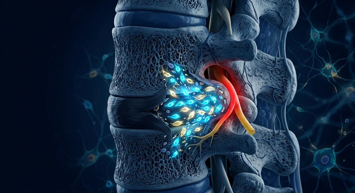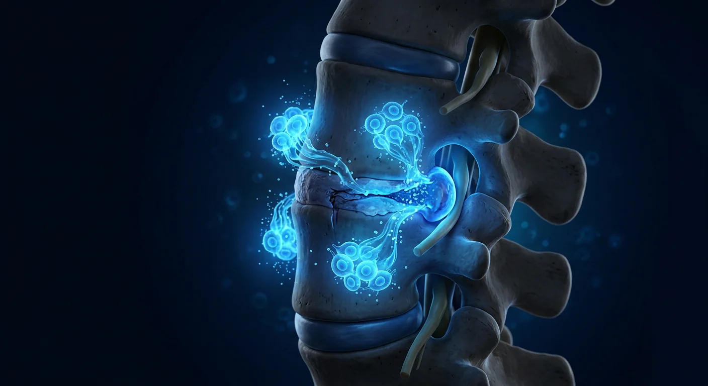
Back pain is one of the most common — and most disruptive — health burdens worldwide. An estimated 80% of adults will experience clinically significant back pain at some point in their lives, and a substantial proportion progress to chronic, life-limiting disability. For many, the underlying driver is degenerative disc disease — a progressive breakdown of the intervertebral discs that cushion the spine, often accompanied by herniation, nerve compression, and persistent inflammation. For decades, the conventional pathway has been linear and limited: anti-inflammatory medication, epidural steroid injections, physical therapy, and eventually spinal surgery. Mesenchymal Stem Cell therapy offers a fundamentally different approach — one focused not on managing pain alone, but on supporting the spine's own regenerative biology.
Understanding what is actually happening in your spine
Healthy spinal discs are composed of a tough outer ring — the annulus fibrosus — and a gel-like inner core, the nucleus pulposus. These structures bear axial load, allow flexion and rotation, and shield spinal nerves as they exit the vertebral column. Degenerative disc disease is the progressive breakdown of this system — driven not just by mechanical wear, but by chronic low-grade inflammation within the disc space. Inflammatory cytokines such as IL-1β, IL-6, and TNF-α are produced by stressed disc cells, accelerate matrix degradation, and sensitize adjacent nerve roots — creating the cycle of pain, muscle spasm, and functional limitation familiar to anyone with chronic back problems.
This matters because traditional treatments target the symptom (pain) without addressing the process (disc degeneration and neuroinflammation). MSC therapy, by contrast, intervenes at the inflammatory and regenerative level — addressing both the pain experience and the underlying spinal biology.
How MSC therapy works in the spine
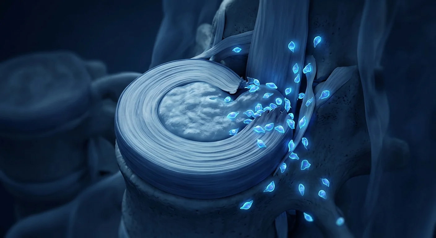
When clinical-grade Mesenchymal Stem Cells are delivered to a degenerated disc or the surrounding epidural space — either via precision injection or systemic infusion — they engage the inflammatory environment in several coordinated ways:
1. They quiet disc inflammation
MSCs respond to the high cytokine environment by releasing anti-inflammatory mediators (TSG-6, PGE2, IDO) that interrupt the degenerative cascade. Within the disc space and surrounding tissue, this can begin reducing pain and radicular symptoms in the early weeks after treatment.
2. They support disc matrix preservation
MSCs secrete growth factors (TGF-β, IGF-1, BMP-7) that signal resident disc cells to maintain proteoglycan and collagen matrix integrity, potentially slowing or partially reversing disc height loss.
3. They modulate neuroinflammation
When a herniated disc compresses a nerve root, the resulting inflammation — not just mechanical pressure — drives the radiating pain, numbness, and weakness of sciatica. MSCs influence local immune cell populations, encouraging a shift from pro-inflammatory to regenerative phenotypes that can reduce nerve root irritation.
4. They support surrounding soft tissue
Paraspinal muscles, ligaments, and facet joints all benefit from MSC paracrine signalling — relevant in the many patients whose back pain stems from a combination of disc, facet, and muscular dysfunction.
What MSC therapy does NOT do
It is important to set honest expectations. MSC therapy does not regrow a completely collapsed disc. It does not reverse severe spinal stenosis or correct structural deformities. What it does is reduce discogenic inflammation, support remaining disc matrix, ease radicular pain, and may delay or potentially avoid the need for spinal surgery in carefully selected candidates with mild-to-moderate degenerative changes.[1]
Who is a good candidate?
The strongest results from MSC therapy for back pain come from patients with mild to moderate degenerative disc disease — typically Pfirrmann grades II, III, and early IV on MRI. These are patients who:[2]
- Have measurable disc height remaining on imaging
- Are experiencing discogenic or radicular pain but do not have severe motor deficits
- Have not responded adequately to conservative treatment (physical therapy, NSAIDs, epidural injections)
- Want to delay or avoid spinal fusion or discectomy surgery
- Are looking for a regenerative rather than purely symptomatic approach
Patients with end-stage disc collapse (severe Pfirrmann grade V), significant spinal instability, or progressive neurological deficits (foot drop, bowel/bladder dysfunction) are typically less suitable for regenerative therapy alone and should be evaluated by a spine surgeon. A reputable clinic will tell you this honestly. The role of the consultation is to determine candidacy through MRI review, neurological examination, and functional assessment.[3]
What the treatment process looks like
At a clinical-grade centre, a typical journey for spine-focused MSC therapy follows a structured path:
Step 1: Consultation and assessment
Medical history review, spinal MRI, neurological examination, inflammatory bio-markers, and functional assessment. The goal is to confirm candidacy and identify the optimal protocol — whether intradiscal injection, epidural delivery, IV infusion, or a combination.
Step 2: Protocol design
The clinical team determines: dose, delivery route (intradiscal injection, transforaminal epidural, IV infusion, or combined), frequency (single session vs. series), and adjunct therapies (physiotherapy, core stabilization, lifestyle adjustments).
Step 3: Treatment delivery
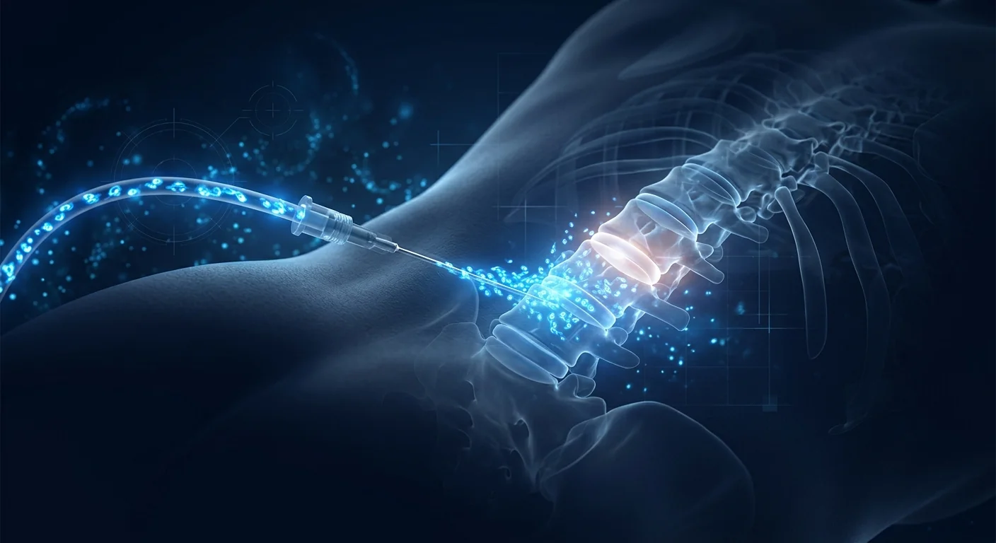
Intradiscal and epidural injections are performed under fluoroscopy or CT guidance for precision placement. Sessions are outpatient, generally 60–90 minutes including preparation. Patients walk out the same day.
Step 4: Recovery period
Light activity restrictions for 48–72 hours, then a graduated return to normal mobility. Most protocols include a structured physiotherapy programme — core stabilization, postural retraining, and gradual loading — alongside the cellular therapy.
Step 5: Outcome monitoring
Follow-up at 4, 12, and 24 weeks tracks pain (using validated scales like VAS and ODI — Oswestry Disability Index), function, range of motion, and where appropriate, repeat MRI.
Realistic timelines: what to expect when
Cellular therapy is not a pharmaceutical — it does not deliver an instant effect. Most patients experience the response in three identifiable phases:[4]
Outcomes vary by patient, severity of disc degeneration, MSC source quality, dosing, and the structure of the surrounding rehabilitation programme. A reputable clinic will be transparent about typical response rates and the proportion of patients who experience strong vs. moderate vs. limited benefit.[5]
Safety and what every patient should know
When delivered with clinical-grade cells, image-guided injection technique, and appropriate clinical oversight, MSC therapy for back pain has an excellent safety profile. The most common side-effects are local: temporary post-injection soreness, mild swelling, or stiffness for 24–72 hours. Serious adverse events are rare in published literature.[6]
The biggest preventable risk in this field is not the therapy itself — it is unregulated providers using non-clinical-grade cells with unverified sterility, identity, or potency. This is why patients should always confirm: source of the cells, laboratory certifications, and access to a Certificate of Analysis for the dose being administered.[7]
For the right patient — with mild to moderate disc degeneration and a goal of preserving spinal function — MSC therapy can be a meaningful alternative to a future of escalating medication and eventual surgery.
— VELAR Clinical Team
The VELAR approach to spinal regeneration
Spine-focused protocols at VELAR Center begin with comprehensive MRI review, neurological assessment, and bio-marker evaluation to confirm candidacy and tailor dosing. Each patient's protocol uses clinical-grade Wharton's jelly–derived MSCs (≥95% MSC marker expression, >95% viability at delivery), delivered via image-guided intradiscal injection, epidural delivery, IV infusion, or combined protocol depending on the indication. Every session is led by an experienced clinician, paired with structured rehabilitation, and monitored across the 1, 3, and 6-month milestones.[8]
If you are considering regenerative therapy for back pain, the most important first step is an honest assessment of your specific candidacy — and a clear understanding of what realistic outcomes look like for your stage of disc disease.
References
- Sakai D, Schol J. Cell therapy for intervertebral disc repair: clinical perspective. Journal of Orthopaedic Translation. 2017;9:8-18. doi:10.1016/j.jot.2017.02.002 ↩
- Pfirrmann CW, Metzdorf A, Zanetti M, Hodler J, Boos N. Magnetic resonance classification of lumbar intervertebral disc degeneration. Spine. 2001;26(17):1873-1878. doi:10.1097/00007632-200109010-00011 ↩
- Orozco L, Soler R, Morera C, Alberca M, Sánchez A, García-Sancho J. Intervertebral disc repair by autologous mesenchymal bone marrow cells: a pilot study. Transplantation. 2011;92(7):822-828. doi:10.1097/TP.0b013e3182298a15 ↩
- Pettine KA, Murphy MB, Suzuki RK, Sand TT. Percutaneous injection of autologous bone marrow concentrate cells significantly reduces lumbar discogenic pain through 12 months. Stem Cells. 2015;33(1):146-156. doi:10.1002/stem.1845 ↩
- Centeno C, Markle J, Dodson E, et al. Treatment of lumbar degenerative disc disease-associated radicular pain with culture-expanded autologous mesenchymal stem cells: a pilot study on safety and efficacy. Journal of Translational Medicine. 2017;15(1):197. doi:10.1186/s12967-017-1300-y ↩
- Meisel HJ, Agarwal N, Hsieh PC, et al. Cell therapy for treatment of intervertebral disc degeneration: a systematic review. Global Spine Journal. 2019;9(1 Suppl):39S-52S. doi:10.1177/2192568219829024 ↩
- Dominici M, Le Blanc K, Mueller I, et al. Minimal criteria for defining multipotent mesenchymal stromal cells. The International Society for Cellular Therapy position statement. Cytotherapy. 2006;8(4):315-317. doi:10.1080/14653240600855905 ↩
- Noriega DC, Ardura F, Hernández-Ramajo R, et al. Intervertebral disc repair by allogeneic mesenchymal bone marrow cells: a randomized controlled trial. Transplantation. 2017;101(8):1945-1951. doi:10.1097/TP.0000000000001484 ↩
- Richardson SM, Kalamegam G, Pushparaj PN, et al. Mesenchymal stem cells in regenerative medicine: focus on articular cartilage and intervertebral disc regeneration. Methods. 2016;99:69-80. doi:10.1016/j.ymeth.2015.09.015 ↩
- Amirdelfan K, Bae H, McJunkin T, et al. Allogeneic mesenchymal precursor cells treatment for chronic low back pain associated with degenerative disc disease: a prospective randomized, placebo-controlled 36-month study of safety and efficacy. Spine Journal. 2021;21(2):212-230. doi:10.1016/j.spinee.2020.10.004 ↩
- Mochida J, Sakai D, Nakamura Y, Watanabe T, Yamamoto Y, Kato S. Intervertebral disc repair with activated nucleus pulposus cell transplantation: a 3-year, prospective clinical study of its safety. European Spine Journal. 2015;24(9):1948-1954. doi:10.1007/s00586-015-4009-8 ↩
- Yoshikawa T, Ueda Y, Miyazaki K, Koizumi M, Takakura Y. Disc regeneration therapy using marrow mesenchymal cell transplantation: a report of two case studies. Spine. 2010;35(11):E475-480. doi:10.1097/BRS.0b013e3181cd2cf4 ↩
背痛是全球最常见——也最具有破坏性——的健康负担之一。据估计,80%的成年人在一生中会经历临床意义上的背痛,其中相当一部分会发展为慢性、限制生活的残疾。对许多人来说,背后的驱动因素是椎间盘退行性疾病——缓冲脊柱的椎间盘进行性退化,常伴椎间盘突出、神经压迫及持续性炎症。数十年来,传统路径线性而有限:抗炎药物、硬膜外类固醇注射、物理治疗,最终是脊柱手术。间充质干细胞疗法提供了一种根本不同的方法——其重点不是单纯管理疼痛,而是支持脊柱自身的再生生物学。
了解您的脊柱中实际正在发生什么
健康的椎间盘由坚韧的外环——纤维环——和凝胶状内核髓核组成。这些结构承受轴向载荷,允许屈曲和旋转,并在神经根离开脊柱时保护它们。椎间盘退行性疾病是这一系统的渐进性破坏——不仅由机械磨损驱动,还由椎间盘空间内的慢性低度炎症驱动。受压椎间盘细胞产生的炎症细胞因子(如 IL-1β、IL-6 与 TNF-α)会加速基质降解,并使邻近神经根敏化——造成任何慢性背痛患者都熟悉的疼痛、肌肉痉挛与功能受限的循环。
这一点很重要,因为传统治疗针对症状(疼痛),却不触及过程(椎间盘退化和神经炎症)。相比之下,MSC 疗法在炎症与再生层面进行干预——同时解决疼痛体验与潜在的脊柱生物学。
MSC 疗法在脊柱中如何运作
当临床级间充质干细胞被递送到退化的椎间盘或周围的硬膜外空间——通过精确注射或全身输注——它们以若干协调的方式与炎症环境互动:
1. 它们抑制椎间盘炎症
MSC 通过释放抗炎介质(TSG-6、PGE2、IDO)来响应高细胞因子环境,打断退化级联反应。在椎间盘空间和周围组织中,这可在治疗后早期数周开始减轻疼痛与根性症状。
2. 它们支持椎间盘基质保护
MSC 分泌生长因子(TGF-β、IGF-1、BMP-7),向常驻椎间盘细胞发出信号以维持蛋白多糖和胶原基质的完整性,可能减缓或部分逆转椎间盘高度丢失。
3. 它们调节神经炎症
当突出的椎间盘压迫神经根时,由此产生的炎症——不仅仅是机械压力——驱动了坐骨神经痛的放射性疼痛、麻木和无力。MSC 影响局部免疫细胞群体,鼓励从促炎表型转向再生表型,可减少神经根刺激。
4. 它们支持周围软组织
椎旁肌肉、韧带和小关节均受益于 MSC 的旁分泌信号传导——对许多背痛源于椎间盘、小关节和肌肉功能障碍组合的患者尤为相关。
MSC 疗法所不能做的事
设定诚实的预期至关重要。MSC 疗法无法重建完全塌陷的椎间盘。它无法逆转严重的椎管狭窄或矫正结构畸形。它能做的是减少椎间盘源性炎症、支持剩余椎间盘基质、缓解根性疼痛,并可能在经过仔细筛选、具有轻度至中度退行性变化的候选者中延迟或潜在避免脊柱手术的需要。[1]
谁是合适的候选者?
对背痛的 MSC 疗法效果最强的来自轻度至中度椎间盘退行性疾病患者——通常在 MRI 上为 Pfirrmann 分级 II、III 与早期 IV。这些患者:[2]
- 影像学上可见可量测的剩余椎间盘高度
- 经历椎间盘源性或根性疼痛但无严重运动功能障碍
- 对保守治疗反应不佳(物理治疗、非甾体抗炎药、硬膜外注射)
- 希望延迟或避免脊柱融合或椎间盘切除术
- 寻找再生而非纯症状性的方法
终末期椎间盘塌陷患者(严重 Pfirrmann V 级)、显著脊柱不稳或进行性神经功能缺损(足下垂、肠/膀胱功能障碍)——通常不太适合单独的再生疗法,应由脊柱外科医生评估。信誉良好的诊所会诚实地告诉您这一点。咨询的作用是通过 MRI 审阅、神经系统检查与功能评估来确定候选资格。[3]
治疗过程是什么样子
在临床级中心,脊柱导向 MSC 疗法的典型旅程遵循结构化路径:
第一步:咨询与评估
病史回顾、脊柱 MRI、神经系统检查、炎症生物标志物以及功能评估。目标是确认候选资格并确定最佳方案——无论是椎间盘内注射、硬膜外递送、IV 输注还是联合方案。
第二步:方案设计
临床团队确定:剂量、递送途径(椎间盘内注射、经椎间孔硬膜外、IV 输注或联合)、频率(单次会话与系列)以及辅助疗法(物理治疗、核心稳定、生活方式调整)。
第三步:治疗递送
椎间盘内和硬膜外注射在荧光透视或 CT 引导下进行以确保精确放置。会话为门诊,通常含准备 60–90 分钟。患者当天即可走出。
第四步:恢复期
48–72 小时轻度活动限制,然后逐渐恢复正常活动。大多数方案包括结构化物理治疗——核心稳定、姿势再训练和渐进负重——配合细胞治疗。
第五步:疗效监测
在 4、12 与 24 周的随访追踪疼痛(使用经验证的量表如 VAS 与 ODI——Oswestry 功能障碍指数)、功能、活动范围,并在适当情况下重复 MRI。
实际时间表:何时期待什么
细胞治疗不是药物——它不会立即产生效果。大多数患者经历的反应分为三个可识别阶段:[4]
疗效因患者、椎间盘退化严重程度、MSC 来源质量、剂量以及周围康复方案的结构而异。信誉良好的诊所会透明地说明典型的反应率以及强、中、有限受益患者的比例。[5]
安全性与每位患者应知道的
当以临床级细胞、影像引导的注射技术与适当的临床监督递送时,背痛的 MSC 疗法具有出色的安全性概况。最常见的副作用是局部的:注射后 24–72 小时的暂时酸痛、轻度肿胀或僵硬。在已发表的文献中,严重不良事件少见。[6]
该领域最大的可预防风险并非疗法本身——而是未受监管的提供者使用未经验证无菌性、身份或效力的非临床级细胞。这就是为什么患者应始终确认:细胞来源、实验室认证,以及所给予剂量的分析证书的获取。[7]
对合适的患者——具有轻度至中度椎间盘退行性变化并以保留脊柱功能为目标——MSC 疗法可以是不断升级的药物治疗与最终手术之未来的有意义替代。
——VELAR 临床团队
VELAR 的脊柱再生方法
VELAR Center 脊柱导向的方案从全面的 MRI 审阅、神经系统评估与生物标志物评估开始,以确认候选资格并定制剂量。每位患者的方案使用临床级华通氏胶来源 MSC(MSC 标志物表达 ≥95%、交付时活性 >95%),通过影像引导的椎间盘内注射、硬膜外递送、IV 输注或联合方案递送,视适应症而定。每次会话由经验丰富的临床医师主导,搭配结构化康复,并在 1、3 与 6 个月里程碑进行监测。[8]
如果您正在考虑背痛的再生疗法,最重要的第一步是对您具体候选资格的诚实评估——以及对您椎间盘疾病阶段的实际疗效的清晰理解。
参考文献
- Sakai D, Schol J. Cell therapy for intervertebral disc repair: clinical perspective. Journal of Orthopaedic Translation. 2017;9:8-18. doi:10.1016/j.jot.2017.02.002 ↩
- Pfirrmann CW, Metzdorf A, Zanetti M, Hodler J, Boos N. Magnetic resonance classification of lumbar intervertebral disc degeneration. Spine. 2001;26(17):1873-1878. doi:10.1097/00007632-200109010-00011 ↩
- Orozco L, Soler R, Morera C, Alberca M, Sánchez A, García-Sancho J. Intervertebral disc repair by autologous mesenchymal bone marrow cells: a pilot study. Transplantation. 2011;92(7):822-828. doi:10.1097/TP.0b013e3182298a15 ↩
- Pettine KA, Murphy MB, Suzuki RK, Sand TT. Percutaneous injection of autologous bone marrow concentrate cells significantly reduces lumbar discogenic pain through 12 months. Stem Cells. 2015;33(1):146-156. doi:10.1002/stem.1845 ↩
- Centeno C, Markle J, Dodson E, et al. Treatment of lumbar degenerative disc disease-associated radicular pain with culture-expanded autologous mesenchymal stem cells: a pilot study on safety and efficacy. Journal of Translational Medicine. 2017;15(1):197. doi:10.1186/s12967-017-1300-y ↩
- Meisel HJ, Agarwal N, Hsieh PC, et al. Cell therapy for treatment of intervertebral disc degeneration: a systematic review. Global Spine Journal. 2019;9(1 Suppl):39S-52S. doi:10.1177/2192568219829024 ↩
- Dominici M, Le Blanc K, Mueller I, et al. Minimal criteria for defining multipotent mesenchymal stromal cells. The International Society for Cellular Therapy position statement. Cytotherapy. 2006;8(4):315-317. doi:10.1080/14653240600855905 ↩
- Noriega DC, Ardura F, Hernández-Ramajo R, et al. Intervertebral disc repair by allogeneic mesenchymal bone marrow cells: a randomized controlled trial. Transplantation. 2017;101(8):1945-1951. doi:10.1097/TP.0000000000001484 ↩
- Richardson SM, Kalamegam G, Pushparaj PN, et al. Mesenchymal stem cells in regenerative medicine: focus on articular cartilage and intervertebral disc regeneration. Methods. 2016;99:69-80. doi:10.1016/j.ymeth.2015.09.015 ↩
- Amirdelfan K, Bae H, McJunkin T, et al. Allogeneic mesenchymal precursor cells treatment for chronic low back pain associated with degenerative disc disease: a prospective randomized, placebo-controlled 36-month study of safety and efficacy. Spine Journal. 2021;21(2):212-230. doi:10.1016/j.spinee.2020.10.004 ↩
- Mochida J, Sakai D, Nakamura Y, Watanabe T, Yamamoto Y, Kato S. Intervertebral disc repair with activated nucleus pulposus cell transplantation: a 3-year, prospective clinical study of its safety. European Spine Journal. 2015;24(9):1948-1954. doi:10.1007/s00586-015-4009-8 ↩
- Yoshikawa T, Ueda Y, Miyazaki K, Koizumi M, Takakura Y. Disc regeneration therapy using marrow mesenchymal cell transplantation: a report of two case studies. Spine. 2010;35(11):E475-480. doi:10.1097/BRS.0b013e3181cd2cf4 ↩
آلام الظهر من أكثر الأعباء الصحية شيوعاً — وإعاقةً — حول العالم. يُقدَّر أن 80% من البالغين سيختبرون آلام ظهر ذات أهمية سريرية في مرحلة ما من حياتهم، وتتطور نسبة كبيرة إلى إعاقة مزمنة محدِّدة للحياة. بالنسبة للكثيرين، المحرك الأساسي هو مرض القرص التنكسي — انهيار تدريجي للأقراص بين الفقرات التي تبطن العمود الفقري، غالباً ما يصاحبه انفتاق وضغط على الأعصاب والتهاب مستمر. لعقود، كان المسار التقليدي خطياً ومحدوداً: أدوية مضادة للالتهاب، حقن الستيرويد فوق الجافية، العلاج الطبيعي، وأخيراً جراحة العمود الفقري. يقدم علاج الخلايا الجذعية الوسيطة منهجاً مختلفاً جذرياً — يركز ليس على إدارة الألم وحده، بل على دعم البيولوجيا التجديدية للعمود الفقري ذاته.
فهم ما يحدث فعلياً في عمودك الفقري
تتكون الأقراص الفقرية السليمة من حلقة خارجية قوية — الحلقة الليفية — ونواة داخلية هلامية هي النواة اللبية. تتحمل هذه الهياكل الحمل المحوري، وتسمح بالثني والدوران، وتحمي الأعصاب الشوكية عند خروجها من العمود الفقري. مرض القرص التنكسي هو الانهيار التدريجي لهذا النظام — مدفوعاً ليس فقط بالاهتراء الميكانيكي، بل بـالالتهاب منخفض الدرجة المزمن داخل حيز القرص. تنتج خلايا القرص المُجهَدة سيتوكينات التهابية مثل IL-1β وIL-6 وTNF-α، تُسرع تدهور المصفوفة، وتُحسِّس جذور الأعصاب المجاورة — مخلقةً دورة الألم والتشنج العضلي والتقييد الوظيفي المألوفة لأي شخص مصاب بمشاكل ظهر مزمنة.
هذا مهم لأن العلاجات التقليدية تستهدف العَرَض (الألم) دون معالجة العملية (تنكس القرص والالتهاب العصبي). علاج MSC، على النقيض، يتدخل على مستوى الالتهاب والتجدد — معالجاً تجربة الألم وكذلك بيولوجيا العمود الفقري الكامنة.
كيف يعمل علاج MSC في العمود الفقري
عندما تُسلَّم الخلايا الجذعية الوسيطة ذات الجودة السريرية إلى قرص متدهور أو الحيز فوق الجافية المحيط — إما عبر الحقن الدقيق أو التسريب الجهازي — فإنها تتفاعل مع البيئة الالتهابية بعدة طرق منسقة:
1. تُهدِّئ التهاب القرص
تستجيب MSC لبيئة السيتوكينات العالية بإطلاق وسطاء مضادين للالتهاب (TSG-6 وPGE2 وIDO) يقطعون السلسلة التنكسية. داخل حيز القرص والأنسجة المحيطة، يمكن أن يبدأ هذا في تقليل الألم والأعراض الجذرية في الأسابيع الأولى بعد العلاج.
2. تدعم الحفاظ على مصفوفة القرص
تفرز MSC عوامل نمو (TGF-β وIGF-1 وBMP-7) تُشير إلى خلايا القرص المقيمة للحفاظ على سلامة مصفوفة البروتيوغليكان والكولاجين، مما قد يُبطئ أو يعكس جزئياً فقدان ارتفاع القرص.
3. تُعدِّل الالتهاب العصبي
عندما يضغط قرص منفتق على جذر عصب، فإن الالتهاب الناتج — وليس فقط الضغط الميكانيكي — هو ما يدفع الألم المنتشر والخدر والضعف في عرق النسا. تؤثر MSC على مجموعات الخلايا المناعية المحلية، مشجعةً الانتقال من أنماط التهابية إلى أنماط تجديدية يمكن أن تقلل من تهيج جذر العصب.
4. تدعم الأنسجة الرخوة المحيطة
تستفيد العضلات المجاورة للفقرات والأربطة والمفاصل الوجيهية جميعها من إشارات MSC شبه الإفرازية — وهذا مهم لدى العديد من المرضى الذين تنبع آلام ظهورهم من مزيج من خلل القرص والمفصل الوجيهي والعضلات.
ما لا يفعله علاج MSC
من المهم وضع توقعات صادقة. علاج MSC لا يُعيد إنماء قرص منهار بالكامل. لا يعكس التضيق الشوكي الشديد أو يصحح التشوهات الهيكلية. ما يفعله هو تقليل الالتهاب القرصي المنشأ، دعم مصفوفة القرص المتبقية، تخفيف الألم الجذري، وقد يؤخر أو يُحتمل أن يتجنب الحاجة إلى جراحة العمود الفقري لدى مرشحين مختارين بعناية مع تغيرات تنكسية خفيفة إلى معتدلة.[1]
من هو المرشح المناسب؟
أقوى نتائج علاج MSC لآلام الظهر تأتي من المرضى المصابين بمرض قرص تنكسي خفيف إلى معتدل — عادة درجات Pfirrmann II وIII وأوائل IV على MRI. هؤلاء المرضى:[2]
- لديهم ارتفاع قرص متبقٍ قابل للقياس على التصوير
- يعانون من ألم قرصي المنشأ أو جذري لكن بدون عجز حركي شديد
- لم يستجيبوا بشكل كافٍ للعلاج المحافظ (العلاج الطبيعي، مضادات الالتهاب غير الستيرويدية، الحقن فوق الجافية)
- يريدون تأخير أو تجنب جراحة دمج الفقرات أو استئصال القرص
- يبحثون عن منهج تجديدي بدلاً من نهج عَرَضي بحت
المرضى الذين يعانون من انهيار قرص في مراحل متأخرة (شديد Pfirrmann V)، عدم استقرار فقري كبير، أو عجز عصبي متقدم (هبوط القدم، خلل الأمعاء/المثانة) — عادة ما يكونون أقل ملاءمة للعلاج التجديدي وحده ويجب تقييمهم من قبل جراح عمود فقري. ستخبرك العيادة ذات السمعة الجيدة بذلك بصدق. دور الاستشارة هو تحديد الأهلية من خلال مراجعة MRI والفحص العصبي والتقييم الوظيفي.[3]
كيف تبدو عملية العلاج
في مركز ذي جودة سريرية، تتبع الرحلة النموذجية لعلاج MSC المتمحور حول العمود الفقري مساراً منظماً:
الخطوة 1: الاستشارة والتقييم
مراجعة التاريخ الطبي، MRI العمود الفقري، الفحص العصبي، المؤشرات الحيوية الالتهابية، والتقييم الوظيفي. الهدف هو تأكيد الأهلية وتحديد البروتوكول الأمثل — سواء كان حقناً داخل القرص، توصيلاً فوق الجافية، تسريباً وريدياً، أو مزيجاً.
الخطوة 2: تصميم البروتوكول
يحدد الفريق السريري: الجرعة، طريقة التوصيل (حقن داخل القرص، فوق الجافية عبر الثقبة، تسريب وريدي، أو مجتمعة)، التكرار (جلسة واحدة مقابل سلسلة)، والعلاجات المساعدة (العلاج الطبيعي، تثبيت الجذع، تعديلات نمط الحياة).
الخطوة 3: تسليم العلاج
تُجرى الحقن داخل القرص وفوق الجافية تحت توجيه التنظير الفلوري أو CT للوضع الدقيق. الجلسات خارجية، عموماً 60–90 دقيقة بما في ذلك التحضير. يخرج المرضى في نفس اليوم.
الخطوة 4: فترة التعافي
قيود نشاط خفيف لمدة 48–72 ساعة، ثم عودة متدرجة إلى الحركة الطبيعية. تشمل معظم البروتوكولات برنامج علاج طبيعي منظم — تثبيت الجذع، إعادة تدريب الوضعية، والتحميل التدريجي — إلى جانب العلاج الخلوي.
الخطوة 5: مراقبة النتائج
المتابعة في 4 و12 و24 أسبوعاً تتتبع الألم (باستخدام مقاييس مُعتمَدة مثل VAS وODI — مؤشر إعاقة Oswestry)، الوظيفة، نطاق الحركة، وحيث يكون مناسباً، إعادة MRI.
جداول زمنية واقعية: ماذا تتوقع ومتى
العلاج الخلوي ليس دواء — لا يعطي تأثيراً فورياً. يختبر معظم المرضى الاستجابة في ثلاث مراحل قابلة للتحديد:[4]
تختلف النتائج حسب المريض، شدة تنكس القرص، جودة مصدر MSC، الجرعات، وهيكل برنامج إعادة التأهيل المحيط. ستكون العيادة ذات السمعة الجيدة شفافة بشأن معدلات الاستجابة النموذجية ونسبة المرضى الذين يختبرون فائدة قوية مقابل معتدلة مقابل محدودة.[5]
السلامة وما يجب أن يعرفه كل مريض
عند التسليم بخلايا ذات جودة سريرية، تقنية حقن مُوجَّهة بالصورة، وإشراف سريري مناسب، يتمتع علاج MSC لآلام الظهر بملف أمان ممتاز. الآثار الجانبية الأكثر شيوعاً هي موضعية: ألم مؤقت بعد الحقن، تورم خفيف، أو تيبس لمدة 24–72 ساعة. الأحداث السلبية الخطيرة نادرة في الأدبيات المنشورة.[6]
أكبر خطر يمكن منعه في هذا المجال ليس العلاج نفسه — بل المزودون غير المُنظَّمين الذين يستخدمون خلايا غير سريرية الجودة بعقامة أو هوية أو فعالية غير مُتحقَّق منها. هذا هو السبب في أنه ينبغي للمرضى دائماً تأكيد: مصدر الخلايا، اعتمادات المختبر، والوصول إلى شهادة تحليل للجرعة المُعطاة.[7]
للمريض المناسب — بتنكس قرص خفيف إلى معتدل وهدف الحفاظ على وظيفة العمود الفقري — يمكن أن يكون علاج MSC بديلاً ذا معنى لمستقبل من الأدوية المتصاعدة والجراحة في النهاية.
— فريق VELAR السريري
منهج VELAR لتجديد العمود الفقري
تبدأ البروتوكولات المتمحورة حول العمود الفقري في VELAR Center بمراجعة MRI شاملة وتقييم عصبي وتقييم المؤشرات الحيوية لتأكيد الأهلية وتخصيص الجرعات. يستخدم بروتوكول كل مريض MSC ذات الجودة السريرية المشتقة من هلام وارتون (تعبير علامات MSC ≥95%، حيوية عند التسليم >95%)، تُسلَّم عبر الحقن داخل القرص المُوجَّه بالصورة، أو التوصيل فوق الجافية، أو التسريب الوريدي، أو بروتوكول مجتمع حسب الدلالة. تُقاد كل جلسة من قبل طبيب ذي خبرة، مقترنةً بإعادة تأهيل منظمة، ومُراقَبة عبر علامات 1 و3 و6 أشهر.[8]
إذا كنت تفكر في العلاج التجديدي لآلام الظهر، فإن أهم خطوة أولى هي تقييم صادق لأهليتك المحددة — وفهم واضح لكيف تبدو النتائج الواقعية لمرحلة مرض قرصك.
المراجع
- Sakai D, Schol J. Cell therapy for intervertebral disc repair: clinical perspective. Journal of Orthopaedic Translation. 2017;9:8-18. doi:10.1016/j.jot.2017.02.002 ↩
- Pfirrmann CW, Metzdorf A, Zanetti M, Hodler J, Boos N. Magnetic resonance classification of lumbar intervertebral disc degeneration. Spine. 2001;26(17):1873-1878. doi:10.1097/00007632-200109010-00011 ↩
- Orozco L, Soler R, Morera C, Alberca M, Sánchez A, García-Sancho J. Intervertebral disc repair by autologous mesenchymal bone marrow cells: a pilot study. Transplantation. 2011;92(7):822-828. doi:10.1097/TP.0b013e3182298a15 ↩
- Pettine KA, Murphy MB, Suzuki RK, Sand TT. Percutaneous injection of autologous bone marrow concentrate cells significantly reduces lumbar discogenic pain through 12 months. Stem Cells. 2015;33(1):146-156. doi:10.1002/stem.1845 ↩
- Centeno C, Markle J, Dodson E, et al. Treatment of lumbar degenerative disc disease-associated radicular pain with culture-expanded autologous mesenchymal stem cells: a pilot study on safety and efficacy. Journal of Translational Medicine. 2017;15(1):197. doi:10.1186/s12967-017-1300-y ↩
- Meisel HJ, Agarwal N, Hsieh PC, et al. Cell therapy for treatment of intervertebral disc degeneration: a systematic review. Global Spine Journal. 2019;9(1 Suppl):39S-52S. doi:10.1177/2192568219829024 ↩
- Dominici M, Le Blanc K, Mueller I, et al. Minimal criteria for defining multipotent mesenchymal stromal cells. The International Society for Cellular Therapy position statement. Cytotherapy. 2006;8(4):315-317. doi:10.1080/14653240600855905 ↩
- Noriega DC, Ardura F, Hernández-Ramajo R, et al. Intervertebral disc repair by allogeneic mesenchymal bone marrow cells: a randomized controlled trial. Transplantation. 2017;101(8):1945-1951. doi:10.1097/TP.0000000000001484 ↩
- Richardson SM, Kalamegam G, Pushparaj PN, et al. Mesenchymal stem cells in regenerative medicine: focus on articular cartilage and intervertebral disc regeneration. Methods. 2016;99:69-80. doi:10.1016/j.ymeth.2015.09.015 ↩
- Amirdelfan K, Bae H, McJunkin T, et al. Allogeneic mesenchymal precursor cells treatment for chronic low back pain associated with degenerative disc disease: a prospective randomized, placebo-controlled 36-month study of safety and efficacy. Spine Journal. 2021;21(2):212-230. doi:10.1016/j.spinee.2020.10.004 ↩
- Mochida J, Sakai D, Nakamura Y, Watanabe T, Yamamoto Y, Kato S. Intervertebral disc repair with activated nucleus pulposus cell transplantation: a 3-year, prospective clinical study of its safety. European Spine Journal. 2015;24(9):1948-1954. doi:10.1007/s00586-015-4009-8 ↩
- Yoshikawa T, Ueda Y, Miyazaki K, Koizumi M, Takakura Y. Disc regeneration therapy using marrow mesenchymal cell transplantation: a report of two case studies. Spine. 2010;35(11):E475-480. doi:10.1097/BRS.0b013e3181cd2cf4 ↩