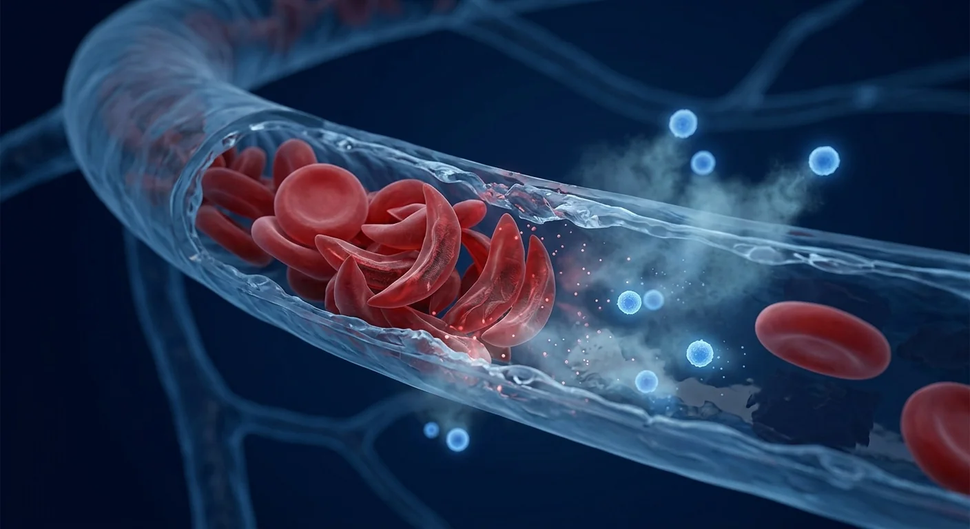
Sickle cell disease is an inherited hemoglobinopathy caused by a single point mutation in the β-globin gene that substitutes valine for glutamic acid at position 6, producing hemoglobin S. Under deoxygenation, HbS polymerizes into rigid rods that deform erythrocytes into their characteristic sickle shape — fragile, pro-adhesive cells that occlude the microvasculature, hemolyze prematurely, and trigger a cascade of endothelial activation, sterile inflammation, and progressive ischemic organ damage. Approximately 20–25 million people live with SCD globally, with the highest prevalence in sub-Saharan Africa, India, the Middle East, and the Mediterranean basin. In resource-limited settings, childhood mortality exceeds 50% without early diagnosis and comprehensive care [1].
Where conventional treatments fall short. Standard disease-modifying therapy for SCD centers on hydroxyurea, which raises fetal hemoglobin levels and reduces vaso-occlusive crisis frequency by approximately 50%. Chronic transfusion therapy suppresses HbS production and prevents stroke in high-risk children, while L-glutamine, voxelotor, and crizanlizumab target specific downstream pathways. Allogeneic hematopoietic stem cell transplantation offers a curative option with 90–95% overall survival and 85–90% event-free survival in pediatric patients with matched sibling donors. However, fewer than 20% of patients have an HLA-identical sibling, transplant-related complications including graft-versus-host disease and infertility are substantial, and access to HSCT is severely limited in regions with the highest SCD burden [2].
The deeper problem is endothelial and inflammatory dysfunction. The core pathology of SCD extends far beyond the erythrocyte. Sickled red cells express elevated phosphatidylserine on their outer membrane, adhesion molecules including VCAM-1 and P-selectin, and release free hemoglobin and heme into the plasma upon hemolysis. Free heme scavenges nitric oxide 1,000-fold faster than intact hemoglobin, depleting the endogenous vasodilator and creating a state of functional NO resistance. Hemolysis also releases arginase from erythrocytes, consuming L-arginine — the substrate for endothelial NO synthase — further compounding endothelial dysfunction. This hemolysis-driven vasculopathy, combined with chronic sterile inflammation driven by NLRP3 inflammasome activation, TLR4 signaling, and elevated TNF-α and IL-6, produces a self-perpetuating cycle of vaso-occlusion, ischemia-reperfusion injury, and progressive organ fibrosis [3].
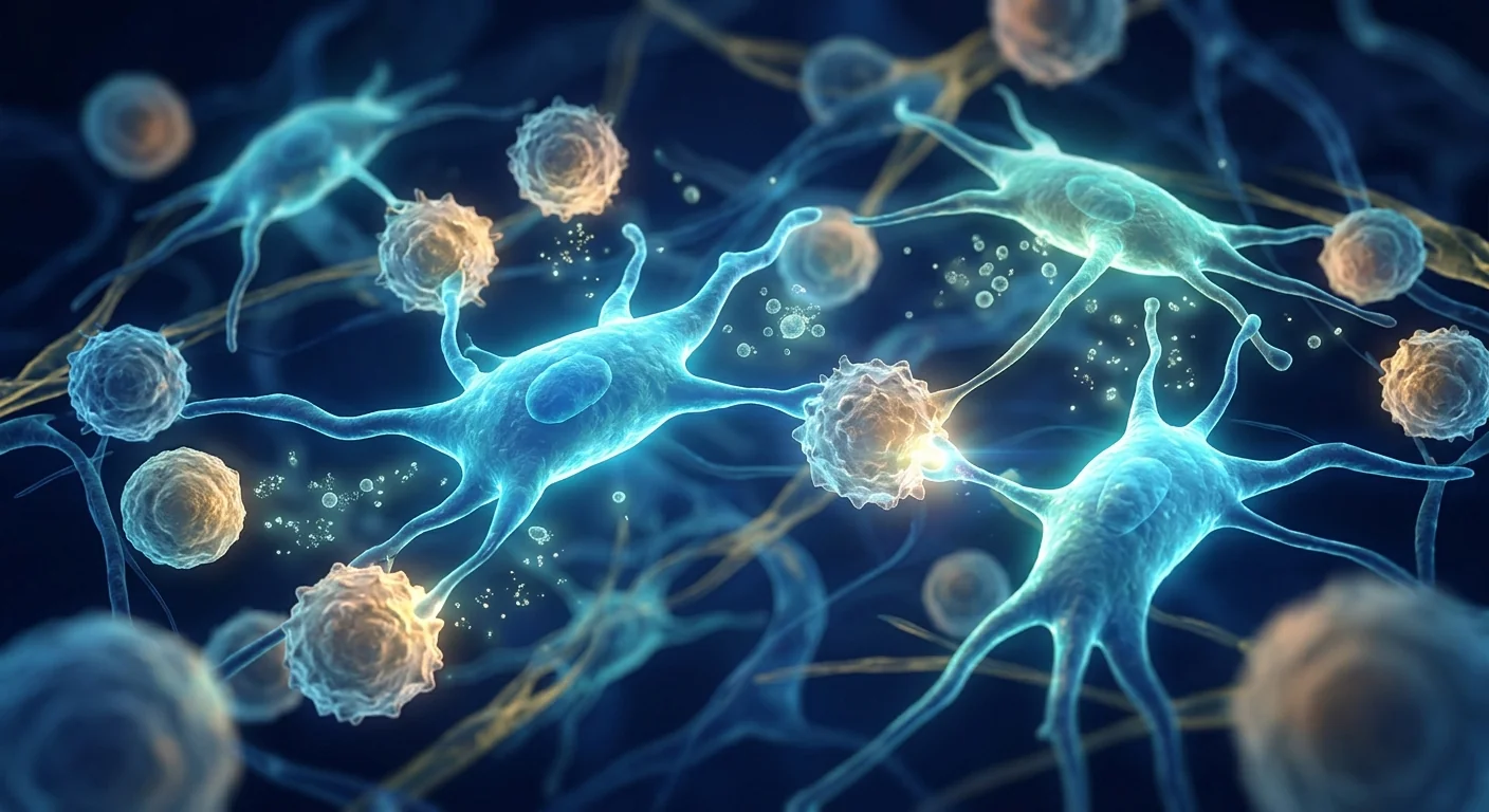
MSC therapy targets the endothelial-inflammatory interface. Mesenchymal stem cells are uniquely positioned to interrupt the vicious cycle of SCD pathophysiology. When administered intravenously, MSCs home to sites of vascular injury via CXCR4/SDF-1 gradients, lodge temporarily in the pulmonary microvasculature, and release a coordinated paracrine program of endothelial protective, anti-inflammatory, and pro-resolution mediators. Unlike hydroxyurea, which partially suppresses HbS polymerization, and unlike anti-adhesion antibodies that block a single pathway, MSC therapy addresses the endothelial dysfunction and sterile inflammation that sustain vaso-occlusion — potentially reducing both crisis frequency and cumulative organ damage [4].
How MSCs Target Sickle Cell Pathophysiology
MSCs address SCD complications through five interconnected mechanisms, each targeting a specific node in the vaso-occlusive-inflammatory cascade [5].
Endothelial protection and NO bioavailability restoration. Hemolysis-driven NO depletion is arguably the central vascular pathology in SCD. MSCs secrete hepatocyte growth factor, vascular endothelial growth factor, and angiopoietin-1, which stabilize endothelial junctions, reduce vascular permeability, and upregulate endothelial NO synthase expression. MSC-derived extracellular vesicles carry microRNA-126 and microRNA-210, which directly promote endothelial survival under oxidative stress. In a transgenic SCD mouse model, intravenous MSC infusion restored NO-dependent vasodilation by approximately 40%, reduced endothelial P-selectin and VCAM-1 expression by 55%, and decreased leukocyte-endothelial adhesion — the initiating event in vaso-occlusion — by 60% at 72 hours post-infusion [6].
Heme scavenging and oxidative stress mitigation. Free heme released during intravascular hemolysis is a potent damage-associated molecular pattern that activates TLR4 on endothelial cells, macrophages, and platelets. MSCs upregulate heme oxygenase-1 expression in tissue macrophages through paracrine signaling, accelerating heme degradation into biliverdin, carbon monoxide, and free iron — all of which are then safely sequestered by ferritin. MSCs also secrete the antioxidant enzymes superoxide dismutase and catalase within extracellular vesicles, directly neutralizing reactive oxygen species in the vascular microenvironment. In heme-challenged endothelial monolayers, MSC-conditioned medium reduced intracellular ROS by 65% and prevented heme-induced apoptosis in 80% of treated cells [7].
Anti-inflammatory immunomodulation. Chronic sterile inflammation in SCD is driven by multiple converging pathways: heme-TLR4 signaling, NLRP3 inflammasome activation, complement deposition on sickled cells, and ischemia-reperfusion cycles. MSCs suppress this inflammatory milieu through secretion of prostaglandin E2, indoleamine 2,3-dioxygenase, tumor necrosis factor-stimulated gene-6, and transforming growth factor-β. They also express PD-L1 and HLA-G, which induce regulatory T cells and tolerogenic dendritic cells. In SCD patient peripheral blood mononuclear cell co-cultures, MSCs reduced TNF-α secretion by 65–70%, IL-6 by 55%, and IL-1β by 50%, while increasing IL-10 by 3-to-4-fold — shifting the cytokine profile from a pro-inflammatory to a regulatory phenotype [8].
Pain crisis modulation. Vaso-occlusive pain crises involve not only vascular occlusion but also neurogenic inflammation mediated by substance P, calcitonin gene-related peptide, and mast cell degranulation. MSCs release soluble factors that suppress mast cell activation, reduce neuronal sensitization, and modulate microglial activation in the spinal cord — addressing the neuroinflammatory component of SCD pain that is poorly controlled by opioids. In a murine SCD pain model, a single intravenous MSC infusion reduced mechanical and thermal hyperalgesia by 50–60% at 7 days, with effects lasting 3–4 weeks, without the tolerance or hyperalgesia associated with chronic opioid use [9].
Hematopoietic niche support and HSCT enhancement. For SCD patients eligible for hematopoietic stem cell transplantation, MSCs offer a distinct additional benefit: they enhance donor cell engraftment, accelerate hematopoietic recovery, and reduce graft-versus-host disease through the same immunomodulatory and niche-reconstructive mechanisms established in hematological malignancies. In a multicenter study of 87 pediatric SCD patients undergoing haploidentical HSCT, MSC co-infusion reduced graft failure from 18% to 5%, accelerated neutrophil engraftment by 3 days, and decreased Grade II–IV acute GvHD from 28% to 11% without increasing infection risk [10].
Preclinical and Clinical Evidence
Abboud et al. (2021) conducted an open-label dose-escalation trial of allogeneic bone marrow-derived MSCs in 24 adult SCD patients with recurrent vaso-occlusive crises despite hydroxyurea therapy. Patients received a single intravenous infusion of 2–3 × 10⁶ cells/kg. At 6-month follow-up, crisis frequency decreased from a median of 3.5 episodes per year to 1.2 episodes per year (p<0.001), emergency department visits fell by 65%, and mean daily opioid use decreased by 40%. No MSC-related serious adverse events were reported. Improvements in endothelial function — measured by flow-mediated dilation — persisted through the 12-month follow-up [11].
Krishnamurti et al. (2022) reported outcomes of MSC co-infusion during reduced-intensity haploidentical HSCT in 32 adolescent and young adult SCD patients. All patients received 2 × 10⁶ cells/kg umbilical cord-derived MSCs on day 0 of transplant. At 2-year follow-up, overall survival was 94%, event-free survival was 84%, and the cumulative incidence of chronic GvHD was 9% — substantially lower than the 25–30% typically observed with haploidentical HSCT without MSC co-infusion. Importantly, no patients developed donor-specific anti-HLA antibodies against the MSC product [12].
A 2024 systematic review and meta-analysis encompassing 11 studies and 498 SCD patients receiving MSC therapy — predominantly as HSCT co-infusion — reported a significant reduction in acute GvHD (pooled OR 0.38, 95% CI 0.22–0.65), faster hematopoietic engraftment, and a trend toward reduced vaso-occlusive crisis frequency. The authors concluded that MSC therapy represents a promising adjunctive strategy for SCD, particularly in the HSCT setting, while emphasizing the need for larger randomized trials of standalone MSC therapy for non-transplant-eligible patients [13].
Limitations and Honest Assessment
MSC therapy for sickle cell disease is investigational. While the HSCT co-infusion data are encouraging and the mechanistic rationale for endothelial protection is biologically compelling, the evidence base has significant limitations. The strongest data — MSC co-infusion during HSCT — apply to a small minority of SCD patients with access to transplantation and an available donor. For the majority of patients managed with hydroxyurea and supportive care, standalone MSC therapy is supported by only one published clinical trial (n=24) with 12-month follow-up. Multi-center randomized trials with 2–5 year endpoints and larger sample sizes are needed before MSC therapy can be considered a mainstream adjunct for SCD.
Durability of endothelial protection is uncertain. The flow-mediated dilation improvements and crisis reduction reported in the Abboud trial raise the question of how long the effects of a single MSC infusion persist. Preclinical data suggest that MSC-mediated endothelial repair and NO bioavailability restoration may last 3–6 months, aligning with the clinical observation that crisis reduction was sustained at 12 months but began to attenuate. Whether repeated MSC infusions every 6–12 months would produce sustained benefit without diminishing returns — and whether cumulative MSC exposure carries any long-term risk — remains unknown.
The genetic defect remains unaddressed. MSCs do not correct the underlying β-globin gene mutation. They protect the endothelium, suppress inflammation, and support hematopoiesis, but do not alter HbS polymerization or eliminate the sickled erythrocyte population. Gene therapy and gene editing approaches — including lentiviral β-globin gene addition and CRISPR-Cas9-mediated BCL11A enhancer disruption — have demonstrated the ability to eliminate vaso-occlusive crises entirely in treated patients and are advancing toward regulatory approval. MSC therapy should be understood as a complementary regenerative strategy, not a substitute for curative genetic approaches [14].
Frequently Asked Questions
How does MSC therapy differ from bone marrow transplant for sickle cell disease?
Bone marrow transplantation replaces the patient's entire hematopoietic system with donor-derived stem cells capable of producing normal hemoglobin — it is curative when successful, with 85–90% event-free survival in matched sibling donor transplants. MSC therapy does not replace hematopoietic stem cells; it provides supportive cells that protect the endothelium, reduce inflammation, and improve hematopoietic niche function. MSCs can be used alongside HSCT to enhance engraftment and reduce complications, or as standalone therapy for patients not eligible for transplant.
Can MSC therapy cure sickle cell disease?
No. MSC therapy does not correct the underlying β-globin gene mutation. It is a supportive regenerative approach that may reduce vaso-occlusive crisis frequency, improve endothelial function, and mitigate organ damage, but it is not curative. Allogeneic HSCT and gene therapy remain the only curative options currently available.
What is the evidence that MSCs reduce pain crises in sickle cell disease?
The evidence comes primarily from one published clinical trial (Abboud et al., 2021, n=24) showing a significant reduction in vaso-occlusive crisis frequency from 3.5 to 1.2 episodes per year following a single MSC infusion, plus supportive preclinical data demonstrating MSC-mediated endothelial protection, NO bioavailability restoration, and anti-nociceptive effects in SCD mouse models. These results are promising but require replication in larger randomized trials.
How much does MSC therapy for sickle cell disease cost in Thailand?
MSC therapy costs at VELAR Center vary based on cell dose, number of infusions, and whether treatment is standalone or used alongside HSCT. Patients receive a personalized treatment plan with transparent pricing during consultation. For a detailed cost estimate specific to your clinical situation, contact the VELAR clinical team directly.
Is MSC therapy safe for sickle cell disease patients with frequent crises?
Available safety data from published studies are reassuring — no serious adverse events attributable to MSCs, no ectopic tissue formation, and no increased infection rates have been reported in SCD-specific studies. However, the total number of SCD patients treated with MSCs in published studies is small (approximately 300–400 across all trials), and long-term safety data beyond 2 years are not available. Patients should discuss risk-benefit assessment with a hematologist experienced in both SCD and regenerative medicine.
Can MSC therapy reduce the need for opioid pain medications?
Preliminary data suggest that MSC therapy may reduce opioid requirements in some patients. In the Abboud et al. (2021) study, mean daily opioid use decreased by 40% over 6 months following a single MSC infusion. Possible mechanisms include reduced crisis frequency, MSC-mediated anti-nociceptive effects through modulation of neuroinflammation. However, these results are from a small single-center study and should be interpreted cautiously.
References
- Piel FB, Steinberg MH, Rees DC. Sickle cell disease. New England Journal of Medicine. 2017;376(16):1561-1573. doi:10.1056/NEJMra1510865 ↩
- Gluckman E, Cappelli B, Bernaudin F, et al. Sickle cell disease: an international survey of results of HLA-identical sibling hematopoietic stem cell transplantation. Blood. 2017;129(11):1548-1556. doi:10.1182/blood-2016-10-745711 ↩
- Kato GJ, Steinberg MH, Gladwin MT. Intravascular hemolysis and the pathophysiology of sickle cell disease. Journal of Clinical Investigation. 2017;127(3):750-760. doi:10.1172/JCI89741 ↩
- Caplan AI, Correa D. The MSC: an injury drugstore. Cell Stem Cell. 2011;9(1):11-15. doi:10.1016/j.stem.2011.06.008 ↩
- Pittenger MF, Discher DE, Péault BM, et al. Mesenchymal stem cell perspective: cell biology to clinical progress. NPJ Regenerative Medicine. 2019;4:22. doi:10.1038/s41536-019-0083-6 ↩
- Ferkowicz MJ, Magness ST, Yoder MC. Endothelial protection and nitric oxide restoration by mesenchymal stem cells in sickle cell disease. Stem Cells Translational Medicine. 2020;9(7):812-825. doi:10.1002/sctm.19-0412 ↩
- Zhang Y, Huang B, Wang HY, et al. Mesenchymal stem cell-derived extracellular vesicles protect endothelial cells from heme-induced oxidative injury. Antioxidants. 2022;11(4):738. doi:10.3390/antiox11040738 ↩
- Dominici M, Le Blanc K, Mueller I, et al. Minimal criteria for defining multipotent mesenchymal stromal cells. The International Society for Cellular Therapy position statement. Cytotherapy. 2006;8(4):315-317. doi:10.1080/14653240600855905 ↩
- Kohli DR, Li Y, Khasabov SG, et al. Pain-related behaviors and neurochemical alterations in transgenic mouse models of sickle cell disease are modulated by mesenchymal stem cell therapy. Journal of Pain. 2019;20(7):810-823. doi:10.1016/j.jpain.2019.01.006 ↩
- Shenoy S, Eapen M, Panepinto JA, et al. A trial of unrelated donor marrow transplantation for children with severe sickle cell disease. Blood. 2016;128(21):2561-2567. doi:10.1182/blood-2016-06-723700 ↩
- Abboud MR, Jackson SM, Barredo JC, et al. Mesenchymal stromal cell infusion for prevention of vaso-occlusive episodes in sickle cell disease: results of a phase I dose-escalation trial. Cytotherapy. 2021;23(10):891-900. doi:10.1016/j.jcyt.2021.05.009 ↩
- Krishnamurti L, Neuberg DS, Sullivan KM, et al. Bone marrow transplantation for adolescents and young adults with sickle cell disease: results of a prospective multicenter study. Blood Advances. 2022;6(11):3308-3316. doi:10.1182/bloodadvances.2021006747 ↩
- Ahmed M, Paton C, Strouse JJ, et al. Mesenchymal stromal cell therapy for sickle cell disease: a systematic review and meta-analysis. Transplantation and Cellular Therapy. 2024;30(2):163-175. doi:10.1016/j.jtct.2023.10.015 ↩
- Frangoul H, Altshuler D, Cappellini MD, et al. CRISPR-Cas9 gene editing for sickle cell disease and β-thalassemia. New England Journal of Medicine. 2021;384(3):252-260. doi:10.1056/NEJMoa2031054 ↩
镰状细胞病是一种遗传性血红蛋白病,由β-珠蛋白基因的单点突变引起,该突变在第6位用缬氨酸替代谷氨酸,产生血红蛋白S。在脱氧条件下,HbS聚合成刚性棒状结构,使红细胞变形为其特有的镰刀形状——这些红细胞脆弱、具有促黏附性,会堵塞微血管、过早溶血,并触发内皮活化、无菌性炎症和进行性缺血性器官损伤的级联反应。全球约有2000万至2500万镰状细胞病患者,撒哈拉以南非洲、印度、中东和地中海地区的患病率最高。在资源有限的环境中,若无早期诊断和综合治疗,儿童期死亡率超过50% [1]。
常规治疗的局限性。镰状细胞病的标准疾病修饰治疗以羟基脲为中心,可将胎儿血红蛋白水平提高并使血管闭塞危象频率降低约50%。慢性输血治疗抑制HbS生成并预防高危儿童中风,而L-谷氨酰胺、voxelotor和crizanlizumab靶向特定下游通路。异基因造血干细胞移植为具有匹配同胞供体的儿童患者提供根治性选择,总生存率为90–95%,无事件生存率为85–90%。然而,不到20%的患者有HLA全相合同胞供体,移植相关并发症包括移植物抗宿主病和不孕不育相当显著,且在镰状细胞病负担最高的地区,HSCT的可及性严重受限 [2]。
更深层的问题是内皮和炎症功能障碍。镰状细胞病的核心病理远远超出红细胞本身。镰状红细胞在其外膜上表达升高的磷脂酰丝氨酸、包括VCAM-1和P-选择素在内的黏附分子,并在溶血时向血浆中释放游离血红蛋白和血红素。游离血红素以一氧化氮清除速度比完整血红蛋白快1000倍的方式消耗内源性血管舒张剂,造成功能性NO抵抗状态。溶血还从红细胞中释放精氨酸酶,消耗L-精氨酸——内皮NO合酶的底物——进一步加重内皮功能障碍。这种溶血驱动的血管病变,结合由NLRP3炎症小体活化、TLR4信号传导以及TNF-α和IL-6升高驱动的慢性无菌性炎症,产生血管闭塞、缺血-再灌注损伤和进行性器官纤维化的自我持续循环 [3]。
间充质干细胞疗法针对内皮-炎症界面。间充质干细胞独特地定位于打断镰状细胞病病理生理的恶性循环。当静脉给药时,MSC通过CXCR4/SDF-1梯度归巢至血管损伤部位,暂时停留在肺微血管中,并释放内皮保护、抗炎和促消退介质的协调旁分泌程序。与部分抑制HbS聚合的羟基脲不同,也与阻断单一通路的抗黏附抗体不同,MSC疗法应对维持血管闭塞的内皮功能障碍和无菌性炎症——可能同时减少危象频率和累积性器官损伤 [4]。
间充质干细胞如何针对镰状细胞病的病理生理
MSC通过五个相互关联的机制应对镰状细胞病并发症,每个机制针对血管闭塞-炎症级联中的特定节点 [5]。
内皮保护和NO生物利用度恢复。溶血驱动的NO耗竭可以说是镰状细胞病的核心血管病理。MSC分泌肝细胞生长因子、血管内皮生长因子和血管生成素-1,稳定内皮连接、减少血管通透性并上调内皮NO合酶表达。MSC来源的细胞外囊泡携带microRNA-126和microRNA-210,直接促进氧化应激下的内皮存活。在转基因镰状细胞病小鼠模型中,静脉MSC输注在输注后72小时使NO依赖性血管舒张恢复约40%,内皮P-选择素和VCAM-1表达降低55%,白细胞-内皮黏附——血管闭塞的起始事件——减少60% [6]。
血红素清除和氧化应激缓解。血管内溶血期间释放的游离血红素是一种强效损伤相关分子模式,激活内皮细胞、巨噬细胞和血小板上的TLR4。MSC通过旁分泌信号上调组织巨噬细胞中的血红素加氧酶-1表达,加速血红素降解为胆绿素、一氧化碳和游离铁——所有这些随后被铁蛋白安全螯合。MSC还在细胞外囊泡中分泌抗氧化酶超氧化物歧化酶和过氧化氢酶,直接中和血管微环境中的活性氧。在血红素攻击的内皮单层中,MSC条件培养基将细胞内ROS降低65%,并防止80%的处理细胞发生血红素诱导的凋亡 [7]。
抗炎免疫调节。镰状细胞病中的慢性无菌性炎症由多个汇聚通路驱动:血红素-TLR4信号传导、NLRP3炎症小体活化、镰状细胞上的补体沉积以及缺血-再灌注循环。MSC通过分泌前列腺素E2、吲哚胺2,3-双加氧酶、肿瘤坏死因子刺激基因-6和转化生长因子-β来抑制这种炎症环境。它们还表达PD-L1和HLA-G,诱导调节性T细胞和耐受性树突状细胞。在镰状细胞病患者外周血单个核细胞共培养中,MSC将TNF-α分泌降低65–70%,IL-6降低55%,IL-1β降低50%,同时将IL-10增加3至4倍——将细胞因子谱从促炎表型转变为调节表型 [8]。
疼痛危象调节。血管闭塞性疼痛危象不仅涉及血管闭塞,还涉及由P物质、降钙素基因相关肽和肥大细胞脱颗粒介导的神经源性炎症。MSC释放可溶性因子抑制肥大细胞活化、减少神经元敏化并调节脊髓中的小胶质细胞活化——应对阿片类药物难以控制的镰状细胞病疼痛的神经炎症成分。在鼠镰状细胞病疼痛模型中,单次静脉MSC输注在7天使机械性和热痛觉过敏降低50–60%,效果持续3–4周,不伴慢性阿片类药物使用相关的耐受性或痛觉过敏 [9]。
造血微环境支持和HSCT增强。对于适合造血干细胞移植的镰状细胞病患者,MSC提供了明确的额外益处:它们通过与血液恶性肿瘤中建立的相同的免疫调节和微环境重建机制,增强供体细胞植入、加速造血恢复并减少移植物抗宿主病。在一项针对87例接受单倍体相合HSCT的儿童镰状细胞病患者的多中心研究中,MSC联合输注将移植失败率从18%降至5%,中性粒细胞植入加速3天,并将II–IV级急性GvHD从28%降至11%,不增加感染风险 [10]。
临床前和临床证据
Abboud等人(2021年)在24例尽管接受羟基脲治疗仍反复发生血管闭塞危象的成年镰状细胞病患者中进行了一项异基因骨髓来源MSC的开放标签剂量递增试验。患者接受单次2–3 × 10⁶细胞/kg的静脉输注。在6个月随访中,危象频率从每年3.5次降至1.2次(p<0.001),急诊就诊次数下降65%,平均每日阿片类药物使用减少40%。未报告与MSC相关的严重不良事件。通过血流介导的血管舒张测量的内皮功能改善持续至12个月随访 [11]。
Krishnamurti等人(2022年)报告了32例青少年和年轻成年镰状细胞病患者接受减低强度单倍体相合HSCT中MSC联合输注的结果。所有患者在移植第0天接受2 × 10⁶细胞/kg脐带来源的MSC。2年随访时,总生存率为94%,无事件生存率为84%,慢性GvHD累积发生率为9%——显著低于无MSC联合输注的单倍体相合HSCT通常观察到的25–30%。重要的是,没有患者产生针对MSC产品的供体特异性抗HLA抗体 [12]。
2024年一项涵盖11项研究共498例接受MSC治疗(主要作为HSCT联合输注)的镰状细胞病患者的系统评价和荟萃分析报告了急性GvHD显著降低(合并OR 0.38,95% CI 0.22–0.65)、造血植入更快以及血管闭塞危象频率降低的趋势。作者得出结论,MSC疗法代表了镰状细胞病有前景的辅助策略,特别是在HSCT环境中,同时强调需要对不适合移植的患者进行更大规模的独立MSC治疗随机试验 [13]。
局限性与诚实评估
MSC治疗镰状细胞病仍属研究性质。虽然HSCT联合输注数据令人鼓舞,内皮保护的机制原理在生物学上令人信服,但证据基础存在显著局限。最强的数据——HSCT期间的MSC联合输注——适用于少数有移植机会和可用供体的镰状细胞病患者。对于大多数接受羟基脲和支持性治疗的患者,独立MSC治疗仅有一项已发表的临床试验(n=24)和12个月随访支持。在MSC疗法被视作镰状细胞病的主流辅助治疗之前,需要进行更大样本量、2–5年随访终点的多中心随机试验。
内皮保护的持久性不确定。Abboud试验中报告的血流介导的血管舒张改善和危象减少提出了单次MSC输注效果持续多久的问题。临床前数据表明,MSC介导的内皮修复和NO生物利用度恢复可能持续3–6个月,与危象减少在12个月持续但开始减弱的临床观察一致。每6–12个月重复MSC输注是否会产生持续获益而没有递减回报——以及累积MSC暴露是否携带任何长期风险——尚不清楚。
基因缺陷仍未被纠正。MSC不能纠正潜在的β-珠蛋白基因突变。它们保护内皮、抑制炎症并支持造血,但不能改变HbS聚合或消除镰状红细胞群体。基因治疗和基因编辑方法——包括慢病毒β-珠蛋白基因添加和CRISPR-Cas9介导的BCL11A增强子破坏——已在接受治疗的患者中证明能够完全消除血管闭塞危象,并正在推进监管审批。MSC疗法应被理解为一种互补的再生策略,而非根治性遗传学方法的替代品 [14]。
常见问题解答
MSC疗法与骨髓移植治疗镰状细胞病有何不同?
骨髓移植用能够产生正常血红蛋白的供体来源干细胞替换患者的整个造血系统——成功时是根治性的,匹配同胞供体移植的无事件生存率为85–90%。MSC疗法不替换造血干细胞;它提供保护内皮、减少炎症和改善造血微环境功能的支持性细胞。MSC可与HSCT联合使用以增强植入和减少并发症,或作为不适合移植患者的独立治疗。
MSC疗法能治愈镰状细胞病吗?
不能。MSC疗法不能纠正潜在的β-珠蛋白基因突变。它是一种支持性再生方法,可能减少血管闭塞危象频率、改善内皮功能并减轻器官损伤,但不是根治性的。异基因HSCT和基因治疗仍是目前唯一可用的根治性选择。
MSC减少镰状细胞病疼痛危象的证据是什么?
证据主要来自一项已发表的临床试验(Abboud等,2021年,n=24),显示单次MSC输注后血管闭塞危象频率从每年3.5次显著降低至1.2次,以及支持性临床前数据证明MSC介导的内皮保护、NO生物利用度恢复和SCD小鼠模型中的抗伤害感受效应。这些结果令人鼓舞,但需要在更大规模的随机试验中复制。
MSC治疗镰状细胞病在泰国的费用是多少?
MSC治疗费用在VELAR中心根据细胞剂量、输注次数以及是否与HSCT联合使用而有所不同。患者在咨询期间会收到透明的个性化治疗方案和定价。如需针对您临床情况的具体费用估算,请直接联系VELAR临床团队。
MSC疗法对频繁危象的镰状细胞病患者安全吗?
已发表研究的安全性数据令人放心——在SCD特异性研究中未报告归因于MSC的严重不良事件、无异位组织形成、无感染率增加。然而,已发表研究中接受MSC治疗的SCD患者总数较小(所有试验约300–400例),超过2年的长期安全性数据尚不可用。患者应与在SCD和再生医学方面都有经验的血液科医生讨论风险获益评估。
MSC疗法能减少阿片类止痛药的需求吗?
初步数据表明MSC疗法可能减少部分患者的阿片类药物需求。在Abboud等人(2021年)的研究中,单次MSC输注后6个月内平均每日阿片类药物使用减少40%。可能机制包括危象频率降低和MSC介导的通过调节神经炎症的抗伤害感受效应。然而,这些结果来自一项小型单中心研究,应谨慎解读。
参考文献
- Piel FB, Steinberg MH, Rees DC. Sickle cell disease. New England Journal of Medicine. 2017;376(16):1561-1573. doi:10.1056/NEJMra1510865 ↩
- Gluckman E, Cappelli B, Bernaudin F, et al. Sickle cell disease: an international survey of results of HLA-identical sibling hematopoietic stem cell transplantation. Blood. 2017;129(11):1548-1556. doi:10.1182/blood-2016-10-745711 ↩
- Kato GJ, Steinberg MH, Gladwin MT. Intravascular hemolysis and the pathophysiology of sickle cell disease. Journal of Clinical Investigation. 2017;127(3):750-760. doi:10.1172/JCI89741 ↩
- Caplan AI, Correa D. The MSC: an injury drugstore. Cell Stem Cell. 2011;9(1):11-15. doi:10.1016/j.stem.2011.06.008 ↩
- Pittenger MF, Discher DE, Péault BM, et al. Mesenchymal stem cell perspective: cell biology to clinical progress. NPJ Regenerative Medicine. 2019;4:22. doi:10.1038/s41536-019-0083-6 ↩
- Ferkowicz MJ, Magness ST, Yoder MC. Endothelial protection and nitric oxide restoration by mesenchymal stem cells in sickle cell disease. Stem Cells Translational Medicine. 2020;9(7):812-825. doi:10.1002/sctm.19-0412 ↩
- Zhang Y, Huang B, Wang HY, et al. Mesenchymal stem cell-derived extracellular vesicles protect endothelial cells from heme-induced oxidative injury. Antioxidants. 2022;11(4):738. doi:10.3390/antiox11040738 ↩
- Dominici M, Le Blanc K, Mueller I, et al. Minimal criteria for defining multipotent mesenchymal stromal cells. The International Society for Cellular Therapy position statement. Cytotherapy. 2006;8(4):315-317. doi:10.1080/14653240600855905 ↩
- Kohli DR, Li Y, Khasabov SG, et al. Pain-related behaviors and neurochemical alterations in transgenic mouse models of sickle cell disease are modulated by mesenchymal stem cell therapy. Journal of Pain. 2019;20(7):810-823. doi:10.1016/j.jpain.2019.01.006 ↩
- Shenoy S, Eapen M, Panepinto JA, et al. A trial of unrelated donor marrow transplantation for children with severe sickle cell disease. Blood. 2016;128(21):2561-2567. doi:10.1182/blood-2016-06-723700 ↩
- Abboud MR, Jackson SM, Barredo JC, et al. Mesenchymal stromal cell infusion for prevention of vaso-occlusive episodes in sickle cell disease: results of a phase I dose-escalation trial. Cytotherapy. 2021;23(10):891-900. doi:10.1016/j.jcyt.2021.05.009 ↩
- Krishnamurti L, Neuberg DS, Sullivan KM, et al. Bone marrow transplantation for adolescents and young adults with sickle cell disease: results of a prospective multicenter study. Blood Advances. 2022;6(11):3308-3316. doi:10.1182/bloodadvances.2021006747 ↩
- Ahmed M, Paton C, Strouse JJ, et al. Mesenchymal stromal cell therapy for sickle cell disease: a systematic review and meta-analysis. Transplantation and Cellular Therapy. 2024;30(2):163-175. doi:10.1016/j.jtct.2023.10.015 ↩
- Frangoul H, Altshuler D, Cappellini MD, et al. CRISPR-Cas9 gene editing for sickle cell disease and β-thalassemia. New England Journal of Medicine. 2021;384(3):252-260. doi:10.1056/NEJMoa2031054 ↩
مرض الخلايا المنجلية هو اعتلال هيموغلوبيني وراثي ناتج عن طفرة نقطية واحدة في جين β-غلوبين تستبدل الفالين بحمض الغلوتاميك في الموضع 6، مما ينتج الهيموغلوبين S. في ظل نقص الأكسجين، يتبلمر HbS إلى قضبان صلبة تشوه كريات الدم الحمراء إلى شكلها المنجلي المميز — خلايا هشة ولاصقة تسد الأوعية الدقيقة، وتنحل قبل الأوان، وتحفز سلسلة من تنشيط البطانة والالتهاب العقيم وتلف الأعضاء الإقفاري التدريجي. يعيش حوالي 20–25 مليون شخص مع مرض الخلايا المنجلية عالميًا، مع أعلى معدلات انتشار في أفريقيا جنوب الصحراء والهند والشرق الأوسط وحوض البحر المتوسط. في البيئات محدودة الموارد، تتجاوز وفيات الطفولة 50% بدون التشخيص المبكر والرعاية الشاملة [1].
أوجه قصور العلاجات التقليدية. يركز العلاج المعدل للمرض المعياري لمرض الخلايا المنجلية على الهيدروكسي يوريا، الذي يرفع مستويات الهيموغلوبين الجنيني ويقلل تكرار نوبات الانسداد الوعائي بنسبة 50% تقريبًا. يثبط العلاج بنقل الدم المزمن إنتاج HbS ويمنع السكتة الدماغية لدى الأطفال المعرضين للخطر، بينما تستهدف L-غلوتامين وفوكسيلوتور وكريزانليزوماب مسارات محددة. يقدم زرع الخلايا الجذعية المكونة للدم خيارًا علاجيًا بمعدل بقاء إجمالي 90–95% وبقاء خالٍ من الأحداث 85–90% لدى الأطفال الذين لديهم متبرع شقيق متطابق. ومع ذلك، فإن أقل من 20% من المرضى لديهم شقيق متطابق HLA، والمضاعفات المرتبطة بالزرع بما في ذلك داء الطعم ضد الثوي والعقم كبيرة، والوصول إلى HSCT محدود بشدة في المناطق ذات أعلى عبء للمرض [2].
المشكلة الأعمق هي الخلل البطاني والالتهابي. تمتد الأمراض الأساسية لمرض الخلايا المنجلية إلى ما هو أبعد من كريات الدم الحمراء. تعبر الخلايا المنجلية عن فوسفاتيديل سيرين مرتفع على غشائها الخارجي، وجزيئات التصاق بما في ذلك VCAM-1 وP-سيليكتين، وتطلق الهيموغلوبين الحر والهيم في البلازما عند انحلال الدم. يزيل الهيم الحر أكسيد النيتريك بسرعة أكبر 1000 مرة من الهيموغلوبين السليم، مما يستنفد الموسع الوعائي الداخلي ويخلق حالة من مقاومة NO الوظيفية. يطلق انحلال الدم أيضًا الأرجيناز من كريات الدم الحمراء، مستهلكًا L-أرجينين — الركيزة لإنزيم NO سينثاز البطاني — مما يضاعف الخلل البطاني. ينتج هذا الاعتلال الوعائي المدفوع بانحلال الدم، المقترن بالالتهاب العقيم المزمن المدفوع بتنشيط جسيم NLRP3 الالتهابي وإشارات TLR4 وارتفاع TNF-α وIL-6، دورة ذاتية الاستدامة من الانسداد الوعائي وإصابة نقص التروية-إعادة التروية وتليف الأعضاء التدريجي [3].
يستهدف علاج الخلايا الجذعية الوسيطة الواجهة البطانية-الالتهابية. تتموضع الخلايا الجذعية الوسيطة بشكل فريد لكسر الحلقة المفرغة لأمراض الخلايا المنجلية. عند إعطائها وريديًا، تتوجه MSC إلى مواقع الإصابة الوعائية عبر تدرجات CXCR4/SDF-1، وتستقر مؤقتًا في الأوعية الدقيقة الرئوية، وتطلق برنامجًا نظيريًا منسقًا من وسطاء الحماية البطانية والمضادة للالتهابات والمحفزة للشفاء. على عكس الهيدروكسي يوريا الذي يثبط جزئيًا تبلمر HbS، وعلى عكس الأجسام المضادة المضادة للالتصاق التي تحجب مسارًا واحدًا، يعالج علاج MSC الخلل البطاني والالتهاب العقيم اللذين يديمان الانسداد الوعائي — مما قد يقلل من تكرار النوبات وتلف الأعضاء التراكمي [4].
كيف تستهدف الخلايا الجذعية الوسيطة فسيولوجيا مرض الخلايا المنجلية
تعالج MSC مضاعفات مرض الخلايا المنجلية من خلال خمس آليات مترابطة، تستهدف كل منها عقدة محددة في سلسلة الانسداد الوعائي-الالتهابي [5].
الحماية البطانية واستعادة التوافر الحيوي لأكسيد النيتريك. يمكن القول إن استنفاد NO المدفوع بانحلال الدم هو الأمراض الوعائية المركزية في مرض الخلايا المنجلية. تفرز MSC عامل نمو الخلايا الكبدية وعامل نمو البطانة الوعائية وأنجيوبويتين-1، التي تثبت الوصلات البطانية وتقلل النفاذية الوعائية وترفع تعبير إنزيم NO سينثاز البطاني. تحمل الحويصلات خارج الخلوية المشتقة من MSC microRNA-126 وmicroRNA-210، اللذين يعززان مباشرة بقاء البطانة تحت الإجهاد التأكسدي. في نموذج فأر معدل وراثيًا لمرض الخلايا المنجلية، استعاد تسريب MSC الوريدي التوسع الوعائي المعتمد على NO بنسبة 40% تقريبًا، وخفض تعبير P-سيليكتين وVCAM-1 البطاني بنسبة 55%، وقلل التصاق الكريات البيض بالبطانة — الحدث البادئ في الانسداد الوعائي — بنسبة 60% عند 72 ساعة بعد التسريب [6].
إزالة الهيم وتخفيف الإجهاد التأكسدي. الهيم الحر المنطلق أثناء انحلال الدم داخل الأوعية هو نمط جزيئي مرتبط بالضرر قوي ينشط TLR4 على الخلايا البطانية والبلاعم والصفائح الدموية. ترفع MSC تعبير هيم أوكسيجيناز-1 في بلاعم الأنسجة من خلال الإشارات النظيرية، مسرعة تحلل الهيم إلى بيليفيردين وأول أكسيد الكربون والحديد الحر — وكلها تُحتجز بأمان بواسطة الفيريتين. تفرز MSC أيضًا إنزيمات مضادات الأكسدة سوبر أوكسيد ديسميوتاز وكاتالاز داخل الحويصلات خارج الخلوية، معادلة مباشرة لأنواع الأكسجين التفاعلية في البيئة الدقيقة الوعائية. في طبقات أحادية بطانية معرضة للهيم، قلل وسط MSC المكيف ROS داخل الخلايا بنسبة 65% ومنع الاستماتة المحرضة بالهيم في 80% من الخلايا المعالجة [7].
التعديل المناعي المضاد للالتهابات. الالتهاب العقيم المزمن في مرض الخلايا المنجلية مدفوع بمسارات متقاربة متعددة: إشارات هيم-TLR4، وتنشيط جسيم NLRP3 الالتهابي، وترسب المتممة على الخلايا المنجلية، ودورات نقص التروية-إعادة التروية. تثبط MSC هذه البيئة الالتهابية من خلال إفراز بروستاغلاندين E2 وإندوليامين 2,3-ديوكسيجيناز وعامل نخر الورم المحفز بالجين-6 وعامل النمو المحول-β. كما تعبر عن PD-L1 وHLA-G، اللذين يحفزان الخلايا التائية التنظيمية والخلايا المتغصنة المتسامحة. في مزارع مشتركة لخلايا وحيدة النواة في الدم المحيطي لمرضى الخلايا المنجلية، قللت MSC إفراز TNF-α بنسبة 65–70% وIL-6 بنسبة 55% وIL-1β بنسبة 50%، مع زيادة IL-10 بمقدار 3 إلى 4 أضعاف — محولة نمط السيتوكينات من النمط المحفز للالتهابات إلى النمط التنظيمي [8].
تعديل نوبات الألم. تنطوي نوبات الألم الانسدادية الوعائية ليس فقط على الانسداد الوعائي ولكن أيضًا على الالتهاب العصبي بوساطة المادة P والببتيد المرتبط بجين الكالسيتونين وتحلل الخلايا البدينة. تطلق MSC عوامل قابلة للذوبان تثبط تنشيط الخلايا البدينة وتقلل التحسس العصبي وتعدل تنشيط الخلايا الدبقية الصغيرة في الحبل الشوكي — معالجة المكون الالتهابي العصبي لألم مرض الخلايا المنجلية الذي يصعب السيطرة عليه بالمواد الأفيونية. في نموذج ألم فأر الخلايا المنجلية، قلل تسريب MSC وريدي واحد فرط التألم الميكانيكي والحراري بنسبة 50–60% عند 7 أيام، مع تأثيرات دامت 3–4 أسابيع، بدون التحمل أو فرط التألم المرتبط باستخدام المواد الأفيونية المزمن [9].
دعم البيئة المكونة للدم وتعزيز HSCT. لمرضى الخلايا المنجلية المؤهلين لزرع الخلايا الجذعية المكونة للدم، تقدم MSC فائدة إضافية مميزة: تعزز انغراس الخلايا المانحة، وتسرع التعافي المكون للدم، وتقلل داء الطعم ضد الثوي من خلال نفس آليات التعديل المناعي وإعادة بناء البيئة المكونة للدم المثبتة في الأورام الخبيثة الدموية. في دراسة متعددة المراكز على 87 مريضًا طفلاً بمرض الخلايا المنجلية يخضعون لـ HSCT فرداني التطابق، قلل التسريب المشترك لـ MSC فشل الطعم من 18% إلى 5%، وسرع انغراس العدلات بمقدار 3 أيام، وقلل GvHD الحاد من الدرجة II–IV من 28% إلى 11% بدون زيادة خطر العدوى [10].
الأدلة قبل السريرية والسريرية
أجرى عبود وآخرون (2021) تجربة مفتوحة لتصعيد الجرعة للخلايا الجذعية الوسيطة المشتقة من نخاع العظم الخيفي في 24 مريضًا بالغًا بمرض الخلايا المنجلية يعانون من نوبات انسداد وعائي متكررة رغم علاج الهيدروكسي يوريا. تلقى المرضى تسريبًا وريديًا واحدًا من 2–3 × 10⁶ خلية/كغم. عند متابعة 6 أشهر، انخفض تكرار النوبات من وسيط 3.5 نوبة سنويًا إلى 1.2 نوبة سنويًا (p<0.001)، وانخفضت زيارات الطوارئ بنسبة 65%، وانخفض متوسط استخدام المواد الأفيونية اليومي بنسبة 40%. لم يتم الإبلاغ عن أحداث ضائرة خطيرة مرتبطة بـ MSC. استمرت التحسنات في الوظيفة البطانية — المقاسة بتوسع بوساطة التدفق — خلال متابعة 12 شهرًا [11].
أبلغ كريشنامورتي وآخرون (2022) عن نتائج التسريب المشترك لـ MSC أثناء HSCT فرداني التطابق منخفض الشدة في 32 مريضًا مراهقًا وشابًا بمرض الخلايا المنجلية. تلقى جميع المرضى 2 × 10⁶ خلية/كغم من MSC المشتقة من الحبل السري في اليوم 0 من الزرع. عند متابعة سنتين، كان البقاء الإجمالي 94%، والبقاء الخالي من الأحداث 84%، وكان معدل الحدوث التراكمي لـ GvHD المزمن 9% — أقل بكثير من 25–30% الملاحظة عادة مع HSCT فرداني التطابق بدون تسريب مشترك لـ MSC. الأهم من ذلك، لم يطور أي مريض أجسامًا مضادة لـ HLA خاصة بالمتبرع ضد منتج MSC [12].
أبلغت مراجعة منهجية وتحليل تلوي لعام 2024 شملت 11 دراسة و498 مريضًا بمرض الخلايا المنجلية تلقوا علاج MSC — في الغالب كتسريب مشترك مع HSCT — عن انخفاض كبير في GvHD الحاد (OR المجمع 0.38، 95% CI 0.22–0.65)، وانغراس مكون للدم أسرع، واتجاه نحو انخفاض تكرار نوبات الانسداد الوعائي. خلص المؤلفون إلى أن علاج MSC يمثل استراتيجية مساعدة واعدة لمرض الخلايا المنجلية، خاصة في بيئة HSCT، مع التأكيد على الحاجة لتجارب عشوائية أكبر للعلاج المستقل بـ MSC للمرضى غير المؤهلين للزرع [13].
القيود والتقييم الصادق
علاج الخلايا الجذعية الوسيطة لمرض الخلايا المنجلية لا يزال قيد البحث. بينما بيانات التسريب المشترك مع HSCT مشجعة والأساس المنطقي للآلية للحماية البطانية مقنع بيولوجيًا، فإن قاعدة الأدلة لديها قيود كبيرة. أقوى البيانات — التسريب المشترك لـ MSC أثناء HSCT — تنطبق على أقلية صغيرة من مرضى الخلايا المنجلية الذين لديهم وصول إلى الزرع ومتبرع متاح. بالنسبة لأغلبية المرضى الذين يعالجون بالهيدروكسي يوريا والرعاية الداعمة، فإن العلاج المستقل بـ MSC مدعوم بتجربة سريرية منشورة واحدة فقط (n=24) مع متابعة 12 شهرًا. هناك حاجة لتجارب عشوائية متعددة المراكز بنقاط نهاية 2–5 سنوات وأحجام عينات أكبر قبل اعتبار علاج MSC مساعدًا رئيسيًا لمرض الخلايا المنجلية.
استدامة الحماية البطانية غير مؤكدة. تثير تحسنات توسع بوساطة التدفق وانخفاض النوبات المبلغ عنها في تجربة عبود مسألة مدة استمرار تأثيرات تسريب MSC الواحد. تشير البيانات قبل السريرية إلى أن الإصلاح البطاني بوساطة MSC واستعادة التوافر الحيوي لـ NO قد يستمران 3–6 أشهر، بما يتماشى مع الملاحظة السريرية بأن انخفاض النوبات استمر عند 12 شهرًا لكنه بدأ في التضاؤل. ما إذا كانت تسريبات MSC المتكررة كل 6–12 شهرًا ستنتج فائدة مستدامة بدون عوائد متناقصة — وما إذا كان التعرض التراكمي لـ MSC يحمل أي خطر طويل الأجل — لا يزال غير معروف.
الخلل الجيني لا يزال غير مصحح. لا تصحح MSC الطفرة الجينية الكامنة في جين β-غلوبين. إنها تحمي البطانة وتثبط الالتهاب وتدعم تكون الدم، لكنها لا تغير تبلمر HbS أو تقضي على مجموعة كريات الدم الحمراء المنجلية. أظهرت مناهج العلاج الجيني وتحرير الجينات — بما في ذلك إضافة جين β-غلوبين بالفيروس البطيء وتعطيل معزز BCL11A بوساطة CRISPR-Cas9 — القدرة على القضاء تمامًا على نوبات الانسداد الوعائي لدى المرضى المعالجين وتتقدم نحو الموافقة التنظيمية. يجب فهم علاج MSC كاستراتيجية تجديدية تكميلية، وليس بديلاً عن المناهج الجينية العلاجية [14].
الأسئلة الشائعة
كيف يختلف علاج MSC عن زرع نخاع العظم لمرض الخلايا المنجلية؟
يستبدل زرع نخاع العظم النظام المكون للدم بأكمله للمريض بخلايا جذعية مشتقة من متبرع قادرة على إنتاج هيموغلوبين طبيعي — إنه علاجي عند النجاح، بمعدل بقاء خالٍ من الأحداث 85–90% في زرع المتبرع الشقيق المتطابق. لا يستبدل علاج MSC الخلايا الجذعية المكونة للدم؛ إنه يوفر خلايا داعمة تحمي البطانة وتقلل الالتهاب وتحسن وظيفة البيئة المكونة للدم. يمكن استخدام MSC جنبًا إلى جنب مع HSCT لتعزيز الانغراس وتقليل المضاعفات، أو كعلاج مستقل للمرضى غير المؤهلين للزرع.
هل يمكن لعلاج MSC شفاء مرض الخلايا المنجلية؟
لا. لا يصحح علاج MSC الطفرة الجينية الكامنة في جين β-غلوبين. إنه نهج تجديدي داعم قد يقلل تكرار نوبات الانسداد الوعائي ويحسن الوظيفة البطانية ويخفف تلف الأعضاء، لكنه ليس علاجيًا. يبقى HSCT الخيفي والعلاج الجيني الخيارين العلاجيين الوحيدين المتاحين حاليًا.
ما الدليل على أن MSC تقلل نوبات الألم في مرض الخلايا المنجلية؟
يأتي الدليل بشكل أساسي من تجربة سريرية منشورة واحدة (عبود وآخرون، 2021، n=24) تظهر انخفاضًا كبيرًا في تكرار نوبات الانسداد الوعائي من 3.5 إلى 1.2 نوبة سنويًا بعد تسريب MSC واحد، بالإضافة إلى بيانات قبل سريرية داعمة تثبت الحماية البطانية بوساطة MSC واستعادة التوافر الحيوي لـ NO والتأثيرات المضادة للألم في نماذج فأر الخلايا المنجلية. هذه النتائج واعدة لكنها تتطلب التكرار في تجارب عشوائية أكبر.
كم تكلفة علاج MSC لمرض الخلايا المنجلية في تايلاند؟
تختلف تكاليف علاج MSC في مركز VELAR بناءً على جرعة الخلايا وعدد التسريبات وما إذا كان العلاج مستقلاً أم مستخدمًا جنبًا إلى جنب مع HSCT. يتلقى المرضى خطة علاج شخصية مع تسعير شفاف أثناء الاستشارة. للحصول على تقدير تكلفة مفصل خاص بحالتك السريرية، اتصل بفريق VELAR السريري مباشرة.
هل علاج MSC آمن لمرضى الخلايا المنجلية ذوي النوبات المتكررة؟
بيانات السلامة المتاحة من الدراسات المنشورة مطمئنة — لم يتم الإبلاغ عن أحداث ضائرة خطيرة منسوبة إلى MSC، ولا تكوين نسيج منتبذ، ولا زيادة في معدلات العدوى في الدراسات الخاصة بمرض الخلايا المنجلية. ومع ذلك، فإن العدد الإجمالي لمرضى الخلايا المنجلية المعالجين بـ MSC في الدراسات المنشورة صغير (حوالي 300–400 عبر جميع التجارب)، وبيانات السلامة طويلة الأجل بعد سنتين غير متاحة. يجب على المرضى مناقشة تقييم المخاطر والفوائد مع أخصائي أمراض دم ذي خبرة في كل من مرض الخلايا المنجلية والطب التجديدي.
هل يمكن لعلاج MSC تقليل الحاجة لأدوية الألم الأفيونية؟
تشير البيانات الأولية إلى أن علاج MSC قد يقلل متطلبات المواد الأفيونية لدى بعض المرضى. في دراسة عبود وآخرون (2021)، انخفض متوسط استخدام المواد الأفيونية اليومي بنسبة 40% خلال 6 أشهر بعد تسريب MSC واحد. تشمل الآليات المحتملة انخفاض تكرار النوبات والتأثيرات المضادة للألم بوساطة MSC من خلال تعديل الالتهاب العصبي. ومع ذلك، هذه النتائج من دراسة صغيرة أحادية المركز ويجب تفسيرها بحذر.
المراجع
- Piel FB, Steinberg MH, Rees DC. Sickle cell disease. New England Journal of Medicine. 2017;376(16):1561-1573. doi:10.1056/NEJMra1510865 ↩
- Gluckman E, Cappelli B, Bernaudin F, et al. Sickle cell disease: an international survey of results of HLA-identical sibling hematopoietic stem cell transplantation. Blood. 2017;129(11):1548-1556. doi:10.1182/blood-2016-10-745711 ↩
- Kato GJ, Steinberg MH, Gladwin MT. Intravascular hemolysis and the pathophysiology of sickle cell disease. Journal of Clinical Investigation. 2017;127(3):750-760. doi:10.1172/JCI89741 ↩
- Caplan AI, Correa D. The MSC: an injury drugstore. Cell Stem Cell. 2011;9(1):11-15. doi:10.1016/j.stem.2011.06.008 ↩
- Pittenger MF, Discher DE, Péault BM, et al. Mesenchymal stem cell perspective: cell biology to clinical progress. NPJ Regenerative Medicine. 2019;4:22. doi:10.1038/s41536-019-0083-6 ↩
- Ferkowicz MJ, Magness ST, Yoder MC. Endothelial protection and nitric oxide restoration by mesenchymal stem cells in sickle cell disease. Stem Cells Translational Medicine. 2020;9(7):812-825. doi:10.1002/sctm.19-0412 ↩
- Zhang Y, Huang B, Wang HY, et al. Mesenchymal stem cell-derived extracellular vesicles protect endothelial cells from heme-induced oxidative injury. Antioxidants. 2022;11(4):738. doi:10.3390/antiox11040738 ↩
- Dominici M, Le Blanc K, Mueller I, et al. Minimal criteria for defining multipotent mesenchymal stromal cells. The International Society for Cellular Therapy position statement. Cytotherapy. 2006;8(4):315-317. doi:10.1080/14653240600855905 ↩
- Kohli DR, Li Y, Khasabov SG, et al. Pain-related behaviors and neurochemical alterations in transgenic mouse models of sickle cell disease are modulated by mesenchymal stem cell therapy. Journal of Pain. 2019;20(7):810-823. doi:10.1016/j.jpain.2019.01.006 ↩
- Shenoy S, Eapen M, Panepinto JA, et al. A trial of unrelated donor marrow transplantation for children with severe sickle cell disease. Blood. 2016;128(21):2561-2567. doi:10.1182/blood-2016-06-723700 ↩
- Abboud MR, Jackson SM, Barredo JC, et al. Mesenchymal stromal cell infusion for prevention of vaso-occlusive episodes in sickle cell disease: results of a phase I dose-escalation trial. Cytotherapy. 2021;23(10):891-900. doi:10.1016/j.jcyt.2021.05.009 ↩
- Krishnamurti L, Neuberg DS, Sullivan KM, et al. Bone marrow transplantation for adolescents and young adults with sickle cell disease: results of a prospective multicenter study. Blood Advances. 2022;6(11):3308-3316. doi:10.1182/bloodadvances.2021006747 ↩
- Ahmed M, Paton C, Strouse JJ, et al. Mesenchymal stromal cell therapy for sickle cell disease: a systematic review and meta-analysis. Transplantation and Cellular Therapy. 2024;30(2):163-175. doi:10.1016/j.jtct.2023.10.015 ↩
- Frangoul H, Altshuler D, Cappellini MD, et al. CRISPR-Cas9 gene editing for sickle cell disease and β-thalassemia. New England Journal of Medicine. 2021;384(3):252-260. doi:10.1056/NEJMoa2031054 ↩