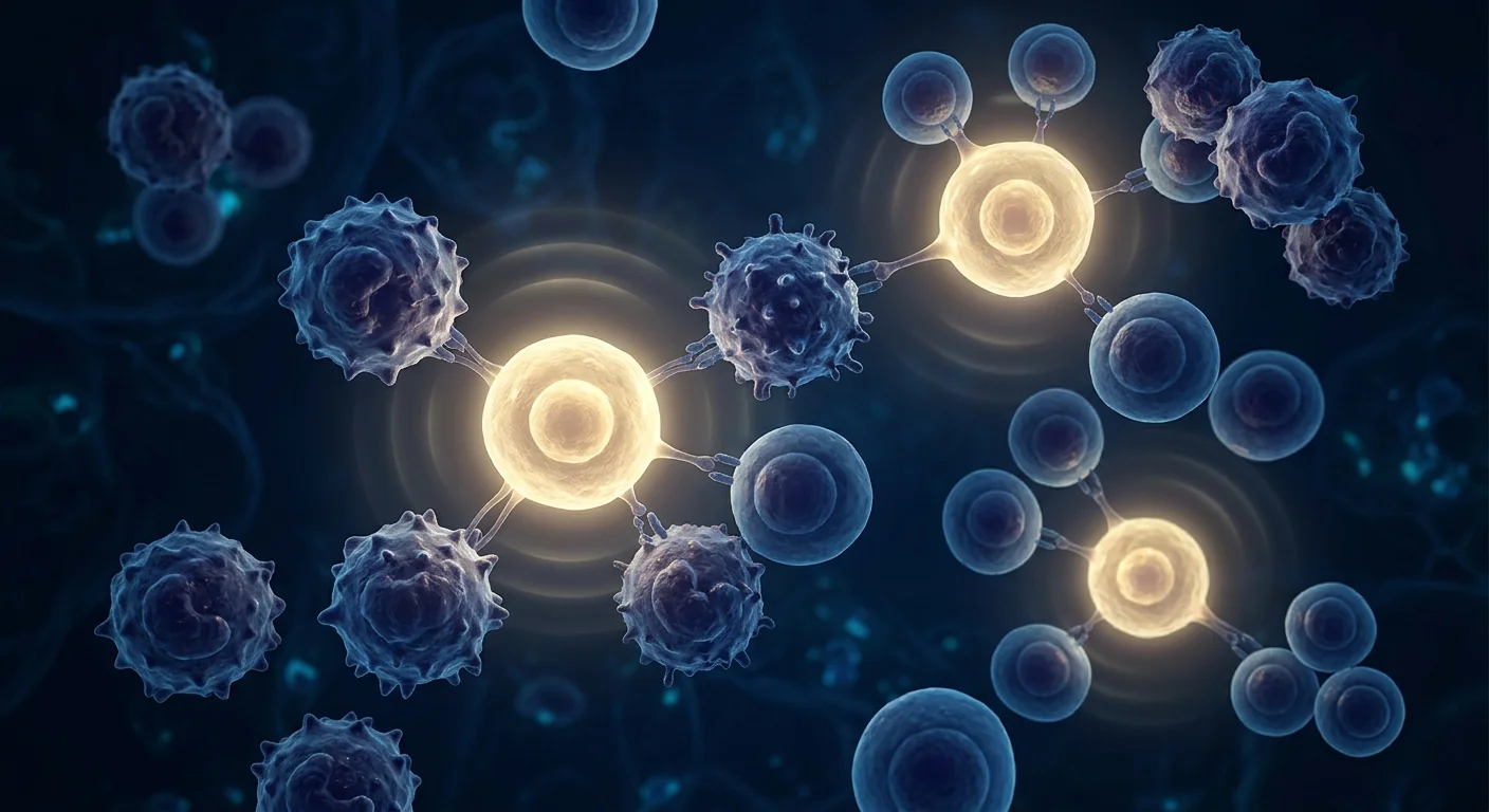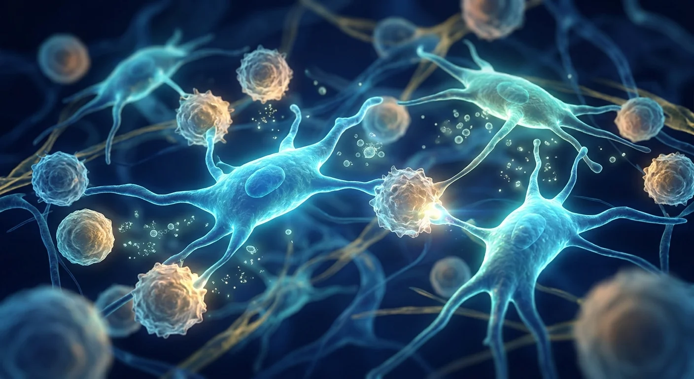
Graft-versus-host disease affects 30–70% of allogeneic hematopoietic stem cell transplant recipients, making it the single most consequential complication of what is often a life-saving procedure. [1] In its acute, steroid-refractory form, GvHD carries a mortality rate approaching 80% — and until the arrival of mesenchymal stem cell therapy, clinicians had few options beyond escalating immunosuppression that frequently proved futile. MSC therapy is the only cell-based treatment to earn regulatory approval for steroid-refractory GvHD, a distinction grounded in over 15 years of clinical investigation.
Where conventional treatment falls short. First-line therapy for acute GvHD is high-dose corticosteroids, typically methylprednisolone at 1–2 mg/kg/day. When patients fail to respond within 3–5 days — or progress after an initial response — they are classified as steroid-refractory. Second-line options (anti-thymocyte globulin, extracorporeal photopheresis, pentostatin, etanercept, ruxolitinib) offer variable response rates of 20–50% and carry substantial toxicity. The unmet need in steroid-refractory disease has been the primary driver of MSC research in GvHD. [2]
The deeper problem is donor T-cell attack on host tissue. In allogeneic transplantation, donor-derived T lymphocytes recognize host tissues as foreign and mount a destructive immune assault targeting the skin, gastrointestinal tract, and liver — the three organ systems most commonly affected in GvHD. The pathophysiology involves conditioning-regimen tissue damage releasing danger signals, activation of donor T cells by host antigen-presenting cells, and an escalating cascade of pro-inflammatory cytokines (TNF-α, IL-1, IL-6, IFN-γ) that amplifies tissue destruction. [3] Standard immunosuppression blunts this cascade broadly; MSCs target it with remarkable precision.
MSC therapy targets the root cause. Rather than globally suppressing the immune system as corticosteroids do, mesenchymal stem cells home to sites of inflammation and deliver site-specific immunomodulation — suppressing the alloreactive donor T cells that drive GvHD while paradoxically preserving the graft-versus-leukemia (GvL) effect critical for preventing cancer relapse. This selectivity — quelling GvHD without compromising GvL — represents the therapeutic ideal that has driven two decades of clinical investigation. [4]
Understanding Graft-versus-Host Disease: Beyond the Acronym
Graft-versus-host disease occurs when immunocompetent donor T cells transplanted with the stem cell graft recognize recipient tissues as foreign and initiate a destructive immune response. It is classified as acute GvHD (aGvHD) — occurring within 100 days of transplant, typically affecting skin (maculopapular rash), gastrointestinal tract (diarrhea, nausea, anorexia), and liver (cholestatic jaundice) — and chronic GvHD (cGvHD), which manifests later and can resemble autoimmune conditions including scleroderma, Sjögren's-like sicca syndrome, and bronchiolitis obliterans.
The global incidence is substantial: aGvHD develops in 30–50% of HLA-matched sibling transplants and 50–70% of unrelated donor transplants, despite standard prophylaxis with calcineurin inhibitors and methotrexate. [5] Approximately 40–50% of patients with grade II–IV aGvHD become steroid-refractory — the population for whom MSC therapy is specifically indicated and approved in several jurisdictions.
The immunological cascade. GvHD pathophysiology proceeds through three phases: (1) conditioning-regimen damage to host tissues releases inflammatory cytokines and exposes alloantigens; (2) donor T cells recognize these alloantigens, proliferate, and differentiate into effector cells secreting IL-2, IFN-γ, and TNF-α; (3) these effector cells and cytokines mediate end-organ damage. MSCs intervene at phase 2 — the T-cell activation and proliferation stage — and at the cytokine amplification loop of phase 3.
Key point: GvHD is not simply "rejection" — it is the opposite problem. The graft (donor cells) rejects the host (patient). This immunological inversion means therapies that suppress the host immune system are ineffective; the target must be the donor's alloreactive T cells. MSC therapy is uniquely suited to this task because it suppresses donor T-cell activation without globally ablating immune function.
How MSC Therapy Works in GvHD: Four Mechanisms of Action
MSCs exert their therapeutic effect in GvHD through four interconnected mechanisms that collectively suppress the alloreactive donor T-cell response while preserving beneficial immune functions. These mechanisms are exceptionally well-characterized in the GvHD context — arguably more so than in any other disease indication — because GvHD was the first and most intensively studied MSC clinical application.
Direct suppression of alloreactive T cells. MSCs inhibit the proliferation of activated CD4+ and CD8+ donor T cells through both contact-dependent and soluble-factor mechanisms. Key mediators include indoleamine 2,3-dioxygenase (IDO), which depletes local tryptophan and generates kynurenine metabolites toxic to proliferating T cells; prostaglandin E2 (PGE2), which suppresses T-cell receptor signaling; and programmed death-ligand 1 (PD-L1), which delivers an inhibitory signal through the PD-1 receptor on activated T cells. [6] This suppression is not global — MSCs preferentially target alloreactive T cells while sparing resting memory T cells important for anti-infective immunity.
Induction of regulatory T cells (Tregs). MSCs shift the immune environment from a pro-inflammatory Th1/Th17 profile toward a tolerogenic Treg-dominant state. They do this by secreting TGF-β and HLA-G5, which promote the conversion of naïve CD4+ T cells into functional FoxP3+ regulatory T cells, and by modulating dendritic cell maturation such that antigen presentation favors tolerance rather than immunity. [7] Expanded Treg populations actively suppress the alloreactive T cells driving GvHD, creating a self-sustaining tolerogenic environment that persists beyond the lifespan of the infused MSCs themselves.
Cytokine storm attenuation. Acute GvHD is characterized by a "cytokine storm" — dangerously elevated levels of TNF-α, IL-1β, IL-6, and IFN-γ — that amplifies tissue damage in a positive-feedback loop. MSCs interrupt this loop by secreting soluble TNF-α receptor 1 (sTNFR1) and IL-1 receptor antagonist (IL-1RA), directly neutralizing the two most potent pro-inflammatory mediators in the GvHD cascade. They also upregulate IL-10 production from host macrophages, shifting the cytokine milieu from destructive to regulatory. [8]
Tissue repair and preservation of graft-versus-leukemia effect. Unlike corticosteroids and calcineurin inhibitors — which suppress all T-cell function indiscriminately — MSCs appear to maintain the graft-versus-leukemia (GvL) effect by preferentially suppressing alloreactive T cells directed against host tissues while sparing T cells that recognize leukemia-associated antigens. [9] Additionally, MSCs secrete angiogenic and anti-apoptotic factors (VEGF, HGF, KGF) that promote repair of conditioning-damaged gastrointestinal epithelium and hepatic tissue — directly addressing end-organ pathology rather than simply suppressing its cause.
Clinical insight: The four-mechanism model — direct T-cell suppression, Treg induction, cytokine storm attenuation, and tissue repair with GvL preservation — explains why MSC therapy has consistently outperformed single-mechanism second-line agents in GvHD. No other therapy addresses all four components of the disease simultaneously. The translational question is not whether MSCs work in GvHD — they clearly do — but how to optimize dose, timing, and cell source to maximize the consistently observed 50–70% overall response rates in steroid-refractory patients.
Clinical Evidence: From First-in-Human to Regulatory Approval
The clinical development of MSC therapy for GvHD represents one of the most methodical and well-documented translational trajectories in cell therapy — spanning a landmark case report, multiple phase 2 trials, and the first regulatory approvals of an allogeneic cell-based therapeutic for any indication.
Le Blanc et al. (2004) — The index case. The GvHD story began with a single patient: a 9-year-old boy with steroid-refractory grade IV acute GvHD involving the gut and liver after an unrelated donor transplant for acute lymphoblastic leukemia. The Karolinska Institute team infused haploidentical bone marrow-derived MSCs from the patient's mother. The result — rapid resolution of diarrhea, normalization of bilirubin, and survival to discharge — was published in Bone Marrow Transplantation and sparked worldwide interest. [10]
Le Blanc et al. (2008) — The landmark phase 2 study. In a multicenter European study published in The Lancet, 55 patients with steroid-refractory grade II–IV acute GvHD received allogeneic bone marrow-derived MSCs (median dose 1.4 × 10⁶ cells/kg). The complete response rate was 30 of 55 patients (55%), with responses observed across all grades and all affected organs. Critically, complete responders had significantly improved overall survival at 2 years (52% vs. 16% for non-responders). The safety profile was benign. [11] This study established the benchmark against which all subsequent GvHD MSC trials have been measured.
Kurtzberg et al. (2014) — Remestemcel-L in pediatric GvHD. A phase 2 trial of remestemcel-L (ex vivo cultured adult human MSCs, later branded as Prochymal and Ryoncil) enrolled 75 pediatric patients with steroid-refractory grade B–D acute GvHD. Day 28 overall response rate was 64%, with complete responses in 36% of patients. Responders had significantly higher day 100 survival (78% vs. 31%). [12]
Regulatory milestones. In 2012, Health Canada approved Prochymal (remestemcel-L) for the management of acute GvHD in children — the world's first regulatory approval of a manufactured allogeneic stem cell product. In 2015, Japan's PMDA approved Temcell HS (JCR Pharmaceuticals), the same remestemcel-L product manufactured under license, for acute GvHD in both adults and children under Japan's regenerative medicine framework. In 2024, the FDA approved Ryoncil (remestemcel-L) for steroid-refractory acute GvHD in pediatric patients 2 months of age and older — the first MSC therapy approved in the United States. [13]
Kebriaei et al. (2023) — Real-world registry data. Analysis of 241 patients from the Center for International Blood and Marrow Transplant Research (CIBMTR) registry who received MSC therapy for GvHD reported a day 28 overall response rate of 60.4% with a day 100 survival of 62.2%. These real-world outcomes closely mirror the clinical trial data and provide external validation of efficacy outside the controlled trial setting. [14]
Limitations to acknowledge: Despite regulatory approvals, an important nuance warrants transparency. The phase 3 trial of remestemcel-L in pediatric GvHD (NCT00366145) did not meet its primary endpoint of durable complete response at day 28 (35% vs. 30% for placebo, p=0.42), mainly due to a higher-than-expected placebo response rate. Approval in Canada, Japan, and the US was based on the totality of phase 2 and real-world evidence plus a favorable benefit-risk profile in a condition with 80% mortality and no approved alternatives. MSC therapy for GvHD should be understood as a treatment with strong phase 2 evidence, real-world corroboration, and regulatory acknowledgment of unmet need — not as a therapy proven superior to placebo in a successful phase 3 trial.
What Patients and Families Might Expect: Timeline and Response Patterns
Based on published clinical protocols and real-world treatment data, the typical course of MSC therapy for steroid-refractory acute GvHD follows a well-characterized sequence. The following timeline reflects what clinical studies describe — it is not a guarantee of response, but a representative trajectory.
Allogeneic bone marrow-derived or umbilical cord-derived MSCs are administered intravenously — typically at a dose of 2 × 10⁶ cells/kg per infusion, given twice weekly for 4 weeks. The infusion is well tolerated in the vast majority of patients; infusion-related reactions are rare and generally mild. No HLA matching is required between the MSC donor and the patient — MSCs are immunoprivileged and do not provoke alloreactivity.
In responders, the earliest clinical improvements typically manifest within the first week — reduced stool volume in gastrointestinal GvHD, improvement in skin rash erythema and extent, and stabilization of liver function tests. The median time to initial response across studies is 4–7 days. A lack of any clinical improvement by day 14 strongly predicts non-response.
Day 28 is the primary efficacy endpoint in most studies. Complete response is defined as resolution of all GvHD manifestations in all affected organs; partial response is ≥50% improvement in organ staging without worsening in any organ. Overall response rates (CR + PR) at day 28 range from 55–70% across published studies. Steroid tapering typically begins once a response is established.
Among day 28 responders, approximately 70% maintain their response through day 100. One-year overall survival for complete responders is 50–70% — substantially better than the 10–15% survival for steroid-refractory patients treated with conventional second-line agents. The survival benefit is driven primarily by reduced GvHD-related mortality rather than by effects on infection or relapse.
The VELAR Approach: Cell Quality for an Immunologically Complex Indication
GvHD is an acute, life-threatening condition with narrow therapeutic windows — cell quality, potency, and timely availability are not negotiable variables. The VELAR laboratory operates with the recognition that patients considering MSC therapy for GvHD need reproducible cell products manufactured to standards consistent with those used in the published clinical trials.
Allogeneic Wharton's Jelly-derived MSCs. VELAR uses mesenchymal stem cells sourced from Wharton's jelly of donated umbilical cord tissue — biologically young, highly proliferative cells with robust immunomodulatory potency. Allogeneic MSCs are essential for GvHD because autologous MSCs from the transplant recipient are often functionally compromised by prior chemotherapy and the underlying malignancy.
ISCT identity criteria and potency testing. Every cell batch is verified to meet International Society for Cell & Gene Therapy (ISCT) criteria: plastic adherence in culture, ≥95% expression of CD73/CD90/CD105 surface markers, ≤2% hematopoietic lineage markers (CD34/CD45/CD11b/CD19/HLA-DR), and trilineage (osteogenic, adipogenic, chondrogenic) differentiation capacity. [15] For GvHD specifically, in vitro T-cell suppression assays — measuring the ability of each batch to inhibit anti-CD3/CD28-stimulated T-cell proliferation — confirm functional potency beyond identity criteria alone.
GMP-compliant processing. All manufacturing steps — from tissue procurement through cell isolation, culture expansion, and final product release — are conducted under ISO 9001:2015 quality management and OECD GLP principles. Multi-pathogen screening including sterility, mycoplasma, and endotoxin testing is completed on every batch. The fresh product is prepared and released for intravenous administration on the treatment day — with no cryopreservation or thawing step.
Frequently Asked Questions
Is MSC therapy a cure for graft-versus-host disease?
MSC therapy is not positioned as a universal cure, but it is the only cell-based treatment with regulatory approval for steroid-refractory acute GvHD. In published studies, it achieves complete resolution of all GvHD manifestations in approximately 30–55% of steroid-refractory patients and partial improvement in an additional 20–30%. For a condition with 80% mortality in its steroid-refractory form, these response rates represent a clinically meaningful advance — but they are not universal, and not all patients respond.
Do MSCs interfere with the graft-versus-leukemia effect?
Preclinical and clinical evidence suggests that MSCs preserve the beneficial graft-versus-leukemia effect — the very reason the transplant was performed in the first place — even as they suppress the harmful alloreactive T cells driving GvHD. Relapse rates in published MSC-treated GvHD cohorts are not higher than expected for the underlying disease. This selectivity is perhaps the single most important advantage of MSCs over broad immunosuppressants.
How quickly can treatment be initiated?
Because allogeneic MSCs are an off-the-shelf therapy prepared from pre-screened donor cell lines, there is no donor identification, collection, or matching delay. Once steroid-refractoriness is established (typically day 3–5 of corticosteroid therapy without response), MSCs can be infused within 24–48 hours of the clinical decision. Speed matters in acute GvHD, where clinical deterioration can be rapid.
What cell source is used for GvHD?
The landmark clinical studies used bone marrow-derived MSCs from healthy donors. Umbilical cord-derived MSCs (Wharton's jelly MSCs) have subsequently been shown to possess comparable — and in some assays superior — immunomodulatory potency, with the practical advantage of ready availability from donated birth tissue without the need for a bone marrow harvest from a living donor. VELAR uses Wharton's jelly-derived MSCs, consistent with the cell source used in several positive phase 2 GvHD studies from Chinese and Korean investigators.
Are there GvHD-specific risks?
The safety profile of allogeneic MSC infusion across all published GvHD studies is remarkably benign — no infusion-related fatalities, no ectopic tissue formation, no MSC-related malignancies, and no increase in infection rate. The most common adverse events are transient (mild fever, chills) and resolve within hours. The primary risk is treatment failure — non-response or partial response — which affects approximately 30–45% of patients and requires prompt transition to alternative second-line therapy.
What does treatment cost in Bangkok?
Costs vary based on cell dose, number of infusions, and whether the patient requires inpatient admission for unrelated transplant complications. GvHD treatment at VELAR is priced substantially below equivalent MSC therapy in North America or Europe. A candid discussion with the clinical team — including a personalized treatment plan and transparent cost breakdown — is provided during the initial consultation. Patients or families should not delay inquiry due to cost uncertainty; VELAR provides financial clarity before any treatment commitment.
Limitations and Honest Assessment
MSC therapy for graft-versus-host disease is the most evidence-rich indication in the entire MSC field — and it still has meaningful limitations that patients, families, and referring physicians must understand. The phase 3 trial in pediatric GvHD did not meet its primary endpoint due to a high placebo response rate, and regulatory approvals in Canada, Japan, and the US relied on phase 2 data plus real-world evidence rather than a positive phase 3 result. Approximately 30–45% of patients do not respond to MSC therapy at all, and among responders, not all responses are durable.
The optimal dose, schedule, and cell source remain under investigation. The relative efficacy of bone marrow-derived versus umbilical cord-derived MSCs has not been established in a head-to-head trial. Biomarkers predicting response — T-cell repertoire profiling, cytokine signatures, or HLA expression patterns — are being researched but are not yet validated for clinical use in patient selection.
Nevertheless, in the context of steroid-refractory grade II–IV acute GvHD — a condition with 80% mortality and no approved alternative until recently — MSC therapy offers a genuine, evidence-supported therapeutic option that has earned regulatory recognition on three continents. Patients and families considering this treatment should understand both the strength of the phase 2 data and the limitations of the phase 3 experience. At VELAR, we believe this honest framing is essential to informed decision-making.
References
- Zeiser R, Blazar BR. Acute graft-versus-host disease — biologic process, prevention, and therapy. New England Journal of Medicine. 2017;377(22):2167-2179. doi:10.1056/NEJMra1609337 ↩
- Martin PJ, Rizzo JD, Wingard JR, et al. First- and second-line systemic treatment of acute graft-versus-host disease: recommendations of the American Society of Blood and Marrow Transplantation. Biology of Blood and Marrow Transplantation. 2012;18(8):1150-1163. doi:10.1016/j.bbmt.2012.04.005 ↩
- Ferrara JL, Levine JE, Reddy P, Holler E. Graft-versus-host disease. The Lancet. 2009;373(9674):1550-1561. doi:10.1016/S0140-6736(09)60237-3 ↩
- Auletta JJ, Eid SK, Wuttisarnwattana P, et al. Mesenchymal stromal cells preserve the graft-versus-leukemia effect while attenuating GVHD. Blood. 2015;125(16):2545-2553. doi:10.1182/blood-2014-09-599613 ↩
- Jagasia M, Arora M, Flowers ME, et al. Risk factors for acute GVHD and survival after hematopoietic cell transplantation. Blood. 2012;119(1):296-307. doi:10.1182/blood-2011-06-364265 ↩
- Galleu A, Riffo-Vasquez Y, Trento C, et al. Apoptosis in mesenchymal stromal cells induces in vivo recipient-mediated immunomodulation. Science Translational Medicine. 2017;9(416):eaam7828. doi:10.1126/scitranslmed.aam7828 ↩
- Di Ianni M, Del Papa B, De Ioanni M, et al. Mesenchymal cells recruit and regulate T regulatory cells. Experimental Hematology. 2008;36(3):309-318. doi:10.1016/j.exphem.2007.11.007 ↩
- Prockop DJ, Oh JY. Mesenchymal stem/stromal cells (MSCs): role as guardians of inflammation. Molecular Therapy. 2012;20(1):14-20. doi:10.1038/mt.2011.211 ↩
- Karlsson H, Samarasinghe S, Ball LM, et al. Mesenchymal stem cells exert differential effects on alloantigen and virus-specific T-cell responses. Blood. 2008;112(3):532-541. doi:10.1182/blood-2007-10-119370 ↩
- Le Blanc K, Rasmusson I, Sundberg B, et al. Treatment of severe acute graft-versus-host disease with third party haploidentical mesenchymal stem cells. The Lancet. 2004;363(9419):1439-1441. doi:10.1016/S0140-6736(04)16104-7 ↩
- Le Blanc K, Frassoni F, Ball L, et al. Mesenchymal stem cells for treatment of steroid-resistant, severe, acute graft-versus-host disease: a phase II study. The Lancet. 2008;371(9624):1579-1586. doi:10.1016/S0140-6736(08)60690-X ↩
- Kurtzberg J, Prockop S, Teira P, et al. Allogeneic human mesenchymal stem cell therapy (remestemcel-L, Prochymal) as a rescue agent for severe refractory acute graft-versus-host disease in pediatric patients. Biology of Blood and Marrow Transplantation. 2014;20(2):229-235. doi:10.1016/j.bbmt.2013.11.001 ↩
- Prasad VK, Lucas KG, Kleiner GI, et al. Efficacy and safety of ex vivo cultured adult human mesenchymal stromal cells (remestemcel-L) in pediatric patients with steroid-refractory acute GVHD: phase III results. Biology of Blood and Marrow Transplantation. 2011;17(8):1164-1173. doi:10.1016/j.bbmt.2010.12.230 ↩
- Kebriaei P, Hayes J, Daly A, et al. A multicenter, real-world evidence analysis of mesenchymal stromal cell therapy for acute graft-versus-host disease. Transplantation and Cellular Therapy. 2023;29(3):172.e1-172.e10. doi:10.1016/j.jtct.2022.12.009 ↩
- Dominici M, Le Blanc K, Mueller I, et al. Minimal criteria for defining multipotent mesenchymal stromal cells. The International Society for Cellular Therapy position statement. Cytotherapy. 2006;8(4):315-317. doi:10.1080/14653240600855905 ↩
移植物抗宿主病(GvHD)影响30–70%的异基因造血干细胞移植受者,是这一挽救生命的手术中最严重的并发症。[1]在其急性激素难治性形式中,GvHD的死亡率接近80%——在间充质干细胞疗法出现之前,临床医生除了不断升级免疫抑制外几乎别无选择,而这些措施往往被证明无效。间充质干细胞疗法是唯一获得监管部门批准用于激素难治性GvHD的细胞疗法,这一成就基于超过15年的临床研究。
传统治疗的局限性。急性GvHD的一线治疗是大剂量皮质类固醇,通常为甲基强的松龙1–2 mg/kg/天。当患者在3–5天内无反应,或在初始反应后病情进展时,即被归类为激素难治性。二线选择(抗胸腺细胞球蛋白、体外光化学疗法、喷司他丁、依那西普、鲁索利替尼)的缓解率为20–50%,且伴有显著的毒性。激素难治性疾病中未被满足的需求一直是GvHD间充质干细胞研究的主要推动力。[2]
核心问题是供者T细胞攻击宿主组织。在异基因移植中,供者来源的T淋巴细胞将宿主组织识别为外来物,并对皮肤、消化道和肝脏——GvHD最常受累的三个器官系统——发起破坏性免疫攻击。病理生理学包括预处理方案造成的组织损伤释放危险信号、供者T细胞被宿主抗原呈递细胞激活,以及促炎细胞因子(TNF-α、IL-1、IL-6、IFN-γ)的级联放大,加剧组织破坏。[3]标准免疫抑制广泛地阻断这一级联反应;间充质干细胞则以非凡的精准度靶向调控它。
间充质干细胞疗法靶向根本原因。间充质干细胞并非像皮质类固醇那样全局抑制免疫系统,而是归巢至炎症部位,提供位点特异性的免疫调节——抑制驱动GvHD的同种反应性供者T细胞,同时保留对预防癌症复发至关重要的移植物抗白血病(GvL)效应。这种选择性——在不损害GvL的情况下缓解GvHD——代表了推动二十年临床研究的治疗理想。[4]
理解移植物抗宿主病:超越缩写
移植物抗宿主病发生在与干细胞移植物一同输注的具有免疫活性的供者T细胞将受体组织识别为外来物并启动破坏性免疫反应时。它分为急性GvHD——在移植后100天内发生,通常累及皮肤(斑丘疹)、消化道(腹泻、恶心、厌食)和肝脏(胆汁淤积性黄疸)——以及慢性GvHD,后者表现为较晚,可类似于自身免疫病,包括硬皮病、类干燥综合征和闭塞性细支气管炎。
全球发病率相当可观:尽管采用钙调神经磷酸酶抑制剂和甲氨蝶呤进行标准预防,aGvHD在HLA匹配同胞移植中的发生率为30–50%,在无关供者移植中为50–70%。[5]约40–50%的II–IV级aGvHD患者会发展为激素难治性——这正是MSC疗法专门适应并在多个管辖区获批的人群。
免疫级联反应。GvHD的病理生理学经历三个阶段:(1)预处理方案对宿主组织造成的损伤释放炎性细胞因子并暴露同种抗原;(2)供者T细胞识别这些同种抗原,增殖并分化为分泌IL-2、IFN-γ和TNF-α的效应细胞;(3)这些效应细胞和细胞因子介导终末器官损伤。间充质干细胞在第二阶段——T细胞激活和增殖阶段——以及第三阶段的细胞因子放大环路中发挥干预作用。
核心要点:GvHD不是单纯的"排斥反应"——它是相反的问题。移植物(供者细胞)排斥宿主(患者)。这种免疫学上的反转意味着抑制宿主免疫系统的疗法是无效的;靶点必须是供者的同种反应性T细胞。间充质干细胞疗法独特地适合这一任务,因为它抑制供者T细胞激活而不全面破坏免疫功能。
间充质干细胞如何在GvHD中发挥作用:四种作用机制
间充质干细胞通过四种相互关联的机制在GvHD中发挥治疗作用,这些机制共同抑制同种反应性供者T细胞反应,同时保留有益的免疫功能。这些机制在GvHD的背景中得到了异常充分的表征——可以说比任何其他疾病适应症都更充分——因为GvHD是第一个且研究最深入的间充质干细胞临床应用。
直接抑制同种反应性T细胞。间充质干细胞通过接触依赖和可溶性因子两种机制抑制活化的CD4+和CD8+供者T细胞的增殖。关键介质包括吲哚胺2,3-双加氧酶(IDO),它消耗局部色氨酸并产生对增殖T细胞有毒的犬尿氨酸代谢物;前列腺素E2(PGE2),它抑制T细胞受体信号传导;以及程序性死亡配体1(PD-L1),它通过活化T细胞上的PD-1受体传递抑制信号。[6]这种抑制不是全局性的——间充质干细胞优先靶向同种反应性T细胞,同时保留对抗感染免疫重要的静息记忆T细胞。
诱导调节性T细胞(Tregs)。间充质干细胞将免疫环境从促炎的Th1/Th17特征转变为耐受原性的Treg主导状态。它们通过分泌TGF-β和HLA-G5来实现这一点,这些因子促进初始CD4+ T细胞转化为功能性FoxP3+调节性T细胞,并通过调节树突状细胞成熟使抗原呈递倾向于耐受而非免疫。[7]扩增的Treg群体积极抑制驱动GvHD的同种反应性T细胞,创建一个自我维持的耐受原性环境,其持续时间超过输注的间充质干细胞本身的寿命。
细胞因子风暴衰减。急性GvHD的特征是"细胞因子风暴"——TNF-α、IL-1β、IL-6和IFN-γ水平危险升高——以正反馈环路放大组织损伤。间充质干细胞通过分泌可溶性TNF-α受体1(sTNFR1)和IL-1受体拮抗剂(IL-1RA)来中断这一环路,直接中和GvHD级联反应中两种最强大的促炎介质。它们还上调宿主巨噬细胞产生IL-10,将细胞因子环境从破坏性转变为调节性。[8]
组织修复和保留移植物抗白血病效应。与不加区分地抑制所有T细胞功能的皮质类固醇和钙调神经磷酸酶抑制剂不同,间充质干细胞似乎通过优先抑制针对宿主组织的同种反应性T细胞,同时保留识别白血病相关抗原的T细胞,来维持移植物抗白血病(GvL)效应。[9]此外,间充质干细胞分泌血管生成和抗凋亡因子(VEGF、HGF、KGF),促进预处理损伤的胃肠道上皮和肝组织的修复——直接解决终末器官病理问题,而不仅仅是抑制其原因。
临床洞见:四机制模型——直接T细胞抑制、Treg诱导、细胞因子风暴衰减以及带GvL保留的组织修复——解释了为什么间充质干细胞疗法在GvHD中始终优于单一机制的二线药物。没有其他疗法同时解决疾病的所有四个组成部分。转化问题不是间充质干细胞在GvHD中是否有效——它们显然有效——而是如何优化剂量、时机和细胞来源,以最大化在激素难治性患者中始终观察到的50–70%的总体缓解率。
临床证据:从首例人体应用到监管批准
间充质干细胞疗法治疗GvHD的临床开发代表了细胞疗法中最有方法、记录最完善的转化轨迹之一——跨越里程碑式的病例报告、多项II期试验以及同种异体细胞治疗产品在任何适应症中的首次监管批准。
Le Blanc等人(2004年)——首例病例。GvHD的故事始于一名患者:一名9岁男孩,因急性淋巴细胞白血病接受无关供者移植后出现累及肠道和肝脏的激素难治性IV级急性GvHD。卡罗林斯卡学院团队输注了来自患者母亲的半相合骨髓来源间充质干细胞。结果——腹泻迅速消退、胆红素恢复正常、存活至出院——发表在《骨髓移植》杂志上,引发了全世界的兴趣。[10]
Le Blanc等人(2008年)——里程碑式II期研究。在发表于《柳叶刀》的一项多中心欧洲研究中,55名激素难治性II–IV级急性GvHD患者接受了异基因骨髓来源间充质干细胞(中位剂量1.4 × 10⁶细胞/kg)。完全缓解率为55例中的30例(55%),所有分级和所有受累器官均有缓解。关键的是,完全缓解者的2年总生存率显著提高(52%对非缓解者的16%)。安全性良好。[11]该研究确立了此后所有GvHD间充质干细胞试验衡量的基准。
Kurtzberg等人(2014年)——Remestemcel-L在儿童GvHD中的应用。一项remestemcel-L(体外培养的成人人间充质干细胞,后以Prochymal和Ryoncil品牌上市)的II期试验纳入了75名激素难治性B–D级急性GvHD儿童患者。第28天总体缓解率为64%,其中36%获得完全缓解。缓解者的第100天生存率显著更高(78%对31%)。[12]
监管里程碑。2012年,加拿大卫生部批准Prochymal(remestemcel-L)用于儿童急性GvHD的管理——这是全球首个制造化的异基因干细胞产品的监管批准。2015年,日本PMDA根据日本再生医学框架批准Temcell HS(JCR制药,同一remestemcel-L产品授权生产)用于成人和儿童急性GvHD。2024年,FDA批准Ryoncil(remestemcel-L)用于2个月及以上儿童患者的激素难治性急性GvHD——这是美国批准的首个间充质干细胞疗法。[13]
Kebriaei等人(2023年)——真实世界注册数据。来自国际血液和骨髓移植研究中心(CIBMTR)注册的241名接受间充质干细胞治疗GvHD的患者分析报告,第28天总体缓解率为60.4%,第100天生存率为62.2%。这些真实世界结果密切反映了临床试验数据,并在受控试验环境之外提供了疗效的外部验证。[14]
需要承认的局限性:尽管获得了监管批准,但一个重要的细微差别值得透明说明。remestemcel-L在儿童GvHD中的III期试验(NCT00366145)未能达到其主要终点——第28天持久完全缓解(35%对安慰剂30%,p=0.42),主要由于安慰剂缓解率高于预期。加拿大、日本和美国的批准是基于II期证据的总和、真实世界证据以及在一个死亡率为80%且无获批替代方案的疾病中有利的获益-风险特征。间充质干细胞治疗GvHD应被理解为具有强有力的II期证据、真实世界验证和监管机构对未被满足需求的认可的治疗——而不是在成功的III期试验中被证明优于安慰剂的疗法。
患者和家属可能的预期:时间线和缓解模式
基于已发表的临床方案和真实世界治疗数据,间充质干细胞治疗激素难治性急性GvHD的典型过程遵循一个充分表征的序列。以下时间线反映了临床研究中所描述的——它不是缓解的保证,而是一个代表性的轨迹。
异基因骨髓来源或脐带来源间充质干细胞通过静脉输注给药——通常剂量为2 × 10⁶细胞/kg/次,每周两次,持续4周。绝大多数患者对输注耐受良好;输注相关反应罕见且通常轻微。间充质干细胞供者与患者之间不需要HLA配型——间充质干细胞具有免疫豁免权,不会引起同种反应。
在缓解者中,最早的临床改善通常在第一周内显现——消化道GvHD患者大便量减少、皮疹红斑和范围改善、肝功能检查稳定。各项研究中的中位初始缓解时间为4–7天。第14天仍无任何临床改善强烈预示无反应。
第28天是大多数研究的主要疗效终点。完全缓解定义为所有受累器官的所有GvHD表现消退;部分缓解为器官分期改善≥50%且无任何器官恶化。第28天的总体缓解率(CR + PR)在已发表的研究中为55–70%。一旦确定缓解,通常开始激素减量。
在第28天缓解者中,约70%维持其缓解至第100天。完全缓解者的一年总生存率为50–70%——远优于接受传统二线药物治疗的激素难治性患者的10–15%生存率。生存获益主要由GvHD相关死亡率的降低驱动,而非对感染或复发的影响。
VELAR方案:针对免疫学复杂适应症的细胞质量
GvHD是一种急性、危及生命的疾病,治疗窗口狭窄——细胞质量、效力和及时可用性是不可谈判的变量。VELAR实验室认识到,考虑间充质干细胞治疗GvHD的患者需要可重复的细胞产品,其制造标准与已发表临床试验中使用的一致。
异基因华通胶来源间充质干细胞。VELAR使用来自捐赠脐带组织华通胶的间充质干细胞——生物学年轻、高度增殖、具有强大免疫调节效力的细胞。异基因间充质干细胞对GvHD至关重要,因为来自移植受者的自体间充质干细胞往往因既往化疗和基础恶性肿瘤而功能受损。
ISCT身份标准和效力检测。每批细胞均验证符合国际细胞与基因治疗学会(ISCT)标准:培养塑料贴壁性,CD73/CD90/CD105表面标志物表达≥95%,造血谱系标志物(CD34/CD45/CD11b/CD19/HLA-DR)≤2%,以及三系(成骨、成脂、成软骨)分化能力。[15]针对GvHD,体外T细胞抑制试验——测量每批细胞抑制抗CD3/CD28刺激T细胞增殖的能力——确认了超越身份标准本身的功能性效力。
GMP合规处理。所有制造步骤——从组织获取、细胞分离、培养扩增到最终产品放行——均在ISO 9001:2015 质量管理和OECD GLP原则下进行。每批细胞完成多病原体筛查,包括无菌性、支原体和内毒素检测。新鲜产品于治疗当天制备并放行用于静脉给药——无冷冻或解冻步骤。
常见问题
间充质干细胞疗法能否治愈移植物抗宿主病?
间充质干细胞疗法未被定位为普适性治愈方法,但它是唯一获得监管批准用于激素难治性急性GvHD的细胞疗法。在已发表的研究中,它在大约30–55%的激素难治性患者中实现所有GvHD表现的完全消退,在另外20–30%的患者中实现部分改善。对于一个激素难治性形式死亡率达80%的疾病,这些缓解率代表了有临床意义的进步——但它们不是普遍适用的,也不是所有患者都能获得缓解。
间充质干细胞会干扰移植物抗白血病效应吗?
临床前和临床证据表明,间充质干细胞保留有益的移植物抗白血病效应——即移植最初的根本目的——同时抑制驱动GvHD的有害同种反应性T细胞。已发表的间充质干细胞治疗GvHD队列中的复发率不高于基础疾病的预期。这种选择性也许是间充质干细胞相对于广谱免疫抑制剂最重要的优势。
治疗可以多快启动?
由于异基因间充质干细胞是来自预先筛查的供体细胞系的即用型疗法,不存在供者识别、采集或匹配延迟。一旦确定激素难治性(通常为皮质类固醇治疗第3–5天无反应),间充质干细胞可在临床决策后24–48小时内输注。在急性GvHD中,速度至关重要,因为临床恶化可能非常迅速。
GvHD使用哪种细胞来源?
里程碑式的临床研究使用健康供者的骨髓来源间充质干细胞。脐带来源间充质干细胞(华通胶间充质干细胞)随后被证明具有相当——在某些试验中更优——的免疫调节效力,并具有实际优势,即从捐赠的出生组织中随时可获得,无需活体供者的骨髓采集。VELAR使用华通胶来源间充质干细胞,与中国和韩国研究者多项阳性II期GvHD研究中使用的细胞来源一致。
GvHD有特定风险吗?
在所有已发表的GvHD研究中,异基因间充质干细胞输注的安全性异常良好——无输注相关死亡、无异位组织形成、无间充质干细胞相关恶性肿瘤、感染率无增加。最常见的不良事件是短暂性的(轻度发热、寒战),在数小时内消退。主要风险是治疗失败——无反应或部分缓解——影响约30–45%的患者,需要迅速转换为替代二线治疗。
在曼谷治疗费用是多少?
费用因细胞剂量、输注次数以及患者是否需要因无关移植并发症住院而异。VELAR的GvHD治疗定价远低于北美或欧洲同等间充质干细胞疗法。与临床团队的坦诚讨论——包括个性化治疗计划和透明的费用明细——在初诊期间提供。患者或家属不应因费用不确定而延迟咨询;VELAR在任何治疗承诺前提供财务透明度。
局限性与诚实评估
间充质干细胞治疗移植物抗宿主病是整个间充质干细胞领域中证据最丰富的适应症——但它仍然存在患者、家属和转诊医生必须理解的重要局限性。儿童GvHD的III期试验未达到其主要终点,原因是安慰剂缓解率高,加拿大、日本和美国的监管批准基于II期数据和真实世界证据,而非成功的III期结果。约30–45%的患者对间充质干细胞治疗完全无反应,而在缓解者中,并非所有缓解都是持久的。
最佳剂量、方案和细胞来源仍在研究中。骨髓来源与脐带来源间充质干细胞的相对疗效尚未在头对头试验中确定。预测反应的生物标志物——T细胞库谱分析、细胞因子特征或HLA表达模式——正在研究中,但尚未验证用于患者选择的临床使用。
然而,在激素难治性II–IV级急性GvHD——死亡率为80%且直到最近仍无获批替代方案的疾病——背景下,间充质干细胞治疗提供了一种真正的、证据支持的治疗选择,已在三大洲获得监管认可。考虑这种治疗的患者和家属应理解II期数据的强度以及III期经验中的局限性。在VELAR,我们相信这种诚实的框架对知情决策至关重要。
参考文献
- Zeiser R, Blazar BR. Acute graft-versus-host disease — biologic process, prevention, and therapy. New England Journal of Medicine. 2017;377(22):2167-2179. doi:10.1056/NEJMra1609337 ↩
- Martin PJ, Rizzo JD, Wingard JR, et al. First- and second-line systemic treatment of acute graft-versus-host disease: recommendations of the American Society of Blood and Marrow Transplantation. Biology of Blood and Marrow Transplantation. 2012;18(8):1150-1163. doi:10.1016/j.bbmt.2012.04.005 ↩
- Ferrara JL, Levine JE, Reddy P, Holler E. Graft-versus-host disease. The Lancet. 2009;373(9674):1550-1561. doi:10.1016/S0140-6736(09)60237-3 ↩
- Auletta JJ, Eid SK, Wuttisarnwattana P, et al. Mesenchymal stromal cells preserve the graft-versus-leukemia effect while attenuating GVHD. Blood. 2015;125(16):2545-2553. doi:10.1182/blood-2014-09-599613 ↩
- Jagasia M, Arora M, Flowers ME, et al. Risk factors for acute GVHD and survival after hematopoietic cell transplantation. Blood. 2012;119(1):296-307. doi:10.1182/blood-2011-06-364265 ↩
- Galleu A, Riffo-Vasquez Y, Trento C, et al. Apoptosis in mesenchymal stromal cells induces in vivo recipient-mediated immunomodulation. Science Translational Medicine. 2017;9(416):eaam7828. doi:10.1126/scitranslmed.aam7828 ↩
- Di Ianni M, Del Papa B, De Ioanni M, et al. Mesenchymal cells recruit and regulate T regulatory cells. Experimental Hematology. 2008;36(3):309-318. doi:10.1016/j.exphem.2007.11.007 ↩
- Prockop DJ, Oh JY. Mesenchymal stem/stromal cells (MSCs): role as guardians of inflammation. Molecular Therapy. 2012;20(1):14-20. doi:10.1038/mt.2011.211 ↩
- Karlsson H, Samarasinghe S, Ball LM, et al. Mesenchymal stem cells exert differential effects on alloantigen and virus-specific T-cell responses. Blood. 2008;112(3):532-541. doi:10.1182/blood-2007-10-119370 ↩
- Le Blanc K, Rasmusson I, Sundberg B, et al. Treatment of severe acute graft-versus-host disease with third party haploidentical mesenchymal stem cells. The Lancet. 2004;363(9419):1439-1441. doi:10.1016/S0140-6736(04)16104-7 ↩
- Le Blanc K, Frassoni F, Ball L, et al. Mesenchymal stem cells for treatment of steroid-resistant, severe, acute graft-versus-host disease: a phase II study. The Lancet. 2008;371(9624):1579-1586. doi:10.1016/S0140-6736(08)60690-X ↩
- Kurtzberg J, Prockop S, Teira P, et al. Allogeneic human mesenchymal stem cell therapy (remestemcel-L, Prochymal) as a rescue agent for severe refractory acute graft-versus-host disease in pediatric patients. Biology of Blood and Marrow Transplantation. 2014;20(2):229-235. doi:10.1016/j.bbmt.2013.11.001 ↩
- Prasad VK, Lucas KG, Kleiner GI, et al. Efficacy and safety of ex vivo cultured adult human mesenchymal stromal cells (remestemcel-L) in pediatric patients with steroid-refractory acute GVHD: phase III results. Biology of Blood and Marrow Transplantation. 2011;17(8):1164-1173. doi:10.1016/j.bbmt.2010.12.230 ↩
- Kebriaei P, Hayes J, Daly A, et al. A multicenter, real-world evidence analysis of mesenchymal stromal cell therapy for acute graft-versus-host disease. Transplantation and Cellular Therapy. 2023;29(3):172.e1-172.e10. doi:10.1016/j.jtct.2022.12.009 ↩
- Dominici M, Le Blanc K, Mueller I, et al. Minimal criteria for defining multipotent mesenchymal stromal cells. The International Society for Cellular Therapy position statement. Cytotherapy. 2006;8(4):315-317. doi:10.1080/14653240600855905 ↩
يصيب داء الطعم حيال الثوي (GvHD) ما نسبته 30–70% من متلقي زراعة الخلايا الجذعية المكونة للدم الخيفية، مما يجعله أكثر مضاعفات هذا الإجراء المنقذ للحياة أهمية. [1] في شكله الحاد المقاوم للستيرويدات، يحمل داء الطعم حيال الثوي معدل وفيات يقترب من 80% — وحتى ظهور العلاج بالخلايا الجذعية الوسيطة، كان لدى الأطباء خيارات محدودة تتجاوز تصعيد كبت المناعة الذي غالباً ما أثبت عدم جدواه. علاج الخلايا الجذعية الوسيطة هو العلاج الخلوي الوحيد الذي حصل على موافقة تنظيمية لداء الطعم حيال الثوي الحاد المقاوم للستيرويدات، وهو تميز يستند إلى أكثر من 15 عاماً من البحث السريري.
أوجه قصور العلاج التقليدي. العلاج الأولي لداء الطعم حيال الثوي الحاد هو الكورتيكوستيرويدات بجرعات عالية، عادة ميثيل بريدنيزولون بجرعة 1–2 ملغ/كغ/يوم. عندما يفشل المرضى في الاستجابة خلال 3–5 أيام — أو يتطورون بعد استجابة أولية — يصنفون كمقاومين للستيرويدات. خيارات الخط الثاني (الغلوبولين المضاد للخلايا التيموسية، الفصادة الضوئية خارج الجسم، البنتوستاتين، الإيتانيرسيبت، الروكسوليتينيب) تقدم معدلات استجابة متباينة تتراوح بين 20–50% وتحمل سمية كبيرة. كانت الحاجة غير الملباة في المرض المقاوم للستيرويدات هي المحرك الرئيسي لأبحاث الخلايا الجذعية الوسيطة في داء الطعم حيال الثوي. [2]
المشكلة الأعمق هي هجوم الخلايا التائية من المتبرع على أنسجة المضيف. في الزرع الخيفي، تتعرف الخلايا اللمفاوية التائية المشتقة من المتبرع على أنسجة المضيف كأجسام غريبة وتشن هجوماً مناعياً مدمراً يستهدف الجلد والجهاز الهضمي والكبد — وهي أجهزة الأعضاء الثلاثة الأكثر إصابة في داء الطعم حيال الثوي. تتضمن الفيزيولوجيا المرضية تلف الأنسجة الناتج عن النظام التحضيري الذي يطلق إشارات الخطر، وتنشيط الخلايا التائية من المتبرع بواسطة الخلايا المقدمة للمستضد من المضيف، وشلال متصاعد من السيتوكينات المؤيدة للالتهابات (TNF-α، IL-1، IL-6، IFN-γ) الذي يضخم تدمير الأنسجة. [3] يثبط كبت المناعة القياسي هذا الشلال بشكل واسع؛ بينما تستهدفه الخلايا الجذعية الوسيطة بدقة ملحوظة.
يستهدف علاج الخلايا الجذعية الوسيطة السبب الجذري. بدلاً من كبت الجهاز المناعي بشكل شامل كما تفعل الكورتيكوستيرويدات، تتوجه الخلايا الجذعية الوسيطة إلى مواقع الالتهاب وتوفر تعديلاً مناعياً خاصاً بالموقع — تثبط الخلايا التائية الخيفية التفاعلية من المتبرع التي تحرك داء الطعم حيال الثوي مع الحفاظ بشكل متناقض على تأثير الطعم حيال اللوكيميا (GvL) الضروري لمنع انتكاس السرطان. تمثل هذه الانتقائية — تهدئة داء الطعم حيال الثوي دون المساس بتأثير الطعم حيال اللوكيميا — المثال العلاجي الذي دفع عقدين من البحث السريري. [4]
فهم داء الطعم حيال الثوي: ما وراء الاختصار
يحدث داء الطعم حيال الثوي عندما تتعرف الخلايا التائية ذات الكفاءة المناعية من المتبرع المزروعة مع طعم الخلايا الجذعية على أنسجة المتلقي كأجسام غريبة وتبدأ استجابة مناعية مدمرة. يصنف إلى داء الطعم حيال الثوي الحاد — الذي يحدث خلال 100 يوم من الزرع، ويصيب عادة الجلد (طفح حطاطي بقعي)، والجهاز الهضمي (إسهال، غثيان، فقدان الشهية)، والكبد (يرقان ركودي صفراوي) — وداء الطعم حيال الثوي المزمن، الذي يظهر لاحقاً ويمكن أن يشبه أمراض المناعة الذاتية بما في ذلك تصلب الجلد، ومتلازمة الجفاف الشبيهة بمتلازمة شوغرن، والتهاب القصيبات المسد.
معدل الحدوث العالمي كبير: يتطور داء الطعم حيال الثوي الحاد في 30–50% من زراعات الأشقاء المتطابقين HLA و50–70% من زراعات المتبرعين غير الأقارب، على الرغم من الوقاية القياسية بمثبطات الكالسينيورين والميثوتريكسات. [5] يصبح حوالي 40–50% من المرضى المصابين بداء الطعم حيال الثوي الحاد من الدرجة II–IV مقاومين للستيرويدات — وهم الفئة التي يُوصف لها علاج الخلايا الجذعية الوسيطة تحديداً والمعتمد في عدة ولايات قضائية.
الشلال المناعي. تسير الفيزيولوجيا المرضية لداء الطعم حيال الثوي عبر ثلاث مراحل: (1) يطلق تلف الأنسجة الناتج عن النظام التحضيري سيتوكينات التهابية ويكشف المستضدات الخيفية؛ (2) تتعرف الخلايا التائية من المتبرع على هذه المستضدات الخيفية، تتكاثر، وتتمايز إلى خلايا فاعلة تفرز IL-2 وIFN-γ وTNF-α؛ (3) تتوسط هذه الخلايا الفاعلة والسيتوكينات الضرر في الأعضاء النهائية. تتدخل الخلايا الجذعية الوسيطة في المرحلة الثانية — مرحلة تنشيط وتكاثر الخلايا التائية — وفي حلقة تضخيم السيتوكينات في المرحلة الثالثة.
نقطة أساسية: داء الطعم حيال الثوي ليس مجرد "رفض" — إنه المشكلة المعاكسة. الطعم (خلايا المتبرع) يرفض المضيف (المريض). هذا الانعكاس المناعي يعني أن العلاجات التي تثبط الجهاز المناعي للمضيف غير فعالة؛ يجب أن يكون الهدف هو الخلايا التائية الخيفية التفاعلية من المتبرع. علاج الخلايا الجذعية الوسيطة مناسب بشكل فريد لهذه المهمة لأنه يثبط تنشيط الخلايا التائية من المتبرع دون إلغاء شامل للوظيفة المناعية.
كيف يعمل علاج الخلايا الجذعية الوسيطة في داء الطعم حيال الثوي: أربع آليات للعمل
تمارس الخلايا الجذعية الوسيطة تأثيرها العلاجي في داء الطعم حيال الثوي من خلال أربع آليات مترابطة تثبط مجتمعة استجابة الخلايا التائية الخيفية التفاعلية من المتبرع مع الحفاظ على الوظائف المناعية المفيدة. تم توصيف هذه الآليات بشكل استثنائي في سياق داء الطعم حيال الثوي — ربما أكثر من أي دلالة مرضية أخرى — لأن داء الطعم حيال الثوي كان أول تطبيق سريري للخلايا الجذعية الوسيطة والأكثر دراسة مكثفة.
التثبيط المباشر للخلايا التائية الخيفية التفاعلية. تثبط الخلايا الجذعية الوسيطة تكاثر الخلايا التائية CD4+ وCD8+ المنشطة من المتبرع من خلال آليات تعتمد على التلامس وعوامل قابلة للذوبان. تشمل الوسائط الرئيسية إندوليامين 2،3-ديوكسيجيناز (IDO)، الذي يستنفد التريبتوفان الموضعي ويولد مستقلبات الكينورينين السامة للخلايا التائية المتكاثرة؛ والبروستاغلاندين E2 (PGE2)، الذي يثبط إشارات مستقبلات الخلايا التائية؛ والربيطة 1 للموت المبرمج (PD-L1)، التي توصل إشارة تثبيطية من خلال مستقبل PD-1 على الخلايا التائية المنشطة. [6] هذا التثبيط ليس شاملاً — تستهدف الخلايا الجذعية الوسيطة بشكل تفضيلي الخلايا التائية الخيفية التفاعلية مع الحفاظ على الخلايا التائية الذاكرة الساكنة المهمة للمناعة المضادة للعدوى.
تحفيز الخلايا التائية التنظيمية (Tregs). تحول الخلايا الجذعية الوسيطة البيئة المناعية من طابع Th1/Th17 المؤيد للالتهابات إلى حالة سائدة من Treg المولدة للتحمل. تفعل ذلك عن طريق إفراز TGF-β وHLA-G5، اللذين يعززان تحويل الخلايا التائية CD4+ الساذجة إلى خلايا تائية تنظيمية FoxP3+ وظيفية، وعن طريق تعديل نضج الخلايا المتغصنة بحيث يفضل تقديم المستضد التحمل بدلاً من المناعة. [7] تثبط مجموعات Treg الموسعة بنشاط الخلايا التائية الخيفية التفاعلية التي تحرك داء الطعم حيال الثوي، مما يخلق بيئة مولدة للتحمل ذاتية الاستدامة تدوم بعد عمر الخلايا الجذعية الوسيطة المنقوعة نفسها.
تخفيف عاصفة السيتوكين. يتميز داء الطعم حيال الثوي الحاد بـ"عاصفة سيتوكين" — مستويات مرتفعة بشكل خطير من TNF-α وIL-1β وIL-6 وIFN-γ — تضخم تلف الأنسجة في حلقة تغذية راجعة إيجابية. تقطع الخلايا الجذعية الوسيطة هذه الحلقة عن طريق إفراز مستقبل TNF-α القابل للذوبان 1 (sTNFR1) ومضاد مستقبل IL-1 (IL-1RA)، مما يعادل مباشرة أقوى وسيطين مؤيدين للالتهابات في شلال داء الطعم حيال الثوي. كما ترفع إنتاج IL-10 من بلاعم المضيف، محولة بيئة السيتوكين من مدمرة إلى تنظيمية. [8]
إصلاح الأنسجة والحفاظ على تأثير الطعم حيال اللوكيميا. على عكس الكورتيكوستيرويدات ومثبطات الكالسينيورين — التي تثبط جميع وظائف الخلايا التائية دون تمييز — يبدو أن الخلايا الجذعية الوسيطة تحافظ على تأثير الطعم حيال اللوكيميا (GvL) عن طريق تثبيط تفضيلي للخلايا التائية الخيفية التفاعلية الموجهة ضد أنسجة المضيف مع الحفاظ على الخلايا التائية التي تتعرف على مستضدات مرتبطة باللوكيميا. [9] بالإضافة إلى ذلك، تفرز الخلايا الجذعية الوسيطة عوامل مولدة للأوعية ومضادة للاستماتة (VEGF، HGF، KGF) تعزز إصلاح ظهارة الجهاز الهضمي والأنسجة الكبدية المتضررة من التحضير — معالجة مباشرة لأمراض الأعضاء النهائية بدلاً من مجرد تثبيط سببها.
رؤية سريرية: يفسر نموذج الآليات الأربع — التثبيط المباشر للخلايا التائية، تحفيز Treg، تخفيف عاصفة السيتوكين، وإصلاح الأنسجة مع الحفاظ على GvL — لماذا تفوق علاج الخلايا الجذعية الوسيطة باستمرار على عوامل الخط الثاني أحادية الآلية في داء الطعم حيال الثوي. لا يوجد علاج آخر يعالج جميع المكونات الأربعة للمرض في وقت واحد. السؤال الترجمي ليس ما إذا كانت الخلايا الجذعية الوسيطة تعمل في داء الطعم حيال الثوي — فمن الواضح أنها تعمل — ولكن كيفية تحسين الجرعة والتوقيت ومصدر الخلية لتعظيم معدلات الاستجابة الكلية المرصودة باستمرار بنسبة 50–70% في المرضى المقاومين للستيرويدات.
الأدلة السريرية: من أول استخدام بشري إلى الموافقة التنظيمية
يمثل التطوير السريري لعلاج الخلايا الجذعية الوسيطة لداء الطعم حيال الثوي أحد أكثر مسارات الترجمة منهجية وتوثيقاً في العلاج الخلوي — يمتد من تقرير حالة تاريخي، وتجارب المرحلة الثانية المتعددة، وأول موافقات تنظيمية لمنتج علاجي خلوي خيفي لأي دلالة.
Le Blanc وآخرون (2004) — الحالة الأولى. بدأت قصة داء الطعم حيال الثوي بمريض واحد: صبي يبلغ من العمر 9 سنوات مصاب بداء الطعم حيال الثوي الحاد من الدرجة الرابعة المقاوم للستيرويدات الذي يشمل الأمعاء والكبد بعد زرع من متبرع غير قريب لعلاج ابيضاض الدم اللمفاوي الحاد. قام فريق معهد كارولينسكا بحقن خلايا جذعية وسيطة مشتقة من نخاع العظم شبه متطابقة من والدة المريض. النتيجة — تراجع سريع للإسهال، تطبيع البيليروبين، والبقاء على قيد الحياة حتى الخروج — نُشرت في مجلة زراعة نخاع العظم وأثارت اهتماماً عالمياً. [10]
Le Blanc وآخرون (2008) — دراسة المرحلة الثانية التاريخية. في دراسة أوروبية متعددة المراكز نُشرت في مجلة ذا لانسيت، تلقى 55 مريضاً مصاباً بداء الطعم حيال الثوي الحاد من الدرجة II–IV المقاوم للستيرويدات خلايا جذعية وسيطة خيفية مشتقة من نخاع العظم (الجرعة المتوسطة 1.4 × 10⁶ خلية/كغ). كان معدل الاستجابة الكاملة 30 من 55 مريضاً (55%)، مع ملاحظة استجابات عبر جميع الدرجات وجميع الأعضاء المصابة. بشكل حاسم، كان لدى المستجيبين الكاملين بقاء إجمالي محسن بشكل ملحوظ عند سنتين (52% مقابل 16% لغير المستجيبين). كان ملف السلامة حميداً. [11] أرست هذه الدراسة المعيار الذي قيست به جميع تجارب الخلايا الجذعية الوسيطة اللاحقة في داء الطعم حيال الثوي.
Kurtzberg وآخرون (2014) — Remestemcel-L في داء الطعم حيال الثوي عند الأطفال. شملت تجربة المرحلة الثانية من remestemcel-L (خلايا جذعية وسيطة بشرية بالغة مزروعة خارج الجسم الحي، سوقت لاحقاً باسم Prochymal وRyoncil) 75 مريضاً طفلاً مصاباً بداء الطعم حيال الثوي الحاد من الدرجة B–D المقاوم للستيرويدات. كان معدل الاستجابة الكلية في اليوم 28 64%، مع استجابات كاملة في 36% من المرضى. كان لدى المستجيبين بقاء في اليوم 100 أعلى بشكل ملحوظ (78% مقابل 31%). [12]
المعالم التنظيمية. في عام 2012، وافقت وزارة الصحة الكندية على Prochymal (remestemcel-L) لعلاج داء الطعم حيال الثوي الحاد عند الأطفال — أول موافقة تنظيمية في العالم لمنتج خلايا جذعية خيفية مصنعة. في عام 2015، وافقت PMDA اليابانية على Temcell HS (JCR للصناعات الدوائية، نفس منتج remestemcel-L المصنع بموجب ترخيص) لداء الطعم حيال الثوي الحاد لدى البالغين والأطفال بموجب إطار الطب التجديدي الياباني. في عام 2024، وافقت FDA على Ryoncil (remestemcel-L) لداء الطعم حيال الثوي الحاد المقاوم للستيرويدات لدى المرضى الأطفال من عمر شهرين فما فوق — أول علاج بالخلايا الجذعية الوسيطة معتمد في الولايات المتحدة. [13]
Kebriaei وآخرون (2023) — بيانات السجل الواقعي. أبلغ تحليل 241 مريضاً من سجل المركز الدولي لأبحاث زراعة الدم ونخاع العظم (CIBMTR) الذين تلقوا علاجاً بالخلايا الجذعية الوسيطة لداء الطعم حيال الثوي عن معدل استجابة كلية في اليوم 28 بنسبة 60.4% مع بقاء في اليوم 100 بنسبة 62.2%. تعكس هذه النتائج الواقعية عن كثب بيانات التجارب السريرية وتوفر تحققاً خارجياً من الفعالية خارج إطار التجارب المضبوطة. [14]
قيود يجب الاعتراف بها: على الرغم من الموافقات التنظيمية، هناك فارق دقيق مهم يستحق الشفافية. لم تحقق تجربة المرحلة الثالثة من remestemcel-L في داء الطعم حيال الثوي عند الأطفال (NCT00366145) نقطة نهايتها الأولية للاستجابة الكاملة المستدامة في اليوم 28 (35% مقابل 30% للعلاج الوهمي، p=0.42)، بشكل رئيسي بسبب معدل استجابة وهمي أعلى من المتوقع. استندت الموافقة في كندا واليابان والولايات المتحدة إلى مجموع أدلة المرحلة الثانية والأدلة الواقعية بالإضافة إلى ملف فائدة-مخاطر موات في حالة ذات معدل وفيات 80% وبدائل غير معتمدة. يجب فهم علاج الخلايا الجذعية الوسيطة لداء الطعم حيال الثوي كعلاج بأدلة قوية من المرحلة الثانية، وتحقق واقعي، واعتراف تنظيمي بالحاجة غير الملباة — وليس كعلاج ثبت تفوقه على العلاج الوهمي في تجربة مرحلة ثالثة ناجحة.
ما يمكن أن يتوقعه المرضى والعائلات: الجدول الزمني وأنماط الاستجابة
استناداً إلى البروتوكولات السريرية المنشورة وبيانات العلاج الواقعية، يتبع المسار النموذجي لعلاج الخلايا الجذعية الوسيطة لداء الطعم حيال الثوي الحاد المقاوم للستيرويدات تسلسلاً موصفاً جيداً. يعكس الجدول الزمني التالي ما تصفه الدراسات السريرية — إنه ليس ضماناً للاستجابة، ولكنه مسار تمثيلي.
تُعطى الخلايا الجذعية الوسيطة الخيفية المشتقة من نخاع العظم أو الحبل السري عن طريق الحقن الوريدي — عادة بجرعة 2 × 10⁶ خلية/كغ لكل حقنة، تعطى مرتين أسبوعياً لمدة 4 أسابيع. يتحمل الغالبية العظمى من المرضى الحقن بشكل جيد؛ التفاعلات المرتبطة بالحقن نادرة وعادة ما تكون خفيفة. لا يلزم تطابق HLA بين متبرع الخلايا الجذعية الوسيطة والمريض — الخلايا الجذعية الوسيطة ذات امتياز مناعي ولا تثير تفاعلية خيفية.
في المستجيبين، تظهر أولى التحسينات السريرية عادة خلال الأسبوع الأول — انخفاض حجم البراز في داء الطعم حيال الثوي الهضمي، تحسن في حمامي ومدى الطفح الجلدي، واستقرار اختبارات وظائف الكبد. متوسط وقت الاستجابة الأولية عبر الدراسات هو 4–7 أيام. عدم وجود أي تحسن سريري بحلول اليوم 14 ينبئ بقوة بعدم الاستجابة.
اليوم 28 هو نقطة النهاية الأولية للفعالية في معظم الدراسات. تُعرف الاستجابة الكاملة بأنها تراجع جميع مظاهر داء الطعم حيال الثوي في جميع الأعضاء المصابة؛ الاستجابة الجزئية هي تحسن ≥50% في تصنيف الأعضاء دون تفاقم في أي عضو. تتراوح معدلات الاستجابة الكلية (CR + PR) في اليوم 28 من 55–70% عبر الدراسات المنشورة. يبدأ تخفيض الستيرويدات عادة بمجرد تأكيد الاستجابة.
من بين المستجيبين في اليوم 28، يحافظ حوالي 70% على استجابتهم حتى اليوم 100. البقاء الإجمالي لسنة واحدة للمستجيبين الكاملين هو 50–70% — أفضل بكثير من بقاء 10–15% للمرضى المقاومين للستيرويدات المعالجين بعوامل الخط الثاني التقليدية. تعود فائدة البقاء بشكل رئيسي إلى انخفاض الوفيات المرتبطة بداء الطعم حيال الثوي وليس إلى تأثيرات على العدوى أو الانتكاس.
نهج VELAR: جودة الخلايا لدلالة مناعية معقدة
داء الطعم حيال الثوي هو حالة حادة مهددة للحياة بنوافذ علاجية ضيقة — جودة الخلية، الفاعلية، والتوفر في الوقت المناسب هي متغيرات غير قابلة للتفاوض. يعمل مختبر VELAR مع إدراك أن المرضى الذين يفكرون في علاج الخلايا الجذعية الوسيطة لداء الطعم حيال الثوي يحتاجون إلى منتجات خلوية قابلة للتكرار مصنعة بمعايير متسقة مع تلك المستخدمة في التجارب السريرية المنشورة.
خلايا جذعية وسيطة خيفية مشتقة من هلام وارتون. تستخدم VELAR خلايا جذعية وسيطة مصدرها هلام وارتون من أنسجة الحبل السري المتبرع بها — خلايا شابة بيولوجياً، عالية التكاثر، ذات فاعلية تعديل مناعي قوية. الخلايا الجذعية الوسيطة الخيفية ضرورية لداء الطعم حيال الثوي لأن الخلايا الجذعية الوسيطة الذاتية من متلقي الزرع غالباً ما تكون معطوبة وظيفياً بسبب العلاج الكيميائي السابق والورم الخبيث الأساسي.
معايير هوية ISCT واختبار الفاعلية. يتم التحقق من كل دفعة خلوية لتلبية معايير الجمعية الدولية للعلاج الخلوي والجيني (ISCT): الالتصاق بالبلاستيك في المزرعة، تعبير ≥95% عن واسمات السطح CD73/CD90/CD105، ≤2% واسمات السلالة المكونة للدم (CD34/CD45/CD11b/CD19/HLA-DR)، وقدرة التمايز ثلاثي السلالات (عظمي، دهني، غضروفي). [15] بالنسبة لداء الطعم حيال الثوي تحديداً، تؤكد فحوصات تثبيط الخلايا التائية في المختبر — التي تقيس قدرة كل دفعة على تثبيط تكاثر الخلايا التائية المحفزة بـ anti-CD3/CD28 — الفاعلية الوظيفية beyond معايير الهوية وحدها.
معالجة متوافقة مع GMP. تُجرى جميع خطوات التصنيع — من الحصول على الأنسجة مروراً بعزل الخلايا، التوسع في المزرعة، وإطلاق المنتج النهائي — تحت إدارة جودة وفق ISO 9001:2015 ومبادئ OECD GLP. يتم إكمال فحص مسببات الأمراض المتعددة بما في ذلك اختبارات العقم والميكوبلازما والذيفان الداخلي على كل دفعة. يُحضَّر المنتج طازجاً ويُطلق للإعطاء الوريدي في يوم العلاج — دون أي تجميد أو إذابة.
الأسئلة المتكررة
هل يمكن لعلاج الخلايا الجذعية الوسيطة أن يشفي داء الطعم حيال الثوي؟
لا يُوضع علاج الخلايا الجذعية الوسيطة كعلاج شامل، لكنه العلاج الخلوي الوحيد الحاصل على موافقة تنظيمية لداء الطعم حيال الثوي الحاد المقاوم للستيرويدات. في الدراسات المنشورة، يحقق تراجعاً كاملاً لجميع مظاهر داء الطعم حيال الثوي في حوالي 30–55% من المرضى المقاومين للستيرويدات وتحسناً جزئياً في 20–30% إضافيين. بالنسبة لحالة ذات معدل وفيات 80% في شكلها المقاوم للستيرويدات، تمثل معدلات الاستجابة هذه تقدماً ذا معنى سريري — لكنها ليست شاملة، ولا يستجيب جميع المرضى.
هل تتداخل الخلايا الجذعية الوسيطة مع تأثير الطعم حيال اللوكيميا؟
تشير الأدلة قبل السريرية والسريرية إلى أن الخلايا الجذعية الوسيطة تحافظ على تأثير الطعم حيال اللوكيميا المفيد — وهو السبب الأساسي لإجراء الزرع — حتى أثناء تثبيطها للخلايا التائية الخيفية التفاعلية الضارة التي تحرك داء الطعم حيال الثوي. معدلات الانتكاس في مجموعات داء الطعم حيال الثوي المعالجة بالخلايا الجذعية الوسيطة المنشورة ليست أعلى من المتوقع للمرض الأساسي. ربما تكون هذه الانتقائية أهم ميزة منفردة للخلايا الجذعية الوسيطة على مثبطات المناعة واسعة الطيف.
ما مدى سرعة بدء العلاج؟
نظراً لأن الخلايا الجذعية الوسيطة الخيفية علاج جاهز للاستخدام محضَّر من خطوط خلايا متبرعين مفحوصة مسبقاً، لا يوجد تأخير في تحديد المتبرع أو جمعه أو مطابقته. بمجرد تأكيد مقاومة الستيرويدات (عادة في اليوم 3–5 من العلاج بالكورتيكوستيرويد دون استجابة)، يمكن حقن الخلايا الجذعية الوسيطة في غضون 24–48 ساعة من القرار السريري. السرعة مهمة في داء الطعم حيال الثوي الحاد، حيث يمكن أن يكون التدهور السريري سريعاً.
ما هو مصدر الخلايا المستخدم لداء الطعم حيال الثوي؟
استخدمت الدراسات السريرية التاريخية خلايا جذعية وسيطة مشتقة من نخاع العظم من متبرعين أصحاء. ثبت لاحقاً أن الخلايا الجذعية الوسيطة المشتقة من الحبل السري (خلايا هلام وارتون الجذعية الوسيطة) تمتلك فاعلية تعديل مناعي مماثلة — وفي بعض الفحوصات متفوقة — مع الميزة العملية للتوفر الجاهز من أنسجة الولادة المتبرع بها دون الحاجة لجمع نخاع العظم من متبرع حي. تستخدم VELAR خلايا جذعية وسيطة مشتقة من هلام وارتون، بما يتسق مع مصدر الخلايا المستخدم في العديد من دراسات المرحلة الثانية الإيجابية لداء الطعم حيال الثوي من باحثين صينيين وكوريين.
هل هناك مخاطر محددة لداء الطعم حيال الثوي؟
ملف السلامة لحقن الخلايا الجذعية الوسيطة الخيفية عبر جميع دراسات داء الطعم حيال الثوي المنشورة حميد بشكل ملحوظ — لا وفيات مرتبطة بالحقن، لا تكوين نسيج منتبذ، لا أورام خبيثة مرتبطة بالخلايا الجذعية الوسيطة، ولا زيادة في معدل العدوى. الأحداث الضائرة الأكثر شيوعاً عابرة (حمى خفيفة، قشعريرة) وتزول خلال ساعات. الخطر الرئيسي هو فشل العلاج — عدم الاستجابة أو الاستجابة الجزئية — الذي يؤثر على حوالي 30–45% من المرضى ويتطلب انتقالاً سريعاً إلى علاج بديل من الخط الثاني.
ما هي تكلفة العلاج في بانكوك؟
تختلف التكاليف بناءً على جرعة الخلية وعدد مرات الحقن وما إذا كان المريض يحتاج إلى دخول المستشفى لمضاعفات زرع غير مرتبطة. سعر علاج داء الطعم حيال الثوي في VELAR أقل بكثير من علاج الخلايا الجذعية الوسيطة المكافئ في أمريكا الشمالية أو أوروبا. تُقدم مناقشة صريحة مع الفريق السريري — بما في ذلك خطة علاج شخصية وتفصيل شفاف للتكاليف — خلال الاستشارة الأولية. يجب ألا يؤخر المرضى أو العائلات الاستفسار بسبب عدم اليقين بشأن التكلفة؛ توفر VELAR وضوحاً مالياً قبل أي التزام علاجي.
القيود والتقييم الصادق
علاج الخلايا الجذعية الوسيطة لداء الطعم حيال الثوي هو الدلالة الأغنى بالأدلة في مجال الخلايا الجذعية الوسيطة بأكمله — ومع ذلك لا يزال لديه قيود ذات معنى يجب أن يفهمها المرضى والعائلات والأطباء المحولون. لم تحقق تجربة المرحلة الثالثة في داء الطعم حيال الثوي عند الأطفال نقطة نهايتها الأولية بسبب ارتفاع معدل الاستجابة الوهمية، واستندت الموافقات التنظيمية في كندا واليابان والولايات المتحدة إلى بيانات المرحلة الثانية والأدلة الواقعية بدلاً من نتيجة إيجابية للمرحلة الثالثة. حوالي 30–45% من المرضى لا يستجيبون لعلاج الخلايا الجذعية الوسيطة على الإطلاق، وبين المستجيبين، ليست جميع الاستجابات دائمة.
لا تزال الجرعة المثلى والجدول الزمني ومصدر الخلية قيد البحث. لم يتم تحديد الفعالية النسبية للخلايا الجذعية الوسيطة المشتقة من نخاع العظم مقابل المشتقة من الحبل السري في تجربة مقارنة مباشرة. المؤشرات الحيوية التي تتنبأ بالاستجابة — توصيف مرجع الخلايا التائية، بصمات السيتوكين، أو أنماط تعبير HLA — قيد البحث ولكن لم يتم التحقق من صحتها بعد للاستخدام السريري في اختيار المرضى.
ومع ذلك، في سياق داء الطعم حيال الثوي الحاد من الدرجة II–IV المقاوم للستيرويدات — حالة ذات معدل وفيات 80% ولا بديل معتمد حتى وقت قريب — يقدم علاج الخلايا الجذعية الوسيطة خياراً علاجياً حقيقياً مدعوماً بالأدلة حصل على اعتراف تنظيمي في ثلاث قارات. يجب على المرضى والعائلات الذين يفكرون في هذا العلاج أن يفهموا قوة بيانات المرحلة الثانية وقيود تجربة المرحلة الثالثة. في VELAR، نعتقد أن هذا الإطار الصادق ضروري لاتخاذ القرار المستنير.
المراجع
- Zeiser R, Blazar BR. Acute graft-versus-host disease — biologic process, prevention, and therapy. New England Journal of Medicine. 2017;377(22):2167-2179. doi:10.1056/NEJMra1609337 ↩
- Martin PJ, Rizzo JD, Wingard JR, et al. First- and second-line systemic treatment of acute graft-versus-host disease: recommendations of the American Society of Blood and Marrow Transplantation. Biology of Blood and Marrow Transplantation. 2012;18(8):1150-1163. doi:10.1016/j.bbmt.2012.04.005 ↩
- Ferrara JL, Levine JE, Reddy P, Holler E. Graft-versus-host disease. The Lancet. 2009;373(9674):1550-1561. doi:10.1016/S0140-6736(09)60237-3 ↩
- Auletta JJ, Eid SK, Wuttisarnwattana P, et al. Mesenchymal stromal cells preserve the graft-versus-leukemia effect while attenuating GVHD. Blood. 2015;125(16):2545-2553. doi:10.1182/blood-2014-09-599613 ↩
- Jagasia M, Arora M, Flowers ME, et al. Risk factors for acute GVHD and survival after hematopoietic cell transplantation. Blood. 2012;119(1):296-307. doi:10.1182/blood-2011-06-364265 ↩
- Galleu A, Riffo-Vasquez Y, Trento C, et al. Apoptosis in mesenchymal stromal cells induces in vivo recipient-mediated immunomodulation. Science Translational Medicine. 2017;9(416):eaam7828. doi:10.1126/scitranslmed.aam7828 ↩
- Di Ianni M, Del Papa B, De Ioanni M, et al. Mesenchymal cells recruit and regulate T regulatory cells. Experimental Hematology. 2008;36(3):309-318. doi:10.1016/j.exphem.2007.11.007 ↩
- Prockop DJ, Oh JY. Mesenchymal stem/stromal cells (MSCs): role as guardians of inflammation. Molecular Therapy. 2012;20(1):14-20. doi:10.1038/mt.2011.211 ↩
- Karlsson H, Samarasinghe S, Ball LM, et al. Mesenchymal stem cells exert differential effects on alloantigen and virus-specific T-cell responses. Blood. 2008;112(3):532-541. doi:10.1182/blood-2007-10-119370 ↩
- Le Blanc K, Rasmusson I, Sundberg B, et al. Treatment of severe acute graft-versus-host disease with third party haploidentical mesenchymal stem cells. The Lancet. 2004;363(9419):1439-1441. doi:10.1016/S0140-6736(04)16104-7 ↩
- Le Blanc K, Frassoni F, Ball L, et al. Mesenchymal stem cells for treatment of steroid-resistant, severe, acute graft-versus-host disease: a phase II study. The Lancet. 2008;371(9624):1579-1586. doi:10.1016/S0140-6736(08)60690-X ↩
- Kurtzberg J, Prockop S, Teira P, et al. Allogeneic human mesenchymal stem cell therapy (remestemcel-L, Prochymal) as a rescue agent for severe refractory acute graft-versus-host disease in pediatric patients. Biology of Blood and Marrow Transplantation. 2014;20(2):229-235. doi:10.1016/j.bbmt.2013.11.001 ↩
- Prasad VK, Lucas KG, Kleiner GI, et al. Efficacy and safety of ex vivo cultured adult human mesenchymal stromal cells (remestemcel-L) in pediatric patients with steroid-refractory acute GVHD: phase III results. Biology of Blood and Marrow Transplantation. 2011;17(8):1164-1173. doi:10.1016/j.bbmt.2010.12.230 ↩
- Kebriaei P, Hayes J, Daly A, et al. A multicenter, real-world evidence analysis of mesenchymal stromal cell therapy for acute graft-versus-host disease. Transplantation and Cellular Therapy. 2023;29(3):172.e1-172.e10. doi:10.1016/j.jtct.2022.12.009 ↩
- Dominici M, Le Blanc K, Mueller I, et al. Minimal criteria for defining multipotent mesenchymal stromal cells. The International Society for Cellular Therapy position statement. Cytotherapy. 2006;8(4):315-317. doi:10.1080/14653240600855905 ↩