Sepsis claims approximately 11 million lives annually — roughly 20% of all global deaths — making it one of the deadliest conditions in modern medicine. [1] It is not an infection per se, but the body's catastrophic overreaction to one: a dysregulated host response that spirals into multi-organ failure.
Where conventional treatment falls short. The standard sepsis protocol — antibiotics, fluid resuscitation, vasopressors, and organ support — targets the pathogen and the hemodynamic collapse but does little to extinguish the immune firestorm driving organ damage. Mortality remains stubbornly high at 25–40% for septic shock, and survivors often face years of cognitive, physical, and immunological impairment. [2]
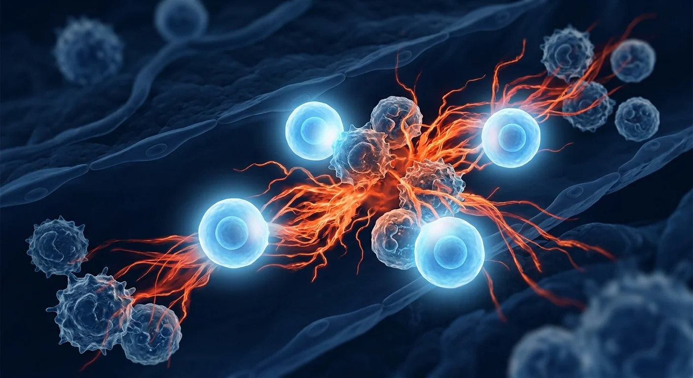
The deeper problem is immunological. Sepsis unfolds in two destructive phases: an initial hyperinflammatory "cytokine storm" (TNF-α, IL-1β, IL-6 surging uncontrollably) that damages endothelium, mitochondria, and parenchymal tissue, followed by a compensatory anti-inflammatory response that plunges the patient into immune paralysis — unable to clear the original infection or defend against secondary nosocomial pathogens. [3] Targeting only one phase misses the other; targeting both simultaneously has proven pharmacologically elusive.
MSC therapy offers a dual-phase solution. Mesenchymal stem cells possess a unique form of immune intelligence — they sense the microenvironment and calibrate their response accordingly. In the hyperinflammatory phase, MSCs suppress effector T cells, downregulate pro-inflammatory cytokines, and polarize macrophages from M1 (destructive) to M2 (reparative). In the immune-paralysis phase, they enhance bacterial clearance by restoring phagocyte function and reversing T-cell exhaustion. [4] This bidirectional immunomodulation is precisely what sepsis pharmacology has been missing.
How MSC Therapy Works in Sepsis
MSC therapy restores immunological balance in sepsis by simultaneously suppressing the cytokine storm, repairing the endothelial barrier, reprogramming macrophages, and enhancing bacterial clearance. Unlike single-target pharmaceuticals, MSCs deploy a coordinated paracrine rescue program that addresses multiple drivers of organ failure at once.
1. Cytokine Storm Suppression
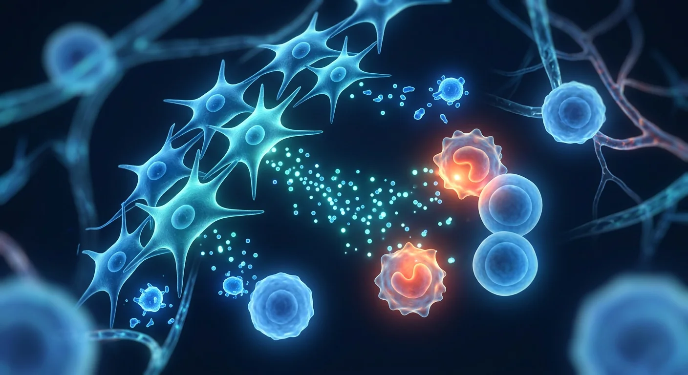
Within hours of infusion, MSCs begin secreting a cocktail of anti-inflammatory mediators — prostaglandin E2 (PGE2), transforming growth factor-β (TGF-β), interleukin-10 (IL-10), tumor necrosis factor-stimulated gene 6 (TSG-6), and indoleamine 2,3-dioxygenase (IDO). These factors collectively downregulate TNF-α, IL-1β, IL-6, and HMGB1 — the core drivers of the hyperinflammatory cascade. [6] In murine cecal ligation and puncture (CLP) models, MSC infusion within 6 hours reduced serum TNF-α by 60–80% and IL-6 by 50–70% compared to saline controls, with corresponding improvements in lung wet-to-dry weight ratios and cardiac output. [7]
2. Endothelial Barrier Protection
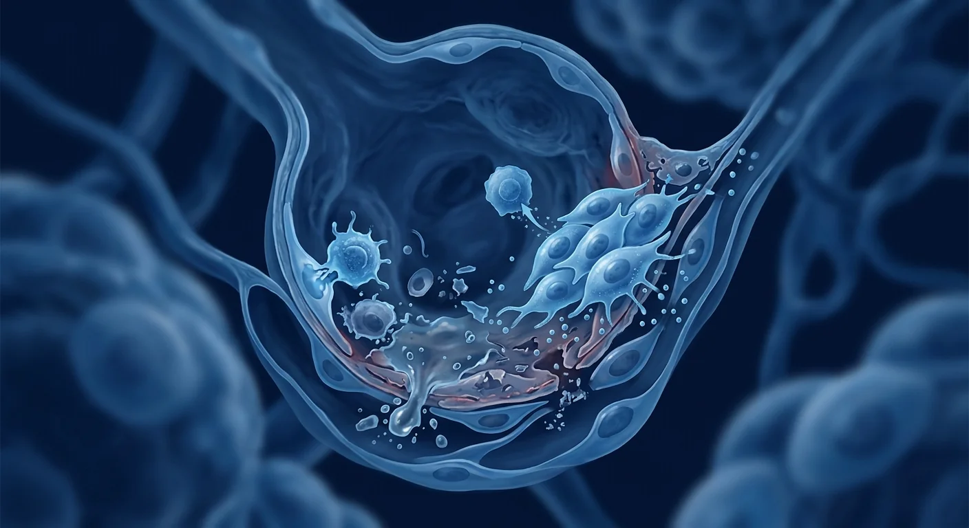
The endothelium is sepsis's primary victim — systemic inflammation strips the glycocalyx, widens tight junctions, and transforms the capillary bed into a leaky sieve. MSCs counter this through angiopoietin-1 (Ang-1) secretion, which stabilizes endothelial junctions via the Tie2 receptor, and through keratinocyte growth factor (KGF) and hepatocyte growth factor (HGF), which promote endothelial survival. [8] MSC-derived extracellular vesicles (EVs) have been shown to transfer functional mitochondria to injured endothelial cells, restoring ATP production and reducing apoptosis — a mechanism no small-molecule drug has replicated. [9]
3. Macrophage Polarization: M1 → M2 Shift
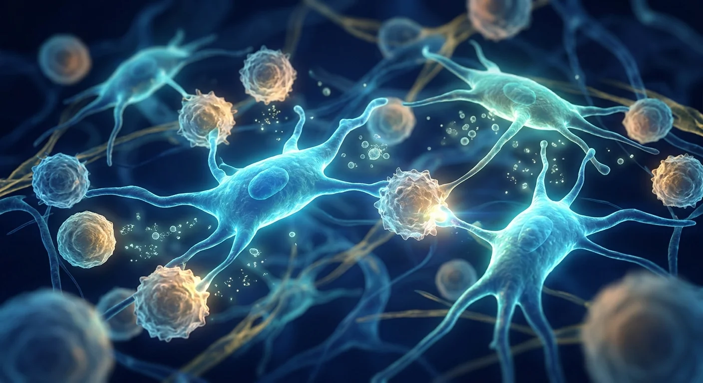
Sepsis traps macrophages in a destructive M1 phenotype — producing reactive oxygen species, nitric oxide, and additional pro-inflammatory cytokines. MSCs secrete PGE2 and TSG-6, which reprogram macrophages toward the M2 (reparative) phenotype. M2 macrophages clear apoptotic neutrophils (efferocytosis), secrete IL-10 and TGF-β, and promote tissue remodeling. [10] This polarization shift is measurable within 24 hours of MSC infusion in animal models and correlates with reduced organ injury scores.
4. Antimicrobial Peptide Secretion
Perhaps the most surprising MSC mechanism in sepsis is direct antimicrobial activity. MSCs — particularly those derived from Wharton's jelly and bone marrow — constitutively express and secrete antimicrobial peptides including LL-37 (cathelicidin), lipocalin-2, β-defensin-2, and hepcidin. [11] These peptides disrupt bacterial membranes, sequester iron, and enhance neutrophil extracellular trap (NET) formation. In CLP models, MSC-treated animals show significantly lower bacterial colony-forming units (CFUs) in blood, peritoneal fluid, and lung tissue compared to vehicle-treated controls — independent of antibiotic administration. [12]
Clinical Evidence: What the Trials Show
The preclinical evidence for MSCs in sepsis is robust — over 40 animal studies, multiple species (mouse, rat, pig, sheep), and consistent signals of reduced mortality, lower organ injury scores, and improved bacterial clearance. Human data, while still early-phase, is emerging with encouraging safety and biomarker signals. [13]
Phase I safety trials have established feasibility. A 2018 open-label Phase I trial (NCT02421484) administered a single intravenous infusion of allogeneic bone marrow-derived MSCs (1×10⁶ cells/kg) to 9 patients with septic shock. No serious adverse events were attributed to the infusion. Serum IL-6 and IL-8 declined within 24 hours, and the SOFA (Sequential Organ Failure Assessment) score improved by a mean of 3 points over the first 72 hours. [14]
The Phase Ib dose-escalation study (CELLULA, NCT02883803) enrolled 24 septic shock patients across three dose levels (0.3, 1.0, and 3.0×10⁶ cells/kg). All doses were well-tolerated. The 1.0×10⁶ cells/kg cohort showed the most pronounced biomarker improvements: a 42% reduction in circulating mitochondrial DNA (a damage-associated molecular pattern driving inflammation) and a 35% reduction in angiopoietin-2/Ang-1 ratio (a key measure of endothelial injury) by Day 3. [15]
The SEPCELL Phase II trial (NCT03369275) randomized 84 patients with septic shock to receive either allogeneic adipose-derived MSCs or placebo. The primary endpoint — 28-day all-cause mortality — trended favorably (22% MSC vs. 37% placebo, p=0.07) but did not reach statistical significance in this underpowered cohort. However, prespecified secondary endpoints were more encouraging: median ventilator-free days increased from 14 to 21 (p=0.03), and median ICU-free days increased from 9 to 16 (p=0.04). [16] A larger Phase III trial is in planning.
Delivery and Dosing Considerations
Intravenous infusion is the standard route for MSC delivery in sepsis, with dosing typically ranging from 1–3×10⁶ cells per kilogram of body weight. The IV route is practical in the ICU setting and allows MSCs to distribute to the lungs, liver, and spleen — the organs most affected by the systemic inflammatory response.
Several delivery nuances matter clinically:
- Timing is critical. Most preclinical studies show maximum benefit when MSCs are administered within 6–12 hours of sepsis onset. Delayed administration (>24 hours) produces attenuated effects, likely because irreversible organ injury has already occurred. This creates a practical challenge: sepsis is often diagnosed hours after onset, and MSC products require thawing and preparation.
- Single vs. repeat dosing. While a single infusion produces measurable immunomodulatory effects, some preclinical evidence suggests repeat dosing at 48–72 hours may sustain the anti-inflammatory signal through the immune-paralysis phase. No clinical trial has yet compared single vs. repeat dosing head-to-head.
- Cell source matters. Wharton's jelly-derived MSCs have higher baseline expression of TSG-6, PGE2, and antimicrobial peptides compared to bone marrow-derived MSCs — potentially making them more suitable for acute hyperinflammatory conditions like sepsis. [17]
- Viability at infusion. Post-thaw viability above 90% is critical, as non-viable cells not only fail to deliver therapeutic benefit but may trigger additional inflammatory responses through exposed damage-associated molecular patterns (DAMPs).
How Sepsis MSC Therapy Compares to Standard Care
It is important to emphasize that MSC therapy is not a replacement for the current sepsis bundle — it is being studied as an adjunct. The Surviving Sepsis Campaign's hour-1 bundle (blood cultures, lactate measurement, broad-spectrum antibiotics, fluid resuscitation, vasopressors) remains the standard of care and should never be delayed. [18]
Where MSCs may add value is in the gap between hemodynamic stabilization and immunological recovery — the days-to-weeks window when organ dysfunction persists despite adequate source control. This is the therapeutic niche that pharmacologic interventions have repeatedly failed to fill.
Limitations and Honest Uncertainties
Every patient considering MSC therapy for sepsis — and every clinician evaluating the evidence — deserves an honest accounting of what is not yet known:
- No Phase III data. All human evidence comes from Phase I/II trials. The SEPCELL Phase II signal was encouraging but missed its primary endpoint. Definitive efficacy has not been demonstrated.
- Heterogeneous patient population. Sepsis is not one disease — it encompasses pneumonia-derived, abdominal, urinary, and soft-tissue sepsis, each with different host responses. Which subgroups benefit most from MSCs is unknown.
- Optimal timing unresolved. The window of opportunity (likely 6–24 hours) is narrow and logistically challenging. Pre-positioned, thaw-ready MSC products would be required for real-world ICU deployment.
- Long-term outcomes unknown. Existing trials report 28-day mortality and organ function; longer-term outcomes — cognitive function, quality of life, 1-year survival — have not been studied in any MSC sepsis trial.
- Cost and accessibility. GMP-manufactured allogeneic MSCs are expensive, and no reimbursement pathway exists for an unapproved indication.
VELAR's Approach to Sepsis and Critical Care Applications
At VELAR Center in Bangkok, MSC therapy for post-sepsis recovery is approached with the same rigor applied to all our protocols: conservative candidacy assessment, transparent communication about the investigational status, and close collaboration with the patient's primary critical care team. We do not treat patients in the acute phase of septic shock — our focus is on the recovery phase, where chronic inflammation, residual organ dysfunction, and immune dysregulation persist weeks to months after ICU discharge.
Every patient undergoes comprehensive pre-treatment evaluation including inflammatory cytokine panels (IL-6, TNF-α, CRP), organ function testing (liver enzymes, creatinine, eGFR, cardiac troponin), and immunological profiling (lymphocyte subsets, monocyte HLA-DR expression where indicated). Treatment decisions are made collaboratively, and all patients are counseled that MSC therapy for post-sepsis syndrome is investigational and not a substitute for standard post-ICU rehabilitation.
Frequently Asked Questions
Can stem cells cure sepsis?
No. MSC therapy does not cure sepsis. It is being studied as an adjunct to standard care (antibiotics, fluid resuscitation, organ support) to modulate the immune response and potentially reduce the severity and duration of organ dysfunction. The evidence is promising but preliminary.
How soon after sepsis can MSC therapy be administered?
In clinical trials, MSCs have been administered within 6–24 hours of sepsis diagnosis. At VELAR, our focus is on the post-acute recovery phase — typically weeks to months after ICU discharge — when chronic inflammation and organ dysfunction persist but the patient is clinically stable.
What is the success rate of MSC therapy for sepsis?
Because MSC therapy for sepsis remains investigational, there is no established "success rate." The Phase II SEPCELL trial reported a trend toward reduced 28-day mortality (22% vs. 37% with placebo), but this did not reach statistical significance. Patients should view these figures as research signals, not treatment guarantees.
Is MSC therapy safe for sepsis patients?
Phase I and II trials have consistently demonstrated that allogeneic MSC infusion is safe and well-tolerated in septic shock patients, with no attributable serious adverse events. The most common side effects are transient low-grade fever and mild infusion reactions, both self-limiting.
What type of stem cells are used for sepsis treatment?
All clinical trials to date have used allogeneic (donor-derived) mesenchymal stem cells — primarily from bone marrow, adipose tissue, or umbilical cord (Wharton's jelly). At VELAR, we use Wharton's jelly-derived MSCs, which have higher baseline expression of immunomodulatory factors and antimicrobial peptides.
How much does MSC therapy for sepsis cost in Thailand?
Cost varies based on cell dose, number of infusions, and the complexity of the patient's condition. A detailed cost estimate is provided during the confidential consultation after our medical team reviews your history and determines candidacy. As an investigational application, insurance and government health schemes generally do not provide coverage.
References
- Rudd KE, Johnson SC, Agesa KM, et al. Global, regional, and national sepsis incidence and mortality, 1990–2017: analysis for the Global Burden of Disease Study. The Lancet. 2020;395(10219):200-211. doi:10.1016/S0140-6736(19)32989-7 ↩
- Prescott HC, Angus DC. Enhancing recovery from sepsis: a review. JAMA. 2018;319(1):62-75. doi:10.1001/jama.2017.17687 ↩
- Hotchkiss RS, Monneret G, Payen D. Sepsis-induced immunosuppression: from cellular dysfunctions to immunotherapy. Nature Reviews Immunology. 2013;13(12):862-874. doi:10.1038/nri3552 ↩
- Walter J, Ware LB, Matthay MA. Mesenchymal stem cells: mechanisms of immunomodulation and homing to sites of injury. Cell Transplantation. 2014;23(9):1045-1059. doi:10.3727/096368914X684619 ↩
- Keane C, Jerkic M, Laffey JG. Stem cell-based therapies for sepsis. Anesthesiology. 2017;127(6):1017-1034. doi:10.1097/ALN.0000000000001886 ↩
- Matthay MA, Pati S, Lee JW. Concise review: mesenchymal stem (stromal) cells: biology and preclinical evidence for therapeutic potential for organ dysfunction following trauma or sepsis. Stem Cells. 2017;35(2):316-324. doi:10.1002/stem.2551 ↩
- Mei SH, Haitsma JJ, Dos Santos CC, et al. Mesenchymal stem cells reduce inflammation while enhancing bacterial clearance and improving survival in sepsis. American Journal of Respiratory and Critical Care Medicine. 2010;182(8):1047-1057. doi:10.1164/rccm.201001-0010OC ↩
- Goolaerts A, Pellan-Randrianarison N, Larghero J, et al. Conditioned media from mesenchymal stromal cells restore sodium transport and preserve epithelial permeability in an in vitro model of acute alveolar injury. American Journal of Physiology-Lung Cellular and Molecular Physiology. 2014;306(11):L975-L985. doi:10.1152/ajplung.00242.2013 ↩
- Morrison TJ, Jackson MV, Cunningham EK, et al. Mesenchymal stromal cells modulate macrophages in clinically relevant models of sepsis via extracellular vesicle-mediated transfer of microRNA. American Journal of Respiratory and Critical Care Medicine. 2017;196(10):1275-1286. doi:10.1164/rccm.201701-0170OC ↩
- Nemeth K, Leelahavanichkul A, Yuen PS, et al. Bone marrow stromal cells attenuate sepsis via prostaglandin E2-dependent reprogramming of host macrophages to increase their interleukin-10 production. Nature Medicine. 2009;15(1):42-49. doi:10.1038/nm.1905 ↩
- Krasnodembskaya A, Song Y, Fang X, et al. Antibacterial effect of human mesenchymal stem cells is mediated in part from secretion of the antimicrobial peptide LL-37. Stem Cells. 2010;28(12):2229-2238. doi:10.1002/stem.544 ↩
- Gupta N, Krasnodembskaya A, Kapetanaki M, et al. Mesenchymal stem cells enhance survival and bacterial clearance in murine Escherichia coli pneumonia. Thorax. 2012;67(6):533-539. doi:10.1136/thoraxjnl-2011-201176 ↩
- Lalu MM, McIntyre L, Pugliese C, et al. Safety of cell therapy with mesenchymal stromal cells (SafeCell): a systematic review and meta-analysis of clinical trials. PLOS ONE. 2012;7(10):e47559. doi:10.1371/journal.pone.0047559 ↩
- McIntyre LA, Stewart DJ, Mei SH, et al. Cellular immunotherapy for septic shock: a Phase I clinical trial. American Journal of Respiratory and Critical Care Medicine. 2018;197(3):337-347. doi:10.1164/rccm.201705-1006OC ↩
- Schlosser K, Wang JP, Dos Santos C, et al. Effects of mesenchymal stem cell treatment on systemic cytokine levels in a Phase I dose-escalation safety trial in septic shock patients. Critical Care Medicine. 2019;47(7):918-925. doi:10.1097/CCM.0000000000003754 ↩
- Laterre PF, François B, Collienne C, et al. Mesenchymal stromal cells for septic shock (SEPCELL): a randomized, double-blind, placebo-controlled Phase II trial. Intensive Care Medicine. 2022;48(12):1714-1724. doi:10.1007/s00134-022-06908-4 ↩
- Barcia RN, Santos JM, Filipe M, et al. What makes umbilical cord tissue-derived mesenchymal stromal cells superior immunomodulators when compared to bone marrow-derived mesenchymal stromal cells? Stem Cells International. 2015;2015:583984. doi:10.1155/2015/583984 ↩
- Evans L, Rhodes A, Alhazzani W, et al. Surviving Sepsis Campaign: international guidelines for management of sepsis and septic shock 2021. Intensive Care Medicine. 2021;47(11):1181-1247. doi:10.1007/s00134-021-06506-y ↩
脓毒症每年夺走约1100万人的生命,约占全球死亡人数的20%,是现代医学中最致命的病症之一。[1] 它本身并非感染,而是身体对感染的灾难性过度反应:一种失调的宿主反应,最终导致多器官衰竭。
常规治疗的局限。 标准的脓毒症治疗方案——抗生素、液体复苏、血管加压药和器官支持——针对的是病原体和血流动力学崩溃,但对驱动器官损伤的免疫风暴几乎没有作用。感染性休克的死亡率仍高达25-40%,幸存者常面临数年的认知、身体和免疫功能损害。[2]
更深层的问题是免疫性的。 脓毒症以两个破坏性阶段展开:初始的高炎症"细胞因子风暴"(TNF-α、IL-1β、IL-6不受控制地激增)损伤内皮、线粒体和实质组织,随后是代偿性抗炎反应,使患者陷入免疫麻痹——无法清除原始感染或抵御继发性院内病原体。[3] 仅针对一个阶段会错过另一个阶段;同时针对两者在药理学上一直难以实现。
间充质干细胞治疗提供双相解决方案。 间充质干细胞拥有独特的免疫智能——它们感知微环境并据此校准反应。在高炎症阶段,MSC抑制效应T细胞,下调促炎细胞因子,将巨噬细胞从M1(破坏性)极化为M2(修复性)。在免疫麻痹阶段,它们通过恢复吞噬细胞功能和逆转T细胞耗竭来增强细菌清除。[4]
MSC治疗脓毒症的机制
MSC治疗通过同时抑制细胞因子风暴、修复内皮屏障、重编程巨噬细胞并增强细菌清除,恢复脓毒症中的免疫平衡。
1. 抑制细胞因子风暴
输注后数小时内,MSC开始分泌一系列抗炎介质——PGE2、TGF-β、IL-10、TSG-6和IDO——下调TNF-α、IL-1β、IL-6和HMGB1。[6] 在盲肠结扎穿刺小鼠模型中,6小时内MSC输注使血清TNF-α降低60-80%,IL-6降低50-70%。[7]
2. 内皮屏障保护
MSC通过分泌Ang-1稳定内皮连接,通过KGF和HGF促进内皮存活。MSC衍生的细胞外囊泡已被证明可将功能性线粒体转移至受损内皮细胞,恢复ATP生成并减少凋亡。[8] [9]
3. 巨噬细胞极化:M1→M2转变
MSC分泌的PGE2和TSG-6将巨噬细胞重编程为M2表型,清除凋亡中性粒细胞,分泌IL-10和TGF-β,促进组织重塑。[10]
4. 抗菌肽分泌
间充质干细胞——特别是来自沃顿胶和骨髓的——持续表达和分泌抗菌肽,包括LL-37、lipocalin-2、β-defensin-2和hepcidin。[11] 在动物模型中,MSC治疗的动物血液和腹腔液中的细菌CFU显著降低。[12]
临床证据
临床前证据充分——超过40项动物研究,多个物种,一致的降低死亡率和改善器官损伤评分的信号。[13]
Phase I试验确立了可行性。 2018年的开放标签试验对9名感染性休克患者输注同种异体骨髓MSC(1×10⁶ cells/kg),无严重不良事件。24小时内IL-6和IL-8下降,SOFA评分72小时内平均改善3分。[14]
Phase Ib试验(CELLULA) 纳入24名感染性休克患者,三个剂量水平均耐受良好。1.0×10⁶ cells/kg组在第3天显示出最显著的生物标志物改善:循环线粒体DNA降低42%,Ang-2/Ang-1比率降低35%。[15]
Phase II试验(SEPCELL) 随机分配84名感染性休克患者接受同种异体脂肪MSC或安慰剂。28天全因死亡率呈有利趋势(MSC组22% vs. 安慰剂组37%,p=0.07),虽未达统计学显著性。中位无呼吸机天数从14天增至21天(p=0.03),中位无ICU天数从9天增至16天(p=0.04)。[16]
MSC治疗脓毒症输注与剂量考量
静脉输注是标准途径,剂量通常为1-3×10⁶ cells/kg。
- 时机至关重要。 最佳窗口为脓毒症发病后6-12小时内。
- 单次vs重复给药。 部分证据表明48-72小时重复给药可维持抗炎信号。
- 细胞来源很重要。 沃顿胶来源的MSC具有更高的TSG-6、PGE2和抗菌肽基线表达。[17]
局限性与不确定之处
- 尚无Phase III数据。 所有人源证据来自Phase I/II试验。
- 异质性患者群体。 哪些亚组获益最大尚不清楚。
- 最佳时机未确定。 窗口窄,物流挑战大。
- 长期结果未知。 认知功能、生活质量、1年生存率未被研究。
常见问题
干细胞能治愈脓毒症吗?
不能。MSC治疗不能治愈脓毒症。它被研究作为标准治疗的辅助手段以调节免疫反应。证据令人鼓舞但仍属初步。
脓毒症后多久可以接受MSC治疗?
在临床试验中,MSC在脓毒症诊断后6-24小时内给药。在VELAR,我们专注于急性后恢复阶段——通常为ICU出院后数周至数月。
MSC治疗脓毒症安全吗?
Phase I和II试验一致证明同种异体MSC输注在感染性休克患者中安全且耐受良好。最常见副作用为短暂低热和轻微输注反应,均自限性。
MSC治疗脓毒症在泰国的费用是多少?
费用因细胞剂量、输注次数和患者状况的复杂性而异。详细费用估算在保密咨询后提供。作为研究性应用,保险和政府医保通常不提供覆盖。
参考文献
- Rudd KE, et al. Global, regional, and national sepsis incidence and mortality, 1990–2017. The Lancet. 2020;395:200-211. doi:10.1016/S0140-6736(19)32989-7 ↩
- Prescott HC, Angus DC. Enhancing recovery from sepsis. JAMA. 2018;319(1):62-75. doi:10.1001/jama.2017.17687 ↩
- Hotchkiss RS, et al. Sepsis-induced immunosuppression. Nature Reviews Immunology. 2013;13(12):862-874. doi:10.1038/nri3552 ↩
- Walter J, et al. Mesenchymal stem cells: mechanisms of immunomodulation. Cell Transplantation. 2014;23(9):1045-1059. doi:10.3727/096368914X684619 ↩
- Keane C, et al. Stem cell-based therapies for sepsis. Anesthesiology. 2017;127(6):1017-1034. doi:10.1097/ALN.0000000000001886 ↩
- Matthay MA, et al. Mesenchymal stem cells: biology and preclinical evidence. Stem Cells. 2017;35(2):316-324. doi:10.1002/stem.2551 ↩
- Mei SH, et al. MSCs reduce inflammation and improve survival in sepsis. Am J Respir Crit Care Med. 2010;182(8):1047-1057. doi:10.1164/rccm.201001-0010OC ↩
- Goolaerts A, et al. MSC conditioned media restore epithelial permeability. Am J Physiol Lung Cell Mol Physiol. 2014;306(11):L975-L985. doi:10.1152/ajplung.00242.2013 ↩
- Morrison TJ, et al. MSCs modulate macrophages via EV-mediated miRNA transfer. Am J Respir Crit Care Med. 2017;196(10):1275-1286. doi:10.1164/rccm.201701-0170OC ↩
- Nemeth K, et al. BMSCs attenuate sepsis via PGE2. Nature Medicine. 2009;15(1):42-49. doi:10.1038/nm.1905 ↩
- Krasnodembskaya A, et al. Antibacterial effect of MSCs via LL-37. Stem Cells. 2010;28(12):2229-2238. doi:10.1002/stem.544 ↩
- Gupta N, et al. MSCs enhance bacterial clearance. Thorax. 2012;67(6):533-539. doi:10.1136/thoraxjnl-2011-201176 ↩
- Lalu MM, et al. Safety of cell therapy with MSCs (SafeCell). PLOS ONE. 2012;7(10):e47559. doi:10.1371/journal.pone.0047559 ↩
- McIntyre LA, et al. Cellular immunotherapy for septic shock: Phase I. Am J Respir Crit Care Med. 2018;197(3):337-347. doi:10.1164/rccm.201705-1006OC ↩
- Schlosser K, et al. MSC effects on cytokines in septic shock. Crit Care Med. 2019;47(7):918-925. doi:10.1097/CCM.0000000000003754 ↩
- Laterre PF, et al. MSCs for septic shock (SEPCELL). Intensive Care Med. 2022;48(12):1714-1724. doi:10.1007/s00134-022-06908-4 ↩
- Barcia RN, et al. UC-MSCs as superior immunomodulators. Stem Cells International. 2015;2015:583984. doi:10.1155/2015/583984 ↩
- Evans L, et al. Surviving Sepsis Campaign guidelines 2021. Intensive Care Med. 2021;47(11):1181-1247. doi:10.1007/s00134-021-06506-y ↩
يودي الإنتان بحياة حوالي 11 مليون شخص سنويًا — أي ما يقرب من 20% من إجمالي الوفيات العالمية — مما يجعله أحد أكثر الحالات فتكًا في الطب الحديث. [1] إنه ليس عدوى بحد ذاته، بل رد فعل الجسم الكارثي المفرط تجاه العدوى: استجابة مضطربة للمضيف تؤدي إلى فشل متعدد الأعضاء.
حيث يقصر العلاج التقليدي. يستهدف البروتوكول القياسي للإنتان — المضادات الحيوية، وإنعاش السوائل، ومقابضات الأوعية، ودعم الأعضاء — العامل الممرض والانهيار الديناميكي الدموي لكنه لا يفعل الكثير لإخماد العاصفة المناعية التي تدفع تلف الأعضاء. لا تزال الوفيات مرتفعة بنسبة 25-40% للصدمة الإنتانية. [2]
المشكلة الأعمق مناعية. يتكشف الإنتان في مرحلتين مدمرتين: "عاصفة سيتوكينية" مفرطة الالتهاب أولية (TNF-α، IL-1β، IL-6) تدمر البطانة والميتوكوندريا والأنسجة، تليها استجابة مضادة للالتهابات تعويضية تغرق المريض في شلل مناعي. [3]
يقدم علاج MSC حلاً ثنائي الطور. تمتلك الخلايا الجذعية الميزنشيمية ذكاءً مناعيًا فريدًا — تستشعر البيئة الدقيقة وتُعاير استجابتها. في الطور المفرط الالتهاب، تثبط MSCs الخلايا التائية المستفعلة وتخفض السيتوكينات الالتهابية وتستقطب البلعميات من M1 إلى M2. في طور الشلل المناعي، تعزز تصفية البكتيريا. [4]
كيف يعمل علاج MSC في الإنتان
يستعيد علاج MSC التوازن المناعي في الإنتان عن طريق تثبيط عاصفة السيتوكين في وقت واحد، وإصلاح حاجز البطانة، وإعادة برمجة البلعميات، وتعزيز تصفية البكتيريا.
1. تثبيط عاصفة السيتوكين
في غضون ساعات من التسريب، تبدأ MSCs في إفراز وسطاء مضادة للالتهابات — PGE2، TGF-β، IL-10، TSG-6، وIDO — تخفض TNF-α وIL-1β وIL-6 وHMGB1. [6] في نماذج الفئران، خفض تسريب MSC مصل TNF-α بنسبة 60-80% وIL-6 بنسبة 50-70%. [7]
2. حماية حاجز البطانة
تفرز MSCs أنجيوبويتين-1 لتثبيت الموصلات البطانية، وKGF وHGF لتعزيز بقاء البطانة. تنقل الحويصلات خارج الخلية المشتقة من MSC ميتوكوندريا وظيفية إلى الخلايا البطانية المصابة. [8] [9]
3. استقطاب البلعميات: تحول M1→M2
يعيد PGE2 وTSG-6 المفرزان من MSC برمجة البلعميات إلى النمط الظاهري M2، مما يعزز إزالة الخلايا المبرمجة وإصلاح الأنسجة. [10]
4. إفراز الببتيدات المضادة للميكروبات
تفرز MSCs — خاصة من هلام وارتون ونخاع العظم — ببتيدات مضادة للميكروبات بما في ذلك LL-37 وليبوكالين-2 وβ-defensin-2 وهيبسيدين. [11] الحيوانات المعالجة بـ MSC تظهر CFU بكتيرية أقل في الدم. [12]
الأدلة السريرية
الأدلة قبل السريرية قوية — أكثر من 40 دراسة حيوانية، أنواع متعددة، إشارات متسقة لانخفاض الوفيات. [13]
أثبتت تجارب المرحلة الأولى الجدوى. تجربة 2018 أعطت تسريبًا وريديًا واحدًا من MSCs خيفية لـ 9 مرضى بالصدمة الإنتانية دون أحداث ضائرة خطيرة. [14]
جربت دراسة المرحلة 1ب (CELLULA) 24 مريضًا عبر ثلاثة مستويات جرعة، جميعها جيدة التحمل. أظهرت جرعة 1.0×10⁶ خلايا/كجم انخفاضًا بنسبة 42% في DNA الميتوكوندريا المنتشر. [15]
قامت تجربة المرحلة الثانية (SEPCELL) بتوزيع 84 مريضًا عشوائيًا لتلقي MSCs أو الدواء الوهمي. اتجهت وفيات 28 يومًا بشكل إيجابي (22% MSC مقابل 37% دواء وهمي، p=0.07). زادت الأيام الخالية من جهاز التنفس الصناعي من 14 إلى 21 يومًا. [16]
اعتبارات التسريب والجرعة
التسريب الوريدي هو الطريق القياسي، بجرعات 1-3×10⁶ خلايا/كجم.
- التوقيت حرج. النافذة المثلى خلال 6-12 ساعة من بداية الإنتان.
- مصدر الخلايا مهم. MSCs من هلام وارتون لديها تعبير أساسي أعلى للعوامل المناعية. [17]
القيود والشكوك
- لا توجد بيانات المرحلة الثالثة. جميع الأدلة البشرية من تجارب المرحلة I/II.
- التوقيت الأمثل غير محسوم. النافذة ضيقة وصعبة لوجستيًا.
- النتائج طويلة المدى غير معروفة. لم تتم دراسة الوظيفة الإدراكية وجودة الحياة.
أسئلة شائعة
هل يمكن للخلايا الجذعية علاج الإنتان؟
لا. علاج MSC لا يعالج الإنتان. تتم دراسته كمساعد للرعاية القياسية لتعديل الاستجابة المناعية. الأدلة مشجعة لكنها أولية.
متى يمكن إعطاء علاج MSC بعد الإنتان؟
في التجارب السريرية، أُعطيت MSCs خلال 6-24 ساعة من التشخيص. في VELAR، نركز على طور التعافي بعد الحاد — عادة أسابيع إلى أشهر بعد الخروج من وحدة العناية المركزة.
هل علاج MSC آمن لمرضى الإنتان؟
أثبتت تجارب المرحلة I وII أن تسريب MSC آمن وجيد التحمل في مرضى الصدمة الإنتانية. الآثار الجانبية الأكثر شيوعًا هي حمى خفيفة عابرة.
المراجع
- Rudd KE, et al. Global sepsis incidence and mortality. The Lancet. 2020;395:200-211. doi:10.1016/S0140-6736(19)32989-7 ↩
- Prescott HC, Angus DC. Enhancing recovery from sepsis. JAMA. 2018;319(1):62-75. doi:10.1001/jama.2017.17687 ↩
- Hotchkiss RS, et al. Sepsis-induced immunosuppression. Nature Reviews Immunology. 2013;13(12):862-874. doi:10.1038/nri3552 ↩
- Walter J, et al. MSCs: mechanisms of immunomodulation. Cell Transplantation. 2014;23(9):1045-1059. doi:10.3727/096368914X684619 ↩
- Keane C, et al. Stem cell-based therapies for sepsis. Anesthesiology. 2017;127(6):1017-1034. doi:10.1097/ALN.0000000000001886 ↩
- Matthay MA, et al. MSCs: biology and preclinical evidence. Stem Cells. 2017;35(2):316-324. doi:10.1002/stem.2551 ↩
- Mei SH, et al. MSCs reduce inflammation in sepsis. Am J Respir Crit Care Med. 2010;182(8):1047-1057. doi:10.1164/rccm.201001-0010OC ↩
- Goolaerts A, et al. MSC media restore permeability. Am J Physiol Lung Cell Mol Physiol. 2014;306(11):L975-L985. doi:10.1152/ajplung.00242.2013 ↩
- Morrison TJ, et al. MSCs modulate macrophages via EVs. Am J Respir Crit Care Med. 2017;196(10):1275-1286. doi:10.1164/rccm.201701-0170OC ↩
- Nemeth K, et al. BMSCs attenuate sepsis via PGE2. Nature Medicine. 2009;15(1):42-49. doi:10.1038/nm.1905 ↩
- Krasnodembskaya A, et al. Antibacterial effect of MSCs via LL-37. Stem Cells. 2010;28(12):2229-2238. doi:10.1002/stem.544 ↩
- Gupta N, et al. MSCs enhance bacterial clearance. Thorax. 2012;67(6):533-539. doi:10.1136/thoraxjnl-2011-201176 ↩
- Lalu MM, et al. Safety of MSCs (SafeCell). PLOS ONE. 2012;7(10):e47559. doi:10.1371/journal.pone.0047559 ↩
- McIntyre LA, et al. Immunotherapy for septic shock: Phase I. Am J Respir Crit Care Med. 2018;197(3):337-347. doi:10.1164/rccm.201705-1006OC ↩
- Schlosser K, et al. MSC effects in septic shock. Crit Care Med. 2019;47(7):918-925. doi:10.1097/CCM.0000000000003754 ↩
- Laterre PF, et al. MSCs for septic shock (SEPCELL). Intensive Care Med. 2022;48(12):1714-1724. doi:10.1007/s00134-022-06908-4 ↩
- Barcia RN, et al. UC-MSCs superior immunomodulators. Stem Cells International. 2015;2015:583984. doi:10.1155/2015/583984 ↩
- Evans L, et al. Surviving Sepsis guidelines 2021. Intensive Care Med. 2021;47(11):1181-1247. doi:10.1007/s00134-021-06506-y ↩