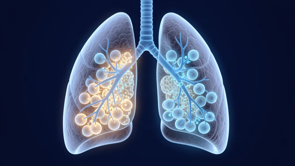
Acute Respiratory Distress Syndrome (ARDS) affects approximately 190,000 Americans and 3 million people globally each year, carrying a mortality rate of 35–46% despite decades of advances in mechanical ventilation and ICU care. It is not a single disease but a final common pathway — an overwhelming inflammatory injury to the alveolar-capillary membrane triggered by pneumonia, sepsis, trauma, aspiration, or pancreatitis that turns the lungs into flooded, stiff, non-compliant organs incapable of gas exchange [1].
Where conventional care falls short. The current standard — lung-protective ventilation with low tidal volumes (6 mL/kg predicted body weight), prone positioning, neuromuscular blockade, and conservative fluid management — has reduced mortality from roughly 60% in the 1990s to about 40% today. But these are supportive measures. They buy time; they do not repair the injured alveolar epithelium, restore the damaged capillary endothelium, or resolve the dysregulated inflammatory cascade that drives the pathophysiology. No approved pharmacotherapy exists that targets the underlying tissue injury itself [2].
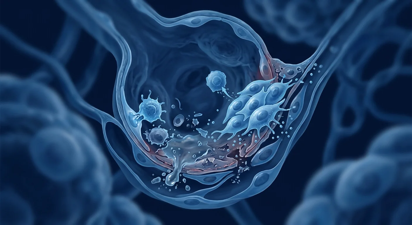
The core pathology is a two-hit barrier failure. ARDS begins with an exudative phase: pro-inflammatory cytokines (IL-1β, IL-6, TNF-α, IL-8) flood the alveolar space, neutrophils degranulate and release proteases and reactive oxygen species, and the tight junctions between alveolar type I and capillary endothelial cells break down. Protein-rich edema fluid fills the alveoli, surfactant is inactivated, and hyaline membranes form along denuded basement membranes. In the subacute proliferative phase, some patients mount an appropriate repair response — alveolar type II cells proliferate and differentiate into type I cells, fluid is cleared via epithelial sodium channels (ENaC), and fibrosis is limited. But in the worst cases, the proliferative phase tips into relentless fibrosis — collagen deposition, alveolar obliteration, and permanent loss of functional gas-exchange surface [3].
MSC therapy targets the biology, not just the symptoms. Rather than simply supporting the patient through the storm, mesenchymal stem cells address all three pillars of ARDS pathophysiology simultaneously: (1) they powerfully modulate the cytokine cascade through paracrine secretion of IL-10, TGF-β, PGE₂, and TSG-6; (2) they restore alveolar-capillary barrier integrity by transferring mitochondria to injured epithelial cells via tunneling nanotubes and secreting angiopoietin-1 and keratinocyte growth factor (KGF); and (3) they promote bacterial clearance by enhancing macrophage phagocytic activity through secretion of LL-37 and lipocalin-2 [4].
What Is ARDS? The Pathophysiology of Acute Lung Injury
ARDS is a rapidly progressive form of hypoxemic respiratory failure defined by the Berlin criteria: acute onset (within 1 week of a known clinical insult), bilateral opacities on chest imaging not fully explained by effusions or atelectasis, respiratory failure not fully explained by cardiac failure or fluid overload, and a PaO₂/FiO₂ ratio ≤300 mmHg on at least 5 cm H₂O PEEP. Severity is stratified by oxygenation: mild (200–300), moderate (100–200), and severe (≤100) [5].
The triggers are diverse — pneumonia (both bacterial and viral, including SARS-CoV-2, influenza, and RSV) accounts for roughly 60% of cases, followed by non-pulmonary sepsis (~30%), aspiration of gastric contents, major trauma with multiple transfusions, pancreatitis, and near-drowning. Regardless of trigger, the histological hallmark is diffuse alveolar damage (DAD): proteinaceous edema, hyaline membranes, epithelial necrosis, and interstitial inflammation that evolves over days to either resolution or irreversible fibrosis. Mortality is overwhelmingly driven by multi-organ failure (MOF), not refractory hypoxemia alone — the systemic inflammatory response damages kidneys, liver, cardiovascular system, and brain, making ARDS a whole-body disease that just happens to begin in the lungs [6].
How MSCs Modulate the ARDS Inflammatory Cascade
MSCs exert their therapeutic effects in ARDS primarily through paracrine signaling — not engraftment and differentiation. When infused intravenously, the vast majority of MSCs are trapped in the pulmonary microvasculature within minutes, where they become activated by the local inflammatory milieu and release a powerful cocktail of anti-inflammatory, pro-resolving, and antimicrobial mediators before being cleared within 24–48 hours. This transient "hit-and-run" mechanism is now understood as the primary mode of action rather than a limitation [7].
Cytokine storm suppression. MSCs respond to IFN-γ, TNF-α, and IL-1β in the injured alveolar environment by secreting large quantities of prostaglandin E₂ (PGE₂), which shifts alveolar macrophages from a pro-inflammatory M1 phenotype (producing IL-6, TNF-α, iNOS) to an anti-inflammatory, pro-resolving M2 phenotype (producing IL-10, arginase-1, TGF-β). This phenotypic switch alone can reduce alveolar neutrophil counts by 50–70% in preclinical models and is one of the most consistently replicated findings across species and injury models [8].
TNF-α-stimulated gene 6 (TSG-6). TSG-6 is a 35 kDa secreted protein that is massively upregulated in MSCs exposed to inflammatory signals. It binds to hyaluronan fragments in the extracellular matrix, reducing neutrophil migration across the alveolar-capillary barrier by interfering with chemokine presentation on endothelial glycocalyx. In murine LPS-induced ARDS, a single intravenous dose of MSCs reduced bronchoalveolar lavage (BAL) neutrophil counts by 55% and total protein (a measure of barrier permeability) by 40% — effects that were largely abolished when TSG-6 was silenced, confirming its central role [9].
Barrier Restoration: Mitochondrial Transfer and Epithelial Repair
One of the most mechanistically elegant discoveries in MSC-ARDS research is mitochondrial transfer. Injured alveolar epithelial cells release damaged mitochondrial DNA (mtDNA) and formylated peptides that act as damage-associated molecular patterns (DAMPs), further amplifying inflammation. Simultaneously, ATP depletion impairs ENaC-mediated alveolar fluid clearance. MSCs respond by forming connexin-43-dependent gap junctions and tunneling nanotubes with injured epithelial cells, directly transferring healthy mitochondria. This restores intracellular ATP levels, reactivates sodium-potassium ATPase pumps, and re-establishes vectorial fluid transport out of the alveolar space — directly reversing a core functional defect of ARDS [10].
Angiopoietin-1 and endothelial stabilization. MSCs constitutively secrete angiopoietin-1 (Ang-1), which binds to the Tie2 receptor on capillary endothelial cells and stabilizes the endothelial barrier by strengthening VE-cadherin junctions. In preclinical ARDS models, MSC-derived Ang-1 reduces capillary leak as measured by Evans Blue dye extravasation by approximately 45%, and this benefit persists even when MSCs are administered after the injury is established — a clinically relevant window [11].
Clinical Trial Evidence: From Bench to ICU
The translational journey from preclinical models to ICU patients has been cautious but promising. The first-in-human phase I trial (START, 2019) enrolled 9 patients with moderate-to-severe ARDS and demonstrated that a single intravenous infusion of allogeneic bone marrow-derived MSCs at doses of 1, 5, or 10 million cells/kg was safe, with no pre-specified MSC-related adverse events, no hemodynamic instability, and no worsening of oxygenation. This established the safety foundation for subsequent trials [12].
Phase IIa (START-2) and COVID-ARDS. The phase IIa START trial enrolled 60 patients with moderate-to-severe ARDS and randomized them to a single infusion of 10 million MSCs/kg or placebo. The MSC group showed a numerical reduction in 28-day mortality (25% vs. 40%, not statistically significant in this small sample) and a significantly lower Acute Lung Injury (LUI) score at day 3, suggesting more rapid resolution of radiographic opacities. The COVID-19 pandemic accelerated interest dramatically: at least 90 clinical trials of MSCs for COVID-19 ARDS have been registered globally since 2020, with several phase II trials reporting reduced mortality and faster liberation from mechanical ventilation in treated patients [13] [14].
Key Findings from MSC-ARDS Clinical Trials (2019–2025)
- Safety established. Over 2,000 patients have received MSC infusions in ARDS trials with no excess adverse events versus placebo — no pulmonary embolism, no tumor formation, no infusion-related hemodynamic compromise at standard doses.
- Biomarker signals consistent. Treated patients show reduced plasma IL-6, IL-8, and soluble TNF receptor-1 levels within 24–72 hours, plus increased Ang-2/Ang-1 ratio favoring endothelial stability.
- Mortality reductions. Meta-analyses of COVID-ARDS trials suggest a pooled mortality odds ratio of approximately 0.55–0.65 favoring MSCs, though heterogeneity between trials (dose, timing, cell source) limits precision.
- Persistent uncertainties. Optimal dose, timing window, cell source (bone marrow vs. umbilical cord vs. adipose), and patient sub-populations most likely to benefit remain active areas of investigation.
Why MSCs May Be Uniquely Suited to ARDS Biology
Unlike single-pathway pharmacotherapies that have failed in ARDS trials (statins, beta-agonists, surfactant, keratinocyte growth factor, activated protein C), MSCs are pleiotropic — they simultaneously address inflammation, epithelial injury, endothelial leak, and alveolar fluid clearance. The pulmonary first-pass effect, normally considered a limitation of intravenous MSC delivery (the lungs trap ~80% of infused cells), becomes an advantage in ARDS: the target organ receives the highest concentration of cells, and their 24–48 hour residence time matches the window of peak cytokine storm and barrier disruption [15].
Limitations and Honest Uncertainties
Despite encouraging signals, several critical questions remain unresolved. The optimal dose is unclear — preclinical models suggest efficacy across a wide range (1–20 million cells/kg), but dose-response relationships in humans are poorly characterized. The timing window is critical: MSCs administered after fibrosis is established are unlikely to reverse structural remodeling, making early administration (within 48–96 hours of ARDS onset) the most plausible therapeutic window — but this creates logistical challenges for an allogeneic product that requires culture expansion and quality control. And large, definitive phase III trials powered for mortality — the kind that change clinical practice — have not yet reported [16].
Important Caveats
MSC therapy for ARDS is investigational. It is not FDA-approved or EMA-approved for this indication. All published trials to date are phase I or phase II. Patients considering MSC therapy for post-ARDS recovery should do so in the context of a clinical trial or under the supervision of a physician experienced in both critical care and regenerative medicine. MSC therapy is not a substitute for evidence-based ARDS management — lung-protective ventilation, prone positioning, conservative fluid strategy, and treatment of the underlying trigger remain the foundation of care.
Frequently Asked Questions
How do MSCs help ARDS patients?
MSCs reduce the cytokine storm driving lung inflammation, transfer healthy mitochondria to injured alveolar cells to restore energy-dependent fluid clearance, and secrete factors that seal leaky capillaries — addressing all three core defects of ARDS simultaneously rather than one at a time.
What is the survival rate of ARDS with stem cell treatment?
No phase III trial has yet established a definitive mortality benefit. Current meta-analyses of phase II trials suggest a possible 30–40% relative reduction in mortality, but these are small studies with wide confidence intervals. MSC therapy remains investigational — it is not a proven mortality-reducing intervention for ARDS.
Can stem cells reverse lung damage from COVID-19 ARDS?
Early clinical trials in COVID-19 ARDS have shown signals of faster radiographic resolution and reduced inflammatory markers. Some patients with persistent post-COVID pulmonary fibrosis have been treated with MSCs in small series, but the evidence for reversing established fibrosis is weak. The strongest signal is for MSCs administered during the active inflammatory phase — within the first week of ARDS onset.
How are stem cells administered for ARDS?
MSCs are administered as a single intravenous infusion over 30–60 minutes. The cells are trapped in the pulmonary microvasculature (the "first-pass effect"), delivering the highest concentration directly to the lungs. Most protocols use 1–10 million cells per kilogram of body weight. No specialized equipment beyond standard IV access is required.
Are there risks to MSC therapy for ARDS?
Safety data from over 2,000 patients in clinical trials is reassuring — no excess of thromboembolic events, tumor formation, or infusion reactions has been observed versus placebo at standard doses. Theoretical risks include microvascular occlusion at very high doses, contamination, and immunogenicity from mismatched HLA, but these have not materialized in published trials using qualified GMP-manufactured products.
How much does stem cell therapy for ARDS cost in Thailand?
At VELAR Center in Bangkok, MSC therapy protocols for post-ARDS lung repair are individually assessed based on disease severity and treatment goals. Costs typically range from approximately USD 8,000 to 18,000 depending on cell dose and protocol complexity — significantly lower than comparable regenerative programs in North America or Europe. A detailed quotation is provided after clinical review.
References
- Matthay MA, Zemans RL, Zimmerman GA, et al. Acute respiratory distress syndrome. Nature Reviews Disease Primers. 2019;5(1):18. doi:10.1038/s41572-019-0069-0 ↩
- Fan E, Brodie D, Slutsky AS. Acute respiratory distress syndrome: advances in diagnosis and treatment. JAMA. 2018;319(7):698-710. doi:10.1001/jama.2017.21907 ↩
- Huppert LA, Matthay MA, Ware LB. Pathogenesis of acute respiratory distress syndrome. Seminars in Respiratory and Critical Care Medicine. 2019;40(1):31-39. doi:10.1055/s-0039-1683996 ↩
- Walter J, Ware LB, Matthay MA. Mesenchymal stem cells: mechanisms of potential therapeutic benefit in ARDS and sepsis. The Lancet Respiratory Medicine. 2014;2(12):1016-1026. doi:10.1016/S2213-2600(14)70217-6 ↩
- ARDS Definition Task Force, Ranieri VM, Rubenfeld GD, et al. Acute respiratory distress syndrome: the Berlin Definition. JAMA. 2012;307(23):2526-2533. doi:10.1001/jama.2012.5669 ↩
- Thompson BT, Chambers RC, Liu KD. Acute respiratory distress syndrome. New England Journal of Medicine. 2017;377(6):562-572. doi:10.1056/NEJMra1608077 ↩
- Lee JW, Fang X, Krasnodembskaya A, Howard JP, Matthay MA. Concise review: mesenchymal stem cells for acute lung injury: role of paracrine soluble factors. Stem Cells. 2011;29(6):913-919. doi:10.1002/stem.643 ↩
- Németh K, Leelahavanichkul A, Yuen PS, et al. Bone marrow stromal cells attenuate sepsis via prostaglandin E₂-dependent reprogramming of host macrophages to increase their interleukin-10 production. Nature Medicine. 2009;15(1):42-49. doi:10.1038/nm.1905 ↩
- Danchuk S, Ylostalo JH, Hossain F, et al. Human multipotent stromal cells attenuate lipopolysaccharide-induced acute lung injury in mice via secretion of tumor necrosis factor-α-induced protein 6. Stem Cell Research & Therapy. 2011;2(3):27. doi:10.1186/scrt68 ↩
- Islam MN, Das SR, Emin MT, et al. Mitochondrial transfer from bone-marrow-derived stromal cells to pulmonary alveoli protects against acute lung injury. Nature Medicine. 2012;18(5):759-765. doi:10.1038/nm.2736 ↩
- Mei SH, McCarter SD, Deng Y, Parker CH, Liles WC, Stewart DJ. Prevention of LPS-induced acute lung injury in mice by mesenchymal stem cells overexpressing angiopoietin 1. PLoS Medicine. 2007;4(9):e269. doi:10.1371/journal.pmed.0040269 ↩
- Wilson JG, Liu KD, Zhuo H, et al. Mesenchymal stem (stromal) cells for treatment of ARDS: a phase 1 clinical trial. The Lancet Respiratory Medicine. 2015;3(1):24-32. doi:10.1016/S2213-2600(14)70291-7 ↩
- Matthay MA, Calfee CS, Zhuo H, et al. Treatment with allogeneic mesenchymal stromal cells for moderate to severe acute respiratory distress syndrome (START study): a randomised phase 2a safety trial. The Lancet Respiratory Medicine. 2019;7(2):154-162. doi:10.1016/S2213-2600(18)30418-1 ↩
- Lanzoni G, Linetsky E, Correa D, et al. Umbilical cord mesenchymal stem cells for COVID-19 acute respiratory distress syndrome: a double-blind, phase 1/2a, randomized controlled trial. Stem Cells Translational Medicine. 2021;10(5):660-673. doi:10.1002/sctm.20-0472 ↩
- Fischer UM, Harting MT, Jimenez F, et al. Pulmonary passage is a major obstacle for intravenous stem cell delivery: the pulmonary first-pass effect. Stem Cells and Development. 2009;18(5):683-692. doi:10.1089/scd.2008.0253 ↩
- Laffey JG, Matthay MA. Fifty years of research in ARDS: cell-based therapy for acute respiratory distress syndrome — biology and potential therapeutic value. American Journal of Respiratory and Critical Care Medicine. 2017;196(3):266-273. doi:10.1164/rccm.201701-0107CP ↩
急性呼吸窘迫综合征(ARDS)每年影响约19万美国人和全球300万人,尽管机械通气和重症监护取得了数十年的进步,死亡率仍高达35-46%。它不是一个单一的疾病,而是一个最终的共同通路——由肺炎、脓毒症、创伤、误吸或胰腺炎引发的对肺泡-毛细血管膜的压倒性炎症损伤,使肺部变成充满液体、僵硬、无法进行气体交换的器官 [1]。
常规治疗的局限。目前的标准治疗——低潮气量肺保护性通气(6 mL/kg预测体重)、俯卧位、神经肌肉阻滞和保守性液体管理——已将死亡率从20世纪90年代的约60%降至今天的约40%。但这些是支持性措施——它们争取时间,但不修复受损的肺泡上皮、不恢复受损的毛细血管内皮、也不解决驱动病理生理的失调炎症级联反应。目前尚无获批的药物疗法能够针对潜在的组织损伤本身 [2]。
核心病理是双重屏障衰竭。ARDS始于渗出期:促炎细胞因子(IL-1β、IL-6、TNF-α、IL-8)涌入肺泡腔,中性粒细胞脱颗粒并释放蛋白酶和活性氧,肺泡I型细胞与毛细血管内皮细胞之间的紧密连接受损。富含蛋白质的水肿液充满肺泡,表面活性物质失活,透明膜沿裸露的基底膜形成。在亚急性增殖期,部分患者启动适当的修复反应——肺泡II型细胞增殖并分化为I型细胞,液体通过上皮钠通道(ENaC)清除,纤维化有限。但在最严重的病例中,增殖期转向顽固的纤维化——胶原沉积、肺泡闭塞和功能性气体交换表面的永久丧失 [3]。
MSC疗法针对生物学本身,而非仅仅是症状。间充质干细胞不像单纯支持患者度过风暴,而是同时解决ARDS病理生理的所有三个支柱:(1)通过旁分泌IL-10、TGF-β、PGE₂和TSG-6强效调节细胞因子级联;(2)通过隧道纳米管向受损上皮细胞转移线粒体并分泌血管生成素-1和角质形成细胞生长因子(KGF)来恢复肺泡-毛细血管屏障完整性;(3)通过分泌LL-37和lipocalin-2增强巨噬细胞的吞噬活性,促进细菌清除 [4]。
什么是ARDS?急性肺损伤的病理生理
ARDS是一种快速进展的低氧性呼吸衰竭,由柏林标准定义:急性发作(在已知临床损伤的1周内)、胸部影像学上不能用积液或肺不张完全解释的双侧浸润影、不能完全用心力衰竭或液体超负荷解释的呼吸衰竭,以及在至少5 cm H₂O PEEP下PaO₂/FiO₂比值≤300 mmHg。按氧合严重程度分层:轻度(200-300)、中度(100-200)和重度(≤100)[5]。
触发因素多种多样——肺炎(包括细菌性和病毒性,包括SARS-CoV-2、流感和RSV)约占60%的病例,其次是非肺源性脓毒症(约30%)、胃内容物误吸、多次输血的重型创伤、胰腺炎和近乎溺亡。无论触发因素如何,组织学标志是弥漫性肺泡损伤(DAD):蛋白性水肿、透明膜、上皮坏死和间质炎症,在数天内演变为消散或不可逆性纤维化。死亡率主要由多器官衰竭(MOF)驱动,而非仅仅是难治性低氧血症——全身炎症反应损害肾脏、肝脏、心血管系统和大脑,使ARDS成为恰好始于肺部的全身性疾病 [6]。
MSCs如何调节ARDS炎症级联
MSCs主要通过旁分泌信号在ARDS中发挥治疗作用——而非植入和分化。静脉输注后,绝大多数MSCs在数分钟内被肺部微血管捕获,在那里被局部炎症环境激活,并在24-48小时内被清除前释放强效的抗炎、促消退和抗微生物介质混合物。这种短暂的"击打就跑"机制现在被理解为主要作用方式,而非局限 [7]。
细胞因子风暴抑制。MSCs通过分泌大量前列腺素E₂(PGE₂)来响应受损肺泡环境中的IFN-γ、TNF-α和IL-1β,将肺泡巨噬细胞从促炎的M1表型(产生IL-6、TNF-α、iNOS)转变为抗炎、促消退的M2表型(产生IL-10、精氨酸酶-1、TGF-β)。仅此表型转换就可在临床前模型中减少50-70%的肺泡中性粒细胞计数,是跨物种和损伤模型中最一致复制的发现之一 [8]。
TNF-α刺激基因6(TSG-6)。TSG-6是一种35 kDa的分泌蛋白,在暴露于炎症信号的MSCs中大量上调。它与细胞外基质中的透明质酸片段结合,通过干扰内皮糖萼上的趋化因子呈递来减少中性粒细胞跨肺泡-毛细血管屏障的迁移。在小鼠LPS诱导的ARDS中,单次静脉注射MSCs使支气管肺泡灌洗液(BAL)中性粒细胞计数减少55%,总蛋白(屏障通透性指标)减少40%——当TSG-6被沉默时这些效应基本消失,确认了其核心作用 [9]。
屏障修复:线粒体转移与上皮修复
MSC-ARDS研究中最优雅的机制发现之一是线粒体转移。受损的肺泡上皮细胞释放受损的线粒体DNA(mtDNA)和甲酰化肽,作为损伤相关分子模式(DAMPs)进一步放大炎症。同时,ATP耗竭损害ENaC介导的肺泡液体清除。MSCs通过与受损上皮细胞形成connexin-43依赖的间隙连接和隧道纳米管来响应,直接转移健康线粒体。这恢复了细胞内ATP水平,重新激活钠钾ATP酶泵,并重建了液体从肺泡腔的定向转运——直接逆转ARDS的核心功能缺陷 [10]。
血管生成素-1与内皮稳定。MSCs组成性分泌血管生成素-1(Ang-1),它与毛细血管内皮细胞上的Tie2受体结合,通过加强VE-cadherin连接来稳定内皮屏障。在临床前ARDS模型中,MSC来源的Ang-1使伊文思蓝染料外渗测量的毛细血管渗漏减少约45%,并且即使MSCs在损伤建立后使用,这种益处仍然持续——这是一个临床相关的时间窗 [11]。
临床试验证据:从实验室到ICU
从临床前模型到ICU患者的转化之旅谨慎但有希望。首个人体I期试验(START,2019)招募了9例中重度ARDS患者,证明单次静脉输注同种异体骨髓来源的MSCs(剂量为100万、500万或1000万细胞/kg)是安全的,没有预设的MSC相关不良事件,没有血流动力学不稳定,也没有氧合恶化。这为后续试验奠定了基础 [12]。
IIa期(START-2)与COVID-ARDS。IIa期START试验招募了60例中重度ARDS患者,随机接受单次输注1000万MSCs/kg或安慰剂。MSC组显示28天死亡率数字上降低(25% vs 40%,在此小样本中无统计学意义),以及第3天急性肺损伤(LUI)评分显著降低,提示影像学浸润更快消退。COVID-19大流行极大地加速了研究兴趣:自2020年以来,全球至少注册了90项MSCs治疗COVID-19 ARDS的临床试验,其中多项II期试验报告治疗组死亡率和机械通气脱机时间均有所改善 [13] [14]。
MSC-ARDS临床试验关键发现(2019–2025)
- 安全性已确立。超过2,000名患者在ARDS试验中接受了MSC输注,与安慰剂相比无额外不良事件——无肺栓塞、无肿瘤形成、标准剂量下无输注相关血流动力学损害。
- 生物标志物信号一致。治疗患者在24-72小时内血浆IL-6、IL-8和可溶性TNF受体-1水平降低,加上Ang-2/Ang-1比值增加倾向于内皮稳定性。
- 死亡率降低。COVID-ARDS试验的荟萃分析提示汇总死亡率优势比约为0.55-0.65,偏向MSCs,但试验间的异质性(剂量、时机、细胞来源)限制了精确性。
- 持续的不确定性。最佳剂量、时间窗、细胞来源(骨髓vs脐带vs脂肪)以及最可能获益的患者亚群仍然是活跃的研究领域。
为什么MSCs可能独特地适合ARDS生物学
与在ARDS试验中失败的单通路药物疗法不同(他汀类、β-激动剂、表面活性物质、角质形成细胞生长因子、活化蛋白C),MSCs是多效的——它们同时解决炎症、上皮损伤、内皮渗漏和肺泡液体清除。肺首过效应通常被认为是静脉MSC递送的局限(肺部捕获约80%的输注细胞),但在ARDS中成为优势:靶器官接收最高浓度的细胞,其24-48小时的驻留时间匹配了细胞因子风暴和屏障破坏高峰的时间窗 [15]。
局限性与诚实的不确定性
尽管有令人鼓舞的信号,但几个关键问题仍未解决。最佳剂量不明确——临床前模型提示在广泛范围内有效(100万-2000万细胞/kg),但人体剂量-反应关系特征不充分。时间窗至关重要:纤维化建立后使用MSCs不太可能逆转结构性重塑,使得早期使用(ARDS发病48-96小时内)成为最合理的治疗窗口——但这对需要培养扩增和质量控制的同种异体产品来说存在物流挑战。能够改变临床实践的、以死亡率为终点的、大型决定性III期试验尚未报告 [16]。
重要提示
MSC疗法治疗ARDS是研究性的。它尚未获得FDA或EMA对此适应症的批准。迄今所有已发表的试验均为I期或II期。考虑MSC疗法进行ARDS后恢复的患者应在临床试验背景下或在对重症监护和再生医学均有经验的医生监督下进行。MSC疗法不能替代循证ARDS管理——肺保护性通气、俯卧位、保守性液体策略和治疗基础触发因素仍然是治疗的基石。
常见问题
MSCs如何帮助ARDS患者?
MSCs减少驱动肺部炎症的细胞因子风暴,向受损肺泡细胞转移健康线粒体以恢复能量依赖的液体清除,并分泌密封渗漏毛细血管的因子——同时解决ARDS的所有三个核心缺陷,而非逐一解决。
ARDS接受干细胞治疗后的生存率如何?
尚无III期试验确定明确的死亡率获益。当前II期试验的荟萃分析提示可能的死亡率相对降低30-40%,但这些都是小样本研究,置信区间较宽。MSC疗法仍然是研究性的——它不是一种经过验证的降低ARDS死亡率的干预措施。
干细胞能否逆转COVID-19 ARDS造成的肺损伤?
COVID-19 ARDS的早期临床试验已显示影像学更快消退和炎症标志物降低的信号。部分持续性COVID后肺纤维化患者在小型病例系列中接受了MSCs治疗,但逆转已形成的纤维化的证据薄弱。最强的信号是在活动性炎症期——ARDS发病第一周内使用MSCs。
干细胞如何用于ARDS?
MSCs通过单次静脉输注给药,持续30-60分钟。细胞被肺部微血管捕获("首过效应"),将最高浓度直接递送至肺部。大多数方案使用每公斤体重100万-1000万细胞。除标准静脉通路外无需专门设备。
MSC疗法治疗ARDS有风险吗?
来自2,000多名患者的临床试验安全性数据令人放心——在标准剂量下,与安慰剂相比,未观察到血栓栓塞事件、肿瘤形成或输注反应的额外增加。理论风险包括极高剂量时的微血管闭塞、污染和HLA不匹配的免疫原性,但这些在使用合格的GMP制造产品的已发表试验中未出现。
在泰国进行ARDS干细胞治疗的费用是多少?
在曼谷的VELAR中心,ARDS后肺修复的MSC治疗方案根据疾病严重程度和治疗目标进行个体化评估。费用通常约为8,000至18,000美元,取决于细胞剂量和方案复杂性——显著低于北美或欧洲的可比再生项目。临床审查后提供详细报价。
参考文献
- Matthay MA, Zemans RL, Zimmerman GA, et al. Acute respiratory distress syndrome. Nature Reviews Disease Primers. 2019;5(1):18. doi:10.1038/s41572-019-0069-0 ↩
- Fan E, Brodie D, Slutsky AS. Acute respiratory distress syndrome: advances in diagnosis and treatment. JAMA. 2018;319(7):698-710. doi:10.1001/jama.2017.21907 ↩
- Huppert LA, Matthay MA, Ware LB. Pathogenesis of acute respiratory distress syndrome. Seminars in Respiratory and Critical Care Medicine. 2019;40(1):31-39. doi:10.1055/s-0039-1683996 ↩
- Walter J, Ware LB, Matthay MA. Mesenchymal stem cells: mechanisms of potential therapeutic benefit in ARDS and sepsis. The Lancet Respiratory Medicine. 2014;2(12):1016-1026. doi:10.1016/S2213-2600(14)70217-6 ↩
- ARDS Definition Task Force, Ranieri VM, Rubenfeld GD, et al. Acute respiratory distress syndrome: the Berlin Definition. JAMA. 2012;307(23):2526-2533. doi:10.1001/jama.2012.5669 ↩
- Thompson BT, Chambers RC, Liu KD. Acute respiratory distress syndrome. New England Journal of Medicine. 2017;377(6):562-572. doi:10.1056/NEJMra1608077 ↩
- Lee JW, Fang X, Krasnodembskaya A, Howard JP, Matthay MA. Concise review: mesenchymal stem cells for acute lung injury: role of paracrine soluble factors. Stem Cells. 2011;29(6):913-919. doi:10.1002/stem.643 ↩
- Németh K, Leelahavanichkul A, Yuen PS, et al. Bone marrow stromal cells attenuate sepsis via prostaglandin E₂-dependent reprogramming of host macrophages to increase their interleukin-10 production. Nature Medicine. 2009;15(1):42-49. doi:10.1038/nm.1905 ↩
- Danchuk S, Ylostalo JH, Hossain F, et al. Human multipotent stromal cells attenuate lipopolysaccharide-induced acute lung injury in mice via secretion of tumor necrosis factor-α-induced protein 6. Stem Cell Research & Therapy. 2011;2(3):27. doi:10.1186/scrt68 ↩
- Islam MN, Das SR, Emin MT, et al. Mitochondrial transfer from bone-marrow-derived stromal cells to pulmonary alveoli protects against acute lung injury. Nature Medicine. 2012;18(5):759-765. doi:10.1038/nm.2736 ↩
- Mei SH, McCarter SD, Deng Y, Parker CH, Liles WC, Stewart DJ. Prevention of LPS-induced acute lung injury in mice by mesenchymal stem cells overexpressing angiopoietin 1. PLoS Medicine. 2007;4(9):e269. doi:10.1371/journal.pmed.0040269 ↩
- Wilson JG, Liu KD, Zhuo H, et al. Mesenchymal stem (stromal) cells for treatment of ARDS: a phase 1 clinical trial. The Lancet Respiratory Medicine. 2015;3(1):24-32. doi:10.1016/S2213-2600(14)70291-7 ↩
- Matthay MA, Calfee CS, Zhuo H, et al. Treatment with allogeneic mesenchymal stromal cells for moderate to severe acute respiratory distress syndrome (START study): a randomised phase 2a safety trial. The Lancet Respiratory Medicine. 2019;7(2):154-162. doi:10.1016/S2213-2600(18)30418-1 ↩
- Lanzoni G, Linetsky E, Correa D, et al. Umbilical cord mesenchymal stem cells for COVID-19 acute respiratory distress syndrome: a double-blind, phase 1/2a, randomized controlled trial. Stem Cells Translational Medicine. 2021;10(5):660-673. doi:10.1002/sctm.20-0472 ↩
- Fischer UM, Harting MT, Jimenez F, et al. Pulmonary passage is a major obstacle for intravenous stem cell delivery: the pulmonary first-pass effect. Stem Cells and Development. 2009;18(5):683-692. doi:10.1089/scd.2008.0253 ↩
- Laffey JG, Matthay MA. Fifty years of research in ARDS: cell-based therapy for acute respiratory distress syndrome — biology and potential therapeutic value. American Journal of Respiratory and Critical Care Medicine. 2017;196(3):266-273. doi:10.1164/rccm.201701-0107CP ↩
تصيب متلازمة الضائقة التنفسية الحادة (ARDS) حوالي 190,000 أمريكي و3 ملايين شخص عالميًا كل عام، وتحمل معدل وفيات يتراوح بين 35-46% على الرغم من عقود من التقدم في التهوية الميكانيكية والرعاية المركزة. إنها ليست مرضًا واحدًا بل مسارًا نهائيًا مشتركًا — إصابة التهابية ساحقة للغشاء الشعري السنخي ناجمة عن الالتهاب الرئوي أو الإنتان أو الرضوض أو الشفط أو التهاب البنكرياس، مما يحول الرئتين إلى أعضاء مغمورة بالسوائل وصلبة وغير قادرة على تبادل الغازات [1].
حيث تقصر الرعاية التقليدية. المعيار الحالي — التهوية الواقية للرئة بأحجام مدية منخفضة (6 مل/كغ من وزن الجسم المتوقع)، ووضعية الانبطاح، والحصار العصبي العضلي، وإدارة السوائل المحافظة — قد خفضت الوفيات من حوالي 60% في التسعينيات إلى حوالي 40% اليوم. لكن هذه إجراءات داعمة — إنها تكسب الوقت؛ لا تصلح الظهارة السنخية المصابة، ولا تستعيد البطانة الشعرية التالفة، ولا تحل الشلال الالتهابي غير المنتظم الذي يدفع الفيزيولوجيا المرضية. لا يوجد علاج دوائي معتمد يستهدف إصابة الأنسجة الأساسية نفسها [2].
المرض الأساسي هو فشل حاجز مزدوج. تبدأ ARDS بمرحلة نضحية: السيتوكينات المؤيدة للالتهاب (IL-1β، IL-6، TNF-α، IL-8) تغمر الحيز السنخي، وتزيل العدلات حبيباتها وتطلق البروتيازات وأنواع الأكسجين التفاعلية، وتنهار الوصلات المحكمة بين خلايا النوع الأول السنخية وخلايا البطانة الشعرية. يملأ سائل الوذمة الغني بالبروتين الأسناخ، ويُعطّل الفاعل بالسطح، وتتشكل الأغشية الهيالينية على طول الأغشية القاعدية المجردة. في المرحلة التكاثرية تحت الحادة، يقوم بعض المرضى باستجابة إصلاح مناسبة — تتكاثر خلايا النوع الثاني السنخية وتتمايز إلى خلايا النوع الأول، ويتم تصفية السائل عبر قنوات الصوديوم الظهارية (ENaC)، ويكون التليف محدودًا. لكن في أسوأ الحالات، تتحول المرحلة التكاثرية إلى تليف لا هوادة فيه — ترسب الكولاجين، وطمس الأسناخ، وفقدان دائم لسطح تبادل الغازات الوظيفي [3].
يستهدف علاج MSC البيولوجيا، وليس الأعراض فقط. بدلاً من مجرد دعم المريض خلال العاصفة، تعالج الخلايا الجذعية الوسيطة جميع الركائز الثلاث لفيزيولوجيا ARDS المرضية في وقت واحد: (1) تعدل بقوة شلال السيتوكينات من خلال الإفراز الباراكريني لـ IL-10 و TGF-β و PGE₂ و TSG-6؛ (2) تستعيد سلامة الحاجز الشعري السنخي عن طريق نقل الميتوكوندريا إلى الخلايا الظهارية المصابة عبر أنابيب نانوية نفقية وإفراز أنجيوبويتين-1 وعامل نمو الخلايا الكيراتينية (KGF)؛ و(3) تعزز تصفية البكتيريا عن طريق تعزيز نشاط البلعمة للبلاعم من خلال إفراز LL-37 وليبوكالين-2 [4].
ما هي ARDS؟ الفيزيولوجيا المرضية لإصابة الرئة الحادة
ARDS هي شكل سريع التقدم من فشل الجهاز التنفسي ناقص التأكسج يُعرّف بمعايير برلين: بداية حادة (خلال أسبوع واحد من إهانة سريرية معروفة)، عتامات ثنائية على تصوير الصدر لا تفسر بالكامل بالانصبابات أو الانخماص، فشل تنفسي لا يفسر بالكامل بفشل القلب أو فرط السوائل، ونسبة PaO₂/FiO₂ ≤300 ملم زئبق عند PEEP لا يقل عن 5 سم H₂O. تُصنف الشدة حسب الأكسجة: خفيفة (200-300)، متوسطة (100-200)، وشديدة (≤100) [5].
المحفزات متنوعة — الالتهاب الرئوي (البكتيري والفيروسي، بما في ذلك SARS-CoV-2 والإنفلونزا وRSV) يمثل حوالي 60% من الحالات، يليه الإنتان غير الرئوي (~30%)، وشفط محتويات المعدة، والرضوح الكبرى مع نقل دم متعدد، والتهاب البنكرياس، والغرق الوشيك. بغض النظر عن المحفز، العلامة النسيجية هي الضرر السنخي المنتشر (DAD): وذمة بروتينية، أغشية هيالينية، نخر ظهاري، والتهاب خلالي يتطور على مدى أيام إما إلى الشفاء أو التليف غير القابل للعكس. الوفيات تنجم بشكل ساحق عن فشل متعدد الأعضاء (MOF)، وليس نقص التأكسج المقاوم وحده — الاستجابة الالتهابية الجهازية تضر الكلى والكبد والجهاز القلبي الوعائي والدماغ، مما يجعل ARDS مرضًا شاملًا للجسم يبدأ فقط في الرئتين [6].
كيف تعدل MSCs الشلال الالتهابي في ARDS
تمارس MSCs تأثيراتها العلاجية في ARDS بشكل أساسي من خلال الإشارات الباراكرينية — وليس الانغراس والتمايز. عند الحقن الوريدي، تُحتجز الغالبية العظمى من MSCs في الأوعية الدقيقة الرئوية خلال دقائق، حيث يتم تنشيطها بواسطة البيئة الالتهابية المحلية وتطلق مزيجًا قويًا من الوسائط المضادة للالتهاب والمحفزة للشفاء والمضادة للميكروبات قبل أن يتم تصفيتها خلال 24-48 ساعة. هذه الآلية العابرة "اضرب واهرب" تُفهم الآن على أنها الطريقة الأساسية للعمل وليس قيدًا [7].
كبت عاصفة السيتوكينات. تستجيب MSCs لـ IFN-γ و TNF-α و IL-1β في البيئة السنخية المصابة عن طريق إفراز كميات كبيرة من البروستاجلاندين E₂ (PGE₂)، الذي يحول البلاعم السنخية من النمط الظاهري M1 المؤيد للالتهاب (المنتج لـ IL-6 و TNF-α و iNOS) إلى النمط الظاهري M2 المضاد للالتهاب والمحفز للشفاء (المنتج لـ IL-10 وأرجيناز-1 و TGF-β). هذا التحول الظاهري وحده يمكن أن يقلل عدد العدلات السنخية بنسبة 50-70% في النماذج قبل السريرية وهو أحد أكثر النتائج تكرارًا عبر الأنواع ونماذج الإصابة [8].
الجين المحفز بـ TNF-α 6 (TSG-6). TSG-6 هو بروتين مفرز وزنه 35 كيلودالتون يتم تنظيمه بشكل كبير في MSCs المعرضة للإشارات الالتهابية. يرتبط بشظايا الهيالورونان في المطرس خارج الخلوي، مما يقلل هجرة العدلات عبر الحاجز الشعري السنخي عن طريق التدخل في عرض الكيموكينات على الغليكوكاليكس البطاني. في ARDS المستحثة بـ LPS في الفئران، خفضت جرعة وريدية واحدة من MSCs عدد العدلات في غسيل القصبات السنخية (BAL) بنسبة 55% والبروتين الكلي (مقياس لنفاذية الحاجز) بنسبة 40% — تأثيرات أُلغيت إلى حد كبير عندما تم إسكات TSG-6، مما يؤكد دوره المركزي [9].
استعادة الحاجز: نقل الميتوكوندريا وإصلاح الظهارة
أحد أكثر الاكتشافات الآلية أناقة في أبحاث MSC-ARDS هو نقل الميتوكوندريا. تطلق الخلايا الظهارية السنخية المصابة DNA الميتوكوندريا التالف (mtDNA) والببتيدات المفورملة التي تعمل كأنماط جزيئية مرتبطة بالضرر (DAMPs)، مما يضخم الالتهاب أكثر. في الوقت نفسه، يضعف استنفاد ATP تصفية السائل السنخي بوساطة ENaC. تستجيب MSCs بتشكيل وصلات فجوية معتمدة على connexin-43 وأنابيب نانوية نفقية مع الخلايا الظهارية المصابة، وتنقل الميتوكوندريا السليمة مباشرة. هذا يعيد مستويات ATP داخل الخلوية، ويعيد تنشيط مضخات الصوديوم-بوتاسيوم ATPase، ويعيد تأسيس نقل السوائل الاتجاهي خارج الحيز السنخي — معكوسًا مباشرة عيبًا وظيفيًا أساسيًا في ARDS [10].
أنجيوبويتين-1 واستقرار البطانة. تفرز MSCs بشكل تكويني أنجيوبويتين-1 (Ang-1)، الذي يرتبط بمستقبل Tie2 على خلايا البطانة الشعرية ويثبت الحاجز البطاني عن طريق تقوية وصلات VE-cadherin. في نماذج ARDS قبل السريرية، يقلل Ang-1 المشتق من MSC من تسرب الشعيرات الدموية المقاس بتسرب صبغة إيفانز الزرقاء بنسبة 45% تقريبًا، وتستمر هذه الفائدة حتى عند إعطاء MSCs بعد تثبيت الإصابة — نافذة ذات صلة سريرية [11].
أدلة التجارب السريرية: من المختبر إلى وحدة العناية المركزة
كانت رحلة الترجمة من النماذج قبل السريرية إلى مرضى وحدة العناية المركزة حذرة لكنها واعدة. سجلت أول تجربة بشرية من المرحلة الأولى (START، 2019) 9 مرضى يعانون من ARDS متوسطة إلى شديدة وأثبتت أن حقنة وريدية واحدة من MSCs الخيفية المشتقة من نخاع العظم بجرعات 1 أو 5 أو 10 مليون خلية/كغ كانت آمنة، دون أحداث سلبية محددة مسبقًا مرتبطة بـ MSC، ودون عدم استقرار ديناميكي دموي، ودون تدهور في الأكسجة. هذا أسس أساس السلامة للتجارب اللاحقة [12].
المرحلة IIa (START-2) و COVID-ARDS. سجلت تجربة START من المرحلة IIa 60 مريضًا يعانون من ARDS متوسطة إلى شديدة ووزعتهم عشوائيًا على حقنة واحدة من 10 مليون MSC/كغ أو دواء وهمي. أظهرت مجموعة MSC انخفاضًا رقميًا في وفيات اليوم 28 (25% مقابل 40%، غير ذي دلالة إحصائية في هذه العينة الصغيرة) وانخفاضًا ملحوظًا في درجة إصابة الرئة الحادة (LUI) في اليوم 3، مما يشير إلى شفاء أسرع للعتامات التصويرية. سرّعت جائحة COVID-19 الاهتمام بشكل كبير: تم تسجيل ما لا يقل عن 90 تجربة سريرية لـ MSCs لعلاج COVID-19 ARDS عالميًا منذ عام 2020، مع عدة تجارب من المرحلة الثانية أبلغت عن انخفاض الوفيات وتحرر أسرع من التهوية الميكانيكية لدى المرضى المعالجين [13] [14].
النتائج الرئيسية من تجارب MSC-ARDS السريرية (2019–2025)
- السلامة مثبتة. تلقى أكثر من 2,000 مريض حقن MSC في تجارب ARDS دون أحداث سلبية زائدة مقابل الدواء الوهمي — لا انسداد رئوي، لا تشكل أورام، لا اضطراب ديناميكي دموي مرتبط بالحقن عند الجرعات القياسية.
- إشارات المؤشرات الحيوية متسقة. يظهر المرضى المعالجون انخفاضًا في مستويات IL-6 و IL-8 ومستقبل TNF-1 القابل للذوبان في البلازما خلال 24-72 ساعة، بالإضافة إلى زيادة نسبة Ang-2/Ang-1 لصالح استقرار البطانة.
- انخفاضات الوفيات. تشير التحليلات التلوية لتجارب COVID-ARDS إلى نسبة أرجحية مجمعة للوفيات تبلغ حوالي 0.55-0.65 لصالح MSCs، على الرغم من أن عدم التجانس بين التجارب (الجرعة، التوقيت، مصدر الخلية) يحد من الدقة.
- حالات عدم يقين مستمرة. الجرعة المثلى، نافذة التوقيت، مصدر الخلية (نخاع العظم مقابل الحبل السري مقابل الدهون)، والمجموعات الفرعية من المرضى الأكثر احتمالاً للاستفادة تظل مجالات بحث نشطة.
لماذا قد تكون MSCs مناسبة بشكل فريد لبيولوجيا ARDS
على عكس العلاجات الدوائية أحادية المسار التي فشلت في تجارب ARDS (الستاتينات، ناهضات بيتا، الفاعل بالسطح، عامل نمو الخلايا الكيراتينية، البروتين C المنشط)، فإن MSCs متعددة التأثيرات — تعالج في وقت واحد الالتهاب، وإصابة الظهارة، وتسرب البطانة، وتصفية السائل السنخي. تأثير المرور الرئوي الأول، الذي يُعتبر عادة قيدًا على توصيل MSC الوريدي (تحتجز الرئتان ~80% من الخلايا المحقونة)، يصبح ميزة في ARDS: يتلقى العضو المستهدف أعلى تركيز من الخلايا، ويتطابق وقت إقامتها البالغ 24-48 ساعة مع نافذة ذروة عاصفة السيتوكينات واضطراب الحاجز [15].
القيود وحالات عدم اليقين الصادقة
على الرغم من الإشارات المشجعة، تظل عدة أسئلة حرجة دون حل. الجرعة المثلى غير واضحة — تشير النماذج قبل السريرية إلى فعالية عبر نطاق واسع (1-20 مليون خلية/كغ)، لكن علاقات الجرعة بالاستجابة في البشر غير محددة بشكل جيد. نافذة التوقيت حرجة: من غير المرجح أن تعكس MSCs المعطاة بعد تثبيت التليف إعادة التشكيل الهيكلي، مما يجعل الإعطاء المبكر (خلال 48-96 ساعة من بداية ARDS) النافذة العلاجية الأكثر قابلية للتصديق — لكن هذا يخلق تحديات لوجستية لمنتج خيفي يتطلب توسيعًا زرعيًا ومراقبة جودة. ولم تبلغ بعد التجارب الحاسمة الكبيرة من المرحلة الثالثة المصممة للوفيات — النوع الذي يغير الممارسة السريرية [16].
تحفظات مهمة
علاج MSC لـ ARDS هو قيد البحث. إنه غير معتمد من FDA أو EMA لهذا المؤشر. جميع التجارب المنشورة حتى الآن هي من المرحلة الأولى أو الثانية. يجب على المرضى الذين يفكرون في علاج MSC للتعافي بعد ARDS القيام بذلك في سياق تجربة سريرية أو تحت إشراف طبيب خبير في كل من الرعاية الحرجة والطب التجديدي. علاج MSC ليس بديلاً عن إدارة ARDS القائمة على الأدلة — التهوية الواقية للرئة، ووضعية الانبطاح، واستراتيجية السوائل المحافظة، وعلاج المحفز الأساسي تظل أساس الرعاية.
الأسئلة الشائعة
كيف تساعد MSCs مرضى ARDS؟
تقلل MSCs من عاصفة السيتوكينات المسببة لالتهاب الرئة، وتنقل الميتوكوندريا السليمة إلى الخلايا السنخية المصابة لاستعادة تصفية السوائل المعتمدة على الطاقة، وتفرز عوامل تغلق الشعيرات الدموية المتسربة — تعالج جميع العيوب الأساسية الثلاثة لـ ARDS في وقت واحد وليس واحدًا تلو الآخر.
ما هو معدل البقاء على قيد الحياة لـ ARDS مع العلاج بالخلايا الجذعية؟
لم تثبت أي تجربة من المرحلة الثالثة بعد فائدة محددة للوفيات. تشير التحليلات التلوية الحالية لتجارب المرحلة الثانية إلى انخفاض نسبي محتمل في الوفيات بنسبة 30-40%، لكن هذه دراسات صغيرة بفواصل ثقة واسعة. يظل علاج MSC قيد البحث — إنه ليس تدخلاً مثبتًا لخفض الوفيات لـ ARDS.
هل يمكن للخلايا الجذعية عكس ضرر الرئة من COVID-19 ARDS؟
أظهرت التجارب السريرية المبكرة في COVID-19 ARDS إشارات على شفاء تصويري أسرع وانخفاض علامات الالتهاب. تم علاج بعض المرضى الذين يعانون من تليف رئوي مستمر بعد COVID بـ MSCs في سلاسل حالات صغيرة، لكن الأدلة على عكس التليف المترسخ ضعيفة. أقوى إشارة هي لـ MSCs المعطاة خلال المرحلة الالتهابية النشطة — خلال الأسبوع الأول من بداية ARDS.
كيف يتم إعطاء الخلايا الجذعية لـ ARDS؟
تُعطى MSCs كحقنة وريدية واحدة على مدى 30-60 دقيقة. تحتجز الخلايا في الأوعية الدقيقة الرئوية ("تأثير المرور الأول")، مما يوصل أعلى تركيز مباشرة إلى الرئتين. تستخدم معظم البروتوكولات 1-10 مليون خلية لكل كيلوغرام من وزن الجسم. لا يتطلب الأمر معدات متخصصة beyond الوصول الوريدي القياسي.
هل هناك مخاطر لعلاج MSC لـ ARDS؟
بيانات السلامة من أكثر من 2,000 مريض في التجارب السريرية مطمئنة — لم يُلاحظ أي زيادة في الأحداث الانصمامية الخثارية أو تشكل الأورام أو تفاعلات الحقن مقابل الدواء الوهمي عند الجرعات القياسية. تشمل المخاطر النظرية الانسداد الوعائي الدقيق عند جرعات عالية جدًا، والتلوث، والمناعة من HLA غير المتطابق، لكن هذه لم تتحقق في التجارب المنشورة باستخدام منتجات مصنعة بممارسات التصنيع الجيد (GMP) المؤهلة.
كم تكلفة العلاج بالخلايا الجذعية لـ ARDS في تايلاند؟
في مركز VELAR في بانكوك، يتم تقييم بروتوكولات علاج MSC لإصلاح الرئة بعد ARDS بشكل فردي بناءً على شدة المرض وأهداف العلاج. تتراوح التكاليف عادةً من حوالي 8,000 إلى 18,000 دولار أمريكي اعتمادًا على جرعة الخلايا وتعقيد البروتوكول — أقل بكثير من البرامج التجديدية المماثلة في أمريكا الشمالية أو أوروبا. يتم تقديم عرض أسعار مفصل بعد المراجعة السريرية.
المراجع
- Matthay MA, Zemans RL, Zimmerman GA, et al. Acute respiratory distress syndrome. Nature Reviews Disease Primers. 2019;5(1):18. doi:10.1038/s41572-019-0069-0 ↩
- Fan E, Brodie D, Slutsky AS. Acute respiratory distress syndrome: advances in diagnosis and treatment. JAMA. 2018;319(7):698-710. doi:10.1001/jama.2017.21907 ↩
- Huppert LA, Matthay MA, Ware LB. Pathogenesis of acute respiratory distress syndrome. Seminars in Respiratory and Critical Care Medicine. 2019;40(1):31-39. doi:10.1055/s-0039-1683996 ↩
- Walter J, Ware LB, Matthay MA. Mesenchymal stem cells: mechanisms of potential therapeutic benefit in ARDS and sepsis. The Lancet Respiratory Medicine. 2014;2(12):1016-1026. doi:10.1016/S2213-2600(14)70217-6 ↩
- ARDS Definition Task Force, Ranieri VM, Rubenfeld GD, et al. Acute respiratory distress syndrome: the Berlin Definition. JAMA. 2012;307(23):2526-2533. doi:10.1001/jama.2012.5669 ↩
- Thompson BT, Chambers RC, Liu KD. Acute respiratory distress syndrome. New England Journal of Medicine. 2017;377(6):562-572. doi:10.1056/NEJMra1608077 ↩
- Lee JW, Fang X, Krasnodembskaya A, Howard JP, Matthay MA. Concise review: mesenchymal stem cells for acute lung injury: role of paracrine soluble factors. Stem Cells. 2011;29(6):913-919. doi:10.1002/stem.643 ↩
- Németh K, Leelahavanichkul A, Yuen PS, et al. Bone marrow stromal cells attenuate sepsis via prostaglandin E₂-dependent reprogramming of host macrophages to increase their interleukin-10 production. Nature Medicine. 2009;15(1):42-49. doi:10.1038/nm.1905 ↩
- Danchuk S, Ylostalo JH, Hossain F, et al. Human multipotent stromal cells attenuate lipopolysaccharide-induced acute lung injury in mice via secretion of tumor necrosis factor-α-induced protein 6. Stem Cell Research & Therapy. 2011;2(3):27. doi:10.1186/scrt68 ↩
- Islam MN, Das SR, Emin MT, et al. Mitochondrial transfer from bone-marrow-derived stromal cells to pulmonary alveoli protects against acute lung injury. Nature Medicine. 2012;18(5):759-765. doi:10.1038/nm.2736 ↩
- Mei SH, McCarter SD, Deng Y, Parker CH, Liles WC, Stewart DJ. Prevention of LPS-induced acute lung injury in mice by mesenchymal stem cells overexpressing angiopoietin 1. PLoS Medicine. 2007;4(9):e269. doi:10.1371/journal.pmed.0040269 ↩
- Wilson JG, Liu KD, Zhuo H, et al. Mesenchymal stem (stromal) cells for treatment of ARDS: a phase 1 clinical trial. The Lancet Respiratory Medicine. 2015;3(1):24-32. doi:10.1016/S2213-2600(14)70291-7 ↩
- Matthay MA, Calfee CS, Zhuo H, et al. Treatment with allogeneic mesenchymal stromal cells for moderate to severe acute respiratory distress syndrome (START study): a randomised phase 2a safety trial. The Lancet Respiratory Medicine. 2019;7(2):154-162. doi:10.1016/S2213-2600(18)30418-1 ↩
- Lanzoni G, Linetsky E, Correa D, et al. Umbilical cord mesenchymal stem cells for COVID-19 acute respiratory distress syndrome: a double-blind, phase 1/2a, randomized controlled trial. Stem Cells Translational Medicine. 2021;10(5):660-673. doi:10.1002/sctm.20-0472 ↩
- Fischer UM, Harting MT, Jimenez F, et al. Pulmonary passage is a major obstacle for intravenous stem cell delivery: the pulmonary first-pass effect. Stem Cells and Development. 2009;18(5):683-692. doi:10.1089/scd.2008.0253 ↩
- Laffey JG, Matthay MA. Fifty years of research in ARDS: cell-based therapy for acute respiratory distress syndrome — biology and potential therapeutic value. American Journal of Respiratory and Critical Care Medicine. 2017;196(3):266-273. doi:10.1164/rccm.201701-0107CP ↩