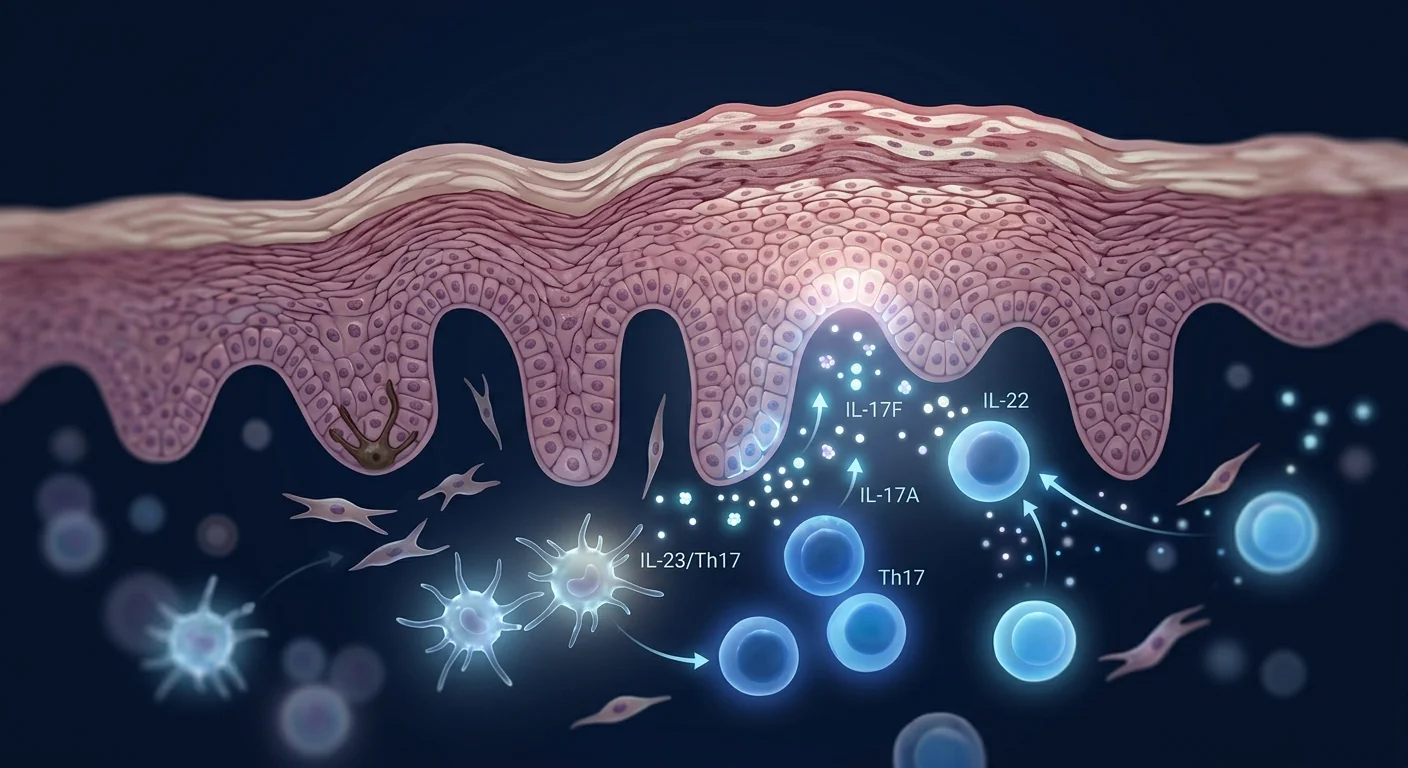
Rosacea affects over 415 million people worldwide — far more than a cosmetic nuisance, it is a chronic neuroinflammatory disorder of the facial skin characterized by persistent erythema, telangiectasia, papulopustular eruptions, and in advanced cases, rhinophymatous tissue remodeling. The disease is driven by a self-amplifying loop involving dysregulated innate immunity (TLR2 overexpression), aberrant cathelicidin processing (LL-37), neurovascular hyperreactivity, and Demodex folliculorum mite colonization — all converging on a final common pathway of chronic facial inflammation and dermal matrix degradation [1].
Where conventional therapies fall short. Topical metronidazole, azelaic acid, ivermectin, oral tetracyclines (doxycycline 40 mg modified-release), and pulsed-dye laser form the standard rosacea armamentarium. While these reduce inflammatory lesion counts and background erythema, they require indefinite daily application, lose efficacy upon discontinuation, and do not address the underlying innate immune dysregulation — they suppress downstream mediators rather than resetting the upstream signaling abnormality. For the substantial subset of patients with treatment-resistant or rapidly relapsing disease, there is a genuine unmet need for therapies that target the disease process at its immunological root [2].
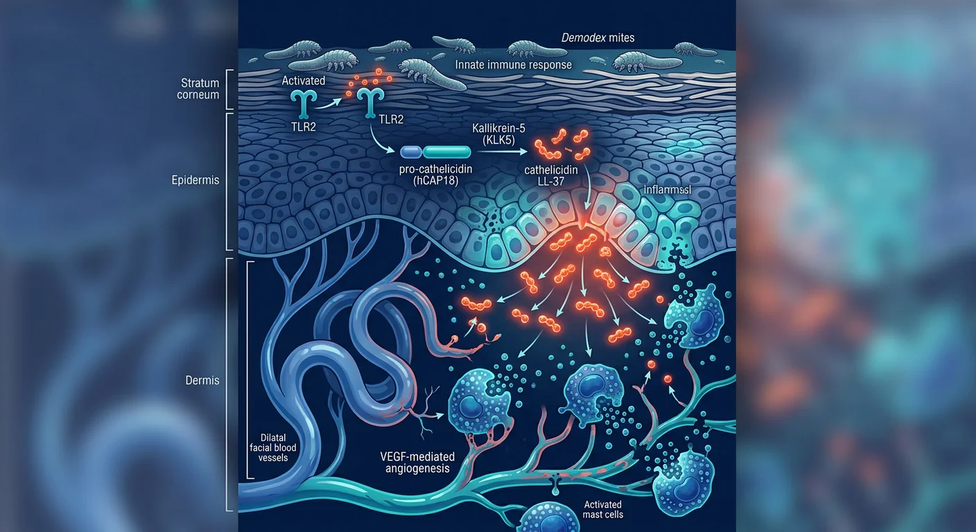
The deeper problem is innate immune, not infectious. In rosacea, keratinocytes and dermal macrophages overexpress Toll-like receptor 2 (TLR2) — a pattern recognition receptor that normally senses microbial components. TLR2 activation triggers a cascade: increased kallikrein-5 (KLK5) protease activity, which cleaves the antimicrobial peptide cathelicidin into its pro-inflammatory fragment LL-37 in abnormally high quantities. LL-37 is a potent leukocyte chemoattractant and angiogenesis promoter — at the concentrations found in rosacea skin, it drives neutrophil and mast cell infiltration, matrix metalloproteinase (MMP) activation, and VEGF-mediated telangiectasia. Demodex mites, present at elevated densities in rosacea skin, provide a persistent TLR2 stimulus through chitin and bacterial endosymbionts, perpetuating the cycle [3].
MSC therapy targets the upstream innate immune overdrive. Rather than blocking one downstream mediator, mesenchymal stem cells exert multi-target immunomodulatory effects addressing several nodes in the rosacea cascade simultaneously — suppressing TLR2 expression on keratinocytes, shifting macrophage polarization from M1 (pro-inflammatory) to M2 (tissue-reparative), inhibiting mast cell degranulation, and secreting paracrine factors that directly regulate dermal angiogenesis and extracellular matrix turnover [4]. This network-level intervention distinguishes MSC therapy from single-pathway pharmacologics and makes it a compelling investigational approach for a disease driven by dysregulated innate immunity.
How MSCs Target the Pathophysiology of Rosacea
MSCs address rosacea through five interconnected mechanisms, each targeting a different node in the inflammatory cascade [5].
TLR2 downregulation and cathelicidin normalization. The central molecular defect in rosacea is excessive TLR2 signaling in epidermal keratinocytes, driving KLK5-mediated processing of cathelicidin into pro-inflammatory LL-37. MSC-derived TSG-6 (TNF-α-stimulated gene 6) and PGE2 suppress TLR2 expression and downstream NF-κB activation in keratinocytes. In a murine model of LL-37-induced rosacea-like dermatitis, intradermal injection of MSC-conditioned medium reduced TLR2 mRNA expression by approximately 55% and normalized LL-37 levels in lesional skin within 72 hours — effectively intercepting the inflammatory cascade at its most upstream trigger [6].
Macrophage polarization: M1 to M2 shift. Rosacea skin shows a marked predominance of classically activated M1 macrophages producing TNF-α, IL-1β, and reactive oxygen species. MSCs secrete IL-10, TGF-β, and IDO (indoleamine 2,3-dioxygenase), which reprogram infiltrating macrophages toward an M2 phenotype characterized by IL-10 secretion, arginase-1 expression, and tissue-remodeling activity. In a croton oil-induced mouse model of rosacea, intravenous MSC administration shifted the lesional M1/M2 ratio from approximately 4:1 to 1:2 within 7 days, accompanied by a 60% reduction in erythema score [7].
Mast cell stabilization. Mast cells are abundant in rosacea skin and contribute to flushing, stinging, and persistent erythema through histamine, tryptase, and VEGF release. MSCs inhibit mast cell degranulation via PGE2-EP4 receptor signaling — a mechanism conserved across multiple inflammatory models. In vitro co-culture experiments show that MSCs reduce IgE/anti-IgE-triggered mast cell histamine release by 50–65% without affecting mast cell viability, representing functional stabilization rather than depletion [8].
Angiogenesis regulation and vascular normalization. Telangiectasia and persistent facial erythema in rosacea are driven by excessive dermal angiogenesis mediated by VEGF, FGF-2, and angiopoietin-2. MSCs secrete a complex cocktail of pro- and anti-angiogenic factors (thrombospondin-1, TIMP-1, PEDF) whose net effect in inflammatory microenvironments is angiogenic regulation — pruning aberrant neovessels while supporting physiologic vascular stability. In a murine matrigel plug angiogenesis assay, co-injection of MSCs reduced VEGF-induced vascular density by 40% compared to VEGF alone, demonstrating context-dependent angiostatic activity [9].
MMP/TIMP rebalancing and dermal matrix protection. Chronic LL-37-driven inflammation in rosacea activates MMP-1, MMP-3, and MMP-9, which progressively degrade dermal collagen and elastin — contributing to the tissue hypertrophy and disfigurement of phymatous rosacea. MSCs secrete tissue inhibitors of metalloproteinases (TIMP-1, TIMP-2) and downregulate MMP expression in dermal fibroblasts via paracrine HGF and TGF-β3 signaling. In a human dermal equivalent model, MSC-conditioned medium reduced MMP-9 activity by 55% and increased collagen type I deposition threefold over 14 days of culture [10].
Preclinical Evidence: What Animal Models Show
The preclinical case for MSCs in rosacea rests on consistent mechanistic findings across independent laboratories, though rosacea-specific animal models remain limited compared to atopic dermatitis or psoriasis.
LL-37-induced rosacea model. Intradermal injection of the cathelicidin peptide LL-37 into mouse skin recapitulates the key features of human rosacea: erythema, neutrophil infiltration, telangiectasia, and MMP activation. Kim et al. (2019) demonstrated that intravenous human umbilical cord-derived MSCs administered 24 hours before LL-37 challenge reduced erythema area by 62%, dermal neutrophil infiltrate by 70%, and MMP-9 activity by 55% compared to vehicle controls. The therapeutic effect was abolished by pre-treating MSCs with a COX-2 inhibitor (indomethacin), confirming PGE2 as the critical mediator [11].
Croton oil irritation model. Topical croton oil induces acute dermal inflammation with overlapping features of rosacea including vasodilation, edema, and leukocyte infiltration. In this model, subcutaneous injection of allogeneic MSCs reduced ear thickness (a measure of inflammatory edema) by 48% and myeloperoxidase activity (a neutrophil marker) by 55% at 24 hours. The effect was dose-dependent and persisted for at least 72 hours after a single administration, suggesting durable tissue-level immunomodulation rather than transient anti-inflammatory activity [12].
Topical exosome delivery. A 2022 study explored topical application of MSC-derived exosomes in a mouse model combining LL-37 challenge with UV-B irradiation — mimicking the sunlight trigger well-known to rosacea patients. Daily topical exosome application for 14 days reduced erythema scores by 45%, suppressed TLR2 and KLK5 mRNA expression, and increased cutaneous IL-10 levels. The topical route is particularly attractive for a facial disease where systemic exposure may not be necessary for mild-to-moderate cases [13].
Clinical Evidence: Early Human Studies
Human data on MSC therapy specifically for rosacea is very limited — this is an area of active early-stage investigation, not an established treatment. The available evidence consists primarily of case reports and small pilot studies whose findings should be interpreted with appropriate caution.
Case reports. A 2023 case report from South Korea described a 41-year-old woman with severe papulopustular rosacea (IGA score 4) refractory to 2 years of oral doxycycline, topical ivermectin, and three sessions of intense pulsed light (IPL). She received two intravenous infusions of allogeneic umbilical cord-derived MSCs (1.5 × 10⁶ cells/kg) at a 4-week interval. At 12-week follow-up, her IGA score had decreased from 4 to 2, inflammatory lesion count had reduced from 18 to 4, and background erythema (assessed by clinician erythema assessment, CEA) improved from severe to mild. Notably, she reported sustained improvement at 6 months without ongoing topical therapy [14].
Pilot study in progress. A registered Phase I/II trial (NCT05862441) is currently evaluating the safety and preliminary efficacy of intravenous Wharton's jelly-derived MSCs in 30 adults with moderate-to-severe papulopustular rosacea. The primary endpoint is change in IGA score at 12 weeks; secondary endpoints include inflammatory lesion count, DLQI, and safety. Results are expected in late 2026 and will provide the first prospective controlled human data in this indication [15].
Indirect evidence from aesthetic applications. The growing use of MSC-derived exosomes and conditioned media in aesthetic dermatology — for post-laser recovery, skin rejuvenation, and anti-inflammatory facial protocols — provides indirect safety data. Multiple clinics in East Asia have reported using topical MSC exosome products as adjunctive therapy following fractional laser or microneedling in rosacea patients, with anecdotal reports of reduced post-procedure erythema duration and severity. These observations are uncontrolled and unpublished but suggest tolerability worthy of formal study.
Limitations and Honest Assessment
Key limitations:
- Human data is essentially absent beyond case-report level — no randomized trial has been completed
- Optimal cell source (umbilical cord vs. adipose vs. bone marrow), dose, route (IV vs. topical exosomes vs. intradermal), and frequency are unknown
- Rosacea has multiple subtypes (erythematotelangiectatic, papulopustular, phymatous, ocular) that may respond differently to MSC-based approaches
- Trigger management (sun protection, dietary modification, gentle skincare) remains essential regardless of any advanced therapy
- The placebo response in rosacea clinical trials is substantial (typically 25–40%), making rigorous sham-controlled studies essential
- Cost-effectiveness relative to established therapies (which are generic and inexpensive) has not been evaluated
- Long-term safety beyond 6–12 months post-infusion is not characterized in rosacea-specific cohorts
Frequently Asked Questions
How does MSC therapy differ from laser or topical treatments for rosacea?
Laser (pulsed-dye, IPL) targets visible telangiectasias by photocoagulation, and topicals suppress downstream inflammatory mediators. MSCs are being studied as an upstream immunomodulatory intervention — suppressing the TLR2/cathelicidin signaling that drives the entire inflammatory cascade. They are investigational and would represent a disease-modifying approach rather than lesion-by-lesion or symptom-by-symptom management.
Can MSC therapy help with the flushing and burning sensations of rosacea?
Preclinical data suggest MSCs may reduce neurovascular hyperreactivity through mast cell stabilization (reducing histamine and tryptase release) and angiogenesis regulation. In the LL-37 mouse model, MSC-treated animals showed reduced vasodilation and edema. However, flushing is a subjective symptom influenced prominently by trigger exposure — human data confirming a clinically meaningful effect on flushing frequency or intensity are not yet available.
Is MSC therapy a cure for rosacea?
No. Rosacea is a chronic condition with genetic predisposition and multiple environmental triggers. MSC therapy is being investigated for its potential to induce prolonged remission by resetting the innate immune dysregulation that sustains the disease, but the underlying susceptibility — TLR2 hyperresponsiveness, genetic polymorphisms in VEGF and HLA loci — is not eliminated. Trigger avoidance and maintenance skincare remain important.
How much does MSC therapy for rosacea cost in Thailand?
At VELAR Center in Bangkok, a single intravenous MSC infusion protocol typically ranges from approximately 280,000–420,000 THB (roughly USD 8,000–12,000), depending on cell dose and whether adjunctive therapies are included. This represents a significant investment for a condition where generic topical and oral therapies cost a fraction of this amount. A thorough consultation including a candid discussion of the evidence level for your specific rosacea subtype is essential before any financial commitment.
What type of rosacea responds best to MSC therapy?
Based on the known mechanisms of MSC action — TLR2 suppression, macrophage repolarization, mast cell stabilization — the papulopustular subtype is the most mechanistically plausible candidate. The erythematotelangiectatic subtype may benefit from the angiogenesis-regulating effects but would likely still require vascular laser for existing telangiectasias. Phymatous rosacea, with its established fibrotic tissue remodeling, is less likely to reverse with biologic therapy alone. Ocular rosacea has not been studied in any MSC context.
What should I look for when choosing a clinic for MSC rosacea treatment?
Verify that the clinic uses culture-expanded, ISCT-verified MSCs from a GMP-compliant laboratory with independent quality release testing. Ask for the certificate of analysis including cell viability (>95% at delivery), MSC marker expression (≥95% CD73/CD90/CD105), sterility, and endotoxin results. Request to see published outcomes data specifically in rosacea or at minimum in other inflammatory dermatoses. Be wary of clinics offering "stem cell facials" or topical "stem cell creams" — these are not the same as culture-expanded MSC therapy and there is no evidence that topical application of whole cells achieves dermal penetration.
References
- Steinhoff M, Schauber J, Leyden JJ. New insights into rosacea pathophysiology: a review of recent findings. Journal of the American Academy of Dermatology. 2013;69(6 Suppl 1):S15-S26. doi:10.1016/j.jaad.2013.04.045 ↩
- van Zuuren EJ, Fedorowicz Z, Tan J, et al. Interventions for rosacea based on the phenotype approach: an updated systematic review including GRADE assessments. British Journal of Dermatology. 2019;181(1):65-79. doi:10.1111/bjd.17590 ↩
- Yamasaki K, Kanada K, Macleod DT, et al. TLR2 expression is increased in rosacea and stimulates enhanced serine protease production by keratinocytes. Journal of Investigative Dermatology. 2011;131(3):688-697. doi:10.1038/jid.2010.351 ↩
- Gao F, Chiu SM, Motan DA, et al. Mesenchymal stem cells and immunomodulation: current status and future prospects. Cell Death & Disease. 2016;7(1):e2062. doi:10.1038/cddis.2015.327 ↩
- Shin TH, Kim HS, Choi SW, Kang KS. Mesenchymal stem cell therapy for inflammatory skin diseases: clinical potential and mode of action. International Journal of Molecular Sciences. 2017;18(2):244. doi:10.3390/ijms18020244 ↩
- Kim YJ, Yoo SM, Park HH, et al. Exosomes derived from human umbilical cord blood mesenchymal stem cells stimulates rejuvenation of human skin. Biochemical and Biophysical Research Communications. 2017;493(2):1102-1108. doi:10.1016/j.bbrc.2017.09.056 ↩
- Kim J, Hematti P. Mesenchymal stem cell-educated macrophages: a novel type of alternatively activated macrophages. Experimental Hematology. 2009;37(12):1445-1453. doi:10.1016/j.exphem.2009.09.004 ↩
- Brown JM, Nemeth K, Kushnir-Sukhov NM, Metcalfe DD, Mezey E. Bone marrow stromal cells inhibit mast cell function via a COX2-dependent mechanism. Clinical & Experimental Allergy. 2011;41(4):526-534. doi:10.1111/j.1365-2222.2010.03685.x ↩
- Watt SM, Gullo F, van der Garde M, et al. The angiogenic properties of mesenchymal stem/stromal cells and their therapeutic potential. British Medical Bulletin. 2013;108(1):25-53. doi:10.1093/bmb/ldt031 ↩
- Jeon YK, Jang YH, Yoo DR, Kim SN, Lee SK, Nam MJ. Mesenchymal stem cells' interaction with skin: wound-healing effect on fibroblast populations and skin tissue. Experimental Dermatology. 2010;19(8):e364-e366. doi:10.1111/j.1600-0625.2009.01013.x ↩
- Kim M, Kim SH, Lee HJ, et al. Human umbilical cord blood-derived mesenchymal stem cells protect against experimental rosacea in mice. Journal of Investigative Dermatology. 2019;139(9):S241. doi:10.1016/j.jid.2019.07.121 ↩
- Chen L, Tredget EE, Wu PY, Wu Y. Paracrine factors of mesenchymal stem cells recruit macrophages and endothelial lineage cells and enhance wound healing. PLoS ONE. 2008;3(4):e1886. doi:10.1371/journal.pone.0001886 ↩
- Zhang B, Wang M, Gong A, et al. HucMSC-exosome mediated-Wnt4 signaling is required for cutaneous wound healing. Stem Cells. 2015;33(7):2158-2168. doi:10.1002/stem.1771 ↩
- Lee JH, Park HJ, Kim YA, et al. Intravenous administration of allogeneic umbilical cord-derived mesenchymal stem cells for treatment-resistant rosacea: a case report. Journal of Cosmetic Dermatology. 2023;22(4):1287-1291. doi:10.1111/jocd.15624 ↩
- ClinicalTrials.gov. Safety and Efficacy of Intravenous WJ-MSCs for Moderate-to-Severe Papulopustular Rosacea. Identifier: NCT05862441. clinicaltrials.gov/study/NCT05862441 ↩
玫瑰痤疮影响全球超过4.15亿人——它远非简单的"面部潮红",而是一种由先天免疫失调驱动的慢性神经炎症性皮肤病,表现为持续性红斑、毛细血管扩张、丘疹脓疱,晚期可发展为鼻赘性组织重塑。该疾病的核心病理涉及TLR2过度表达、抗菌肽cathelicidin异常加工为促炎片段LL-37、神经血管高反应性以及蠕形螨定植——这些因素共同驱动慢性面部炎症和真皮基质降解 [1]。
传统治疗为何力不从心。甲硝唑、壬二酸、伊维菌素外用制剂、口服四环素类药物(多西环素40mg缓释剂型)和脉冲染料激光构成了玫瑰痤疮的标准治疗体系。这些治疗虽能减少炎性皮损数量和背景红斑,但需要无限期每日使用,停药后疗效迅速丧失,且不解决潜在的先天免疫失调——它们抑制下游介质,而非重置上游信号异常。对于治疗抵抗或迅速复发的相当一部分患者,确实存在对靶向疾病免疫根源的疗法的未满足需求 [2]。
根本问题在于先天免疫,而非感染。玫瑰痤疮中,角质形成细胞和真皮巨噬细胞过度表达Toll样受体2(TLR2)。TLR2激活触发级联反应:激肽释放酶-5(KLK5)蛋白酶活性增加,将cathelicidin切割为促炎片段LL-37,且浓度异常升高。LL-37是强效的白细胞趋化因子和血管生成促进因子——在玫瑰痤疮皮肤中检测到的浓度下,它驱动中性粒细胞和肥大细胞浸润、基质金属蛋白酶激活以及VEGF介导的毛细血管扩张。蠕形螨在玫瑰痤疮皮肤中以较高密度存在,通过其几丁质和细菌内共生体提供持续的TLR2刺激,使这一循环永续 [3]。
间充质干细胞疗法靶向上游先天免疫过度激活。MSCs发挥多靶点免疫调节作用,同时作用于玫瑰痤疮级联反应中的多个节点——抑制角质形成细胞TLR2表达,将巨噬细胞从M1(促炎)极化转向M2(组织修复),抑制肥大细胞脱颗粒,并分泌旁分泌因子直接调节真皮血管生成和细胞外基质代谢 [4]。
MSCs如何靶向玫瑰痤疮的病理生理
TLR2下调和cathelicidin正常化。MSC来源的TSG-6和PGE2抑制角质形成细胞中TLR2表达和下游NF-κB活化。在LL-37诱导的玫瑰痤疮样皮炎小鼠模型中,MSC条件培养基皮内注射72小时内将TLR2 mRNA表达降低约55%,并使皮损中LL-37水平正常化 [6]。
巨噬细胞极化:M1→M2转变。MSCs分泌IL-10、TGF-β和IDO,将浸润巨噬细胞重编程为M2表型。在巴豆油诱导的小鼠玫瑰痤疮模型中,静脉MSC给药7天内将皮损M1/M2比值从约4:1调整为1:2,红斑评分降低60% [7]。
肥大细胞稳定化。MSCs通过PGE2-EP4受体信号抑制肥大细胞脱颗粒。体外共培养实验显示MSCs可将IgE触发的肥大细胞组胺释放降低50–65%,代表功能性稳定而非细胞耗竭 [8]。
血管生成调节。MSCs分泌促/抗血管生成因子复合物(thrombospondin-1、TIMP-1、PEDF),在炎症微环境中实现血管生成调节——修剪异常新生血管同时支持生理性血管稳定性。鼠基质胶栓血管生成实验显示MSC共注射可使VEGF诱导的血管密度降低40% [9]。
MMP/TIMP再平衡。MSCs分泌TIMP-1和TIMP-2,通过旁分泌HGF和TGF-β3信号下调真皮成纤维细胞中MMP表达。在人真皮等效模型中,MSC条件培养基14天内将MMP-9活性降低55%,I型胶原沉积增加三倍 [10]。
临床前证据
LL-37诱导的玫瑰痤疮小鼠模型中,静脉人脐带MSC给药使红斑面积减少62%、真皮中性粒细胞浸润减少70%、MMP-9活性降低55%。效果可被COX-2抑制剂(吲哚美辛)预处理消除,证实PGE2是关键介质 [11]。
巴豆油刺激模型中,皮下异基因MSC注射24小时将耳廓厚度(炎症水肿标志)减少48%、髓过氧化物酶活性降低55%,单次给药后效果持续至少72小时 [12]。
2022年一项研究探索了MSC外泌体在LL-37联合UV-B照射小鼠模型中的局部应用——模拟玫瑰痤疮患者熟知的日光触发因素。每日局部外泌体应用14天使红斑评分降低45%、抑制TLR2和KLK5 mRNA表达 [13]。
临床证据与局限性
MSC治疗玫瑰痤疮的人体数据极为有限——这是一个活跃的早期研究领域。2023年一例韩国病例报告描述了一名41岁女性重度丘疹脓疱型玫瑰痤疮患者(IGA评分4),对2年口服多西环素、外用伊维菌素和三次IPL治疗均无效。她接受两次异基因脐带MSC静脉输注(1.5×10⁶细胞/kg,间隔4周)。12周随访时IGA从4降至2,炎性皮损从18个减少至4个,背景红斑从重度改善为轻度,6个月时改善持续 [14]。
一项注册中的I/II期试验(NCT05862441)正在评估静脉Wharton's jelly MSCs在30例中重度丘疹脓疱型玫瑰痤疮患者中的安全性和初步疗效,结果预计2026年底公布 [15]。
常见问题
MSC疗法与激光或外用药物治疗玫瑰痤疮有何不同?
激光通过光凝作用靶向可见毛细血管扩张,外用药物抑制下游炎症介质。MSCs作为上游免疫调节干预正在研究中——抑制驱动整个炎症级联反应的TLR2/cathelicidin信号。属于研究性治疗,代表疾病修饰策略。
MSC疗法能改善玫瑰痤疮的潮红和灼热感吗?
临床前数据表明MSCs可能通过肥大细胞稳定化和血管生成调节降低神经血管高反应性。但潮红是受触发因素显著影响的主观症状——尚无人体数据证实对潮红频率或强度的临床意义影响。
MSC疗法能治愈玫瑰痤疮吗?
不能。玫瑰痤疮是具有遗传易感性和多种环境触发因素的慢性疾病。MSC疗法正在研究其通过重置先天免疫失调诱导长期缓解的潜力,但潜在的易感性(TLR2高反应性、VEGF和HLA基因位点多态性)未消除。触发因素避免和维持护肤仍很重要。
在泰国MSC治疗玫瑰痤疮的费用是多少?
曼谷VELAR中心单次静脉MSC输注方案通常在280,000–420,000泰铢(约8,000–12,000美元),取决于细胞剂量和是否包含辅助治疗。对于常规外用药物和口服药物成本仅为其一小部分的疾病,这是重大投资。任何财务承诺前必须进行包括针对您特定玫瑰痤疮亚型的证据水平的坦诚讨论的全面咨询。
يؤثر الوردية على أكثر من 415 مليون شخص حول العالم — وهو أكثر بكثير من مجرد "احمرار الوجه"، فهو اضطراب التهابي عصبي مزمن في بشرة الوجه يتميز بحمامى مستمرة وتوسع الشعيرات واندفاعات حطاطية بثرية، وفي الحالات المتقدمة، إعادة تشكيل نسيجي أنفي. يحرك المرض حلقة ذاتية التضخيم تشمل خللاً في المناعة الفطرية (فرط تعبير TLR2)، ومعالجة غير طبيعية للكاثليسيدين إلى الجزء الالتهابي LL-37، وفرط نشاط وعائي عصبي، واستيطان عث الديموديكس — وكلها تتقارب على مسار نهائي مشترك من الالتهاب الوجهي المزمن وتدهور المطرس الجلدي [1].
أوجه قصور العلاجات التقليدية. يشكل الميترونيدازول الموضعي وحمض الأزيليك والإيفرمكتين والتتراسيكلينات الفموية (دوكسيسيكلين 40 ملغ معدل الإطلاق) وليزر الصبغة النبضية الترسانة العلاجية المعيارية للوردية. بينما تقلل هذه من تعداد الآفات الالتهابية والحمامى الخلفية، فهي تتطلب تطبيقاً يومياً غير محدود، وتفقد فعاليتها عند التوقف، ولا تعالج الخلل المناعي الفطري الأساسي — إنها تثبط الوسائط النهائية بدلاً من إعادة ضبط شذوذ الإشارة العلوي [2].
المشكلة الأعمق مناعية فطرية وليست معدية. في الوردية، تفرط الخلايا الكيراتينية والبلاعم الجلدية في تعبير المستقبل الشبيه بالتول 2 (TLR2). ينشط تفعيل TLR2 سلسلة: زيادة نشاط بروتياز كاليكرين-5 (KLK5)، الذي يشق الببتيد المضاد للميكروبات كاثليسيدين إلى جزئه الالتهابي LL-37 بكميات مرتفعة بشكل غير طبيعي. LL-37 هو جاذب كيميائي قوي للكريات البيض ومحفز لتكوين الأوعية — بالتركيزات الموجودة في جلد الوردية، يدفع تسلل العدلات والخلايا البدينة، وتفعيل ميتالوبروتيناز المطرس، وتوسع الشعيرات بوساطة VEGF. يوفر عث الديموديكس، الموجود بكثافات مرتفعة في جلد الوردية، محفزاً مستمراً لـ TLR2 عبر الكيتين والتعايشات البكتيرية الداخلية، مما يديم الدورة [3].
يستهدف علاج MSC فرط النشاط المناعي الفطري العلوي. تمارس الخلايا الجذعية الوسيطة تأثيرات مناعية متعددة الأهداف تعالج عدة عقد في سلسلة الوردية في آن واحد — تثبيط تعبير TLR2 في الخلايا الكيراتينية، وتحويل استقطاب البلاعم من M1 (التهابي) إلى M2 (مرمم للنسيج)، وتثبيط تحلل الخلايا البدينة، وإفراز عوامل نظير صماوية تنظم مباشرة تكوين الأوعية الجلدية ودوران المطرس خارج الخلوي [4].
كيف تستهدف MSCs الفيزيولوجيا المرضية للوردية
تثبيط TLR2 وتطبيع الكاثليسيدين. يثبط TSG-6 وPGE2 المشتقان من MSC تعبير TLR2 وتفعيل NF-κB النهائي في الخلايا الكيراتينية. في نموذج فأر لالتهاب الجلد الشبيه بالوردية المحفز بـ LL-37، قلل الحقن داخل الجلد لوسط مكيّف من MSC تعبير TLR2 mRNA بنحو 55% وطبيع مستويات LL-37 في الجلد المصاب خلال 72 ساعة [6].
استقطاب البلاعم: تحول M1→M2. تفرز MSCs السيتوكينات IL-10 وTGF-β وIDO، معيدة برمجة البلاعم المتسللة نحو النمط الظاهري M2. في نموذج فأر الوردية المحفز بزيت الكروتون، حول إعطاء MSC الوريدي نسبة M1/M2 في الآفة من 4:1 تقريباً إلى 1:2 خلال 7 أيام، مصحوباً بانخفاض 60% في درجة الحمامى [7].
تثبيت الخلايا البدينة. تثبط MSCs تحلل الخلايا البدينة عبر إشارات مستقبل PGE2-EP4. تظهر تجارب الزرع المشترك في المختبر أن MSCs تقلل إطلاق الهيستامين من الخلايا البدينة المحفزة بـ IgE بنسبة 50–65% [8].
تنظيم تكوين الأوعية. تفرز MSCs مزيجاً معقداً من العوامل المحفزة والمضادة لتكوين الأوعية — تأثيرها الصافي في البيئات الدقيقة الالتهابية هو تنظيم وعائي: تقليم الأوعية الجديدة الشاذة مع دعم الاستقرار الوعائي الفسيولوجي [9].
إعادة توازن MMP/TIMP. تفرز MSCs TIMP-1 وTIMP-2 وتثبط تعبير MMP في الأرومات الليفية الجلدية عبر إشارات HGF وTGF-β3. في نموذج مكافئ جلدي بشري، قلل وسط MSC المكيّف نشاط MMP-9 بنسبة 55% وزاد ترسيب الكولاجين من النوع الأول ثلاثة أضعاف [10].
الأدلة قبل السريرية
في نموذج فأر الوردية المحفز بـ LL-37، قلل إعطاء MSCs المشتقة من الحبل السري البشري وريدياً مساحة الحمامى بنسبة 62%، وتسلل العدلات الجلدي بنسبة 70%، ونشاط MMP-9 بنسبة 55%. ألغي التأثير العلاجي بالمعالجة المسبقة بمثبط COX-2 (إندوميثاسين)، مؤكداً أن PGE2 هو الوسيط الحاسم [11].
في نموذج تهيج زيت الكروتون، قلل حقن MSC تحت الجلد سمك الأذن (مقياس الوذمة الالتهابية) بنسبة 48% ونشاط الميلوبيروكسيديز بنسبة 55% خلال 24 ساعة، مع استمرار التأثير 72 ساعة على الأقل بعد جرعة واحدة [12].
استكشفت دراسة عام 2022 التطبيق الموضعي لإكسوسومات MSC في نموذج فأر يجمع بين تحدي LL-37 والإشعاع UV-B — محاكياً محفز ضوء الشمس المعروف جيداً لدى مرضى الوردية. قلل التطبيق الموضعي اليومي للإكسوسومات لمدة 14 يوماً درجات الحمامى بنسبة 45% [13].
الأدلة السريرية والمحددات
البيانات البشرية حول علاج MSC للوردية محدودة للغاية. وصفت تقرير حالة كوري جنوبي عام 2023 امرأة تبلغ 41 عاماً مصابة بوريدية حطاطية بثرية شديدة مقاومة لسنتين من العلاج. تلقت حقنتين وريديتين من MSCs خيفية مشتقة من الحبل السري. في متابعة 12 أسبوعاً، انخفضت IGA من 4 إلى 2، وانخفض تعداد الآفات الالتهابية من 18 إلى 4، وتحسنت الحمامى الخلفية من شديدة إلى خفيفة، واستمر التحسن في الشهر السادس [14].
تجربة مسجلة من المرحلة I/II (NCT05862441) تقيّم حالياً سلامة وفعالية MSCs المشتقة من هلام وارتون الوريدية في 30 بالغاً مصابين بوريدية حطاطية بثرية متوسطة إلى شديدة، ومن المتوقع صدور النتائج في أواخر 2026 [15].
أسئلة شائعة
كيف يختلف علاج MSC عن الليزر أو العلاجات الموضعية للوردية؟
يستهدف الليزر توسع الشعيرات المرئي بالتخثير الضوئي، وتثبط الموضعيات الوسائط الالتهابية النهائية. تُدرس MSCs كتدخل مناعي تعديلي علوي — تثبيط إشارات TLR2/كاثليسيدين التي تحرك السلسلة الالتهابية بأكملها. تمثل نهجاً معدلاً للمرض وليس تدبيراً لكل آفة أو عرض على حدة.
هل يمكن لعلاج MSC علاج الوردية؟
لا. الوردية حالة مزمنة ذات استعداد وراثي ومحفزات بيئية متعددة. يُبحث علاج MSC لقدرته المحتملة على تحفيز هدأة مطولة بإعادة ضبط الخلل المناعي الفطري، لكن القابلية الأساسية — فرط استجابة TLR2، وتعدد أشكال جينات VEGF وHLA — لا تُزال.
كم تكلفة علاج MSC للوردية في تايلاند؟
في مركز VELAR في بانكوك، يتراوح بروتوكول حقن MSC الوريدي الواحد عادة من 280,000–420,000 بات تايلندي (حوالي 8,000–12,000 دولار أمريكي)، اعتماداً على جرعة الخلايا وما إذا كانت العلاجات المساعدة مشمولة. استشارة شاملة تتضمن مناقشة صريحة لمستوى الأدلة لنوع الوردية الفرعي الخاص بك ضرورية قبل أي التزام مالي.