Retinitis pigmentosa (RP) is not one disease but a family of inherited retinal degenerations — collectively the most common cause of inherited blindness — affecting roughly 1 in 4,000 people worldwide. [1] The unifying feature is progressive loss of photoreceptor cells (rods first, then cones), leading to night blindness, tunnel vision, and — in many cases — legal blindness by middle age. Despite decades of research, no approved therapy halts RP progression for the majority of patients.
Where conventional approaches fall short. Gene therapy has emerged for a handful of specific mutations (RPE65, approved as voretigene neparvovec), but this covers less than 2% of RP patients. Vitamin A supplementation and retinal prostheses offer modest benefits at best. For the overwhelming majority of RP patients, the therapeutic landscape remains a waiting room — monitoring progression without a meaningful intervention.
The deeper problem is cellular collapse, not just mutation. Regardless of which gene is mutated, RP converges on a shared final pathway: photoreceptor apoptosis, oxidative stress, chronic neuroinflammation, and progressive remodeling of the retinal microenvironment that accelerates further cell death. This convergence suggests a therapeutic opportunity — an intervention that targets these downstream processes could benefit patients across multiple RP genotypes.
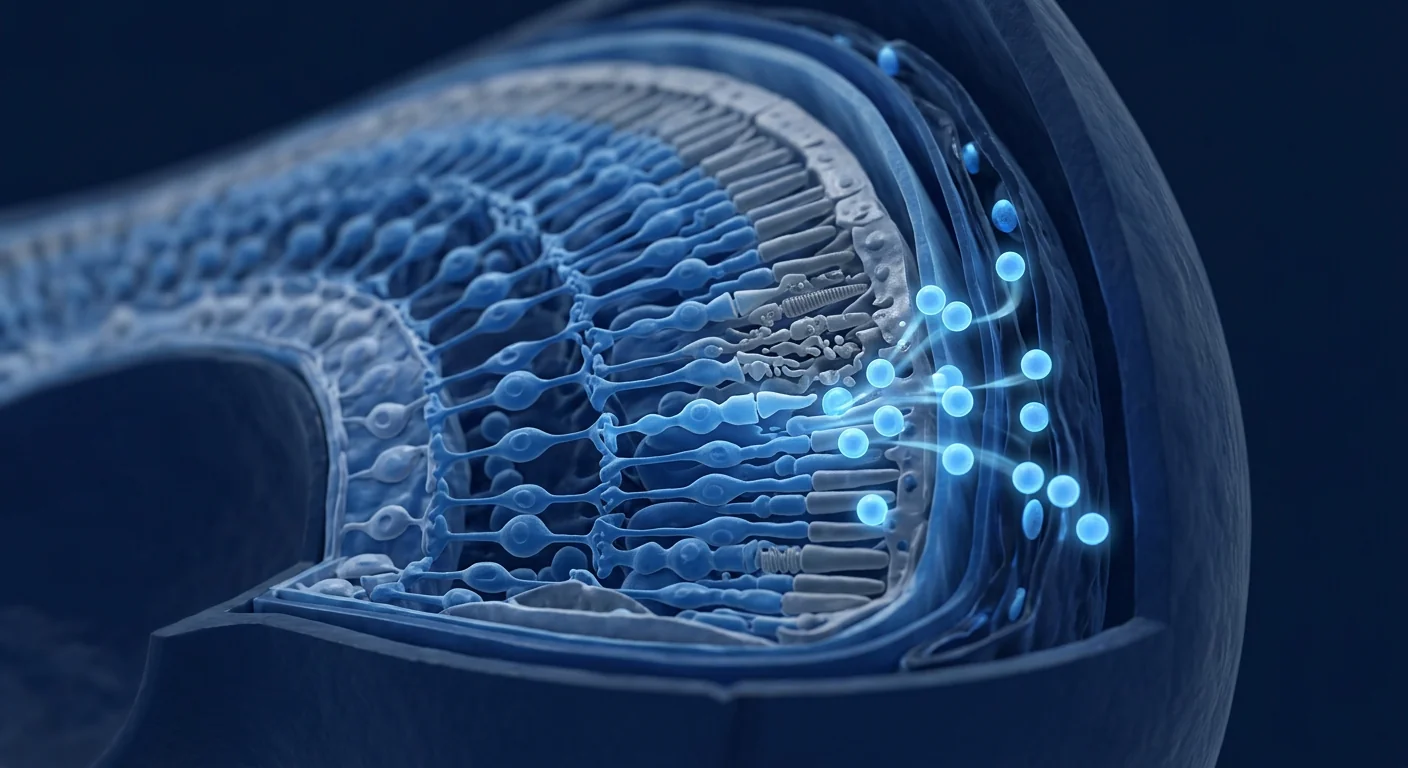
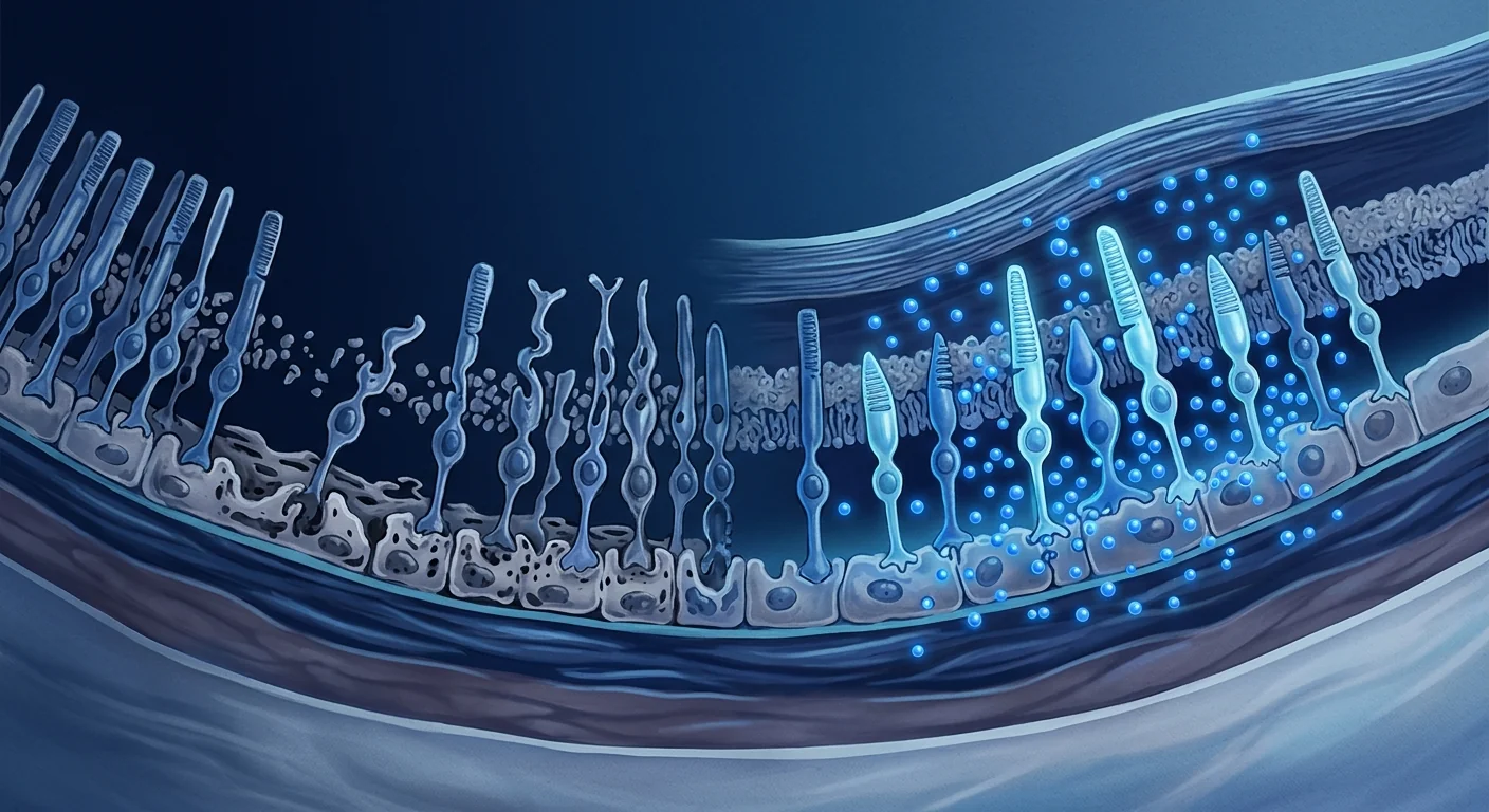
MSC therapy targets the shared biology of retinal degeneration. Rather than correcting a specific gene, mesenchymal stem cells address the microenvironment in which photoreceptors are dying — delivering neurotrophic factors, damping microglial activation, reducing oxidative stress, and preserving the retinal pigment epithelium (RPE) that photoreceptors depend on for survival. [2]
What is happening at the cellular level in retinitis pigmentosa?
Retinitis pigmentosa is fundamentally a disease of progressive photoreceptor death driven by inherited mutations in over 80 identified genes. The cellular hallmarks that recur across most forms of RP include:
- Photoreceptor apoptosis — rod photoreceptors die first (explaining the characteristic night blindness), followed by cone degeneration as the retinal microenvironment deteriorates [1][3]
- Chronic neuroinflammation — activated microglia and Müller glial cells release pro-inflammatory cytokines (TNF-α, IL-1β) that accelerate photoreceptor death
- Oxidative stress — the retina's extremely high metabolic rate and oxygen consumption make it uniquely vulnerable to cumulative oxidative damage, which builds as rods die and oxygen tension in the outer retina paradoxically increases [4]
- RPE dysfunction — the retinal pigment epithelium, which nourishes and supports photoreceptors, degenerates alongside them, creating a vicious cycle of mutual deterioration
- Trophic factor depletion — as cells die, the retina's endogenous supply of survival factors (BDNF, CNTF, GDNF) dwindles, removing a critical support system for remaining photoreceptors
Therapeutic logic follows from this biology: if you cannot fix the mutation, you intervene at the level of the microenvironment to preserve the photoreceptors that are still alive. This is precisely the space where MSC therapy is being investigated.
What MSCs may biologically contribute to retinal preservation
Mesenchymal Stem Cells do not become photoreceptors. They are not designed to replace retinal cells directly. What they do — and the reason research interest in MSC therapy for retinal degeneration continues to grow — is alter the cellular environment around stressed photoreceptors. Their relevant actions in RP biology include:
Neurotrophic factor secretion
MSCs secrete a rich cocktail of neurotrophic factors — BDNF, CNTF, GDNF, NGF, and VEGF — that directly support photoreceptor survival. [5][6] In animal models of RP, MSC-derived BDNF and CNTF have been shown to delay photoreceptor apoptosis and preserve outer nuclear layer thickness — a direct structural correlate of retained vision. This is the single most convincing mechanism by which MSCs could slow RP progression.
Anti-inflammatory and immunomodulatory signalling
MSCs shift microglia from a pro-inflammatory (M1) to a neuroprotective (M2) phenotype in the retina, reducing the chronic low-grade inflammation that accelerates photoreceptor loss. [7] MSC-secreted factors including IDO, PGE₂, and TGF-β suppress activated microglia and reduce TNF-α and IL-1β levels in the retinal microenvironment — directly addressing one of the key drivers of cone death after rod loss.
Oxidative stress reduction
The retina's uniquely high oxygen consumption makes oxidative damage a central driver of RP progression. MSCs upregulate endogenous antioxidant enzymes (SOD, catalase, glutathione peroxidase) in host retinal cells and directly scavenge reactive oxygen species via paracrine signalling. [4][8]
RPE support and mitochondrial transfer
Emerging research shows MSCs can transfer healthy mitochondria to stressed RPE cells via tunneling nanotubes — directly rescuing the metabolic support system that photoreceptors depend on. [9] Additionally, MSCs secrete factors that promote RPE cell survival and barrier function, preserving the critical RPE-photoreceptor metabolic axis.

What the evidence supports — and what it doesn't
An honest summary of the published research landscape for MSC therapy in retinitis pigmentosa:
What is plausible: A meaningful slowing of photoreceptor loss — measured by preserved outer nuclear layer thickness on OCT imaging, maintained visual field area, or delayed decline in electroretinography (ERG) amplitudes — particularly in patients treated in early to mid-stage disease when substantial photoreceptor populations remain viable. This is consistent with what MSC-derived neurotrophins and anti-inflammatory factors could realistically achieve. [10][11]
What is not supported: Restoration of lost vision, regeneration of photoreceptors already dead, normalization of ERG responses in advanced disease, or halting of progression entirely. Any clinic claiming to "restore sight" in RP with MSC therapy is overstating what biology and current research can deliver. The honest goal — even in the best-case scenario — is preservation of remaining vision, not recovery of what has been lost.
What remains under active investigation: Optimal delivery route (intravitreal injection vs subretinal vs systemic IV — each with different risk-benefit profiles for an ocular target), cell dosage, frequency of repeat administration, and which RP genotypes respond best to neurotrophic support. The field has not yet conducted the large, randomized trials needed to establish treatment guidelines.
A Hard but Important Distinction
MSC therapy for retinitis pigmentosa is, at present, an investigational neuroprotective strategy — a possible addition to ophthalmological care, not a replacement for it. Standard low-vision rehabilitation, genetic counselling, vitamin A supplementation (where indicated), and regular ophthalmological monitoring remain the primary care framework. Regenerative therapy is best considered alongside that framework, not instead of it.
Who is most likely to benefit from MSC therapy in RP?
Within the boundaries of what is realistic, the patients with the strongest case for considering MSC therapy in retinitis pigmentosa are those who:
- Are in early to mid-stage disease — still retain measurable visual field (ideally >20 degrees) and have preserved outer nuclear layer thickness on OCT imaging, indicating substantial viable photoreceptor populations
- Have documented progression — a history of measurable decline over 12–24 months provides a baseline against which to assess any treatment effect
- Have no mutation eligible for approved gene therapy — if RPE65 or another approved gene therapy target is present, that should be pursued first as the evidence base is stronger
- Understand the goal is preservation, not restoration — and have realistic expectations for what neuroprotection can achieve
- Have no contraindicating ocular conditions — active ocular infection, uncontrolled glaucoma, or retinal detachment would preclude intravitreal or subretinal delivery
Delivery routes for retinal conditions — what is known
Getting MSCs to the retina presents unique challenges not faced in systemic conditions. Three delivery routes are under investigation, each with distinct advantages and limitations:
| Route | Advantages | Limitations |
|---|---|---|
| Intravitreal injection | Minimally invasive; broad distribution across retina; office-based procedure | Inner limiting membrane barrier; short retinal residence time; risk of endophthalmitis |
| Subretinal injection | Direct access to photoreceptors and RPE; highest local concentration | Surgical procedure; risk of retinal detachment; focal delivery may miss peripheral degeneration |
| Systemic intravenous | Non-invasive to the eye; systemic anti-inflammatory benefit; may reach both eyes | Blood-retinal barrier limits retinal penetration; lower local concentration; first-pass lung trapping |
Most preclinical RP studies have used intravitreal or subretinal delivery. [12] Systemic IV delivery is being explored for its broader immunomodulatory benefits, but whether sufficient MSC-derived factors cross the blood-retinal barrier to meaningfully impact photoreceptor survival remains an open question. At VELAR, delivery route selection is individualized based on disease stage, retinal anatomy on OCT, and a comprehensive risk-benefit assessment.
VELAR's approach to RP — coordinated ophthalmological care
RP protocols at VELAR Center are designed in coordination with each patient's existing ophthalmologist and retinal specialist — never as a replacement for that care. Every programme uses clinical-grade Wharton's jelly-derived MSCs (≥95% MSC identity marker expression, >95% viability at delivery) delivered via a personalized route and dosing schedule, paired with comprehensive biomarker tracking and validated ophthalmic outcome measures (OCT retinal layer thickness, visual field perimetry, ETDRS visual acuity) at scheduled timepoints.
For patients and families in the difficult position of watching RP progress with limited conventional options, our consultations focus on three questions: where are you in the disease course, what does the honest evidence support for someone in your position, and is regenerative neuroprotection a sensible addition to the ophthalmological care you already have? These answers — not commercial promotion — should drive any treatment decision.
Limitations and honest uncertainties
Several important limitations must be acknowledged openly:
- No large randomized controlled trials in RP. The MSC evidence base for RP is predominantly preclinical (animal models: rd1, rd10, RCS rats) with a small number of early-phase human studies. [13] Meaningful clinical efficacy data in human RP patients is extremely limited.
- Delivery remains a challenge. The optimal route, dose, and frequency for retinal conditions has not been established. Each route involves trade-offs between invasiveness, local concentration, and safety.
- Neuroprotection is not cure. Even in the best-case scenario, MSC therapy aims to slow the rate of photoreceptor loss — it does not reverse existing damage or restore lost visual field. Patients must understand this distinction clearly before committing to treatment.
- Genotype matters — and we don't yet know how. RP is genetically heterogeneous. Whether certain mutations respond better to neurotrophic support than others is unknown. This is a significant knowledge gap.
- Systemic vs local immune privilege. The eye is an immune-privileged site, which complicates predictions about how systemically delivered MSCs interact with retinal immune cells.
Frequently Asked Questions
Can stem cell therapy restore vision lost to retinitis pigmentosa?
No. MSC therapy does not regenerate photoreceptors that have already died. The realistic goal is neuroprotection — preserving the photoreceptors that remain functional and slowing the rate of further vision loss. Any claim of "vision restoration" in RP is not supported by current evidence.
How are MSCs delivered for retinal conditions?
Three routes are under investigation: intravitreal injection (into the vitreous cavity of the eye), subretinal injection (under the retina, requiring a surgical procedure), and systemic intravenous infusion. Each has distinct risk-benefit profiles, and route selection should be individualized based on retinal anatomy and disease stage.
What results can I realistically expect from MSC therapy for RP?
In the most optimistic evidence-based scenario: a modest slowing of photoreceptor loss, potentially measurable as preserved outer nuclear layer thickness on OCT or stabilized visual field area over time. Some patients report subjective improvements in light sensitivity or peripheral awareness, but these are not consistently documented in published studies. Complete stabilization or improvement of vision is not a supported expectation.
Is MSC therapy safe for the eye?
The safety profile of intravitreal and systemic MSC administration appears favorable in published studies, with low rates of serious adverse events. [14] However, any intraocular procedure carries risks including endophthalmitis, retinal detachment, elevated intraocular pressure, and cataract formation. These risks must be weighed against the potential benefit in each individual case.
How much does stem cell therapy for retinitis pigmentosa cost in Thailand?
MSC therapy costs vary based on cell dosage, delivery route, and protocol duration. At VELAR Center in Bangkok, a typical RP-directed protocol is priced significantly below equivalent care in North America or Western Europe, reflecting Thailand's position as a leading medical tourism destination. A detailed cost breakdown is provided during the initial consultation after a clinical assessment determines the appropriate protocol.
Should I try gene therapy first before considering MSCs?
Yes — if your RP is caused by a mutation for which an approved gene therapy exists (currently RPE65, with others in clinical trials), gene therapy should be pursued first. The evidence base for gene-specific therapies, where available, is substantially stronger than the current evidence for MSC therapy in RP. MSC therapy is best considered when gene therapy is not an option — which is the case for the vast majority of RP patients.
References
- Verbakel SK, van Huet RAC, Boon CJF, et al. Non-syndromic retinitis pigmentosa. Progress in Retinal and Eye Research. 2018;66:157-186. doi:10.1016/j.preteyeres.2018.03.005 ↩
- Ding SLS, Kumar S, Mok PL. Cellular reparative mechanisms of mesenchymal stem cells for retinal diseases. International Journal of Molecular Sciences. 2017;18(8):1406. doi:10.3390/ijms18081406 ↩
- Hartong DT, Berson EL, Dryja TP. Retinitis pigmentosa. The Lancet. 2006;368(9549):1795-1809. doi:10.1016/S0140-6736(06)69740-7 ↩
- Punzo C, Kornacker K, Cepko CL. Stimulation of the insulin/mTOR pathway delays cone death in a mouse model of retinitis pigmentosa. Nature Neuroscience. 2009;12(1):44-52. doi:10.1038/nn.2234 ↩
- Mead B, Berry M, Logan A, Scott RAH, Leadbeater W, Scheven BA. Stem cell treatment of degenerative eye disease. Stem Cell Research. 2015;14(3):243-257. doi:10.1016/j.scr.2015.02.003 ↩
- Inoue Y, Iriyama A, Ueno S, et al. Subretinal transplantation of bone marrow mesenchymal stem cells delays retinal degeneration in the RCS rat model of retinal degeneration. Experimental Eye Research. 2007;85(2):234-241. doi:10.1016/j.exer.2007.04.007 ↩
- Jha KA, Pentecost M, Lenin R, et al. TSG-6 in conditioned media from adipose mesenchymal stem cells protects against visual deficits in mild traumatic brain injury model through neurovascular modulation. Stem Cell Research & Therapy. 2019;10:267. doi:10.1186/s13287-019-1380-0 ↩
- Usategui-Martín R, Puertas-Neyra K, García-Gutiérrez MT, et al. Human mesenchymal stem cell secretome exhibits a neuroprotective effect in an in vitro model of retinitis pigmentosa. International Journal of Molecular Sciences. 2021;22(21):11794. doi:10.3390/ijms222111794 ↩
- Jiang D, Chen FX, Zhou H, et al. Mitochondrial transfer of mesenchymal stem cells effectively protects corneal epithelial cells from mitochondrial damage. Cell Death & Disease. 2016;7(11):e2467. doi:10.1038/cddis.2016.358 ↩
- Tzameret A, Sher I, Belkin M, et al. Transplantation of human bone marrow mesenchymal stem cells as a thin subretinal layer ameliorates retinal degeneration in a rat model of retinal dystrophy. Experimental Eye Research. 2014;118:135-144. doi:10.1016/j.exer.2013.10.006 ↩
- Leow SN, Luu CD, Nizam MH, et al. Safety and efficacy of human Wharton's jelly-derived mesenchymal stem cells therapy for retinal degeneration in Royal College of Surgeons rats. Stem Cells Translational Medicine. 2021;10(5):689-701. doi:10.1002/sctm.20-0370 ↩
- Öner A. Stem cell treatment in retinal diseases: recent developments. Turkish Journal of Ophthalmology. 2018;48(1):33-38. doi:10.4274/tjo.89972 ↩
- Park SS, Moisseiev E, Bauer G, et al. Advances in bone marrow stem cell therapy for retinal dysfunction. Progress in Retinal and Eye Research. 2017;56:148-165. doi:10.1016/j.preteyeres.2016.10.002 ↩
- Satarian L, Nourinia R, Safi S, et al. Intravitreal injection of bone marrow mesenchymal stem cells in patients with advanced retinitis pigmentosa: a safety study. Journal of Ophthalmic and Vision Research. 2017;12(1):58-64. doi:10.4103/2008-322X.200164 ↩
视网膜色素变性(RP)不是一个单一的疾病,而是一组遗传性视网膜退行性疾病——共同构成了遗传性失明最常见的原因——全球约每4,000人中就有1人受影响。[1] 其共同特征是光感受器细胞(先是视杆细胞,然后是视锥细胞)的进行性丧失,导致夜盲、管状视野,许多病例在中年时达到法定失明。尽管经过数十年的研究,目前尚无获批的疗法能够阻止大多数RP患者的疾病进展。
传统方法的局限性。基因治疗已针对少数特定突变(RPE65,已获批为voretigene neparvovec)问世,但这仅覆盖不到2%的RP患者。维生素A补充和视网膜假体充其量只能提供有限的益处。对于绝大多数RP患者而言,治疗前景仍然是一个等待室——监测进展而没有有意义的干预措施。
更根本的问题是细胞崩溃,而不仅仅是突变。无论哪个基因发生突变,RP都汇聚于一个共同的最终通路:光感受器凋亡、氧化应激、慢性神经炎症以及视网膜微环境的进行性重塑,从而加速进一步的细胞死亡。这种汇聚性提示了一个治疗机会——针对这些下游过程的干预可能使多种RP基因型的患者受益。
间充质干细胞疗法靶向视网膜退行性变的共同生物学。间充质干细胞不是纠正特定的基因,而是改善光感受器正在死亡的微环境——递送神经营养因子、抑制小胶质细胞活化、减少氧化应激,并保护光感受器赖以生存的视网膜色素上皮(RPE)。[2]
视网膜色素变性在细胞层面发生了什么?
视网膜色素变性从根本上说是一种由超过80个已鉴定基因的遗传突变驱动的进行性光感受器死亡疾病。在大多数RP形式中反复出现的细胞标志包括:
- 光感受器凋亡——视杆光感受器首先死亡(解释了特征性的夜盲),随后随着视网膜微环境的恶化,视锥细胞发生退行性变 [1][3]
- 慢性神经炎症——活化的小胶质细胞和Müller胶质细胞释放促炎细胞因子(TNF-α、IL-1β),加速光感受器死亡
- 氧化应激——视网膜极高的代谢率和氧耗使其对累积性氧化损伤具有独特的脆弱性,随着视杆细胞死亡和外层视网膜氧张力反常增加,这种损伤不断积累 [4]
- RPE功能障碍——滋养和支持光感受器的视网膜色素上皮与之一同退化,形成相互恶化的恶性循环
- 神经营养因子耗竭——随着细胞死亡,视网膜内源性存活因子(BDNF、CNTF、GDNF)的供应减少,剥夺了剩余光感受器的关键支持系统
治疗逻辑源于这一生物学:如果无法修复突变,就在微环境层面进行干预,以保护仍然存活的光感受器。这正是间充质干细胞疗法正在被研究的领域。
间充质干细胞对视网膜保护的生物学贡献
间充质干细胞不会变成光感受器。它们的设计目的不是直接替代视网膜细胞。它们所做的——也是对视网膜退行性变中间充质干细胞疗法的研究兴趣持续增长的原因——是改变受压光感受器周围的细胞环境。它们在RP生物学中的相关作用包括:
神经营养因子分泌
间充质干细胞分泌丰富的神经营养因子混合物——BDNF、CNTF、GDNF、NGF和VEGF——直接支持光感受器存活。[5][6] 在RP动物模型中,间充质干细胞来源的BDNF和CNTF已被证明能够延缓光感受器凋亡并保持外核层厚度——这是保留视力的直接结构性指标。这是间充质干细胞可能延缓RP进展的最有说服力的机制。
抗炎和免疫调节信号
间充质干细胞将视网膜中的小胶质细胞从促炎性(M1)表型转变为神经保护性(M2)表型,减少加速光感受器丧失的慢性低度炎症。[7] 间充质干细胞分泌的因子(包括IDO、PGE₂和TGF-β)抑制活化的小胶质细胞,降低视网膜微环境中的TNF-α和IL-1β水平——直接针对视杆细胞丧失后视锥细胞死亡的关键驱动因素之一。
氧化应激减少
视网膜独特的高氧耗使氧化损伤成为RP进展的核心驱动因素。间充质干细胞上调宿主视网膜细胞中的内源性抗氧化酶(SOD、过氧化氢酶、谷胱甘肽过氧化物酶),并通过旁分泌信号直接清除活性氧。[4][8]
RPE支持和线粒体转移
新兴研究表明,间充质干细胞可通过隧道纳米管将健康线粒体转移至受压的RPE细胞——直接挽救光感受器所依赖的代谢支持系统。[9] 此外,间充质干细胞分泌促进RPE细胞存活和屏障功能的因子,保护关键的RPE-光感受器代谢轴。
证据支持什么——不支持什么
对视网膜色素变性中间充质干细胞疗法已发表研究现状的诚实总结:
什么是合理的:有意义地延缓光感受器丧失——通过OCT成像测量的外核层厚度保持、视野面积维持或视网膜电图(ERG)振幅下降延迟来衡量——特别是在疾病早中期接受治疗的患者中,此时仍有大量可存活的光感受器群体。这与间充质干细胞来源的神经营养因子和抗炎因子所能实际达到的效果一致。[10][11]
没有证据支持的:恢复已丧失的视力、再生已经死亡的光感受器、晚期疾病中ERG反应正常化或完全阻止疾病进展。任何声称能用间充质干细胞疗法"恢复视力"的诊所都是在夸大生物学和当前研究所能提供的效果。诚实的目标——即使在最佳情况下——是保护剩余的视力,而非恢复已经失去的。
仍在积极研究中的:最佳递送途径(玻璃体内注射 vs 视网膜下注射 vs 全身静脉注射——每种途径对眼部靶标具有不同的风险-获益特征)、细胞剂量、重复给药的频率,以及哪些RP基因型对神经营养支持反应最佳。该领域尚未进行建立治疗指南所需的大型随机试验。
一个重要但困难的区分
间充质干细胞疗法治疗视网膜色素变性目前是一种研究性的神经保护策略——是眼科护理的可能补充,而非替代。标准的低视力康复、遗传咨询、维生素A补充(在适用情况下)和定期眼科监测仍然是主要护理框架。再生疗法最好在这一框架的基础上考虑,而非取而代之。
谁最有可能从间充质干细胞疗法中获益?
在现实可行的范围内,最有可能从间充质干细胞疗法治疗视网膜色素变性中获益的患者是那些:
- 处于疾病早中期——仍保留可测量的视野(理想情况下>20度),OCT成像显示外核层厚度保持,表明存在大量可存活的光感受器群体
- 有记录在案的疾病进展——12-24个月内可测量的衰退史,为评估任何治疗效果提供基线
- 没有符合条件的获批基因治疗突变——如果存在RPE65或其他获批的基因治疗靶点,应首先追求该疗法,因为证据基础更强
- 理解目标是保护而非恢复——并对神经保护可能达到的效果有现实的期望
- 没有禁忌性眼部疾病——活动性眼部感染、未控制的青光眼或视网膜脱离将排除玻璃体内或视网膜下给药
视网膜疾病的递送途径——已知情况
将间充质干细胞送达视网膜面临全身性疾病所没有的独特挑战。三种递送途径正在研究中,各具不同的优势和局限性:
| 途径 | 优势 | 局限性 |
|---|---|---|
| 玻璃体内注射 | 微创;广泛分布于视网膜;可在门诊进行 | 内界膜屏障;视网膜停留时间短;眼内炎风险 |
| 视网膜下注射 | 直接接触光感受器和RPE;局部浓度最高 | 需手术操作;视网膜脱离风险;局灶性递送可能遗漏周边退行性变 |
| 全身静脉注射 | 对眼部无创;系统性抗炎获益;可能同时到达双眼 | 血-视网膜屏障限制视网膜渗透;局部浓度较低;首过肺捕获 |
大多数临床前RP研究使用了玻璃体内或视网膜下递送。[12] 全身静脉递送正在探索其更广泛的免疫调节获益,但间充质干细胞来源的因子是否足以穿过血-视网膜屏障以对光感受器存活产生有意义的影响,仍然是一个悬而未决的问题。在VELAR,递送途径的选择根据疾病阶段、OCT视网膜解剖结构和全面的风险-获益评估进行个体化制定。
VELAR的RP方法——协调眼科护理
VELAR中心的RP方案是与每位患者现有的眼科医生和视网膜专家协调设计的——绝不替代该护理。每个项目使用临床级Wharton's jelly来源的间充质干细胞(≥95%间充质干细胞身份标志物表达,>95%交付时活力),通过个性化的递送途径和剂量方案进行,结合全面的生物标志物追踪和经过验证的眼科结果测量(OCT视网膜层厚度、视野检查、ETDRS视力)在预定时间点进行评估。
对于在常规选择有限的情况下目睹RP进展的患者和家庭,我们的咨询聚焦于三个问题:您处于疾病进程的哪个阶段,诚实的证据支持对您这种情况的人有什么作用,以及再生性神经保护是否是对您现有眼科护理的合理补充?这些答案——而非商业促销——应驱动任何治疗决策。
局限性与诚实的不确定性
必须公开承认若干重要局限性:
- RP中缺乏大型随机对照试验。间充质干细胞治疗RP的证据基础主要是临床前的(动物模型:rd1、rd10、RCS大鼠),仅有少量早期人类研究。[13] 在人类RP患者中有意义的临床疗效数据极为有限。
- 递送仍是一个挑战。针对视网膜疾病的最佳途径、剂量和频率尚未确定。每种途径都涉及侵入性、局部浓度和安全性之间的权衡。
- 神经保护不是治愈。即使在最佳情况下,间充质干细胞疗法的目标是减缓光感受器丧失的速度——它不能逆转现有损伤或恢复已丧失的视野。患者必须在承诺治疗前清楚地理解这一区别。
- 基因型很重要——我们尚不清楚如何重要。RP在遗传上是异质性的。某些突变是否比其他的对神经营养支持反应更好,目前尚不清楚。这是一个重大的知识空白。
- 全身与局部免疫特权。眼睛是一个免疫特权部位,这使关于全身递送的间充质干细胞如何与视网膜免疫细胞相互作用的预测变得复杂。
常见问题
干细胞疗法能否恢复因视网膜色素变性而丧失的视力?
不能。间充质干细胞疗法不会再生已经死亡的光感受器。现实的目标是神经保护——保护仍然有功能的光感受器并减缓进一步视力丧失的速度。任何"视力恢复"的说法在RP中都不被当前证据支持。
如何将间充质干细胞递送至视网膜?
三种途径正在研究中:玻璃体内注射(注入眼球的玻璃体腔)、视网膜下注射(在视网膜下方,需要手术操作)以及全身静脉输注。每种途径具有不同的风险-获益特征,途径选择应根据视网膜解剖结构和疾病阶段进行个体化制定。
间充质干细胞疗法治疗RP可以期待什么现实结果?
在最乐观的循证情况下:适度减缓光感受器丧失,可能通过OCT上外核层厚度的保持或视野面积的稳定来衡量。一些患者报告了光敏感度或周边感知的主观改善,但这些在已发表的研究中并未得到一致的记录。视力的完全稳定或改善不是被支持的期望。
眼部间充质干细胞疗法安全吗?
在已发表的研究中,玻璃体内和全身间充质干细胞给药的安全性特征似乎是良好的,严重不良事件发生率低。[14] 然而,任何眼内操作都带有风险,包括眼内炎、视网膜脱离、眼压升高和白内障形成。这些风险必须在每个个案中与潜在获益权衡。
在泰国,视网膜色素变性的干细胞疗法费用是多少?
间充质干细胞疗法费用因细胞剂量、递送途径和方案持续时间而异。在曼谷VELAR中心,典型的RP定向方案价格显著低于北美或西欧的等效护理,反映了泰国作为领先医疗旅游目的地的地位。在初步咨询后,当临床评估确定了适当的方案时,会提供详细的费用明细。
在考虑间充质干细胞之前,我应该先尝试基因治疗吗?
是的——如果您的RP是由存在获批基因疗法的突变引起的(目前是RPE65,其他正在临床试验中),应首先追求基因治疗。对于特定基因的疗法,其证据基础在适用情况下比间充质干细胞疗法治疗RP的当前证据要强得多。间充质干细胞疗法最好在基因治疗不是选项时考虑——这正是绝大多数RP患者的情况。
参考文献
- Verbakel SK, van Huet RAC, Boon CJF, et al. Non-syndromic retinitis pigmentosa. Progress in Retinal and Eye Research. 2018;66:157-186. doi:10.1016/j.preteyeres.2018.03.005 ↩
- Ding SLS, Kumar S, Mok PL. Cellular reparative mechanisms of mesenchymal stem cells for retinal diseases. International Journal of Molecular Sciences. 2017;18(8):1406. doi:10.3390/ijms18081406 ↩
- Hartong DT, Berson EL, Dryja TP. Retinitis pigmentosa. The Lancet. 2006;368(9549):1795-1809. doi:10.1016/S0140-6736(06)69740-7 ↩
- Punzo C, Kornacker K, Cepko CL. Stimulation of the insulin/mTOR pathway delays cone death in a mouse model of retinitis pigmentosa. Nature Neuroscience. 2009;12(1):44-52. doi:10.1038/nn.2234 ↩
- Mead B, Berry M, Logan A, Scott RAH, Leadbeater W, Scheven BA. Stem cell treatment of degenerative eye disease. Stem Cell Research. 2015;14(3):243-257. doi:10.1016/j.scr.2015.02.003 ↩
- Inoue Y, Iriyama A, Ueno S, et al. Subretinal transplantation of bone marrow mesenchymal stem cells delays retinal degeneration in the RCS rat model of retinal degeneration. Experimental Eye Research. 2007;85(2):234-241. doi:10.1016/j.exer.2007.04.007 ↩
- Jha KA, Pentecost M, Lenin R, et al. TSG-6 in conditioned media from adipose mesenchymal stem cells protects against visual deficits in mild traumatic brain injury model through neurovascular modulation. Stem Cell Research & Therapy. 2019;10:267. doi:10.1186/s13287-019-1380-0 ↩
- Usategui-Martín R, Puertas-Neyra K, García-Gutiérrez MT, et al. Human mesenchymal stem cell secretome exhibits a neuroprotective effect in an in vitro model of retinitis pigmentosa. International Journal of Molecular Sciences. 2021;22(21):11794. doi:10.3390/ijms222111794 ↩
- Jiang D, Chen FX, Zhou H, et al. Mitochondrial transfer of mesenchymal stem cells effectively protects corneal epithelial cells from mitochondrial damage. Cell Death & Disease. 2016;7(11):e2467. doi:10.1038/cddis.2016.358 ↩
- Tzameret A, Sher I, Belkin M, et al. Transplantation of human bone marrow mesenchymal stem cells as a thin subretinal layer ameliorates retinal degeneration in a rat model of retinal dystrophy. Experimental Eye Research. 2014;118:135-144. doi:10.1016/j.exer.2013.10.006 ↩
- Leow SN, Luu CD, Nizam MH, et al. Safety and efficacy of human Wharton's jelly-derived mesenchymal stem cells therapy for retinal degeneration in Royal College of Surgeons rats. Stem Cells Translational Medicine. 2021;10(5):689-701. doi:10.1002/sctm.20-0370 ↩
- Öner A. Stem cell treatment in retinal diseases: recent developments. Turkish Journal of Ophthalmology. 2018;48(1):33-38. doi:10.4274/tjo.89972 ↩
- Park SS, Moisseiev E, Bauer G, et al. Advances in bone marrow stem cell therapy for retinal dysfunction. Progress in Retinal and Eye Research. 2017;56:148-165. doi:10.1016/j.preteyeres.2016.10.002 ↩
- Satarian L, Nourinia R, Safi S, et al. Intravitreal injection of bone marrow mesenchymal stem cells in patients with advanced retinitis pigmentosa: a safety study. Journal of Ophthalmic and Vision Research. 2017;12(1):58-64. doi:10.4103/2008-322X.200164 ↩
التهاب الشبكية الصباغي (RP) ليس مرضاً واحداً بل عائلة من التنكسات الشبكية الوراثية — وهي مجتمعة السبب الأكثر شيوعاً للعمى الوراثي — حيث يصيب حوالي 1 من كل 4,000 شخص حول العالم. [1] السمة الموحدة هي الفقدان التدريجي لخلايا المستقبلات الضوئية (العصي أولاً، ثم المخاريط)، مما يؤدي إلى العمى الليلي، الرؤية النفقية، وفي كثير من الحالات — العمى القانوني بحلول منتصف العمر. على الرغم من عقود من البحث، لا يوجد علاج معتمد يوقف تطور RP لغالبية المرضى.
أوجه قصور العلاجات التقليدية. ظهر العلاج الجيني لعدد قليل من الطفرات المحددة (RPE65، المعتمد باسم voretigene neparvovec)، لكن هذا يغطي أقل من 2% من مرضى RP. مكملات فيتامين A والبدائل الشبكية تقدم فوائد متواضعة في أحسن الأحوال. بالنسبة للأغلبية الساحقة من مرضى RP، يظل المشهد العلاجي غرفة انتظار — مراقبة التقدم دون تدخل ذي معنى.
المشكلة الأعمق هي الانهيار الخلوي، وليس الطفرة فقط. بغض النظر عن الجين المتحور، يتقارب RP في مسار نهائي مشترك: موت الخلايا المبرمج للمستقبلات الضوئية، الإجهاد التأكسدي، الالتهاب العصبي المزمن، وإعادة التشكيل التدريجي للبيئة الدقيقة الشبكية التي تسرع المزيد من موت الخلايا. يشير هذا التقارب إلى فرصة علاجية — تدخل يستهدف هذه العمليات اللاحقة يمكن أن يفيد المرضى عبر أنماط جينية متعددة من RP.
يستهدف علاج MSC البيولوجيا المشتركة للتنكس الشبكي. بدلاً من تصحيح جين محدد، تعالج الخلايا الجذعية الوسيطة البيئة الدقيقة التي تموت فيها المستقبلات الضوئية — توصيل عوامل التغذية العصبية، تثبيط تنشيط الخلايا الدبقية الصغيرة، تقليل الإجهاد التأكسدي، والحفاظ على الظهارة الصبغية الشبكية (RPE) التي تعتمد عليها المستقبلات الضوئية للبقاء. [2]
ما الذي يحدث على المستوى الخلوي في التهاب الشبكية الصباغي؟
التهاب الشبكية الصباغي هو في الأساس مرض موت تدريجي للمستقبلات الضوئية مدفوع بطفرات وراثية في أكثر من 80 جيناً محدداً. السمات الخلوية التي تتكرر عبر معظم أشكال RP تشمل:
- موت الخلايا المبرمج للمستقبلات الضوئية — تموت المستقبلات الضوئية العصوية أولاً (مفسرة العمى الليلي المميز)، يليها تنكس المخاريط مع تدهور البيئة الدقيقة الشبكية [1][3]
- الالتهاب العصبي المزمن — تطلق الخلايا الدبقية الصغيرة المنشطة وخلايا Müller الدبقية سيتوكينات التهابية (TNF-α، IL-1β) تسرع موت المستقبلات الضوئية
- الإجهاد التأكسدي — المعدل الأيضي العالي للغاية واستهلاك الأكسجين في الشبكية يجعلها عرضة بشكل فريد للضرر التأكسدي التراكمي، الذي يتراكم مع موت العصي وزيادة توتر الأكسجين في الشبكية الخارجية بشكل متناقض [4]
- خلل RPE — تتنكس الظهارة الصبغية الشبكية، التي تغذي وتدعم المستقبلات الضوئية، جنباً إلى جنب معها، مما يخلق حلقة مفرغة من التدهور المتبادل
- استنفاد عوامل التغذية — مع موت الخلايا، يتضاءل الإمداد الداخلي للشبكية بعوامل البقاء (BDNF، CNTF، GDNF)، مما يزيل نظام دعم حاسم للمستقبلات الضوئية المتبقية
يتبع المنطق العلاجي من هذه البيولوجيا: إذا لم تتمكن من إصلاح الطفرة، تتدخل على مستوى البيئة الدقيقة للحفاظ على المستقبلات الضوئية التي لا تزال حية. هذا هو بالضبط المجال الذي يتم فيه دراسة علاج MSC.
ما الذي قد تساهم به MSCs بيولوجياً في الحفاظ على الشبكية
لا تتحول الخلايا الجذعية الوسيطة إلى مستقبلات ضوئية. ليست مصممة لتحل محل الخلايا الشبكية مباشرة. ما تفعله — والسبب في استمرار نمو الاهتمام البحثي بعلاج MSC للتنكس الشبكي — هو تغيير البيئة الخلوية حول المستقبلات الضوئية المجهدة. تشمل أفعالها ذات الصلة في بيولوجيا RP:
إفراز عوامل التغذية العصبية
تفرز MSCs مزيجاً غنياً من عوامل التغذية العصبية — BDNF، CNTF، GDNF، NGF، و VEGF — التي تدعم مباشرة بقاء المستقبلات الضوئية. [5][6] في نماذج حيوانية من RP، ثبت أن BDNF و CNTF المشتقين من MSC يؤخران موت الخلايا المبرمج للمستقبلات الضوئية ويحافظان على سمك الطبقة النووية الخارجية — وهو مؤشر هيكلي مباشر للرؤية المحفوظة. هذه هي الآلية الوحيدة الأكثر إقناعاً التي يمكن من خلالها لـ MSCs إبطاء تقدم RP.
الإشارات المضادة للالتهاب والمناعية
تحول MSCs الخلايا الدبقية الصغيرة في الشبكية من النمط الظاهري المؤيد للالتهاب (M1) إلى النمط الواقي للأعصاب (M2)، مما يقلل الالتهاب المزمن منخفض الدرجة الذي يسرع فقدان المستقبلات الضوئية. [7] عوامل MSC المفرزة بما في ذلك IDO و PGE₂ و TGF-β تثبط الخلايا الدبقية الصغيرة المنشطة وتقلل مستويات TNF-α و IL-1β في البيئة الدقيقة الشبكية — مستهدفة مباشرة أحد المحركات الرئيسية لموت المخاريط بعد فقدان العصي.
تقليل الإجهاد التأكسدي
الاستهلاك العالي الفريد للأكسجين في الشبكية يجعل الضرر التأكسدي محركاً مركزياً لتقدم RP. ترفع MSCs من مستوى إنزيمات مضادات الأكسدة الداخلية (SOD، الكاتالاز، الجلوتاثيون بيروكسيديز) في خلايا الشبكية المضيفة وتزيل أنواع الأكسجين التفاعلية مباشرة عبر إشارات الباراكرين. [4][8]
دعم RPE ونقل الميتوكوندريا
تظهر الأبحاث الناشئة أن MSCs يمكنها نقل ميتوكوندريا سليمة إلى خلايا RPE المجهدة عبر أنابيب نانوية نفقية — لإنقاذ نظام الدعم الأيضي الذي تعتمد عليه المستقبلات الضوئية مباشرة. [9] بالإضافة إلى ذلك، تفرز MSCs عوامل تعزز بقاء خلايا RPE ووظيفة الحاجز، محافظة على المحور الأيضي الحرج RPE-مستقبلات ضوئية.
ما تدعمه الأدلة — وما لا تدعمه
ملخص صادق لمشهد البحث المنشور لعلاج MSC في التهاب الشبكية الصباغي:
ما هو معقول: إبطاء ذو معنى لفقدان المستقبلات الضوئية — يقاس بالحفاظ على سمك الطبقة النووية الخارجية في تصوير OCT، أو الحفاظ على مساحة المجال البصري، أو تأخير الانخفاض في سعات تخطيط كهربية الشبكية (ERG) — خاصة في المرضى المعالجين في المراحل المبكرة إلى المتوسطة من المرض عندما تبقى مجموعات كبيرة من المستقبلات الضوئية قابلة للحياة. هذا متسق مع ما يمكن أن تحققه عوامل التغذية العصبية والعوامل المضادة للالتهاب المشتقة من MSC بشكل واقعي. [10][11]
ما هو غير مدعوم: استعادة الرؤية المفقودة، تجديد المستقبلات الضوئية الميتة بالفعل، تطبيع استجابات ERG في المرض المتقدم، أو وقف التقدم تماماً. أي عيادة تدعي "استعادة البصر" في RP بعلاج MSC تبالغ في تقدير ما يمكن أن تقدمه البيولوجيا والبحوث الحالية. الهدف الصادق — حتى في أفضل السيناريوهات — هو الحفاظ على الرؤية المتبقية، وليس استعادة ما فُقد.
ما يزال قيد التحقيق النشط: طريق التوصيل الأمثل (الحقن داخل الجسم الزجاجي مقابل تحت الشبكي مقابل الوريدي الجهازي — لكل منها ملفات مخاطر-فوائد مختلفة للهدف العيني)، جرعة الخلايا، تكرار الإعطاء المتكرر، وأي الأنماط الجينية لـ RP تستجيب بشكل أفضل للدعم الغذائي العصبي. لم يجرِ المجال بعد التجارب العشوائية الكبيرة اللازمة لوضع إرشادات العلاج.
تمييز صعب لكنه مهم
علاج MSC لالتهاب الشبكية الصباغي هو، في الوقت الحالي، استراتيجية حماية عصبية استقصائية — إضافة محتملة للرعاية العينية، وليس بديلاً عنها. يظل التأهيل القياسي لضعف البصر، الاستشارة الوراثية، مكملات فيتامين A (حيث يُشار إليها)، والمراقبة العينية المنتظمة هي إطار الرعاية الأساسي. يُفضل النظر في العلاج التجديدي إلى جانب هذا الإطار، وليس بدلاً منه.
من هم الأكثر احتمالاً للاستفادة من علاج MSC في RP؟
ضمن حدود ما هو واقعي، المرضى الذين لديهم أقوى حالة للنظر في علاج MSC في التهاب الشبكية الصباغي هم أولئك الذين:
- في المراحل المبكرة إلى المتوسطة من المرض — لا يزالون يحتفظون بمجال بصري قابل للقياس (بشكل مثالي >20 درجة) ولديهم سمك طبقة نووية خارجية محفوظ في تصوير OCT، مما يشير إلى مجموعات كبيرة من المستقبلات الضوئية القابلة للحياة
- لديهم تقدم موثق — تاريخ من التدهور القابل للقياس على مدى 12-24 شهراً يوفر خط أساس يمكن من خلاله تقييم أي تأثير علاجي
- ليس لديهم طفرة مؤهلة للعلاج الجيني المعتمد — إذا كان RPE65 أو هدف آخر معتمد للعلاج الجيني موجوداً، يجب متابعته أولاً لأن قاعدة الأدلة أقوى
- يفهمون أن الهدف هو الحفظ، وليس الاستعادة — ولديهم توقعات واقعية لما يمكن أن تحققه الحماية العصبية
- ليس لديهم حالات عينية مانعة — العدوى العينية النشطة، الجلوكوما غير المسيطر عليها، أو انفصال الشبكية ستمنع التوصيل داخل الجسم الزجاجي أو تحت الشبكي
طرق التوصيل للحالات الشبكية — ما هو معروف
يطرح إيصال MSCs إلى الشبكية تحديات فريدة لا تواجه في الحالات الجهازية. هناك ثلاثة طرق توصيل قيد التحقيق، لكل منها مزايا وقيود مميزة:
| الطريق | المزايا | القيود |
|---|---|---|
| الحقن داخل الجسم الزجاجي | أقل توغلاً؛ توزيع واسع عبر الشبكية؛ إجراء في العيادة | حاجز الغشاء المحدد الداخلي؛ مدة بقاء شبكية قصيرة؛ خطر التهاب باطن المقلة |
| الحقن تحت الشبكي | وصول مباشر إلى المستقبلات الضوئية و RPE؛ أعلى تركيز موضعي | إجراء جراحي؛ خطر انفصال الشبكية؛ قد يفوت التوصيل البؤري التنكس المحيطي |
| التسريب الوريدي الجهازي | غير جراحي للعين؛ فائدة جهازية مضادة للالتهاب؛ قد يصل إلى كلتا العينين | حاجز الدم-الشبكية يحد من الاختراق الشبكي؛ تركيز موضعي أقل؛ احتجاز الرئة في المرور الأول |
استخدمت معظم الدراسات قبل السريرية لـ RP التوصيل داخل الجسم الزجاجي أو تحت الشبكي. [12] يتم استكشاف التوصيل الوريدي الجهازي لفوائده المناعية الأوسع، لكن ما إذا كانت عوامل MSC المشتقة كافية لعبور حاجز الدم-الشبكية للتأثير بشكل ذي معنى على بقاء المستقبلات الضوئية يظل سؤالاً مفتوحاً. في VELAR، يتم تخصيص اختيار طريق التوصيل بناءً على مرحلة المرض، تشريح الشبكية في OCT، وتقييم شامل للمخاطر والفوائد.
نهج VELAR لـ RP — رعاية عينية منسقة
تُصمم بروتوكولات RP في مركز VELAR بالتنسيق مع طبيب العيون وأخصائي الشبكية الحالي لكل مريض — وليس كبديل لتلك الرعاية أبداً. يستخدم كل برنامج MSCs مشتقة من هلام وارتون ذات الجودة السريرية (تعبير علامة هوية MSC ≥95%، حيوية عند التسليم >95%) تُعطى عبر طريق وجرعة مخصصين، مقترنة بتتبع شامل للمؤشرات الحيوية ومقاييس نتائج عينية معتمدة (سمك طبقة الشبكية في OCT، قياس المجال البصري، حدة البصر ETDRS) في نقاط زمنية مجدولة.
للمرضى والعائلات في الموقف الصعب لمشاهدة RP يتقدم مع خيارات تقليدية محدودة، تركز استشاراتنا على ثلاثة أسئلة: أين أنت في مسار المرض، ما الذي تدعمه الأدلة الصادقة لشخص في وضعك، وهل الحماية العصبية التجديدية إضافة معقولة للرعاية العينية التي لديك بالفعل؟ هذه الإجابات — وليس الترويج التجاري — يجب أن تقود أي قرار علاجي.
القيود والشكوك الصادقة
يجب الاعتراف بعدة قيود مهمة بصراحة:
- لا توجد تجارب عشوائية محكومة كبيرة في RP. قاعدة أدلة MSC لـ RP هي في الغالب قبل سريرية (نماذج حيوانية: فئران rd1، rd10، RCS) مع عدد صغير من الدراسات البشرية في المراحل المبكرة. [13] بيانات الفعالية السريرية ذات المعنى في مرضى RP البشريين محدودة للغاية.
- يظل التوصيل تحدياً. لم يتم تحديد الطريق والجرعة والتكرار الأمثل للحالات الشبكية. كل طريق ينطوي على مقايضات بين التوغل، التركيز الموضعي، والسلامة.
- الحماية العصبية ليست علاجاً. حتى في أفضل السيناريوهات، يهدف علاج MSC إلى إبطاء معدل فقدان المستقبلات الضوئية — لا يعكس الضرر الموجود أو يستعيد المجال البصري المفقود. يجب على المرضى فهم هذا التمييز بوضوح قبل الالتزام بالعلاج.
- النمط الجيني مهم — ولا نعرف بعد كيف. RP غير متجانس وراثياً. ما إذا كانت طفرات معينة تستجيب بشكل أفضل للدعم الغذائي العصبي من غيرها غير معروف. هذه فجوة معرفية كبيرة.
- الامتياز المناعي الجهازي مقابل الموضعي. العين موقع ذو امتياز مناعي، مما يعقد التنبؤات حول كيفية تفاعل MSCs المعطاة جهازياً مع الخلايا المناعية الشبكية.
الأسئلة الشائعة
هل يمكن أن يستعيد العلاج بالخلايا الجذعية الرؤية المفقودة بسبب التهاب الشبكية الصباغي؟
لا. علاج MSC لا يجدد المستقبلات الضوئية التي ماتت بالفعل. الهدف الواقعي هو الحماية العصبية — الحفاظ على المستقبلات الضوئية التي تظل وظيفية وإبطاء معدل فقدان الرؤية الإضافي. أي ادعاء بـ "استعادة البصر" في RP غير مدعوم بالأدلة الحالية.
كيف يتم توصيل MSCs للحالات الشبكية؟
هناك ثلاثة طرق قيد التحقيق: الحقن داخل الجسم الزجاجي (في التجويف الزجاجي للعين)، الحقن تحت الشبكي (تحت الشبكية، يتطلب إجراء جراحياً)، والتسريب الوريدي الجهازي. لكل منها ملفات مخاطر-فوائد مميزة، ويجب تخصيص اختيار الطريق بناءً على تشريح الشبكية ومرحلة المرض.
ما النتائج التي يمكن أن أتوقعها بشكل واقعي من علاج MSC لـ RP؟
في أكثر السيناريوهات تفاؤلاً القائمة على الأدلة: إبطاء متواضع لفقدان المستقبلات الضوئية، يمكن قياسه على أنه حفاظ على سمك الطبقة النووية الخارجية في OCT أو استقرار مساحة المجال البصري مع مرور الوقت. يبلغ بعض المرضى عن تحسينات ذاتية في حساسية الضوء أو الوعي المحيطي، لكن هذه لم توثق باستمرار في الدراسات المنشورة. الاستقرار الكامل أو تحسين الرؤية ليس توقعاً مدعوماً.
هل علاج MSC آمن للعين؟
يبدو ملف سلامة إعطاء MSC داخل الجسم الزجاجي والجهازي مواتياً في الدراسات المنشورة، مع معدلات منخفضة من الأحداث الضائرة الخطيرة. [14] ومع ذلك، فإن أي إجراء داخل العين يحمل مخاطر تشمل التهاب باطن المقلة، انفصال الشبكية، ارتفاع ضغط العين، وتكون الساد. يجب موازنة هذه المخاطر مقابل الفائدة المحتملة في كل حالة فردية.
كم تكلفة العلاج بالخلايا الجذعية لالتهاب الشبكية الصباغي في تايلاند؟
تختلف تكاليف علاج MSC بناءً على جرعة الخلايا، طريق التوصيل، ومدة البروتوكول. في مركز VELAR في بانكوك، سعر البروتوكول النموذجي الموجه لـ RP أقل بكثير من الرعاية المكافئة في أمريكا الشمالية أو أوروبا الغربية، مما يعكس مكانة تايلاند كوجهة رائدة للسياحة العلاجية. يتم تقديم تفصيل مفصل للتكلفة خلال الاستشارة الأولية بعد أن يحدد التقييم السريري البروتوكول المناسب.
هل يجب أن أجرب العلاج الجيني أولاً قبل التفكير في MSCs؟
نعم — إذا كان RP لديك ناتجاً عن طفرة يوجد لها علاج جيني معتمد (حالياً RPE65، مع غيرها في التجارب السريرية)، يجب متابعة العلاج الجيني أولاً. قاعدة الأدلة للعلاجات الخاصة بالجينات، حيثما كانت متاحة، أقوى بكثير من الأدلة الحالية لعلاج MSC في RP. يُفضل النظر في علاج MSC عندما لا يكون العلاج الجيني خياراً — وهو الحال بالنسبة للأغلبية الساحقة من مرضى RP.
المراجع
- Verbakel SK, van Huet RAC, Boon CJF, et al. Non-syndromic retinitis pigmentosa. Progress in Retinal and Eye Research. 2018;66:157-186. doi:10.1016/j.preteyeres.2018.03.005 ↩
- Ding SLS, Kumar S, Mok PL. Cellular reparative mechanisms of mesenchymal stem cells for retinal diseases. International Journal of Molecular Sciences. 2017;18(8):1406. doi:10.3390/ijms18081406 ↩
- Hartong DT, Berson EL, Dryja TP. Retinitis pigmentosa. The Lancet. 2006;368(9549):1795-1809. doi:10.1016/S0140-6736(06)69740-7 ↩
- Punzo C, Kornacker K, Cepko CL. Stimulation of the insulin/mTOR pathway delays cone death in a mouse model of retinitis pigmentosa. Nature Neuroscience. 2009;12(1):44-52. doi:10.1038/nn.2234 ↩
- Mead B, Berry M, Logan A, Scott RAH, Leadbeater W, Scheven BA. Stem cell treatment of degenerative eye disease. Stem Cell Research. 2015;14(3):243-257. doi:10.1016/j.scr.2015.02.003 ↩
- Inoue Y, Iriyama A, Ueno S, et al. Subretinal transplantation of bone marrow mesenchymal stem cells delays retinal degeneration in the RCS rat model of retinal degeneration. Experimental Eye Research. 2007;85(2):234-241. doi:10.1016/j.exer.2007.04.007 ↩
- Jha KA, Pentecost M, Lenin R, et al. TSG-6 in conditioned media from adipose mesenchymal stem cells protects against visual deficits in mild traumatic brain injury model through neurovascular modulation. Stem Cell Research & Therapy. 2019;10:267. doi:10.1186/s13287-019-1380-0 ↩
- Usategui-Martín R, Puertas-Neyra K, García-Gutiérrez MT, et al. Human mesenchymal stem cell secretome exhibits a neuroprotective effect in an in vitro model of retinitis pigmentosa. International Journal of Molecular Sciences. 2021;22(21):11794. doi:10.3390/ijms222111794 ↩
- Jiang D, Chen FX, Zhou H, et al. Mitochondrial transfer of mesenchymal stem cells effectively protects corneal epithelial cells from mitochondrial damage. Cell Death & Disease. 2016;7(11):e2467. doi:10.1038/cddis.2016.358 ↩
- Tzameret A, Sher I, Belkin M, et al. Transplantation of human bone marrow mesenchymal stem cells as a thin subretinal layer ameliorates retinal degeneration in a rat model of retinal dystrophy. Experimental Eye Research. 2014;118:135-144. doi:10.1016/j.exer.2013.10.006 ↩
- Leow SN, Luu CD, Nizam MH, et al. Safety and efficacy of human Wharton's jelly-derived mesenchymal stem cells therapy for retinal degeneration in Royal College of Surgeons rats. Stem Cells Translational Medicine. 2021;10(5):689-701. doi:10.1002/sctm.20-0370 ↩
- Öner A. Stem cell treatment in retinal diseases: recent developments. Turkish Journal of Ophthalmology. 2018;48(1):33-38. doi:10.4274/tjo.89972 ↩
- Park SS, Moisseiev E, Bauer G, et al. Advances in bone marrow stem cell therapy for retinal dysfunction. Progress in Retinal and Eye Research. 2017;56:148-165. doi:10.1016/j.preteyeres.2016.10.002 ↩
- Satarian L, Nourinia R, Safi S, et al. Intravitreal injection of bone marrow mesenchymal stem cells in patients with advanced retinitis pigmentosa: a safety study. Journal of Ophthalmic and Vision Research. 2017;12(1):58-64. doi:10.4103/2008-322X.200164 ↩