Age-related macular degeneration (AMD) is the leading cause of irreversible vision loss in adults over 50, affecting an estimated 196 million people globally — a figure projected to rise to 288 million by 2040. Yet AMD is frequently misunderstood as an inevitable consequence of aging. In reality, it is a complex, multifactorial disease in which oxidative stress, chronic low-grade inflammation, and dysregulated complement activation converge to destroy the retinal pigment epithelium (RPE) and the photoreceptors it supports. Unlike cataracts, which can be surgically corrected, AMD involves the gradual death of neurons that cannot be replaced by any approved therapy. The question that mesenchymal stem cell (MSC) research is now asking is whether the degenerative cascade can be interrupted — and whether surviving retinal cells can be supported — before vision is irreversibly lost.
What actually happens inside the degenerating macula
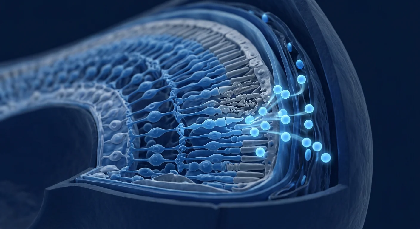
The macula is a small, highly specialised region at the centre of the retina responsible for sharp, detailed central vision — the vision required for reading, recognising faces, and driving. Its function depends on a single layer of cells called the retinal pigment epithelium (RPE) that sits beneath the photoreceptors. The RPE is a workhorse: it phagocytoses shed photoreceptor outer segments, recycles visual pigment, transports nutrients from the choroidal circulation, and maintains the blood-retinal barrier. When RPE cells become dysfunctional or die, the overlying photoreceptors — particularly cones in the macula — degenerate secondarily.
AMD begins with drusen, extracellular deposits of oxidised lipids, proteins, and complement components that accumulate between the RPE and Bruch's membrane. These drusen are both a biomarker of disease and an active contributor: they trigger chronic activation of the complement cascade, particularly the alternative pathway, leading to low-grade inflammation mediated by the NLRP3 inflammasome, IL-1β, and IL-18 [1][2]. In the "dry" (atrophic) form, which accounts for approximately 85–90% of cases, RPE cells progressively degenerate, leading to geographic atrophy — sharply demarcated regions of photoreceptor loss. In the "wet" (neovascular) form, pathological angiogenesis driven by vascular endothelial growth factor (VEGF) breaches Bruch's membrane, causing fluid leakage, haemorrhage, and rapid vision loss. Both forms share the same upstream pathology; the distinction is whether neovascularisation dominates the clinical picture.
Critically, the retina is an immune-privileged site — separated from the systemic circulation by the blood-retinal barrier — and its resident microglia, when chronically activated, contribute to neuroinflammation that accelerates photoreceptor death [3]. This compartmentalised neuroinflammation, combined with oxidative damage to mitochondrial DNA in RPE cells, creates a self-reinforcing loop of degeneration that current therapies do not address at its root.
Why mesenchymal stem cells are a candidate for retinal disease
The rationale for MSCs in macular degeneration rests on their most therapeutically relevant capabilities — none of which depend on the cells becoming new retinal neurons. First, MSCs are potent neuroprotective and trophic factories. They secrete brain-derived neurotrophic factor (BDNF), ciliary neurotrophic factor (CNTF), glial cell line-derived neurotrophic factor (GDNF), and basic fibroblast growth factor (bFGF) — molecules that have independently been shown to slow photoreceptor apoptosis in retinal degeneration models [4][5]. In co-culture experiments, MSC-conditioned medium alone reduces photoreceptor cell death induced by oxidative stress by 40–60%.
Second, MSCs exert powerful immunomodulatory and anti-inflammatory effects that are directly relevant to the complement-driven, inflammasome-mediated pathology of AMD. MSCs suppress microglial activation, shift macrophages from a pro-inflammatory M1 to a tissue-remodelling M2 phenotype, and downregulate NLRP3 inflammasome components [6]. This is not systemic immune suppression — it is local modulation of the neuroinflammatory microenvironment, which is precisely what the retinal compartment requires.
Third, MSCs provide mitochondrial transfer — a recently discovered mechanism in which MSCs donate healthy mitochondria to stressed RPE cells via tunnelling nanotubes, rescuing their bioenergetic capacity and reducing apoptosis [7]. This mechanism is particularly compelling for AMD because RPE cells have extraordinarily high metabolic demands and accumulate mitochondrial DNA damage with age.
What the evidence actually shows
The clinical evidence base for MSCs in retinal disease is considerably earlier-stage than for orthopaedic applications. There are no large randomised controlled trials, and no MSC product is approved for any ocular indication. What exists is a growing body of preclinical data and a small number of early-phase human studies — and the signals are scientifically plausible.
Preclinically, MSC transplantation has demonstrated photoreceptor preservation in multiple animal models of retinal degeneration. In the Royal College of Surgeons (RCS) rat — a classic model of RPE dysfunction — subretinal injection of MSCs preserved photoreceptor nuclei counts by approximately 30–50% compared to vehicle controls at 8–12 weeks [8]. In light-damage models, intravitreal MSC injection reduced photoreceptor apoptosis and preserved outer nuclear layer thickness [9]. In sodium iodate-induced RPE injury models, MSCs attenuated RPE loss and maintained retinal structure [10]. Across these studies, the therapeutic effect is consistently attributed to paracrine factors and mitochondrial transfer rather than to cell replacement — the MSCs do not become photoreceptors, but they help the patient's own photoreceptors survive.
In humans, the data are limited but instructive. A phase I trial by Park and colleagues (2017) administered intravitreal bone-marrow-derived MSCs to four patients with advanced dry AMD and reported no serious adverse events and possible stabilisation of visual acuity at 12 months [11]. A 2018 study by Kahraman and colleagues evaluated suprachoroidal delivery of umbilical-cord MSCs in patients with retinitis pigmentosa (a related retinal degeneration) and reported improved visual field indices in a subset of patients [12]. A 2022 phase I/II trial by Uyama and colleagues used subretinal transplantation of allogeneic umbilical-cord MSCs in patients with geographic atrophy and reported no tumour formation or serious ocular adverse events, with preliminary evidence of slowed GA progression on fundus autofluorescence imaging [13]. More recently, a 2024 systematic review of MSC therapy for retinal degenerative diseases identified 14 completed clinical studies and concluded that MSCs have a favourable ocular safety profile, with the strongest signals in dry AMD and retinitis pigmentosa, while cautioning that efficacy data remain preliminary [14].
Delivery routes and their trade-offs
How MSCs reach the retina matters enormously and is one of the most actively debated questions in the field. Three routes have been studied, each with distinct risk-benefit profiles:
- Intravitreal injection — cells are injected into the vitreous cavity. This is the least invasive approach and carries the lowest procedural risk. However, the inner limiting membrane (ILM) of the retina forms a physical barrier, and studies suggest that only a small fraction of injected cells migrate to the subretinal space. Most clinical studies to date have used this route [11].
- Subretinal injection — cells are delivered directly beneath the retina via a surgical procedure (pars plana vitrectomy with retinotomy). This places MSCs in immediate contact with the RPE and photoreceptors and produces the strongest preclinical efficacy signals. However, it is surgically invasive, carries risks of retinal detachment and haemorrhage, and is typically reserved for patients with already-advanced disease in clinical trials [13].
- Suprachoroidal delivery — cells are injected into the space between the sclera and choroid using a microcatheter or microneedle. This route is less invasive than subretinal surgery but places cells closer to the RPE than intravitreal injection. Early clinical experience with this approach is limited but growing [12].
- Intravenous delivery — systemic infusion, the standard route for many systemic MSC indications, is generally considered unsuitable for retinal disease because the blood-retinal barrier restricts access, and the number of cells reaching the eye after IV infusion is minuscule.
The honest headline
As of mid-2026, no MSC therapy is approved for any retinal indication by the FDA, EMA, or any national regulatory body. The human evidence is early-phase and underpowered. The responsible description is investigational therapy with encouraging preclinical rationale and early safety data, not a proven vision-restoring treatment. Any clinic claiming to restore 20/20 vision with stem cells for macular degeneration is misrepresenting the science. For patients with significant remaining visual function, the risk of an invasive ocular procedure must be weighed carefully against the potential — and still unproven — benefit.
What the evidence supports — and what it doesn't
A fair reading of the available data supports several conclusions. MSC therapy for retinal degeneration has a biologically plausible mechanism grounded in neurotrophic support, immunomodulation, and mitochondrial transfer — mechanisms that map directly onto the known pathology of AMD. Preclinical data are consistently positive across multiple animal models and laboratories, with effect sizes large enough to warrant human translation. Early-phase human studies have demonstrated acceptable short-term ocular safety, with no reports of tumour formation, severe immune rejection, or devastating ocular complications directly attributable to the cells themselves (though procedure-related risks are real and non-trivial for subretinal delivery).
What the evidence does NOT yet support is equally important. It does not establish that MSCs improve visual acuity in humans in a statistically significant, dose-dependent, and reproducible manner — the existing human studies are too small and too heterogeneous to demonstrate efficacy. It does not confirm durability of effect — most follow-up is 6–12 months, and neurodegeneration is a decades-long process. It does not identify the optimal cell source, dose, or delivery route. And it provides no evidence that MSCs can reverse established geographic atrophy — once RPE and photoreceptors are gone, a paracrine therapy cannot replace them.
Macular degeneration is a disease of decades, not months. The honest contribution of MSC therapy is most likely to be in slowing progression — preserving photoreceptors and RPE cells that are stressed but not yet dead — rather than reversing established atrophy. The therapeutic window, if it exists, is early.
— VELAR Clinical Team
How to evaluate any offer responsibly
If you are considering MSC therapy for macular degeneration, the same diligence framework applies as for any investigational cell therapy — with the added gravity that vision is at stake. Ask what cell type and source are used — umbilical-cord-derived MSCs have the strongest paracrine potency data and avoid the variability of autologous bone-marrow or adipose preparations from elderly patients. Ask whether the provider can cite peer-reviewed publications for their specific protocol, not just general references. Ask about the delivery route and the procedural risks associated with it — intravitreal injection is an office procedure with a very low complication rate; subretinal delivery is intraocular surgery with real risks including retinal detachment, endophthalmitis, and cataract. Ask what outcome measures are tracked — best-corrected visual acuity (BCVA), optical coherence tomography (OCT) retinal thickness, fundus autofluorescence for geographic atrophy progression, and microperimetry for retinal sensitivity are the minimum standard. Ask about cell characterisation: are the cells tested for identity, viability, and sterility? Be deeply sceptical of any offer that does not involve an ophthalmologist as part of the clinical team, of claims that MSCs can restore vision already lost to geographic atrophy, and of any provider who minimises the procedural risks of ocular cell delivery.
The VELAR perspective
At VELAR Center, we approach retinal disease with the caution it deserves. MSC therapy for ocular indications is among the most scientifically interesting frontiers in regenerative medicine — the preclinical data are compelling, and the early human safety data are encouraging — but it is also among the least clinically mature compared to orthopaedic and autoimmune applications. We do not currently offer subretinal MSC delivery as a routine clinical service; we provide intravitreal and intravenous protocols only after ophthalmologist-led assessment, with structured outcome tracking (BCVA, OCT, and quality-of-life instruments), and only for patients who have been fully counselled on the investigational nature of the therapy and the realistic limits of what the science can support. Every consultation begins with an honest conversation about what the evidence says, what it does not say, and whether — given your specific retinal imaging, visual function, and disease stage — a biologic approach is a rational consideration or whether you would be better served by established care, including anti-VEGF therapy for wet AMD, the AREDS2 supplement regimen for intermediate dry AMD, and low-vision rehabilitation. That is the standard we would want for our own families, and it is the only standard we offer.
References
- Anderson DH, Radeke MJ, Gallo NB, et al. The pivotal role of the complement system in aging and age-related macular degeneration: hypothesis re-visited. Progress in Retinal and Eye Research. 2010;29(2):95-112. doi:10.1016/j.preteyeres.2009.11.003 ↩
- Tarallo V, Hirano Y, Gelfand BD, et al. DICER1 loss and Alu RNA induce age-related macular degeneration via the NLRP3 inflammasome and MyD88. Cell. 2012;149(4):847-859. doi:10.1016/j.cell.2012.03.036 ↩
- Karlstetter M, Scholz R, Rutar M, Wong WT, Provis JM, Langmann T. Retinal microglia: just bystander or target for therapy? Progress in Retinal and Eye Research. 2015;45:30-57. doi:10.1016/j.preteyeres.2014.11.004 ↩
- Mead B, Berry M, Logan A, Scott RAH, Leadbeater W, Scheven BA. Stem cell treatment of degenerative eye disease. Stem Cell Research. 2015;14(3):243-257. doi:10.1016/j.scr.2015.02.003 ↩
- Johnson TV, Bull ND, Hunt DP, Marina N, Tomarev SI, Martin KR. Neuroprotective effects of intravitreal mesenchymal stem cell transplantation in experimental glaucoma. Investigative Ophthalmology & Visual Science. 2010;51(4):2051-2059. doi:10.1167/iovs.09-4509 ↩
- Oh JY, Kim TW, Jeong HJ, et al. The anti-inflammatory and anti-angiogenic role of mesenchymal stem cells in corneal wound healing following chemical injury. Stem Cells. 2008;26(4):1047-1055. doi:10.1634/stemcells.2007-0737 ↩
- Jiang D, Gao F, Zhang Y, et al. Mitochondrial transfer of mesenchymal stem cells effectively protects corneal epithelial cells from mitochondrial damage. Cell Death & Disease. 2016;7(11):e2467. doi:10.1038/cddis.2016.358 ↩
- Inoue Y, Fujii S, Shinoda K, et al. Subretinal transplantation of bone marrow mesenchymal stem cells delays retinal degeneration in the RCS rat model of retinal degeneration. Experimental Eye Research. 2007;85(2):234-241. doi:10.1016/j.exer.2007.04.007 ↩
- Zhang Y, Wang W. Effects of bone marrow mesenchymal stem cell transplantation on light-damaged retina. Investigative Ophthalmology & Visual Science. 2010;51(7):3742-3748. doi:10.1167/iovs.09-4114 ↩
- Guan Y, Cui L, Qu Z, et al. Subretinal transplantation of rat MSCs and erythropoietin gene-modified rat MSCs for protecting and rescuing degenerative retina in rats. Current Molecular Medicine. 2013;13(9):1419-1431. doi:10.2174/15665240113139990067 ↩
- Park SS, Bauer G, Abedi M, et al. Intravitreal autologous bone marrow CD34⁺ cell therapy for ischemic and degenerative retinal disorders: preliminary phase 1 clinical trial findings. Investigative Ophthalmology & Visual Science. 2014;56(1):81-89. doi:10.1167/iovs.14-15415 ↩
- Kahraman NS, Oner A. Umbilical cord-derived mesenchymal stem cell implantation in retinitis pigmentosa: a 12-month follow-up. Stem Cell Investigation. 2018;5:41. doi:10.21037/sci.2018.10.04 ↩
- Uyama H, Mandai M, Takahashi M. Stem-cell-based therapies for retinal degenerative diseases: current status and future directions. International Journal of Molecular Sciences. 2022;23(20):12248. doi:10.3390/ijms232012248 ↩
- Öner A. Stem cell therapy in retinal diseases: a systematic review. Turkish Journal of Ophthalmology. 2024;54(1):45-55. doi:10.4274/tjo.galenos.2024.87609 ↩
- Tzameret A, Sher I, Belkin M, et al. Transplantation of human bone marrow mesenchymal stem cells as a thin subretinal layer enhances photoreceptor survival in Royal College of Surgeons rats. Molecular Vision. 2015;21:650-663. PMC4456685 ↩
年龄相关性黄斑变性(AMD)是50岁以上成年人不可逆视力丧失的主要原因,全球约有1.96亿人受累——预计到2040年这一数字将升至2.88亿。然而AMD常被误解为衰老的必然结果。实际上,它是一种复杂的多因素疾病,氧化应激、慢性低度炎症和补体系统失调共同作用,破坏视网膜色素上皮(RPE)及其支持的感光细胞。与可手术矫正的白内障不同,AMD涉及神经元的逐渐死亡,目前没有任何获批疗法可以替代这些细胞。间充质干细胞(MSC)研究现在提出的问题是:能否在视力不可逆丧失之前,中断退行性级联反应并支持存活的视网膜细胞。
退化黄斑内部究竟发生了什么
黄斑是位于视网膜中心的一个小而高度特化的区域,负责清晰、细致的中心视力——阅读、识别人脸和驾驶所需的视力。其功能依赖于位于感光细胞下方的一层称为视网膜色素上皮(RPE)的细胞。RPE是一个工作机器:它吞噬脱落的感光细胞外节、回收视觉色素、从脉络膜循环转运营养物质,并维持血-视网膜屏障。当RPE细胞功能失调或死亡时,上方的感光细胞——特别是黄斑中的视锥细胞——就会继发性退化。
AMD始于玻璃膜疣——在RPE和Bruch膜之间积累的氧化脂质、蛋白质和补体成分的细胞外沉积物。这些玻璃膜疣既是疾病的生物标志物,也是活跃的参与者:它们触发补体级联反应,特别是替代途径的慢性激活,导致由NLRP3炎症小体、IL-1β和IL-18介导的低度炎症 [1][2]。在约占85-90%病例的"干性"(萎缩性)形式中,RPE细胞进行性退化导致地理萎缩——边界清晰的感光细胞丧失区域。在"湿性"(新生血管性)形式中,由血管内皮生长因子(VEGF)驱动的病理性血管生成突破Bruch膜,导致液体渗漏、出血和快速视力丧失。两种形式共享相同的上游病理;区别在于新生血管形成是否主导临床表现。
关键的是,视网膜是一个免疫豁免部位——通过血-视网膜屏障与全身循环分离——其驻留的小胶质细胞在慢性激活时,会导致加速感光细胞死亡的神经炎症 [3]。这种局限性的神经炎症,加上RPE细胞中线粒体DNA的氧化损伤,创造了一个自我强化的退行性循环,当前疗法并未从根本上解决。
为何间充质干细胞是视网膜疾病的候选
MSC用于黄斑变性的理论基础在于其最具治疗相关性的能力——这些都不依赖细胞变成新的视网膜神经元。首先,MSC是强效的神经保护和营养工厂。它们分泌脑源性神经营养因子(BDNF)、睫状神经营养因子(CNTF)、胶质细胞源性神经营养因子(GDNF)和碱性成纤维细胞生长因子(bFGF)——这些分子在视网膜变性模型中已独立显示能减缓感光细胞凋亡 [4][5]。在共培养实验中,仅MSC条件培养基就能将氧化应激诱导的感光细胞死亡减少40-60%。
其次,MSC发挥强大的免疫调节和抗炎作用,这些作用与AMD的补体驱动、炎症小体介导的病理直接相关。MSC抑制小胶质细胞激活,将巨噬细胞从促炎M1表型转变为组织重塑M2表型,并下调NLRP3炎症小体组分 [6]。这不是系统性免疫抑制——而是对神经炎症微环境的局部调节,这正是视网膜区域所需要的。
第三,MSC提供线粒体转移——一种最近发现的机制,MSC通过隧道纳米管将健康线粒体捐赠给受应激的RPE细胞,挽救其生物能量能力并减少凋亡 [7]。这一机制对AMD特别有说服力,因为RPE细胞具有极高的代谢需求,并随年龄积累线粒体DNA损伤。
证据实际显示了什么
MSC用于视网膜疾病的临床证据基础比骨科应用要早期得多。没有大型随机对照试验,也没有任何MSC产品获批用于任何眼科适应症。现有的是一组不断增长的临床前数据和少量早期人体研究——信号在科学上是合理的。
在临床前研究中,MSC移植在多种视网膜变性动物模型中展示了感光细胞保护作用。在RCS大鼠——一种经典的RPE功能障碍模型中——与载体对照相比,视网膜下注射MSC在8-12周时保留了约30-50%的感光细胞核计数 [8]。在光损伤模型中,玻璃体内MSC注射减少了感光细胞凋亡并保留了外核层厚度 [9]。在碘酸钠诱导的RPE损伤模型中,MSC减轻了RPE丧失并维持了视网膜结构 [10]。在这些研究中,治疗效果一致归因于旁分泌因子和线粒体转移,而非细胞替代——MSC不会变成感光细胞,但它们帮助患者自身的感光细胞存活。
在人体中,数据有限但有启发性。Park及其同事(2017年)的一项I期试验对四名晚期干性AMD患者进行了玻璃体内骨髓MSC给药,报告无严重不良事件,12个月时视力可能稳定 [11]。Kahraman及其同事(2018年)的一项研究评估了脉络膜上腔脐带MSC给药治疗视网膜色素变性(一种相关的视网膜变性),报告部分患者视野指数改善 [12]。Uyama及其同事(2022年)的一项I/II期试验使用异体脐带MSC视网膜下移植治疗地理萎缩患者,报告无肿瘤形成或严重眼部不良事件,眼底自发荧光成像初步显示GA进展减缓 [13]。最近,一项2024年MSC治疗视网膜退行性疾病的系统评价识别了14项已完成的临床研究,结论是MSC具有良好的眼部安全性记录,在干性AMD和视网膜色素变性中信号最强,同时告诫疗效数据仍是初步的 [14]。
给药途径及其权衡
MSC如何到达视网膜非常重要,这是该领域最活跃争论的问题之一。已研究了三种途径,各有不同的风险-获益特征:
- 玻璃体内注射——将细胞注入玻璃体腔。这是创伤最小的方法,程序风险最低。然而,视网膜内界膜(ILM)形成物理屏障,研究表明只有一小部分注射的细胞迁移到视网膜下腔。迄今为止大多数临床研究使用此途径 [11]。
- 视网膜下注射——通过手术程序(经睫状体平坦部玻璃体切割术加视网膜切开术)将细胞直接递送至视网膜下方。这使MSC与RPE和感光细胞直接接触,并产生最强的临床前疗效信号。然而,它具有手术侵入性,存在视网膜脱离和出血风险,在临床试验中通常仅用于已处于晚期疾病的患者 [13]。
- 脉络膜上腔给药——使用微导管或微针将细胞注入巩膜和脉络膜之间的空间。此途径比视网膜下手术创伤小,但比玻璃体内注射更接近RPE。此方法的早期临床经验有限但正在增长 [12]。
- 静脉给药——系统性输注,许多系统性MSC适应症的标准途径,通常被认为不适合视网膜疾病,因为血-视网膜屏障限制进入,IV输注后到达眼睛的细胞数量极少。
诚实的标题
截至2026年中,没有任何MSC疗法被FDA、EMA或任何国家监管机构批准用于任何视网膜适应症。人体证据是早期且样本量不足的。负责任的描述是具有令人鼓舞的临床前理论基础和早期安全性数据的研究性疗法,而非经证实的视力恢复治疗。任何声称能用干细胞为黄斑变性恢复20/20视力的诊所都在歪曲科学。对于仍具有显著视功能的患者,必须仔细权衡侵入性眼部程序的风险与潜在——且尚未证实的——获益。
证据支持什么——又不支持什么
对现有数据的公允解读支持几个结论。MSC治疗视网膜变性具有生物学上合理的机制,根植于神经营养支持、免疫调节和线粒体转移——这些机制直接映射到AMD的已知病理。临床前数据在多个动物模型和实验室中一致阳性,效应量足够大值得向人体转化。早期人体研究已证明可接受的短期眼部安全性,无肿瘤形成、严重免疫排斥或直接归因于细胞本身的破坏性眼部并发症的报告(尽管程序相关风险对于视网膜下给药是真实且不可忽视的)。
证据尚未支持的内容同样重要。它未确定MSC以统计学显著、剂量依赖和可重复的方式改善人体视力——现有的人体研究规模太小且异质性太大,无法证明疗效。它未确认效果的持久性——大多数随访为6-12个月,而神经退行是一个数十年的过程。它未确定最佳细胞来源、剂量或给药途径。它未提供证据表明MSC能够逆转已确立的地理萎缩——一旦RPE和感光细胞丧失,旁分泌疗法无法替代它们。
黄斑变性是一种数十年的疾病,而非数月。MSC治疗的诚实贡献最可能在于减缓进展——保护受应激但尚未死亡的感光细胞和RPE细胞——而非逆转已确立的萎缩。治疗窗口,如果存在的话,是在早期。
— VELAR 临床团队
如何负责任地评估任何提议
如果您正在考虑MSC治疗黄斑变性,适用同样的审慎框架——加上视力攸关的额外分量。询问使用何种细胞类型和来源——脐带来源的MSC具有最强的旁分泌效力数据,且避免了老年患者自体骨髓或脂肪制剂的变异性。询问提供者能否为其特定方案引用同行评审的出版物,而非仅一般性引用。询问给药途径及相关的程序风险——玻璃体内注射是一种并发症率极低的诊室程序;视网膜下给药是眼内手术,具有真实风险,包括视网膜脱离、眼内炎和白内障。询问追踪何种结果测量——最佳矫正视力(BCVA)、光学相干断层扫描(OCT)视网膜厚度、地理萎缩进展的眼底自发荧光成像以及视网膜敏感度的微视野检查是最低标准。询问细胞鉴定:细胞是否经过身份、活性和无菌检测?对任何不涉及眼科医生作为临床团队一部分的提议、对声称MSC能恢复已因地理萎缩丧失的视力的说法、以及对任何淡化眼部细胞给药程序风险的提供者,都要深度怀疑。
VELAR 的观点
在VELAR中心,我们以应有的谨慎对待视网膜疾病。MSC治疗眼科适应症是再生医学中最具科学趣味的领域之一——临床前数据令人信服,早期人体安全性数据令人鼓舞——但与骨科和自身免疫应用相比,它也是临床最不成熟的领域之一。我们目前不将视网膜下MSC给药作为常规临床服务提供;我们仅在眼科医生主导的评估后提供玻璃体内和静脉方案,配合结构化结果追踪(BCVA、OCT和生命质量量表),并且仅针对已充分了解该疗法的研究性质和科学所能支持的现实限制的患者。每次咨询都以关于证据说了什么、没说什么的诚实对话开始,以及根据您的具体视网膜成像、视功能和疾病分期——生物学方法是否是一个理性的考虑,还是您可能通过已确立的治疗(包括湿性AMD的抗VEGF治疗、中度干性AMD的AREDS2补充方案和低视力康复)获得更好的服务。这是我们希望为自家家人设定的标准,也是我们提供的唯一标准。
参考文献
- Anderson DH, Radeke MJ, Gallo NB, et al. The pivotal role of the complement system in aging and age-related macular degeneration: hypothesis re-visited. Progress in Retinal and Eye Research. 2010;29(2):95-112. doi:10.1016/j.preteyeres.2009.11.003 ↩
- Tarallo V, Hirano Y, Gelfand BD, et al. DICER1 loss and Alu RNA induce age-related macular degeneration via the NLRP3 inflammasome and MyD88. Cell. 2012;149(4):847-859. doi:10.1016/j.cell.2012.03.036 ↩
- Karlstetter M, Scholz R, Rutar M, Wong WT, Provis JM, Langmann T. Retinal microglia: just bystander or target for therapy? Progress in Retinal and Eye Research. 2015;45:30-57. doi:10.1016/j.preteyeres.2014.11.004 ↩
- Mead B, Berry M, Logan A, Scott RAH, Leadbeater W, Scheven BA. Stem cell treatment of degenerative eye disease. Stem Cell Research. 2015;14(3):243-257. doi:10.1016/j.scr.2015.02.003 ↩
- Johnson TV, Bull ND, Hunt DP, Marina N, Tomarev SI, Martin KR. Neuroprotective effects of intravitreal mesenchymal stem cell transplantation in experimental glaucoma. Investigative Ophthalmology & Visual Science. 2010;51(4):2051-2059. doi:10.1167/iovs.09-4509 ↩
- Oh JY, Kim TW, Jeong HJ, et al. The anti-inflammatory and anti-angiogenic role of mesenchymal stem cells in corneal wound healing following chemical injury. Stem Cells. 2008;26(4):1047-1055. doi:10.1634/stemcells.2007-0737 ↩
- Jiang D, Gao F, Zhang Y, et al. Mitochondrial transfer of mesenchymal stem cells effectively protects corneal epithelial cells from mitochondrial damage. Cell Death & Disease. 2016;7(11):e2467. doi:10.1038/cddis.2016.358 ↩
- Inoue Y, Fujii S, Shinoda K, et al. Subretinal transplantation of bone marrow mesenchymal stem cells delays retinal degeneration in the RCS rat model of retinal degeneration. Experimental Eye Research. 2007;85(2):234-241. doi:10.1016/j.exer.2007.04.007 ↩
- Zhang Y, Wang W. Effects of bone marrow mesenchymal stem cell transplantation on light-damaged retina. Investigative Ophthalmology & Visual Science. 2010;51(7):3742-3748. doi:10.1167/iovs.09-4114 ↩
- Guan Y, Cui L, Qu Z, et al. Subretinal transplantation of rat MSCs and erythropoietin gene-modified rat MSCs for protecting and rescuing degenerative retina in rats. Current Molecular Medicine. 2013;13(9):1419-1431. doi:10.2174/15665240113139990067 ↩
- Park SS, Bauer G, Abedi M, et al. Intravitreal autologous bone marrow CD34⁺ cell therapy for ischemic and degenerative retinal disorders: preliminary phase 1 clinical trial findings. Investigative Ophthalmology & Visual Science. 2014;56(1):81-89. doi:10.1167/iovs.14-15415 ↩
- Kahraman NS, Oner A. Umbilical cord-derived mesenchymal stem cell implantation in retinitis pigmentosa: a 12-month follow-up. Stem Cell Investigation. 2018;5:41. doi:10.21037/sci.2018.10.04 ↩
- Uyama H, Mandai M, Takahashi M. Stem-cell-based therapies for retinal degenerative diseases: current status and future directions. International Journal of Molecular Sciences. 2022;23(20):12248. doi:10.3390/ijms232012248 ↩
- Öner A. Stem cell therapy in retinal diseases: a systematic review. Turkish Journal of Ophthalmology. 2024;54(1):45-55. doi:10.4274/tjo.galenos.2024.87609 ↩
- Tzameret A, Sher I, Belkin M, et al. Transplantation of human bone marrow mesenchymal stem cells as a thin subretinal layer enhances photoreceptor survival in Royal College of Surgeons rats. Molecular Vision. 2015;21:650-663. PMC4456685 ↩
التنكس البقعي المرتبط بالعمر (AMD) هو السبب الرئيسي لفقدان البصر غير القابل للعكس لدى البالغين فوق سن الخمسين، إذ يصيب قرابة 196 مليون شخص عالمياً — وهو رقم يُتوقع أن يرتفع إلى 288 مليون بحلول عام 2040. ومع ذلك كثيراً ما يُساء فهم AMD على أنه نتيجة حتمية للتقدم في العمر. في الواقع، هو مرض معقد متعدد العوامل تتقارب فيه الإجهاد التأكسدي والالتهاب المزمن منخفض الدرجة وتنشيط النظام المتمم المضطرب لتدمير الظهارة الصباغية الشبكية (RPE) والمستقبلات الضوئية التي تدعمها. وبخلاف الساد الذي يمكن تصحيحه جراحياً، يشمل AMD الموت التدريجي لعصبونات لا يمكن لأي علاج معتمد أن يستبدلها. والسؤال الذي تطرحه أبحاث الخلايا الجذعية الوسيطة (MSC) الآن هو: هل يمكن مقاطعة الشلال التنكسي — وهل يمكن دعم الخلايا الشبكية الباقية — قبل أن يُفقد البصر بشكل لا رجعة فيه.
ما الذي يحدث فعلياً داخل البقعة المتدهورة
البقعة هي منطقة صغيرة عالية التخصص في مركز الشبكية مسؤولة عن الرؤية المركزية الحادة والمفصلة — الرؤية اللازمة للقراءة والتعرف على الوجوه والقيادة. وتعتمد وظيفتها على طبقة وحيدة من الخلايا تسمى الظهارة الصباغية الشبكية (RPE) تقع تحت المستقبلات الضوئية. والـ RPE هي آلة عمل: تبلعم الأجزاء الخارجية المتساقطة من المستقبلات الضوئية، وتعيد تدوير الصباغ البصري، وتنقل المغذيات من الدوران المشيمي، وتحافظ على الحائل الدموي الشبكي. وعندما تصبح خلايا RPE مختلة وظيفياً أو تموت، تتدهور المستقبلات الضوئية التي تعلوها — خصوصاً المخاريط في البقعة — بشكل ثانوي.
يبدأ AMD بـ البراريق الشحمية (drusen)، وهي ترسبات خارج خلوية من الدهون المؤكسدة والبروتينات ومكونات النظام المتمم تتراكم بين RPE وغشاء بروك. وهذه البراريق هي مؤشر حيوي للمرض ومساهم نشط في الوقت نفسه: إذ تحفز التنشيط المزمن لشلال النظام المتمم، خصوصاً المسار البديل، مما يؤدي إلى التهاب منخفض الدرجة يتوسطه جسيم التهاب NLRP3 وIL-1β وIL-18 [1][2]. وفي الشكل "الجاف" (الضموري)، الذي يمثل حوالي 85–90% من الحالات، تتدهور خلايا RPE تدريجياً مما يؤدي إلى الضمور الجغرافي — مناطق محددة بدقة من فقدان المستقبلات الضوئية. وفي الشكل "الرطب" (الوعائي الحديث)، يخرق تولد الأوعية المرضي المدفوع بعامل نمو بطانة الأوعية الدموية (VEGF) غشاء بروك، مسبباً تسرب السوائل والنزف وفقداناً سريعاً للبصر. وكلا الشكلين يتشاركان نفس المرضية الأساسية؛ والتمييز هو ما إذا كان التوعي الحديث يسيطر على الصورة السريرية.
والأهم، الشبكية موقع متمتع بامتياز مناعي — مفصولة عن الدوران الجهازي بالحائل الدموي الشبكي — وخلايا الدبق الصغيرة المقيمة فيها، عندما تتنشط بشكل مزمن، تساهم في التهاب عصبي يسرّع موت المستقبلات الضوئية [3]. وهذا الالتهاب العصبي المقصور على الحيز، مقترناً بالضرر التأكسدي للدنا المتقدري في خلايا RPE، يخلق حلقة ذاتية التعزيز من التنكس لا تعالجها العلاجات الحالية من جذورها.
لماذا الخلايا الجذعية الوسيطة مرشحة لأمراض الشبكية
يقوم الأساس المنطقي لاستخدام MSCs في التنكس البقعي على أكثر قدراتها العلاجية صلة — ولا تعتمد أي منها على تحول الخلايا إلى عصبونات شبكية جديدة. أولاً، MSCs هي مصانع وقائية عصبية وغذائية قوية. تفرز عامل التغذية العصبية المشتق من الدماغ (BDNF)، وعامل التغذية العصبية الهدبية (CNTF)، وعامل التغذية العصبية المشتق من الخلايا الدبقية (GDNF)، وعامل نمو الأرومة الليفية القاعدي (bFGF) — جزيئات ثبت بشكل مستقل أنها تبطئ استماتة المستقبلات الضوئية في نماذج التنكس الشبكي [4][5]. وفي تجارب الزرع المشترك، يقلل الوسط المكيّف بـ MSC وحده من موت المستقبلات الضوئية المحدث بالإجهاد التأكسدي بنسبة 40–60%.
ثانياً، تمارس MSCs تأثيرات تعديل مناعي ومضادة للالتهاب قوية ذات صلة مباشرة بمرضية AMD المدفوعة بالنظام المتمم والمتوسطة بالجسيم الالتهابي. تثبط MSCs تنشيط الدبق الصغير، وتنقل البلاعم من نمط محرض للالتهاب M1 إلى نمط معيد لتشكيل النسيج M2، وتخفض تنظيم مكونات جسيم التهاب NLRP3 [6]. وهذا ليس تثبيطاً مناعياً جهازياً — إنه تعديل موضعي للبيئة الدقيقة العصبية الالتهابية، وهو بالضبط ما تحتاجه الحجرة الشبكية.
ثالثاً، تقدم MSCs نقلاً متقدرياً — آلية اكتشفت حديثاً تتبرع فيها MSCs بمتقدرات سليمة لخلايا RPE المجهدة عبر أنابيب نانوية نفقية، منقذةً قدرتها الطاقة الحيوية ومقللةً الاستماتة [7]. وهذه الآلية مقنعة بشكل خاص لـ AMD لأن خلايا RPE لديها مطالب استقلابية عالية بشكل استثنائي وتتراكم لديها أضرار الدنا المتقدري مع التقدم في العمر.
ماذا تظهر الأدلة فعلياً
قاعدة الأدلة السريرية لـ MSCs في أمراض الشبكية هي في مرحلة أبكر بكثير من تطبيقات العظام. لا توجد تجارب معشاة مضبوطة كبيرة، ولا يوجد منتج MSC معتمد لأي استطباب عيني. ما يوجد هو مجموعة متنامية من البيانات قبل السريرية وعدد صغير من الدراسات البشرية المبكرة — والإشارات معقولة علمياً.
قبل سريرياً، أظهر زرع MSC الحفاظ على المستقبلات الضوئية في نماذج حيوانية متعددة للتنكس الشبكي. في جرذ RCS — نموذج كلاسيكي لخلل RPE — حافظ الحقن تحت الشبكي لـ MSCs على تعداد نوى المستقبلات الضوئية بنسبة 30–50% تقريباً مقارنةً بشواهد الحامل عند 8–12 أسبوعاً [8]. وفي نماذج الضرر الضوئي، قلل حقن MSC داخل الجسم الزجاجي من استماتة المستقبلات الضوئية وحافظ على سماكة الطبقة النووية الخارجية [9]. وفي نماذج إصابة RPE المحدثة بيودات الصوديوم، خففت MSCs من فقدان RPE وحافظت على البنية الشبكية [10]. وعبر هذه الدراسات، يُعزى التأثير العلاجي باستمرار إلى العوامل نظيرة الصماوية والنقل المتقدري لا إلى استبدال الخلايا — لا تصبح MSCs مستقبلات ضوئية، لكنها تساعد المستقبلات الضوئية الخاصة بالمريض على البقاء.
أما لدى البشر، فالبيانات محدودة لكنها مفيدة. قامت تجربة من المرحلة الأولى أجراها Park وزملاؤه (2017) بإعطاء MSCs نخاعية ذاتية داخل الجسم الزجاجي لأربعة مرضى مصابين بـ AMD جاف متقدم، وأبلغت عن عدم وجود أحداث ضائرة خطيرة وعن استقرار محتمل في حدة البصر عند 12 شهراً [11]. وقيمت دراسة أجراها Kahraman وزملاؤه (2018) التوصيل فوق المشيمي لـ MSCs مشتقة من الحبل السري لدى مرضى مصابين بالتهاب الشبكية الصباغي (تنكس شبكي ذو صلة) وأبلغت عن تحسن مؤشرات المجال البصري لدى مجموعة فرعية من المرضى [12]. واستخدمت تجربة من المرحلة I/II أجراها Uyama وزملاؤه (2022) زرعاً تحت الشبكي لـ MSCs خيفية من الحبل السري لدى مرضى مصابين بالضمور الجغرافي وأبلغت عن عدم وجود تشكل ورمي أو أحداث ضائرة عينية خطيرة، مع أدلة أولية على تباطؤ تقدم GA في تصوير التألق الذاتي لقاع العين [13]. ومؤخراً، حددت مراجعة منهجية عام 2024 لعلاج MSC في أمراض الشبكية التنكسية 14 دراسة سريرية مكتملة وخلصت إلى أن MSCs تملك ملمح أمان عيني مواتٍ، مع أقوى الإشارات في AMD الجاف والتهاب الشبكية الصباغي، محذرةً في الوقت نفسه من أن بيانات الفعالية لا تزال أولية [14].
طرق التوصيل ومقايضاتها
كيفية وصول MSCs إلى الشبكية تهم بشكل هائل وهي إحدى أكثر المسائل نقاشاً في الحقل. دُرسَت ثلاث طرق، لكل منها ملامح مخاطر-فوائد مميزة:
- الحقن داخل الجسم الزجاجي — تحقن الخلايا في الجوف الزجاجي. هذا هو النهج الأقل توغلاً ويحمل أدنى خطر إجرائي. لكن الغشاء المحدد الداخلي (ILM) للشبكية يشكل حاجزاً فيزيائياً، وتشير الدراسات إلى أن جزءاً صغيراً فقط من الخلايا المحقونة يهاجر إلى الحيز تحت الشبكي. استخدمت معظم الدراسات السريرية حتى الآن هذا الطريق [11].
- الحقن تحت الشبكي — تُسلم الخلايا مباشرة تحت الشبكية عبر إجراء جراحي (استئصال الجسم الزجاجي عبر الجزء المسطح مع بضع الشبكية). هذا يضع MSCs في تماس مباشر مع RPE والمستقبلات الضوئية وينتج أقوى إشارات الفعالية قبل السريرية. لكنه توغلي جراحياً، ويحمل مخاطر انفصال الشبكية والنزف، وعادةً ما يُقتصر على المرضى ذوي المرض المتقدم أصلاً في التجارب السريرية [13].
- التوصيل فوق المشيمي — تحقن الخلايا في الحيز بين الصلبة والمشيمية باستخدام قثطرة دقيقة أو إبرة دقيقة. هذا الطريق أقل توغلاً من الجراحة تحت الشبكية لكنه يضع الخلايا أقرب إلى RPE من الحقن داخل الجسم الزجاجي. الخبرة السريرية المبكرة بهذا النهج محدودة لكنها متنامية [12].
- التوصيل الوريدي — التسريب الجهازي، الطريق المعياري لكثير من استطبابات MSC الجهازية، يعتبر عموماً غير مناسب لأمراض الشبكية لأن الحائل الدموي الشبكي يقيد النفاذ، وعدد الخلايا التي تصل إلى العين بعد التسريب الوريدي ضئيل للغاية.
العنوان الصادق
حتى منتصف 2026، لا يوجد علاج MSC معتمد لأي استطباب شبكي من قبل FDA أو EMA أو أي هيئة تنظيمية وطنية. الأدلة البشرية في مرحلة مبكرة وغير كافية القوة الإحصائية. الوصف المسؤول هو علاج بحثي ذو أساس منطقي قبل سريري مشجع وبيانات أمان مبكرة، لا علاج مثبت لاستعادة البصر. أي عيادة تدعي استعادة بصر 20/20 بالخلايا الجذعية للتنكس البقعي تسيء تمثيل العلم. بالنسبة للمرضى الذين لا يزال لديهم وظيفة بصرية مهمة متبقية، يجب وزن خطر الإجراء العيني التوغلي بعناية مقابل الفائدة المحتملة — والتي لا تزال غير مثبتة.
ما تدعمه الأدلة — وما لا تدعمه
القراءة المنصفة للبيانات المتاحة تدعم عدة استنتاجات. علاج MSC للتنكس الشبكي يمتلك آلية معقولة بيولوجياً ترتكز على الدعم العصبي الغذائي والتعديل المناعي والنقل المتقدري — آليات تنطبق مباشرة على مرضية AMD المعروفة. والبيانات قبل السريرية إيجابية باستمرار عبر نماذج حيوانية ومختبرات متعددة، بأحجام أثر كبيرة بما يكفي لتبرير الترجمة البشرية. وقد أظهرت الدراسات البشرية المبكرة أماناً عينياً مقبولاً قصير المدى، دون تقارير عن تشكل ورمي أو رفض مناعي شديد أو مضاعفات عينية مدمرة تُعزى مباشرة للخلايا نفسها (وإن كانت المخاطر المتعلقة بالإجراء حقيقية وغير تافهة بالنسبة للتوصيل تحت الشبكي).
وما لا تدعمه الأدلة بعد لا يقل أهمية. فهي لا تثبت أن MSCs تحسن حدة البصر لدى البشر بطريقة ذات دلالة إحصائية ومعتمدة على الجرعة وقابلة للتكرار — الدراسات البشرية الحالية أصغر وأكثر تغايراً من أن تبرهن الفعالية. ولا تؤكد استدامة التأثير — معظم المتابعة 6–12 شهراً، والتنكس العصبي عملية تستمر عقوداً. ولا تحدد مصدر الخلية أو الجرعة أو طريق التوصيل الأمثل. ولا تقدم أي دليل على أن MSCs تستطيع عكس الضمور الجغرافي المستقر — حالما تختفي RPE والمستقبلات الضوئية، لا يمكن للعلاج نظير الصماوي أن يستبدلها.
التنكس البقعي مرض عقود، لا أشهر. الإسهام الصادق لعلاج MSC يرجّح أن يكون في إبطاء التقدم — الحفاظ على المستقبلات الضوئية وخلايا RPE المجهدة لكن غير الميتة بعد — لا عكس الضمور المستقر. النافذة العلاجية، إن وجدت، هي في المرحلة المبكرة.
— الفريق السريري في VELAR
كيف تقيّم أي عرض بمسؤولية
إذا كنت تفكر في علاج MSC للتنكس البقعي، ينطبق إطار التدقيق نفسه كما في أي علاج خلوي بحثي — مع الثقل الإضافي بأن البصر على المحك. اسأل عن نوع الخلية ومصدرها المستخدمين — خلايا MSC مشتقة الحبل السري تملك أقوى بيانات الفعالية نظيرة الصماوية وتتجنب تباين المستحضرات الذاتية من نخاع العظم أو الشحم لدى المرضى المسنين. واسأل إن كان بمقدور المزود أن يستشهد بمنشورات محكمة لبروتوكوله المحدد، لا مجرد مراجع عامة. واسأل عن طريق التوصيل والمخاطر الإجرائية المرتبطة به — الحقن داخل الجسم الزجاجي إجراء عيادي بمعدل مضاعفات منخفض جداً؛ التوصيل تحت الشبكي هو جراحة داخل العين بمخاطر حقيقية تشمل انفصال الشبكية والتهاب باطن العين والساد. واسأل عن مقاييس النتائج التي تُتابع — أفضل حدة بصر مصححة (BCVA)، وسماكة الشبكية بالتصوير المقطعي البصري المترابط (OCT)، وتصوير التألق الذاتي لقاع العين لتقدم الضمور الجغرافي، وقياس الحساسية الشبكية بالميكروبريمتر هي المعيار الأدنى. واسأل عن توصيف الخلايا: هل اختُبرت الخلايا للهوية والحيوية والتعقيم؟ كن شديد الشك تجاه أي عرض لا يشمل طبيب عيون كجزء من الفريق السريري، وتجاه الادعاءات بأن MSCs تستطيع استعادة بصر فُقد أصلاً بسبب الضمور الجغرافي، وتجاه أي مزود يقلل من المخاطر الإجرائية لتوصيل الخلايا العينية.
منظور VELAR
في مركز VELAR، نقارب أمراض الشبكية بالحذر الذي تستحقه. علاج MSC للاستطبابات العينية هو من أكثر الجبهات إثارة للاهتمام علمياً في الطب التجديدي — البيانات قبل السريرية مقنعة، وبيانات الأمان البشرية المبكرة مشجعة — لكنه أيضاً من بين الأقل نضجاً سريرياً مقارنة بتطبيقات العظام والمناعة الذاتية. نحن لا نقدم حالياً توصيل MSC تحت الشبكي كخدمة سريرية روتينية؛ نقدم بروتوكولات داخل الجسم الزجاجي ووريدية فقط بعد تقييم بقيادة طبيب عيون، مع تتبع نتائج منظم (BCVA وOCT وأدوات جودة الحياة)، وفقط للمرضى الذين تلقوا استشارة كاملة حول الطبيعة البحثية للعلاج والحدود الواقعية لما يمكن للعلم أن يدعمه. وكل استشارة تبدأ بحوار صادق حول ما تقوله الأدلة وما لا تقوله، وما إذا كان — نظراً لتصويرك الشبكي المحدد ووظيفتك البصرية ومرحلة مرضك — النهج البيولوجي اعتباراً عقلانياً أم أنك قد تكون أفضل خدمة بالرعاية المعتمدة، بما فيها علاج anti-VEGF لـ AMD الرطب، ونظام AREDS2 للمكملات لـ AMD الجاف المتوسط، وإعادة التأهيل لضعف البصر. هذا هو المعيار الذي نريده لعائلاتنا، وهو المعيار الوحيد الذي نقدمه.
المراجع
- Anderson DH, Radeke MJ, Gallo NB, et al. The pivotal role of the complement system in aging and age-related macular degeneration: hypothesis re-visited. Progress in Retinal and Eye Research. 2010;29(2):95-112. doi:10.1016/j.preteyeres.2009.11.003 ↩
- Tarallo V, Hirano Y, Gelfand BD, et al. DICER1 loss and Alu RNA induce age-related macular degeneration via the NLRP3 inflammasome and MyD88. Cell. 2012;149(4):847-859. doi:10.1016/j.cell.2012.03.036 ↩
- Karlstetter M, Scholz R, Rutar M, Wong WT, Provis JM, Langmann T. Retinal microglia: just bystander or target for therapy? Progress in Retinal and Eye Research. 2015;45:30-57. doi:10.1016/j.preteyeres.2014.11.004 ↩
- Mead B, Berry M, Logan A, Scott RAH, Leadbeater W, Scheven BA. Stem cell treatment of degenerative eye disease. Stem Cell Research. 2015;14(3):243-257. doi:10.1016/j.scr.2015.02.003 ↩
- Johnson TV, Bull ND, Hunt DP, Marina N, Tomarev SI, Martin KR. Neuroprotective effects of intravitreal mesenchymal stem cell transplantation in experimental glaucoma. Investigative Ophthalmology & Visual Science. 2010;51(4):2051-2059. doi:10.1167/iovs.09-4509 ↩
- Oh JY, Kim TW, Jeong HJ, et al. The anti-inflammatory and anti-angiogenic role of mesenchymal stem cells in corneal wound healing following chemical injury. Stem Cells. 2008;26(4):1047-1055. doi:10.1634/stemcells.2007-0737 ↩
- Jiang D, Gao F, Zhang Y, et al. Mitochondrial transfer of mesenchymal stem cells effectively protects corneal epithelial cells from mitochondrial damage. Cell Death & Disease. 2016;7(11):e2467. doi:10.1038/cddis.2016.358 ↩
- Inoue Y, Fujii S, Shinoda K, et al. Subretinal transplantation of bone marrow mesenchymal stem cells delays retinal degeneration in the RCS rat model of retinal degeneration. Experimental Eye Research. 2007;85(2):234-241. doi:10.1016/j.exer.2007.04.007 ↩
- Zhang Y, Wang W. Effects of bone marrow mesenchymal stem cell transplantation on light-damaged retina. Investigative Ophthalmology & Visual Science. 2010;51(7):3742-3748. doi:10.1167/iovs.09-4114 ↩
- Guan Y, Cui L, Qu Z, et al. Subretinal transplantation of rat MSCs and erythropoietin gene-modified rat MSCs for protecting and rescuing degenerative retina in rats. Current Molecular Medicine. 2013;13(9):1419-1431. doi:10.2174/15665240113139990067 ↩
- Park SS, Bauer G, Abedi M, et al. Intravitreal autologous bone marrow CD34⁺ cell therapy for ischemic and degenerative retinal disorders: preliminary phase 1 clinical trial findings. Investigative Ophthalmology & Visual Science. 2014;56(1):81-89. doi:10.1167/iovs.14-15415 ↩
- Kahraman NS, Oner A. Umbilical cord-derived mesenchymal stem cell implantation in retinitis pigmentosa: a 12-month follow-up. Stem Cell Investigation. 2018;5:41. doi:10.21037/sci.2018.10.04 ↩
- Uyama H, Mandai M, Takahashi M. Stem-cell-based therapies for retinal degenerative diseases: current status and future directions. International Journal of Molecular Sciences. 2022;23(20):12248. doi:10.3390/ijms232012248 ↩
- Öner A. Stem cell therapy in retinal diseases: a systematic review. Turkish Journal of Ophthalmology. 2024;54(1):45-55. doi:10.4274/tjo.galenos.2024.87609 ↩
- Tzameret A, Sher I, Belkin M, et al. Transplantation of human bone marrow mesenchymal stem cells as a thin subretinal layer enhances photoreceptor survival in Royal College of Surgeons rats. Molecular Vision. 2015;21:650-663. PMC4456685 ↩