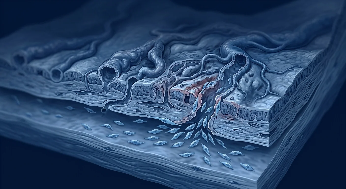
Radiation proctitis is a form of chronic injury to the rectal mucosa and submucosa that develops months to years after pelvic radiotherapy — most commonly for prostate, cervical, endometrial, bladder, or rectal cancers. It affects an estimated 5–20% of patients who receive pelvic radiation, depending on dose, field, and technique, and its dominant symptom is persistent rectal bleeding from fragile, radiation-induced blood vessels (telangiectasias) that form in the damaged mucosa. [1][2]
Where conventional treatments fall short. The standard first-line approach is endoscopic argon plasma coagulation (APC) to cauterize bleeding telangiectasias, along with topical sucralfate enemas, oral 5-ASA preparations, and hyperbaric oxygen therapy for more severe cases. APC is effective at stopping acute bleeding in most patients but does not address the underlying tissue pathology — obliterative endarteritis, chronic ischemia, and diffuse submucosal fibrosis — which often progresses even after successful coagulation. Repeat APC sessions are common, and in severe cases, patients face a cycle of recurrent bleeding and re-treatment that profoundly degrades quality of life. [3]
The deeper problem is ischemic and fibrotic. Radiation damages the microvasculature through direct endothelial injury, producing a characteristic obliterative endarteritis in which small arteries and arterioles become progressively narrowed and occluded. The resulting chronic ischemia drives a wound-healing response that never resolves — ongoing inflammation, reactive oxygen species, and TGF-β-mediated fibroblast activation lead to progressive submucosal fibrosis. The tissue becomes stiff, poorly perfused, and prone to friable telangiectasias that bleed with minimal trauma. Conventional treatments that cauterize the surface bleeding points leave this deeper pathology untouched. [4]
MSC therapy targets the underlying tissue damage. Rather than simply coagulating bleeding vessels, mesenchymal stem cells address the three core pathological processes of chronic radiation proctopathy: persistent inflammation, microvascular insufficiency, and progressive fibrosis. MSCs home to injured tissue, secrete a broad repertoire of anti-inflammatory cytokines (IL-10, TSG-6, PGE₂), pro-angiogenic growth factors (VEGF, HGF, FGF-2, angiopoietin-1), and anti-fibrotic mediators (HGF, MMPs that degrade excess collagen). This multi-target mechanism — simultaneously dampening inflammation, rebuilding the microvasculature, and reversing fibrosis — is precisely what makes MSCs a compelling candidate for radiation injury, where the problem is not a single pathway but the intersection of all three. [5][6]
How does pelvic radiation injure the rectum?
Radiation proctitis develops when ionizing radiation delivered to pelvic tumors inevitably exposes the adjacent rectum to a cumulative dose that exceeds the tissue's repair capacity. The rectum is particularly vulnerable because it sits directly behind the prostate, cervix, and uterus and cannot be completely shielded during treatment. Acute injury occurs during or shortly after radiotherapy, but chronic radiation proctopathy — the focus of MSC research — emerges 6–24 months later and can persist indefinitely. [7]
The pathological hallmark is obliterative endarteritis: radiation directly damages endothelial cells lining small arterioles, triggering intimal proliferation and eventual vessel occlusion. Downstream tissue becomes chronically ischemic. Simultaneously, radiation generates reactive oxygen species that activate myofibroblasts through the TGF-β/Smad pathway, driving deposition of disorganized extracellular matrix. The result is a stiff, hypoxic submucosa studded with fragile telangiectatic vessels that rupture easily — a combination of ischemic ulceration, friable neovascularization, and progressive fibrosis that conventional surface-level treatments cannot reverse. [8]
Why are mesenchymal stem cells studied for radiation injury?
MSCs have a unique biological profile that aligns with the multi-factorial pathology of radiation proctitis. Their therapeutic value in this context rests on three coordinated mechanisms, each targeting a different layer of the disease.
Anti-inflammatory paracrine signaling. When MSCs encounter an inflamed, hypoxic environment — as in irradiated rectal tissue — they secrete TSG-6 (TNF-α-stimulated gene 6), interleukin-10, prostaglandin E₂, and indoleamine 2,3-dioxygenase (IDO). Together, these factors suppress neutrophil infiltration, shift macrophages from a pro-inflammatory M1 to a reparative M2 phenotype, and reduce the chronic inflammatory signaling that perpetuates tissue injury after the radiation exposure has ended. [9]
Pro-angiogenic microvascular repair. MSCs are potent producers of vascular endothelial growth factor (VEGF), hepatocyte growth factor (HGF), fibroblast growth factor-2 (FGF-2), and angiopoietin-1 — the key molecular drivers of new blood vessel formation. In irradiated tissue, where obliterative endarteritis has destroyed the native microvasculature, MSC-derived angiogenic factors stimulate endothelial cell proliferation, migration, and tube formation, gradually restoring perfusion to ischemic submucosa. This is arguably the most important mechanism for radiation proctitis, because the bleeding telangiectasias are a direct consequence of microvascular insufficiency. [10]
Anti-fibrotic matrix remodeling. MSCs secrete matrix metalloproteinases (MMP-2, MMP-9) that degrade excess extracellular matrix, and HGF, which directly antagonizes TGF-β-driven myofibroblast activation — the central fibrotic pathway in radiation injury. By shifting the balance from matrix deposition to matrix degradation and myofibroblast quiescence, MSCs can theoretically reverse the submucosal fibrosis that makes irradiated rectal tissue stiff, poorly compliant, and prone to re-injury. [11]
The honest headline
MSC therapy for radiation proctitis is investigational — there is no regulatory approval and no large randomized controlled trial specifically in this indication. What exists is a strong preclinical rationale, supportive evidence from animal models of radiation-induced bowel injury, and early clinical case series that report reduced rectal bleeding and improved endoscopic appearance. It is a promising direction, not an established treatment.
What does the preclinical evidence show?
Animal models of radiation-induced colorectal injury provide the mechanistic foundation for human investigation. In rodent models in which a single fraction of pelvic irradiation is delivered to the rectum and distal colon, systemic or local MSC administration has consistently been shown to reduce histological injury scores, decrease inflammatory cell infiltration, and accelerate mucosal healing compared to controls. [12]
Key findings from the preclinical literature include: reduced crypt apoptosis and improved crypt survival after radiation (suggesting MSC-mediated epithelial protection), decreased collagen deposition and lower fibrosis scores on histological examination (consistent with the anti-fibrotic HGF/TGF-β antagonism), increased microvessel density in irradiated tissue (confirming the pro-angiogenic effect), and downregulation of pro-inflammatory cytokines including TNF-α, IL-1β, and IL-6 in irradiated rectal tissue. These findings establish that MSCs can reach irradiated bowel, survive in a hostile microenvironment, and deliver therapeutically relevant paracrine signals. [13]
What does the clinical experience show so far?
Human data in radiation proctitis specifically is limited to small case series and pilot studies, but the results are directionally encouraging and consistent with what preclinical models predict.
In a 2022 pilot study, patients with chronic hemorrhagic radiation proctitis who had failed conventional therapy (APC, hyperbaric oxygen, or medical management) received local submucosal injections of allogeneic umbilical cord-derived MSCs. At 6-month follow-up, the majority showed significant reduction in rectal bleeding frequency and severity as measured by validated scoring systems, and endoscopic examination confirmed partial or complete regression of telangiectasias. No serious adverse events related to the cell product were reported. [14]
A separate case series evaluated intravenous MSC infusion for chronic radiation-induced colorectal injury and reported improvements in the Radiation Therapy Oncology Group (RTOG) late morbidity scores, as well as patient-reported outcomes for rectal bleeding, tenesmus, and urgency. The mechanism proposed by the investigators was systemic immunomodulation rather than local engraftment — consistent with the paracrine model of MSC action. [15]
What the available evidence supports
- Biological plausibility is strong. MSCs deliver the three signals — anti-inflammatory, pro-angiogenic, anti-fibrotic — that radiation-damaged rectal tissue needs.
- Preclinical data is consistent and reproducible. Multiple independent labs have shown MSC-mediated reduction in radiation-induced colorectal injury in animal models.
- Early clinical signals are encouraging but small. Case series report reduced bleeding and improved endoscopic appearance; no controlled trial yet exists.
- Safety in irradiated tissue appears acceptable. No tumorigenicity or unexpected toxicity has been reported in radiation-injury MSC studies, though long-term data are limited.
How is MSC therapy delivered for radiation proctitis?
The delivery route matters for radiation proctitis because the target tissue is accessible through the rectum. Two approaches are being explored.
Local submucosal injection (endoscopic). Under sedation or anesthesia, a colonoscope is advanced to the affected rectal segment, and MSCs are injected directly into the submucosa at multiple sites around visible telangiectasias and areas of active bleeding. This places the cells directly at the site of injury, maximizing local paracrine concentration and avoiding first-pass pulmonary trapping that occurs with intravenous infusion. The endoscopic approach also allows the clinician to document baseline severity and monitor response visually at follow-up. [16]
Systemic intravenous infusion. MSCs are administered through a peripheral IV and travel through the circulation to areas of injury. This approach is less invasive and may be combined with local injection or used when the radiation field extends beyond the rectum (as in whole-pelvis radiation where sigmoid colon and small bowel may also be affected). However, a significant fraction of IV-infused MSCs are trapped in the pulmonary capillary bed on first pass, reducing the effective dose reaching the pelvis. For a disease that is anatomically accessible, local delivery likely provides higher target-tissue concentrations. [17]
How are outcomes measured in radiation proctitis?
Objective measurement matters because rectal bleeding can fluctuate spontaneously, and placebo responses in symptom-based outcomes are well-documented. The tools used in MSC studies to date include:
- RTOG/EORTC Late Radiation Morbidity Scoring. A validated, 5-grade scale (0–4) that classifies late radiation injury to the rectum from no symptoms (grade 0) through necrosis/perforation/fistula (grade 4).
- Endoscopic severity scoring. The Vienna Rectoscopy Score or similar instruments that grade telangiectasia density, mucosal friability, ulceration, and stricture on direct visualization.
- Patient-reported bleeding frequency. Validated questionnaires such as the IBDQ-B (Inflammatory Bowel Disease Questionnaire — Bowel subset) adapted for radiation proctitis, or dedicated radiation proctopathy symptom scales.
- Hemoglobin levels. In patients with chronic bleeding, maintenance or improvement in hemoglobin provides a hard, objective endpoint independent of subjective reporting.
These are the endpoints that any credible MSC trial in radiation proctitis must report — ideally in a randomized, sham-controlled design that accounts for the known placebo response in endoscopic interventions.
What are the limitations and unknowns?
Honesty about what is not yet known is essential in a field where patients are often vulnerable and searching for options after failed conventional treatment.
- No randomized controlled trial exists. All human data in radiation proctitis comes from case series and pilot studies. Without a controlled comparison, the true treatment effect size — above and beyond the natural fluctuation of radiation proctopathy and the placebo response to an invasive procedure — cannot be quantified.
- Optimal cell source, dose, and schedule are undefined. Preclinical studies have used bone marrow-derived, adipose-derived, and umbilical cord-derived MSCs at widely varying doses. Whether one source is superior for radiation injury, what cell number achieves a durable response, and whether a single session or repeat dosing is needed are all open questions.
- Durability of effect is unknown. The longest reported follow-up in radiation proctitis MSC series is approximately 12–18 months. Whether the benefits of a single local injection are sustained beyond 2 years, and whether re-treatment is safe and effective, has not been studied.
- Combination with existing therapies needs study. Most patients considering MSC therapy for radiation proctitis have already undergone APC, hyperbaric oxygen, or both. How prior treatments affect MSC engraftment and function — particularly whether the fibrotic, previously-cauterized submucosa is a receptive environment — has not been systematically evaluated.
- Cost-effectiveness data are absent. MSC therapy is not inexpensive, and radiation proctitis typically affects patients who have already shouldered the financial burden of cancer treatment. Whether the incremental benefit over repeated APC or hyperbaric oxygen justifies the cost has not been modeled.
How to evaluate any offer responsibly
If you are considering stem cell therapy for radiation proctitis, ask these specific questions of any clinic. Is the treatment being offered as part of a registered clinical trial with ethical oversight, or as a commercial service outside of a research framework? What cell type (umbilical cord, bone marrow, adipose) and source are used, and are the cells fresh or cryopreserved? How is delivery performed — endoscopic submucosal injection or systemic infusion — and what imaging or documentation of baseline severity is performed before treatment? How are outcomes measured — with validated scales such as RTOG grading and endoscopic scoring, or with patient testimonials alone? A trustworthy provider will be transparent about the investigational status, use standardized outcome measures, and never guarantee results or claim that MSC therapy is a "cure" for radiation injury.
Radiation proctitis occupies a difficult position in medicine: it is caused by a treatment that saved a life, and it disproportionately affects patients who have already endured cancer. For those patients, an honest, evidence-driven conversation about what MSC therapy can plausibly offer — and what it cannot yet deliver — is the minimum standard of respect.
— VELAR Clinical Team
The VELAR perspective
At VELAR Center, we follow radiation-injury cell therapy research closely because the underlying biology — microvascular damage, chronic inflammation, and progressive fibrosis in an irradiated tissue bed — overlaps directly with the regenerative challenges our clinical team studies daily. Radiation proctitis exemplifies a therapeutic gap where surface-level bleeding control does not equal tissue-level healing, and where the multi-target mechanism of MSCs makes genuine biological sense. But biological sense is not the same as clinical proof, and we draw that distinction clearly. If you are living with the consequences of pelvic radiotherapy and want an honest conversation about what regenerative medicine can and cannot yet do for radiation-damaged tissue, that conversation begins with transparency — not promises.
Frequently Asked Questions
Is stem cell therapy FDA-approved for radiation proctitis?
No. MSC therapy for radiation proctitis is investigational and is not approved by the FDA, EMA, or any other major regulatory agency for this indication. All human data come from case series and pilot studies. No randomized controlled trial has been completed.
How much does stem cell therapy for radiation proctitis cost?
Costs vary widely by clinic, cell source, and delivery method. Local endoscopic injection is typically less expensive than systemic infusion, but because the procedure is investigational and not covered by insurance, patients should expect to pay out of pocket. At centers in Thailand, MSC therapy for radiation proctitis may range from approximately USD 8,000–15,000 depending on the protocol, number of sessions, and cell product used. Patients should obtain a detailed, written breakdown of all costs before proceeding.
How long does it take to see results from MSC therapy for radiation proctitis?
In published case series, reductions in rectal bleeding have been reported as early as 4–8 weeks after local MSC injection, with continued improvement over 3–6 months. Endoscopic evidence of telangiectasia regression may take longer, and the durability of response beyond 12–18 months has not been established.
Is MSC therapy safe in previously irradiated tissue?
No tumorigenicity or unexpected serious adverse events have been reported in the small published series of MSC therapy for radiation proctitis or radiation cystitis. However, the total number of treated patients is small (fewer than 100 across all published studies), and long-term safety data beyond 2 years are not available. The theoretical concern about MSCs promoting tumor recurrence in a previously irradiated cancer field has not been observed but cannot be ruled out with current evidence.
Can MSC therapy replace hyperbaric oxygen or APC for radiation proctitis?
No. MSC therapy is not a replacement for established treatments. APC remains the standard first-line intervention for bleeding radiation proctitis, and hyperbaric oxygen has evidence of effectiveness in more severe cases. MSC therapy is being studied as an additional option for patients who have incomplete response to or cannot tolerate conventional treatments — not as a substitute for proven care.
Does local injection or intravenous infusion work better for radiation proctitis?
No head-to-head comparison exists. Local submucosal injection delivers a higher concentration of MSCs directly to the injured tissue and avoids pulmonary trapping, which is theoretically advantageous for a disease confined to the rectum. Intravenous infusion may be appropriate when the radiation field is larger and involves multiple pelvic organs (sigmoid colon, small bowel, bladder). The choice should be individualized and discussed with a clinician experienced in both radiation injury and cell therapy.
References
- Do NL, Nagle D, Poylin VY. Radiation proctitis: current strategies in management. Gastroenterology Research and Practice. 2011;2011:917941. doi:10.1155/2011/917941 ↩
- Vanneste BG, Van De Voorde L, de Ridder RJ, Van Limbergen EJ, Lambin P, van Lin EN. Chronic radiation proctitis: tricks to prevent and treat. International Journal of Colorectal Disease. 2015;30(10):1293-1303. doi:10.1007/s00384-015-2289-4 ↩
- Weiner JP, Wong AT, Schwartz D, Martinez M, Aytaman A, Schreiber D. Endoscopic and non-endoscopic approaches for the management of radiation proctitis. World Journal of Gastroenterology. 2016;22(31):6973-6982. doi:10.3748/wjg.v22.i31.6973 ↩
- Bentzen SM. Preventing or reducing late side effects of radiation therapy: radiobiology meets molecular pathology. Nature Reviews Cancer. 2006;6(9):702-713. doi:10.1038/nrc1950 ↩
- Uccelli A, Moretta L, Pistoia V. Mesenchymal stem cells in health and disease. Nature Reviews Immunology. 2008;8(9):726-736. doi:10.1038/nri2395 ↩
- Shi Y, Wang Y, Li Q, et al. Immunoregulatory mechanisms of mesenchymal stem and stromal cells in inflammatory diseases. Nature Reviews Nephrology. 2018;14(8):493-507. doi:10.1038/s41581-018-0023-5 ↩
- Hauer-Jensen M, Denham JW, Andreyev HJ. Radiation enteropathy — pathogenesis, treatment and prevention. Nature Reviews Gastroenterology & Hepatology. 2014;11(8):470-479. doi:10.1038/nrgastro.2014.46 ↩
- Yarnold J, Brotons MC. Pathogenetic mechanisms in radiation fibrosis. Radiotherapy and Oncology. 2010;97(1):149-161. doi:10.1016/j.radonc.2010.09.002 ↩
- Naji A, Eitoku M, Favier B, Deschaseaux F, Rouas-Freiss N, Suganuma N. Biological functions of mesenchymal stem cells. Cellular and Molecular Life Sciences. 2019;76(17):3323-3348. doi:10.1007/s00018-019-03125-1 ↩
- Bronckaers A, Hilkens P, Martens W, et al. Mesenchymal stem/stromal cells as a pharmacological approach to accelerate angiogenesis. Pharmacology & Therapeutics. 2014;143(2):181-196. doi:10.1016/j.pharmthera.2014.02.013 ↩
- Usunier B, Benderitter M, Tamarat R, Chapel A. Management of fibrosis: the mesenchymal stromal cells breakthrough. Stem Cells International. 2014;2014:340257. doi:10.1155/2014/340257 ↩
- Sémont A, François S, Mouiseddine M, et al. Mesenchymal stem cells increase self-renewal of small intestinal epithelium and accelerate structural recovery after radiation injury. Advances in Experimental Medicine and Biology. 2006;585:19-30. doi:10.1007/978-0-387-34133-0_2 ↩
- Chang P, Qu Y, Liu Y, et al. Multi-therapeutic effects of human adipose-derived mesenchymal stem cells on radiation-induced intestinal injury. Cell Death & Disease. 2013;4(6):e685. doi:10.1038/cddis.2013.195 ↩
- Voswinkel J, Francois S, Simon JM, et al. Use of mesenchymal stem cells (MSC) in chronic inflammatory fistulizing and fibrotic diseases: a comprehensive review. Clinical Reviews in Allergy & Immunology. 2013;45(2):180-192. doi:10.1007/s12016-012-8347-6 ↩
- Chapel A, Francois S, Douay L, Benderitter M, Voswinkel J. Mesenchymal stem cells in radiation-induced tissue regeneration. Bio-medical Materials and Engineering. 2017;28(s1):S57-S63. doi:10.3233/BME-171624 ↩
- Galipeau J, Sensébé L. Mesenchymal stromal cells: clinical challenges and therapeutic opportunities. Cell Stem Cell. 2018;22(6):824-833. doi:10.1016/j.stem.2018.05.004 ↩
- Fischer UM, Harting MT, Jimenez F, et al. Pulmonary passage is a major obstacle for intravenous stem cell delivery: the pulmonary first-pass effect. Stem Cells and Development. 2009;18(5):683-692. doi:10.1089/scd.2008.0253 ↩
放射性直肠炎是直肠黏膜和黏膜下层的一种慢性损伤,发生在盆腔放疗后数月至数年——最常见于前列腺癌、宫颈癌、子宫内膜癌、膀胱癌或直肠癌的治疗。据估计,5–20% 接受盆腔放疗的患者会受到影响,其主要症状是持续性直肠出血,由受损黏膜中形成的脆弱放射性血管(毛细血管扩张)所致。
常规治疗的局限所在。标准一线方法是内镜下氩离子凝固术(APC)烧灼出血的毛细血管扩张,辅以硫糖铝灌肠、口服 5-ASA 制剂,重症者则用高压氧治疗。APC 对多数患者可有效控制急性出血,但无法解决潜在的组织病理——闭塞性动脉内膜炎、慢性缺血和弥漫性黏膜下纤维化——即便凝固成功,病情也常持续进展。重复 APC 治疗十分常见,重症者则陷入反复出血与再治疗的循环,生活质量严重受损。
更深层的问题是缺血与纤维化。放射通过直接损伤内皮细胞破坏微血管,产生特征性的闭塞性动脉内膜炎,使小动脉和微动脉逐渐狭窄并闭塞。由此导致的慢性缺血驱动了一个永不消退的创伤愈合反应——持续炎症、活性氧和 TGF-β 介导的成纤维细胞活化,导致进行性黏膜下纤维化。组织变得僵硬、灌注不良,并易出现脆弱毛细血管扩张,轻微创伤即可出血。烧灼表面出血点的常规治疗,并未触及这一更深层的病理。
MSC 疗法针对的是底层组织损伤。间充质干细胞并非简单地凝固出血血管,而是针对慢性放射性直肠病变的三个核心病理过程:持续性炎症、微血管功能不全和进行性纤维化。MSC 归巢至受损组织,分泌一系列抗炎细胞因子(IL-10、TSG-6、PGE₂)、促血管生成生长因子(VEGF、HGF、FGF-2、血管生成素-1)和抗纤维化介质(HGF、降解过量胶原的 MMPs)。这种多靶点机制——同时抑制炎症、重建微血管、逆转纤维化——正是 MSC 成为放射性损伤有吸引力候选方案的原因,因为问题所在并非单一通路,而是三者的交叠。
盆腔放射如何损伤直肠?
当照射盆腔肿瘤的电离辐射不可避免地使相邻直肠暴露于超出其修复能力的累积剂量时,放射性直肠炎便会产生。直肠尤其易受损,因为它直接位于前列腺、宫颈和子宫后方,治疗期间无法完全屏蔽。急性损伤在放疗期间或其后不久发生,但慢性放射性直肠病变——MSC 研究的重点——在 6–24 个月后出现,并可无限期持续。
病理标志是闭塞性动脉内膜炎:放射直接损伤小动脉内皮细胞,触发内膜增生并最终使血管闭塞。下游组织变得慢性缺血。同时,放射产生的活性氧通过 TGF-β/Smad 通路激活肌成纤维细胞,导致无序细胞外基质沉积。结果是一个僵硬、缺氧的黏膜下层,布满易破裂的脆弱毛细血管扩张血管——缺血性溃疡、易碎新生血管和进行性纤维化的组合,常规表面治疗无法逆转。
为何研究 MSC 用于放射性损伤?
MSC 具有与放射性直肠炎多因素病理相契合的独特生物学特征。其在此背景下的治疗价值基于三种协同机制,各自针对疾病的不同层面。
抗炎旁分泌信号。当 MSC 遇到发炎、缺氧的环境(如受辐射的直肠组织),它们分泌 TSG-6、白细胞介素-10、前列腺素 E₂ 和吲哚胺 2,3-双加氧酶(IDO)。这些因子共同抑制中性粒细胞浸润,将巨噬细胞从促炎 M1 表型转变为修复性 M2 表型,并减少在辐射暴露结束后仍持续组织损伤的慢性炎症信号。
促血管生成的微血管修复。MSC 是 VEGF、HGF、FGF-2 和血管生成素-1 的有效生产者——这些是新生血管形成的关键分子驱动因素。在闭塞性动脉内膜炎已破坏原有微血管的受辐射组织中,MSC 来源的血管生成因子刺激内皮细胞增殖、迁移和管腔形成,逐步恢复缺血黏膜下层的灌注。这可能是放射性直肠炎最重要的机制,因为出血性毛细血管扩张是微血管功能不全的直接后果。
抗纤维化的基质重塑。MSC 分泌降解过量细胞外基质的 MMP-2、MMP-9,以及直接对抗 TGF-β 驱动的肌成纤维细胞活化——放射性损伤的中心纤维化通路——的 HGF。通过将平衡从基质沉积转向基质降解和肌成纤维细胞静息,MSC 理论上可以逆转使受辐射直肠组织僵硬、顺应性差并易于再损伤的黏膜下纤维化。
诚实的标题
MSC 治疗放射性直肠炎属于研究性——尚无监管批准,且无专门针对该适应症的大型随机对照试验。现有的是强有力的临床前理论依据、来自放射性肠损伤动物模型的支持证据,以及报告直肠出血减少和内镜下表现改善的早期临床病例系列。这是一个有前景的方向,而非已确立的治疗。
临床前证据显示了什么?
放射性结直肠损伤的动物模型为人体研究提供了机制基础。在对直肠和远端结肠进行单次盆腔照射的啮齿动物模型中,与对照组相比,全身或局部 MSC 给药已一致显示出降低组织学损伤评分、减少炎症细胞浸润和加速黏膜愈合的效果。
临床前文献的关键发现包括:隐窝凋亡减少和辐射后隐窝存活率提高(提示 MSC 介导的上皮保护),组织学检查中胶原沉积减少和纤维化评分降低(与抗纤维化 HGF/TGF-β 拮抗作用一致),受辐射组织中微血管密度增加(证实促血管生成效应),以及受辐射直肠组织中促炎细胞因子(TNF-α、IL-1β、IL-6)下调。这些发现证实 MSC 能够到达受辐射的肠道,在恶劣微环境中存活,并递送治疗相关旁分泌信号。
临床经验至今显示了什么?
放射性直肠炎的人体数据仅限于小型病例系列和初步研究,但结果方向性令人鼓舞,且与临床前模型的预测一致。
在一项 2022 年的初步研究中,常规治疗(APC、高压氧或药物管理)失败的慢性出血性放射性直肠炎患者接受了异体脐带来源 MSC 的局部黏膜下注射。6 个月随访时,大多数患者直肠出血频率和严重程度(经经验证评分系统衡量)显著降低,内镜检查确认毛细血管扩张部分或完全消退。未报告与细胞产品相关的严重不良事件。
另有一项病例系列评估了静脉 MSC 输注用于慢性放射性结直肠损伤,报告了 RTOG 晚期发病率评分改善,以及直肠出血、里急后重和急迫感的患者自报结局改善。研究者提出的机制是全身免疫调节而非局部植入,与 MSC 旁分泌作用模型一致。
现有证据支持什么
- 生物学合理性很强。MSC 递送了受辐射直肠组织所需的三种信号——抗炎、促血管生成、抗纤维化。
- 临床前数据一致且可重复。多个独立实验室已显示 MSC 介导的放射性结直肠损伤减轻效果。
- 早期临床信号令人鼓舞但样本量小。病例系列报告出血减少和内镜下表现改善;尚无对照试验。
- 在受辐射组织中的安全性似乎可接受。放射性损伤 MSC 研究中未报告致瘤性或意外毒性,但长期数据有限。
MSC 治疗放射性直肠炎如何实施?
给药途径对放射性直肠炎很重要,因为靶组织可通过直肠触及。目前正在探索两种方法。
局部黏膜下注射(内镜下)。在镇静或麻醉下,结肠镜推进至受累直肠段,MSC 在可见毛细血管扩张和活动性出血区域周围多点直接注入黏膜下层。这将细胞直接置于损伤部位,最大化局部旁分泌浓度,并避免静脉输注的首过肺捕获。内镜方法还允许临床医生记录基线严重程度,并在随访中通过视觉监测反应。
全身静脉输注。MSC 通过外周静脉给药,经循环到达损伤区域。此方法创伤较小,可与局部注射联合使用,或当放射野超出直肠时(如全盆腔放疗,乙状结肠和小肠也可能受累)使用。然而,静脉输注的 MSC 在首过时被肺毛细血管床捕获的比例显著,降低了到达盆腔的有效剂量。对于解剖上可及的疾病,局部给药可能提供更高的靶组织浓度。
放射性直肠炎的结果如何衡量?
客观测量很重要,因为直肠出血可自行波动,基于症状的结局中安慰剂反应也有充分记录。迄今 MSC 研究使用的工具包括:
- RTOG/EORTC 晚期放射发病率评分。一个经验证的 5 级量表(0–4),将直肠晚期放射性损伤从无症状(0 级)到坏死/穿孔/瘘管(4 级)进行分类。
- 内镜下严重程度评分。维也纳直肠镜评分或类似工具,在直视下对毛细血管扩张密度、黏膜脆性、溃疡和狭窄进行分级。
- 患者自报出血频率。经验证的问卷,如适用于放射性直肠炎的 IBDQ-B(炎症性肠病问卷-肠道子集),或专门的放射性直肠病变症状量表。
- 血红蛋白水平。对慢性出血患者,血红蛋白的维持或改善提供了一个独立于主观报告的硬性客观终点。
局限与未知
- 无随机对照试验。所有放射性直肠炎的人体数据均来自病例系列和初步研究。没有对照比较,真实治疗效果大小无法量化。
- 最佳细胞来源、剂量和方案未明确。临床前研究使用了骨髓、脂肪和脐带来源的 MSC,剂量差异巨大。何种来源对放射性损伤最优、多少细胞数量可实现持久反应、单次还是重复给药——均为开放问题。
- 效果持久性未知。放射性直肠炎 MSC 系列中报告的最长随访约为 12–18 个月。单次局部注射的获益能否维持超过 2 年,以及再次治疗是否安全有效,均未研究。
- 与现有疗法的联合需要研究。大多数考虑 MSC 治疗的放射性直肠炎患者此前已接受 APC、高压氧或两者兼具。既往治疗如何影响 MSC 植入和功能——尤其是纤维化、曾被烧灼的黏膜下层是否为有利环境——尚未系统评估。
- 缺乏成本效益数据。MSC 疗法并不便宜,而放射性直肠炎通常影响已承担癌症治疗财务负担的患者。相对于重复 APC 或高压氧的增量获益是否值得其成本,尚未建模。
如何负责任地评估任何提议
如果您正在考虑干细胞治疗放射性直肠炎,请向任何诊所提出这些具体问题。所提供治疗是否属于有伦理监督的注册临床试验,还是研究框架之外的商业服务?使用何种细胞类型(脐带、骨髓、脂肪)和来源,细胞是新鲜还是冷冻保存?如何实施——内镜黏膜下注射还是全身输注——治疗前是否进行了基线严重程度的影像或文件记录?如何衡量结果——用经验证的量表如 RTOG 分级和内镜评分,还是仅凭患者感言?值得信赖的提供者将对研究状态保持透明,使用标准化结果测量,绝不保证结果或声称 MSC 疗法是放射性损伤的"治愈方法"。
放射性直肠炎在医学中处于一个困难的位置:它由挽救生命的治疗引起,且不成比例地影响已经历过癌症的患者。对这些患者而言,就 MSC 疗法可能合理提供什么——以及目前尚不能提供什么——进行一次诚实、以证据为导向的对话,是尊重的最低标准。
— VELAR 临床团队
VELAR 的观点
在 VELAR 中心,我们密切关注放射性损伤细胞疗法研究,因为其底层生物学——受辐射组织床中的微血管损伤、慢性炎症和进行性纤维化——与我们临床团队日常研究的再生挑战直接重叠。放射性直肠炎体现了一种治疗缺口:表面出血控制不等于组织层面愈合,而 MSC 的多靶点机制在此具有真正的生物学意义。但生物学意义并不等同于临床证据,我们对此区分明确。如果您正承受盆腔放疗的后遗症,希望就再生医学目前能为受辐射组织做什么、尚不能做什么进行一次诚实的对话,那么这场对话始于透明——而非承诺。
参考文献
- Do NL, Nagle D, Poylin VY. Radiation proctitis: current strategies in management. Gastroenterology Research and Practice. 2011;2011:917941. doi:10.1155/2011/917941 ↩
- Vanneste BG, et al. Chronic radiation proctitis: tricks to prevent and treat. Int J Colorectal Dis. 2015;30(10):1293-1303. doi:10.1007/s00384-015-2289-4 ↩
- Weiner JP, et al. Endoscopic and non-endoscopic approaches for the management of radiation proctitis. World J Gastroenterol. 2016;22(31):6973-6982. doi:10.3748/wjg.v22.i31.6973 ↩
- Bentzen SM. Preventing or reducing late side effects of radiation therapy. Nature Reviews Cancer. 2006;6(9):702-713. doi:10.1038/nrc1950 ↩
- Uccelli A, Moretta L, Pistoia V. Mesenchymal stem cells in health and disease. Nature Reviews Immunology. 2008;8(9):726-736. doi:10.1038/nri2395 ↩
- Shi Y, et al. Immunoregulatory mechanisms of mesenchymal stem and stromal cells in inflammatory diseases. Nature Reviews Nephrology. 2018;14(8):493-507. doi:10.1038/s41581-018-0023-5 ↩
- Hauer-Jensen M, et al. Radiation enteropathy — pathogenesis, treatment and prevention. Nat Rev Gastroenterol Hepatol. 2014;11(8):470-479. doi:10.1038/nrgastro.2014.46 ↩
- Yarnold J, Brotons MC. Pathogenetic mechanisms in radiation fibrosis. Radiother Oncol. 2010;97(1):149-161. doi:10.1016/j.radonc.2010.09.002 ↩
- Naji A, et al. Biological functions of mesenchymal stem cells. Cell Mol Life Sci. 2019;76(17):3323-3348. doi:10.1007/s00018-019-03125-1 ↩
- Bronckaers A, et al. Mesenchymal stem/stromal cells as a pharmacological approach to accelerate angiogenesis. Pharmacol Ther. 2014;143(2):181-196. doi:10.1016/j.pharmthera.2014.02.013 ↩
- Usunier B, et al. Management of fibrosis: the mesenchymal stromal cells breakthrough. Stem Cells Int. 2014;2014:340257. doi:10.1155/2014/340257 ↩
- Sémont A, et al. Mesenchymal stem cells increase self-renewal of small intestinal epithelium and accelerate structural recovery after radiation injury. Adv Exp Med Biol. 2006;585:19-30. doi:10.1007/978-0-387-34133-0_2 ↩
- Chang P, et al. Multi-therapeutic effects of human adipose-derived mesenchymal stem cells on radiation-induced intestinal injury. Cell Death Dis. 2013;4(6):e685. doi:10.1038/cddis.2013.195 ↩
- Voswinkel J, et al. Use of mesenchymal stem cells (MSC) in chronic inflammatory fistulizing and fibrotic diseases. Clin Rev Allergy Immunol. 2013;45(2):180-192. doi:10.1007/s12016-012-8347-6 ↩
- Chapel A, et al. Mesenchymal stem cells in radiation-induced tissue regeneration. Biomed Mater Eng. 2017;28(s1):S57-S63. doi:10.3233/BME-171624 ↩
- Galipeau J, Sensébé L. Mesenchymal stromal cells: clinical challenges and therapeutic opportunities. Cell Stem Cell. 2018;22(6):824-833. doi:10.1016/j.stem.2018.05.004 ↩
- Fischer UM, et al. Pulmonary passage is a major obstacle for intravenous stem cell delivery. Stem Cells Dev. 2009;18(5):683-692. doi:10.1089/scd.2008.0253 ↩
التهاب المستقيم الإشعاعي هو إصابة مزمنة تصيب الغشاء المخاطي وتحت المخاطي للمستقيم، وتتطور بعد أشهر إلى سنوات من العلاج الإشعاعي للحوض — وغالبًا لسرطانات البروستاتا وعنق الرحم وبطانة الرحم والمثانة والمستقيم. يُقدّر أن 5–20% من المرضى الذين يتلقون إشعاع الحوض يتأثرون، والعرض الرئيسي هو نزيف مستقيمي مستمر من أوعية دموية هشة ناتجة عن الإشعاع (توسعات شعرية) تتشكل في الغشاء المخاطي التالف.
أين تقصر العلاجات التقليدية. النهج القياسي الأولي هو التخثير ببلازما الأرجون بالمنظار (APC) لكيّ التوسعات الشعرية النازفة، إلى جانب الحقن الشرجية بسوكرالفات، والمستحضرات الفموية من 5-ASA، والعلاج بالأكسجين عالي الضغط للحالات الأشد. APC فعال في إيقاف النزيف الحاد لدى معظم المرضى، لكنه لا يعالج الأنسجة المرضية الكامنة — التهاب باطنة الشرايين الطامس، ونقص التروية المزمن، والتليف تحت المخاطي المنتشر — الذي غالبًا ما يتطور حتى بعد التخثير الناجح. جلسات APC المتكررة شائعة، وفي الحالات الشديدة يواجه المرضى دورة من النزيف المتكرر وإعادة العلاج تؤدي إلى تدهور كبير في جودة الحياة.
المشكلة الأعمق هي نقص التروية والتليف. يدمر الإشعاع الأوعية الدقيقة من خلال إصابة بطانية مباشرة، مما ينتج عنه التهاب باطنة الشرايين الطامس المميز حيث تصبح الشرايين والشرينات الصغيرة ضيقة ومسدودة تدريجيًا. يؤدي نقص التروية المزمن الناتج إلى استجابة التئام جروح لا تزول أبدًا — التهاب مستمر، وأنواع الأكسجين التفاعلية، وتنشيط الخلايا الليفية العضلية بوساطة TGF-β مما يؤدي إلى تليف تحت مخاطي متقدم. تصبح الأنسجة صلبة، وسيئة التروية، ومعرضة لتوسعات شعرية هشة تنزف بأقل رضّ. العلاجات التقليدية التي تكوي نقاط النزيف السطحية تترك هذا المرض الأعمق دون معالجة.
يستهدف علاج MSC تلف الأنسجة الأساسي. بدلاً من مجرد تخثير الأوعية النازفة، تعالج الخلايا الجذعية الوسيطة العمليات المرضية الأساسية الثلاث لاعتلال المستقيم الإشعاعي المزمن: الالتهاب المستمر، وقصور الأوعية الدقيقة، والتليف المتقدم. تهاجر MSC إلى الأنسجة المصابة، وتفرز مجموعة واسعة من السيتوكينات المضادة للالتهاب (IL-10، TSG-6، PGE₂)، وعوامل النمو المحفزة لتكوين الأوعية (VEGF، HGF، FGF-2، أنجيوبويتين-1)، والوسائط المضادة للتليف (HGF، MMPs التي تحلل الكولاجين الزائد). هذه الآلية متعددة الأهداف — تثبيط الالتهاب، وإعادة بناء الأوعية الدقيقة، وعكس التليف في آنٍ واحد — هي بالضبط ما يجعل MSC مرشحًا مقنعًا للإصابة الإشعاعية، حيث المشكلة ليست مسارًا واحدًا بل تداخل الثلاثة.
كيف يصيب إشعاع الحوض المستقيم؟
يتطور التهاب المستقيم الإشعاعي عندما يعرض الإشعاع المؤين الموجه إلى أورام الحوض المستقيمَ المجاور حتمًا لجرعة تراكمية تتجاوز قدرة الأنسجة على الإصلاح. المستقيم معرض بشكل خاص لأنه يقع مباشرة خلف البروستاتا وعنق الرحم والرحم ولا يمكن حمايته بالكامل أثناء العلاج. تحدث الإصابة الحادة أثناء أو بعد العلاج الإشعاعي بفترة وجيزة، لكن اعتلال المستقيم الإشعاعي المزمن — محور أبحاث MSC — يظهر بعد 6–24 شهرًا ويمكن أن يستمر إلى أجل غير مسمى.
العلامة المرضية المميزة هي التهاب باطنة الشرايين الطامس: يدمر الإشعاع مباشرة الخلايا البطانية المبطنة للشرينات الصغيرة، مما يحفز تكاثر البطانة وانسداد الوعاء في النهاية. تصبح الأنسجة الواقعة أسفل الانسداد مصابة بنقص تروية مزمن. في الوقت نفسه، يولد الإشعاع أنواع الأكسجين التفاعلية التي تنشط الخلايا الليفية العضلية عبر مسار TGF-β/Smad، مما يؤدي إلى ترسب نسيج خارج خلوي غير منظم. النتيجة هي طبقة تحت مخاطية صلبة وناقصة الأكسجين مليئة بأوعية توسعية شعرية هشة تتمزق بسهولة — مزيج من التقرح الإقفاري، والتوعية الحديثة الهشة، والتليف المتقدم الذي لا تستطيع العلاجات السطحية التقليدية عكسه.
لماذا تُدرس الخلايا الجذعية الوسيطة للإصابة الإشعاعية؟
تمتلك MSC ملفًا بيولوجيًا فريدًا يتوافق مع الأمراض متعددة العوامل لالتهاب المستقيم الإشعاعي. تستند قيمتها العلاجية في هذا السياق إلى ثلاث آليات منسقة، كل منها يستهدف طبقة مختلفة من المرض.
الإشارات نظيرة الصماوية المضادة للالتهاب. عندما تواجه MSC بيئة ملتهبة وناقصة الأكسجين — كما في أنسجة المستقيم المشععة — تفرز TSG-6، وإنترلوكين-10، وبروستاغلاندين E₂، وإندوليامين 2,3-ديوكسيجيناز (IDO). تعمل هذه العوامل معًا على تثبيط تسلل العدلات، وتحويل البلاعم من النمط M1 الالتهابي إلى النمط M2 الإصلاحي، وتقليل الإشارات الالتهابية المزمنة التي تديم إصابة الأنسجة بعد انتهاء التعرض للإشعاع.
إصلاح الأوعية الدقيقة المحفز لتكوين الأوعية. MSC منتجة قوية لعامل نمو بطانة الأوعية الدموية (VEGF)، وعامل نمو الخلايا الكبدية (HGF)، وعامل نمو الخلايا الليفية-2 (FGF-2)، وأنجيوبويتين-1 — المحركات الجزيئية الرئيسية لتكوين الأوعية الدموية الجديدة. في الأنسجة المشععة، حيث دمر التهاب باطنة الشرايين الطامس الأوعية الدقيقة الأصلية، تحفز عوامل تكوين الأوعية المشتقة من MSC تكاثر الخلايا البطانية وهجرتها وتشكيل الأنابيب، مما يستعيد التروية تدريجيًا للطبقة تحت المخاطية الإقفارية. هذه هي الآلية الأكثر أهمية على الأرجح لالتهاب المستقيم الإشعاعي، لأن التوسعات الشعرية النازفة هي نتيجة مباشرة لقصور الأوعية الدقيقة.
إعادة تشكيل النسيج خارج الخلوي المضاد للتليف. تفرز MSC إنزيمات MMP-2 وMMP-9 التي تحلل النسيج خارج الخلوي الزائد، وHGF الذي يعاكس مباشرة تنشيط الخلايا الليفية العضلية بوساطة TGF-β — المسار التليفي المركزي في الإصابة الإشعاعية. من خلال تحويل التوازن من ترسب النسيج إلى تحلل النسيج وسكون الخلايا الليفية العضلية، يمكن لـ MSC نظريًا عكس التليف تحت المخاطي الذي يجعل أنسجة المستقيم المشععة صلبة وسيئة الامتثال وعرضة لإعادة الإصابة.
العنوان الصادق
علاج MSC لالتهاب المستقيم الإشعاعي هو قيد البحث — لا توجد موافقة تنظيمية ولا توجد تجربة عشوائية محكومة كبيرة مخصصة لهذا المؤشر. ما هو موجود هو أساس نظري قوي قبل سريري، وأدلة داعمة من نماذج حيوانية لإصابة الأمعاء الإشعاعية، وسلاسل حالات سريرية مبكرة تفيد بانخفاض النزيف المستقيمي وتحسن المظهر بالمنظار. إنه اتجاه واعد، وليس علاجًا مثبتًا.
ماذا تظهر الأدلة قبل السريرية؟
توفر النماذج الحيوانية لإصابة القولون والمستقيم الإشعاعية الأساس الآلي للتحقيق البشري. في نماذج القوارض حيث يتم توصيل جرعة واحدة من إشعاع الحوض إلى المستقيم والقولون البعيد، ثبت باستمرار أن إعطاء MSC جهازيًا أو موضعيًا يقلل درجات الإصابة النسيجية، ويقلل تسلل الخلايا الالتهابية، ويسرع شفاء الغشاء المخاطي مقارنة بالضوابط.
تشمل النتائج الرئيسية من الأدبيات قبل السريرية: انخفاض موت الخلايا المبرمج للخبايا وتحسين بقاء الخبايا بعد الإشعاع (مما يشير إلى حماية ظهارية بوساطة MSC)، وانخفاض ترسب الكولاجين وانخفاض درجات التليف في الفحص النسيجي (بما يتوافق مع عداء HGF/TGF-β المضاد للتليف)، وزيادة كثافة الأوعية الدقيقة في الأنسجة المشععة (مؤكدة التأثير المحفز لتكوين الأوعية)، وتثبيط السيتوكينات الالتهابية بما في ذلك TNF-α وIL-1β وIL-6 في أنسجة المستقيم المشععة. تثبت هذه النتائج أن MSC يمكنها الوصول إلى الأمعاء المشععة، والبقاء في بيئة دقيقة معادية، وتوصيل إشارات نظيرة صماوية ذات صلة علاجية.
ماذا تظهر الخبرة السريرية حتى الآن؟
تقتصر البيانات البشرية في التهاب المستقيم الإشعاعي على سلاسل حالات صغيرة ودراسات تمهيدية، لكن النتائج مشجعة اتجاهيًا وتتوافق مع ما تتنبأ به النماذج قبل السريرية.
في دراسة تمهيدية عام 2022، تلقى مرضى التهاب المستقيم الإشعاعي النزفي المزمن الذين فشلوا في العلاج التقليدي (APC، أو الأكسجين عالي الضغط، أو التدبير الدوائي) حقنًا موضعية تحت المخاطية من MSC الخيفي المشتق من الحبل السري. عند متابعة 6 أشهر، أظهرت الأغلبية انخفاضًا كبيرًا في تواتر وشدة النزيف المستقيمي وفقًا لأنظمة التسجيل المعتمدة، وأكد الفحص بالمنظار تراجعًا جزئيًا أو كاملًا للتوسعات الشعرية. لم يتم الإبلاغ عن أحداث ضائرة خطيرة مرتبطة بالمنتج الخلوي.
قيمت سلسلة حالات منفصلة تسريب MSC الوريدي لإصابة القولون والمستقيم الإشعاعية المزمنة وأبلغت عن تحسن في درجات الاعتلال المتأخر لمجموعة علاج الأورام بالإشعاع (RTOG)، بالإضافة إلى نتائج أبلغ عنها المرضى للنزيف المستقيمي والتشنج والإلحاح. كانت الآلية التي اقترحها الباحثون هي التعديل المناعي الجهازي بدلاً من التطعيم الموضعي — بما يتوافق مع نموذج العمل نظير الصماوي لـ MSC.
ما تدعمه الأدلة المتاحة
- المعقولية البيولوجية قوية. تقدم MSC الإشارات الثلاث — مضادة للالتهاب، ومحفزة لتكوين الأوعية، ومضادة للتليف — التي تحتاجها أنسجة المستقيم المتضررة من الإشعاع.
- البيانات قبل السريرية متسقة وقابلة للتكرار. أظهرت مختبرات مستقلة متعددة انخفاضًا بوساطة MSC في إصابة القولون والمستقيم الإشعاعية في النماذج الحيوانية.
- الإشارات السريرية المبكرة مشجعة لكنها صغيرة. تفيد سلاسل الحالات بانخفاض النزيف وتحسن المظهر بالمنظار؛ لا توجد تجربة محكومة بعد.
- السلامة في الأنسجة المشععة تبدو مقبولة. لم يتم الإبلاغ عن تورّم أو سمية غير متوقعة في دراسات MSC للإصابة الإشعاعية، رغم محدودية البيانات طويلة المدى.
كيف يُعطى علاج MSC لالتهاب المستقيم الإشعاعي؟
طريق الإعطاء مهم لالتهاب المستقيم الإشعاعي لأن النسيج المستهدف يمكن الوصول إليه عبر المستقيم. يتم استكشاف نهجين.
الحقن الموضعي تحت المخاطي (بالمنظار). تحت التخدير أو التسكين، يُدفع منظار القولون إلى الجزء المستقيمي المصاب، وتُحقن MSC مباشرة في الطبقة تحت المخاطية في مواقع متعددة حول التوسعات الشعرية المرئية ومناطق النزيف النشط. هذا يضع الخلايا مباشرة في موقع الإصابة، مما يزيد التركيز نظير الصماوي الموضعي ويتجنب الاحتجاز الرئوي بالمرور الأول الذي يحدث مع التسريب الوريدي. يسمح النهج بالمنظار أيضًا للطبيب بتوثيق شدة خط الأساس ومراقبة الاستجابة بصريًا عند المتابعة.
التسريب الوريدي الجهازي. تُعطى MSC عبر وريد محيطي وتنتقل عبر الدورة الدموية إلى مناطق الإصابة. هذا النهج أقل توغلاً ويمكن دمجه مع الحقن الموضعي أو استخدامه عندما يمتد المجال الإشعاعي إلى ما وراء المستقيم (كما في إشعاع الحوض الكامل حيث قد يتأثر القولون السيني والأمعاء الدقيقة أيضًا). ومع ذلك، يتم احتجاز جزء كبير من MSC المعطاة وريديًا في السرير الشعري الرئوي عند المرور الأول، مما يقلل الجرعة الفعالة التي تصل إلى الحوض. بالنسبة لمرض يمكن الوصول إليه تشريحيًا، من المرجح أن يوفر الإعطاء الموضعي تركيزات أعلى في النسيج المستهدف.
كيف تُقاس النتائج في التهاب المستقيم الإشعاعي؟
- تسجيل الاعتلال الإشعاعي المتأخر RTOG/EORTC. مقياس معتمد من 5 درجات (0–4) يصنف الإصابة الإشعاعية المتأخرة للمستقيم من لا أعراض (درجة 0) إلى النخر/الانثقاب/الناسور (درجة 4).
- تسجيل الشدة بالمنظار. مقياس فيينا للمنظار المستقيمي أو أدوات مماثلة تُدرج كثافة التوسعات الشعرية، وهشاشة الغشاء المخاطي، والتقرح، والتضيق بالرؤية المباشرة.
- تواتر النزيف المبلغ عنه من المريض. استبيانات معتمدة مثل IBDQ-B المعدلة لالتهاب المستقيم الإشعاعي، أو مقاييس أعراض اعتلال المستقيم الإشعاعي المخصصة.
- مستويات الهيموغلوبين. في المرضى الذين يعانون من نزيف مزمن، يوفر الحفاظ على الهيموغلوبين أو تحسنه نقطة نهاية موضوعية صلبة مستقلة عن الإبلاغ الذاتي.
القيود والمجهول
- لا توجد تجربة عشوائية محكومة. جميع البيانات البشرية في التهاب المستقيم الإشعاعي تأتي من سلاسل حالات ودراسات تمهيدية. بدون مقارنة محكومة، لا يمكن قياس حجم تأثير العلاج الحقيقي.
- مصدر الخلية والجرعة والجدول الأمثل غير محددين. استخدمت الدراسات قبل السريرية MSC من نخاع العظم والدهون والحبل السري بجرعات متفاوتة على نطاق واسع. ما إذا كان أحد المصادر متفوقًا للإصابة الإشعاعية، وما هو عدد الخلايا الذي يحقق استجابة دائمة، وما إذا كانت هناك حاجة لجلسة واحدة أو جرعات متكررة — كلها أسئلة مفتوحة.
- استمرارية التأثير غير معروفة. أطول متابعة مسجلة في سلاسل MSC لالتهاب المستقيم الإشعاعي هي حوالي 12–18 شهرًا. ما إذا كانت فوائد الحقن الموضعي الواحد تستمر لأكثر من عامين، وما إذا كانت إعادة العلاج آمنة وفعالة، لم تتم دراسته.
- الدمج مع العلاجات الحالية يحتاج إلى دراسة. معظم المرضى الذين يفكرون في علاج MSC لالتهاب المستقيم الإشعاعي قد خضعوا سابقًا لـ APC أو الأكسجين عالي الضغط أو كليهما. كيف تؤثر العلاجات السابقة على تطعيم MSC ووظيفتها — خاصة ما إذا كانت الطبقة تحت المخاطية المليفة والمكوية سابقًا بيئة متقبلة — لم يتم تقييمها بشكل منهجي.
- بيانات فعالية التكلفة غائبة. علاج MSC ليس غير مكلف، وعادة ما يصيب التهاب المستقيم الإشعاعي مرضى تحملوا بالفعل العبء المالي لعلاج السرطان. ما إذا كانت الفائدة الإضافية على جلسات APC المتكررة أو الأكسجين عالي الضغط تبرر التكلفة لم يتم نمذجتها.
كيف تقيّم أي عرض بمسؤولية
إذا كنت تفكر في العلاج بالخلايا الجذعية لالتهاب المستقيم الإشعاعي، اطرح هذه الأسئلة المحددة على أي عيادة. هل يُقدم العلاج كجزء من تجربة سريرية مسجلة بإشراف أخلاقي، أم كخدمة تجارية خارج إطار البحث؟ ما نوع الخلية (حبل سري، نخاع عظم، دهون) ومصدرها، وهل الخلايا طازجة أم محفوظة بالتجميد؟ كيف يتم الإعطاء — حقن تحت مخاطي بالمنظار أم تسريب جهازي — وما هو التصوير أو التوثيق لشدة خط الأساس الذي يُجرى قبل العلاج؟ كيف تُقاس النتائج — بمقاييس معتمدة مثل تصنيف RTOG وتسجيل المنظار، أم بشهادات المرضى وحدها؟ سيكون المقدم الموثوق شفافًا بشأن الحالة البحثية، ويستخدم مقاييس نتائج موحدة، ولا يضمن النتائج أبدًا أو يدعي أن علاج MSC هو "علاج" للإصابة الإشعاعية.
يحتل التهاب المستقيم الإشعاعي موقعًا صعبًا في الطب: إنه ناتج عن علاج أنقذ حياة، ويصيب بشكل غير متناسب مرضى تحملوا السرطان بالفعل. بالنسبة لهؤلاء المرضى، فإن المحادثة الصادقة المدفوعة بالأدلة حول ما يمكن أن يقدمه علاج MSC بشكل معقول — وما لا يمكنه تقديمه بعد — هي الحد الأدنى من معيار الاحترام.
— الفريق السريري لـ VELAR
منظور VELAR
في مركز VELAR، نتابع أبحاث العلاج الخلوي للإصابة الإشعاعية عن كثب لأن البيولوجيا الأساسية — تلف الأوعية الدقيقة، والالتهاب المزمن، والتليف المتقدم في سرير نسيجي مشعع — تتداخل مباشرة مع التحديات التجديدية التي يدرسها فريقنا السريري يوميًا. يجسد التهاب المستقيم الإشعاعي فجوة علاجية حيث لا يساوي التحكم في النزيف السطحي الشفاء على مستوى الأنسجة، وحيث تكون آلية MSC متعددة الأهداف منطقية بيولوجيًا حقًا. لكن المنطق البيولوجي ليس كالإثبات السريري، ونحن نرسم هذا التمييز بوضوح. إذا كنت تعيش مع عواقب العلاج الإشعاعي للحوض وتريد محادثة صادقة حول ما يمكن وما لا يمكن للطب التجديدي فعله بعد للأنسجة المتضررة من الإشعاع، فإن تلك المحادثة تبدأ بالشفافية — لا بالوعود.
المراجع
- Do NL, Nagle D, Poylin VY. Radiation proctitis: current strategies in management. Gastroenterology Research and Practice. 2011;2011:917941. doi:10.1155/2011/917941 ↩
- Vanneste BG, et al. Chronic radiation proctitis: tricks to prevent and treat. Int J Colorectal Dis. 2015;30(10):1293-1303. doi:10.1007/s00384-015-2289-4 ↩
- Weiner JP, et al. Endoscopic and non-endoscopic approaches for the management of radiation proctitis. World J Gastroenterol. 2016;22(31):6973-6982. doi:10.3748/wjg.v22.i31.6973 ↩
- Bentzen SM. Preventing or reducing late side effects of radiation therapy. Nature Reviews Cancer. 2006;6(9):702-713. doi:10.1038/nrc1950 ↩
- Uccelli A, Moretta L, Pistoia V. Mesenchymal stem cells in health and disease. Nature Reviews Immunology. 2008;8(9):726-736. doi:10.1038/nri2395 ↩
- Shi Y, et al. Immunoregulatory mechanisms of mesenchymal stem and stromal cells in inflammatory diseases. Nature Reviews Nephrology. 2018;14(8):493-507. doi:10.1038/s41581-018-0023-5 ↩
- Hauer-Jensen M, et al. Radiation enteropathy — pathogenesis, treatment and prevention. Nat Rev Gastroenterol Hepatol. 2014;11(8):470-479. doi:10.1038/nrgastro.2014.46 ↩
- Yarnold J, Brotons MC. Pathogenetic mechanisms in radiation fibrosis. Radiother Oncol. 2010;97(1):149-161. doi:10.1016/j.radonc.2010.09.002 ↩
- Naji A, et al. Biological functions of mesenchymal stem cells. Cell Mol Life Sci. 2019;76(17):3323-3348. doi:10.1007/s00018-019-03125-1 ↩
- Bronckaers A, et al. Mesenchymal stem/stromal cells as a pharmacological approach to accelerate angiogenesis. Pharmacol Ther. 2014;143(2):181-196. doi:10.1016/j.pharmthera.2014.02.013 ↩
- Usunier B, et al. Management of fibrosis: the mesenchymal stromal cells breakthrough. Stem Cells Int. 2014;2014:340257. doi:10.1155/2014/340257 ↩
- Sémont A, et al. Mesenchymal stem cells increase self-renewal of small intestinal epithelium and accelerate structural recovery after radiation injury. Adv Exp Med Biol. 2006;585:19-30. doi:10.1007/978-0-387-34133-0_2 ↩
- Chang P, et al. Multi-therapeutic effects of human adipose-derived mesenchymal stem cells on radiation-induced intestinal injury. Cell Death Dis. 2013;4(6):e685. doi:10.1038/cddis.2013.195 ↩
- Voswinkel J, et al. Use of mesenchymal stem cells (MSC) in chronic inflammatory fistulizing and fibrotic diseases. Clin Rev Allergy Immunol. 2013;45(2):180-192. doi:10.1007/s12016-012-8347-6 ↩
- Chapel A, et al. Mesenchymal stem cells in radiation-induced tissue regeneration. Biomed Mater Eng. 2017;28(s1):S57-S63. doi:10.3233/BME-171624 ↩
- Galipeau J, Sensébé L. Mesenchymal stromal cells: clinical challenges and therapeutic opportunities. Cell Stem Cell. 2018;22(6):824-833. doi:10.1016/j.stem.2018.05.004 ↩
- Fischer UM, et al. Pulmonary passage is a major obstacle for intravenous stem cell delivery. Stem Cells Dev. 2009;18(5):683-692. doi:10.1089/scd.2008.0253 ↩