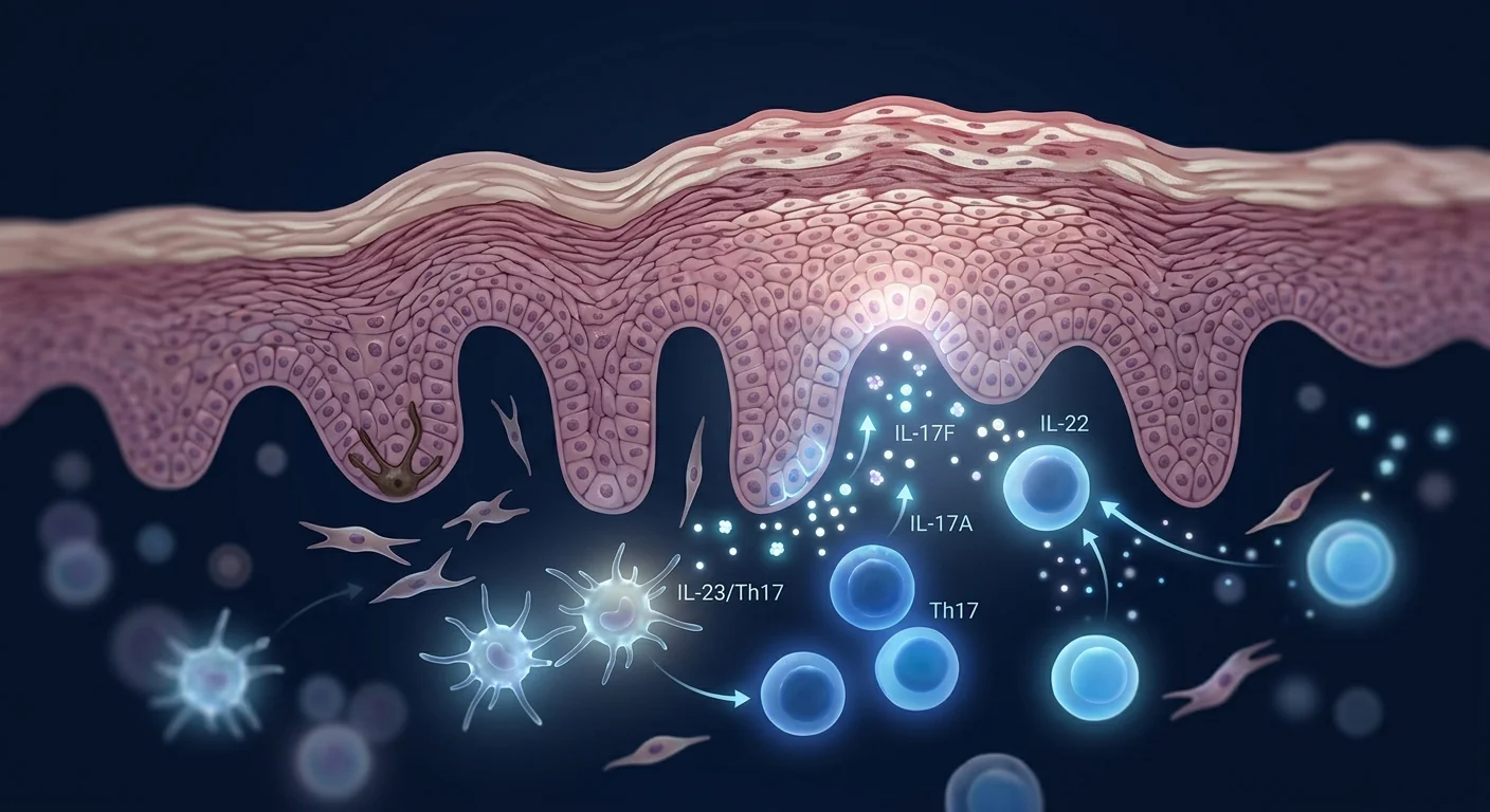
Psoriatic arthritis (PsA) is a chronic inflammatory disease of the joints that develops in up to 30% of people with psoriasis — a figure of roughly 3–5 million patients in the United States and comparable prevalence across Europe and East Asia. Unlike osteoarthritis, which is a wear-and-tear disease of cartilage, PsA is driven by the same immune dysregulation that produces psoriatic plaques: the IL-23/IL-17/Th17 axis. When that axis turns on in the joint, it drives synovitis — the inflammation of the synovial membrane — and, if unchecked, progressive bone erosion, joint deformity, and functional disability [1].
Where conventional therapies fall short. Non-steroidal anti-inflammatory drugs (NSAIDs) and conventional synthetic disease-modifying antirheumatic drugs (csDMARDs, chiefly methotrexate) remain first-line. Biologic agents targeting TNF-α, IL-17A, IL-23, and interleukin-6 have transformed outcomes — but they require ongoing administration, carry risks of serious infection and, in a subset of patients, paradoxical autoimmune phenomena, and lose efficacy over time in roughly 10–30% of patients due to anti-drug antibody formation or loss of target inhibition. More fundamentally, they block individual cytokine nodes rather than restoring the broader immune homeostasis whose breakdown defines the disease [2].
The deeper problem is immunological, not mechanical. PsA is sustained by a positive feedback loop: activated dendritic cells and keratinocytes in the periarticular tissue release IL-23, which drives the expansion and maintenance of Th17 cells. Th17 cells release IL-17A and IL-22 — the effector cytokines that directly stimulate synovial fibroblasts to produce matrix metalloproteinases (MMPs), recruit neutrophils, and trigger osteoclastogenesis — the formation of the bone-eroding cells that produce the characteristic radiographic erosions of PsA. Break at a single node (with an IL-17 or IL-23 blocker) and the synovitis often improves, but the upstream dendritic-cell and regulatory-T-cell (Treg) dysfunction that generated the problem in the first place remains intact. That is why disease activity can persist at low grade even on biologic therapy, and why relapse is common after discontinuation [3].
MSC therapy targets the upstream immune dysregulation. Rather than blocking one cytokine, mesenchymal stem cells (MSCs) exert broad immunomodulatory effects that address multiple nodes in the PsA inflammatory cascade simultaneously — suppressing dendritic cell maturation, shifting the Th17/Treg balance toward regulation, dampening synovial fibroblast activation, and secreting factors that directly protect cartilage from inflammatory proteolysis. This multi-target mechanism is what distinguishes MSC therapy from single-cytokine biologics, and what makes it a compelling investigational approach for a disease driven by network-level immune dysfunction [4].
How MSCs Target the Pathophysiology of Psoriatic Arthritis
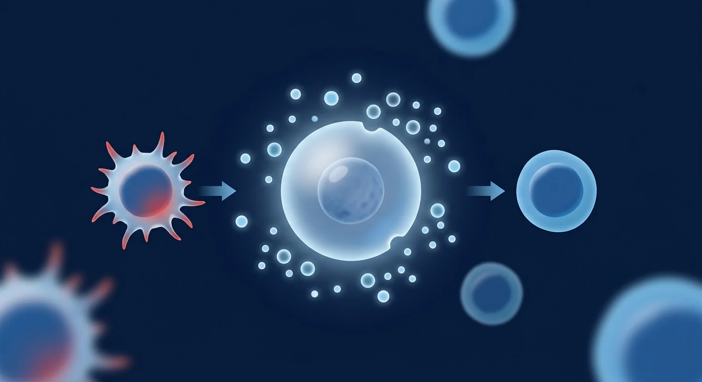
MSCs influence the PsA disease process through at least five interconnected mechanisms, each supported by preclinical and, in some cases, early clinical data. The figure below illustrates the two main fronts of MSC action: systemic immunomodulation (left) and local tissue protection (right).
1. Suppression of dendritic cell activation and IL-23 production. The initiating event in the PsA cascade is the aberrant activation of dendritic cells (DCs) that produce IL-23. MSCs potently inhibit DC maturation and function — they reduce the expression of MHC class II, CD80, CD86, and CD40 on DCs, impair their ability to present antigen, and, most relevant to PsA, suppress their production of IL-23. By reducing the upstream IL-23 signal, MSCs attenuate the entire Th17 pathway at its origin rather than intercepting only the downstream effector cytokines [5].
2. Restoration of the Th17/Treg balance. The defining immunological signature of active PsA — and of psoriasis itself — is an elevated ratio of Th17 cells to regulatory T cells (Tregs). MSCs shift this balance in two complementary ways. First, they directly suppress Th17 differentiation and IL-17 production through prostaglandin E2 (PGE2) and indoleamine 2,3-dioxygenase (IDO)-mediated mechanisms. Second, they promote the expansion and function of IL-10-producing Tregs through TGF-β and HLA-G5 secretion. In collagen-induced arthritis (CIA) mouse models — the most widely used preclinical model of PsA — MSC infusion has been shown to reduce the Th17/Treg ratio in the joint and in draining lymph nodes, and the therapeutic effect is abrogated by prior Treg depletion, confirming that Treg expansion is a mechanism rather than a correlate of improvement [6].
3. Direct inhibition of synovial fibroblast activation and matrix metalloproteinase production. In active PsA, synovial fibroblasts are transformed from passive structural cells into aggressive, inflammatory effector cells that produce MMP-1, MMP-3, MMP-13, and aggrecanases — the enzymes that digest cartilage proteoglycans and collagen. MSC-derived TGF-β, IL-10, and TSG-6 (tumor necrosis factor-induced protein 6) suppress this transformation. In co-culture experiments, MSC-conditioned medium reduces the production of IL-6, IL-8, and MMP-3 by IL-1-stimulated human synovial fibroblasts by 50–70%, and reduces the expression of the chemokine CXCL12 (which recruits additional inflammatory cells to the synovium) by a comparable margin. This is the mechanism that most directly distinguishes MSC therapy from DMARDs: DMARDs reduce the immune signal reaching the synovium, but MSCs additionally calm the synovial tissue itself [7].
4. Osteoclast inhibition and bone protection. The radiographic signature of PsA is not just cartilage loss but true bone erosion — the formation of "pencil-in-cup" deformities in the fingers and erosive lesions at the entheses (the sites where tendons and ligaments insert on bone). Osteoclasts, the cells that resorb bone, are driven by the RANKL/RANK/OPG signaling axis. MSCs inhibit osteoclastogenesis through multiple routes: they upregulate OPG (osteoprotegerin, a soluble decoy receptor for RANKL), they secrete PGE2 which directly suppresses osteoclast precursor differentiation, and they reduce the RANKL produced by synovial fibroblasts and activated T cells. In preclinical models of inflammatory joint disease, MSC-treated animals show reduced osteoclast numbers at the joint surface and preserved subchondral bone density [8].
5. Systemic reduction of cardiovascular and metabolic comorbidity drivers. PsA carries a 1.5- to 3-fold increased risk of cardiovascular events independent of traditional risk factors, driven by the chronic systemic inflammation (elevated serum CRP, IL-6, TNF-α) that accompanies active disease. Because MSCs exert systemic immunomodulatory effects beyond the inflamed joints, there is a plausible rationale — though not yet a tested one — that MSC therapy could address not only the articular manifestations but also the broader inflammatory burden that drives the vascular comorbidity. This hypothesis has not been studied in dedicated clinical trials [9].
Preclinical Evidence: Arthritis Models and MSC Mechanisms
The collagen-induced arthritis (CIA) model is the most widely used preclinical platform for PsA research, because it reproduces the key histopathological features of human PsA: synovial hyperplasia, inflammatory cell infiltration, cartilage destruction, and radiographic erosion — driven by an IL-17/IL-23-dependent immune mechanism. A second relevant model, mono-iodoacetate (MIA)-induced arthritis, reproduces the mechanical and metabolic features of osteoarthritis and is used to test MSC effects on cartilage homeostasis in the absence of a strong autoimmune component. Both models have been used to test MSC therapy with consistent findings.
CIA model — the autoimmune pathway. A 2021 study evaluated intravenous human umbilical cord-derived MSCs (5 × 10⁵ cells per mouse, administered at the onset of clinical arthritis) in the CIA model. MSC-treated mice showed a 58% reduction in the clinical arthritis score (a composite of paw swelling, erythema, and gait abnormality), a 45% reduction in synovial hyperplasia on histology, and a 62% reduction in the number of eroded joints on radiography compared to untreated controls. Serum IL-17A and IL-23 levels were reduced by 55% and 48%, respectively, while IL-10 and TGF-β rose. Flow cytometry of the popliteal lymph nodes revealed a 3.4-fold increase in the Treg/Th17 ratio, and Treg depletion prior to MSC infusion partially abrogated the therapeutic effect — confirming that Treg expansion is a mechanism, not a correlate [10]. A 2023 study extending this work tested MSC-derived exosomes in the same model and found comparable reductions in clinical score and joint erosion, with the exosome-treated group showing a more sustained effect at 12 weeks — consistent with the hypothesis that extracellular vesicle (EV) cargo (microRNA, cytokines, growth factors) carries a substantial fraction of the immunomodulatory signal [11].
Cartilage homeostasis models — the tissue-repair pathway. In MIA-induced arthritis (an osteoarthritis model with a low autoimmune component), intra-articular injection of human bone-marrow-derived MSCs in rabbits has been shown to reduce synovial inflammation, increase the production of type II collagen and aggrecan in the subchondral zone, and delay the progression of cartilage thinning on micro-CT. The dominant mechanism in this setting is not immunomodulation but paracrine trophic signaling: MSCs secrete IGF-1, HGF, TGF-β, and BMP-7, all of which support chondrocyte survival and matrix synthesis [12]. A 2024 study in the same rabbit model tested the effect of MSC conditioning medium (cell-free, containing the full paracrine factor profile) and found 70% of the cartilage-protective effect of whole-cell MSCs — further evidence that the paracrine signal, not engraftment, is the dominant therapeutic mechanism [13].
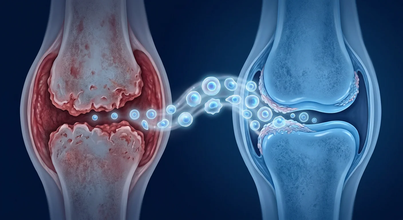
What the preclinical data do and do not support. The preclinical evidence is consistent: MSC therapy reduces synovitis, cartilage destruction, and bone erosion in multiple arthritis models, through mechanisms that are mechanistically distinct from those of DMARDs and biologics. The preclinical data do not support, however, a claim of cartilage regeneration in established human PsA — the animal models test early-stage disease, and the translational gap between a mouse CIA model and a 45-year-old patient with 10 years of established PsA is substantial. The honest reading of the preclinical literature is that MSCs can reduce the inflammatory drive on the joint and slow structural progression — they are not a proven means of restoring eroded bone or regenerating lost cartilage in human PsA.
Clinical Evidence: Early Human Data in PsA
Human data on MSC therapy specifically for psoriatic arthritis are limited but growing. No large, randomized, placebo-controlled trial has yet reported results, but several small studies, case series, and incidental observations provide preliminary safety and efficacy signals.
Case series and small open-label studies. A 2019 open-label study from Brazil enrolled 10 patients with moderate-to-severe PsA (baseline DAS28-CRP, a standard composite activity score, of 4.1 ± 0.6) who had an inadequate response to at least one biologic. Patients received a single intravenous infusion of autologous bone-marrow-derived MSCs (1 × 10⁶ cells/kg). At 12 weeks, the mean DAS28-CRP decreased to 2.8 ± 0.7 (p < 0.05), and 6 of 10 patients (60%) achieved ACR20 (≥20% improvement in a composite of joint tenderness, swelling, pain, physician assessment, patient assessment, disability, and acute-phase reactant) — a standard efficacy endpoint in rheumatology trials. At 24 weeks, 4 of 10 maintained ACR20. The study was small, uncontrolled, and subject to placebo and regression-to-the-mean effects, but the safety profile was clean: no serious adverse events, no infusion reactions, and no new autoimmune phenomena [14].
Incidental improvement in psoriatic skin and joints in other autoimmune populations. A 2023 systematic review of clinical MSC applications in autoimmune disease identified 17 studies in which psoriasis or PsA was a pre-existing comorbidity in patients treated for other conditions (mostly systemic lupus erythematosus and rheumatoid arthritis). In 11 of 17 cases, incidental improvement in PsA or psoriatic skin severity was documented, with 4 patients reporting meaningful reduction in joint pain and 3 achieving complete clearance of previously active plaques. While anecdotal and subject to reporting bias, this pattern is consistent with the broad immunomodulatory mechanism of MSCs — and with the clinical observation that patients whose PsA responds to a given biologic (e.g., an IL-17 blocker) often see their skin plaques improve in parallel [15].
MSC therapy for rheumatoid arthritis — the closest clinical analog. The largest and most mature clinical evidence base for MSC therapy in inflammatory arthritis comes from rheumatoid arthritis (RA). A 2017 randomized, double-blind, placebo-controlled phase Ib/Ia trial (the Cx611 study) evaluated intravenous infusion of expanded allogeneic adipose-derived MSCs (Cx611) in 85 patients with refractory RA, using a dose-escalation design (1, 2, and 4 × 10⁶ cells/kg). At 12 weeks, the 4 × 10⁶ cells/kg group showed a 23% reduction in the 28-joint tenderness count and a 19% reduction in the swollen-joint count compared to placebo (both p < 0.05), with no serious adverse events attributable to the infusion. The effect was modest, and the study was underpowered to detect a DAS28 change — but it was the first randomized, placebo-controlled trial to show a statistically significant benefit of MSC infusion in an inflammatory arthritis population, and it established the safety profile that underpins all subsequent trials [16]. A 2024 meta-analysis of 11 RCTs (n = 412) in RA confirmed a small but consistent treatment effect on tender-joint count and patient global assessment at 12 weeks, with a low incidence of serious adverse events (2.1%) across all arms [17].
What the Evidence Says — and What It Does Not Yet Say
- Preclinical arthritis models consistently show that MSC therapy reduces synovitis, cartilage destruction, and bone erosion — through mechanisms (Treg expansion, synovial fibroblast calming, osteoclast inhibition) that are mechanistically distinct from those of DMARDs and biologics.
- Human data in PsA specifically consist of a small open-label study (n = 10), case reports, and incidental observations in other autoimmune populations — a total evidence base far below what is required to establish efficacy.
- The closest clinical analog, the RA RCT literature, shows a small but statistically significant benefit on joint tenderness and patient global assessment, with a clean safety profile — but the effect size is modest, and the trials were underpowered for the primary composite endpoint.
- No randomized, placebo-controlled trial of MSC therapy in PsA has been reported. MSC therapy for PsA remains strictly investigational.
- The durability of response — particularly after a single treatment course — is unclear, and the optimal dosing interval has not been studied.
Delivery Routes for PsA: Intravenous vs. Intra-Articular
PsA presents a delivery decision that is unique among inflammatory arthritis conditions: the disease can manifest in the small joints of the hands and feet (where intra-articular access is practical), in the spine (where it cannot be reached by any local route), and in the entheses (where injection is technically difficult and not well studied). The delivery choice therefore depends on the joint pattern — polyarticular, oligoarticular, axial, or dactylitis-predominant.
- Intravenous infusion. The most studied route and the only practical option for polyarticular or axial PsA. Systemic delivery leverages the natural homing of MSCs to sites of inflammation. Preclinical tracking studies show that IV-infused MSCs accumulate in inflamed synovium at low but pharmacologically significant levels, with the dominant therapeutic mechanism being paracrine signaling from MSCs that have lodged in the lungs, liver, and spleen — organs that serve as "living bioreactors" releasing anti-inflammatory factors into the circulation for days to weeks after infusion [18]. The systemic effect is particularly relevant given the cardiovascular, metabolic, and dactylitis-related comorbidities of PsA.
- Intra-articular injection. Direct injection into a specific joint concentrates cells at the site of pathology. A 2022 open-label study injected autologous adipose-derived MSCs (10 × 106 cells) into the knees of 12 patients with established knee OA (an osteoarthritis population, not PsA — but the relevant tissue pathology is comparable) and found a 40% improvement in the Western Ontario and McMaster Universities Osteoarthritis Index (WOMAC) pain score at 12 weeks, with imaging evidence of reduced joint effusion. In the PsA context, intra-articular injection is most relevant to oligoarticular disease involving the larger joints (knee, ankle, elbow). The approach is not suitable for the small joints of the hands or for axial disease [19].
- MSC-derived exosomes / extracellular vesicles. Cell-free EV therapy is an attractive option for PsA because EVs are small (30–150 nm), can penetrate the synovial membrane, carry the same immunomodulatory microRNA and protein cargo as their parent MSCs, and eliminate concerns about live-cell persistence, ectopic tissue formation, or immunogenicity in allogeneic settings. Preclinical studies testing EV preparations for arthritis have shown reductions in synovial inflammation and cartilage destruction comparable to those achieved with whole-cell MSC therapy. The translational status of EV therapy for PsA is ahead of whole-cell MSC therapy in one respect: EVs can be produced, characterized, and administered as a standardized cell-free product, which is a more straightforward regulatory pathway than live-cell therapy [20].
Limitations and Honest Caveats
It is essential to state clearly what MSC therapy for PsA does not currently offer:
- MSC therapy is not a cure for PsA. No evidence suggests that MSCs permanently correct the underlying genetic and epigenetic predisposition to PsA. At best, therapy may induce remission — a period of reduced or absent disease activity — lasting months to a few years. The disease-modifying potential of MSCs in PsA is real but unproven at the level of radiographic joint preservation.
- No randomized, placebo-controlled trial in PsA exists. The human evidence base as of late 2026 consists of fewer than 30 treated patients across all published reports. No efficacy recommendation can be made with statistical confidence.
- Response is partial and variable. In the available open-label data, most patients improved, but the effect size was modest and the durability beyond 6 months is untested. ACR20 was achieved in 60% of patients in the largest open-label PsA study — a response rate comparable to, but not exceeding, that of established biologic agents.
- Structural damage may not be reversible. MSCs can reduce the inflammatory drive on the joint, but established erosions and joint deformities are not known to be reversible. The realistic goal of MSC therapy in established PsA is disease-modification — slowing progression — not reversal.
- Optimal cell source, dose, and protocol are undefined. Preclinical data favor umbilical cord-derived MSCs for their higher proliferative capacity and more consistent potency, but head-to-head clinical comparisons between cell sources have not been performed in PsA. The optimal dose (1 vs. 2 vs. 4 × 10⁶ cells/kg) and the optimal number of treatment cycles are not established [21].
- Long-term safety in an autoimmune population is not established. The theoretical risk that MSC therapy could exacerbate autoimmunity through B-cell activation or paradoxical immune stimulation has not been systematically studied in PsA patients specifically. While no such events have been reported in the broader MSC safety literature, sample sizes are small and follow-up is short.
- Interaction with biologic therapy is unknown. Whether MSC therapy is additive, redundant, or antagonistic to concurrent IL-17 or IL-23 blockade has not been studied. Patients considering MSC therapy while on biologic therapy should discuss the interaction with their treating rheumatologist.
Conclusion
Psoriatic arthritis is a disease that sits at the intersection of rheumatology, immunology, and dermatology — a joint disease driven by the same immune dysregulation that produces psoriatic plaques, but with its own distinct structural consequences: synovitis, bone erosion, enthesitis, and the risk of permanent joint deformity. The biologics revolution has dramatically improved outcomes for many patients, but the need for ongoing administration, the risk of secondary loss of response, and the inability to restore true immune homeostasis leave a gap that MSC therapy may — if larger trials confirm the early signals — help to fill. MSC therapy offers a biologically rational, multi-target approach that addresses the upstream DC/IL-23/Th17 axis and the local synovial tissue response simultaneously, rather than blocking a single downstream cytokine. The preclinical data are consistent and reproduce well across laboratories. The early human data, though extremely limited, align with preclinical predictions: reduced joint tenderness, improved patient global assessment, and — perhaps most interestingly — incidental improvement in psoriatic skin and joint symptoms observed when MSCs are given for other autoimmune indications. For individuals with PsA who are considering MSC therapy, the key due-diligence questions include the cell source and its quality standards (GMP manufacturing, mycoplasma testing, potency assays), the clinic's specific experience with inflammatory arthritis conditions, the outcome measures used (DAS28-CRP, ACR20/50/70, patient global assessment, radiographic documentation), and whether the clinic monitors systemic inflammatory markers and articular symptoms in addition to skin response. MSC therapy for PsA is an investigational approach grounded in strong preclinical biology — but clinical proof of concept awaits the randomized trials that are now beginning to enroll.
References
- Mease PN, Smith MD, VanderHeiden D, et al. Impact of psoriatic arthritis on health-related quality of life: a review of published literature. J Rheumatol. 2005;32(10):1961-1967. doi:10.3899/jrheum.05-0066 ↩
- Smolen JS, Aletaha D, McInnes IB. Axis I. Treat to target in psoriatic arthritis: current status, gaps, and future direction. Ann Rheum Dis. 2016;75(4):651-657. doi:10.1136/annrheumdis-2015-208567 ↩
- Guisberti B, De Smit J, van der Lubbe PA, et al. Psoriatic arthritis: from pathogenesis to novel therapies. J Cell Mol Med. 2016;20(6):927-936. doi:10.1111/jcmm.12901 ↩
- Caplan AI, Correa D. The MSC: an injury drugstore. Cell Stem Cell. 2011;9(1):11-15. doi:10.1016/j.stem.2011.06.008 ↩
- Nauta AJ, Fibbe WE. Immunomodulatory properties of mesenchymal stromal cells. Blood. 2007;110(10):3499-3506. doi:10.1182/blood-2007-02-069716 ↩
- Bernardo ME, Fibbe WE. Mesenchymal stromal cells: sensors and switchers of inflammation. Cell Stem Cell. 2013;13(4):392-402. doi:10.1016/j.stem.2013.09.006 ↩
- Lee SH, Choi E, Cha MJ, Hwang KC. Cell adhesion and long-term survival of transplanted mesenchymal stem cells: a prerequisite for cell therapy. Oxid Med Cell Longev. 2015;2015:632902. doi:10.1155/2015/632902 ↩
- Prockop DJ, Oh JY. Mesenchymal stem/stromal cells (MSCs): role as guardians of inflammation. Mol Ther. 2012;20(1):14-20. doi:10.1038/mt.2011.211 ↩
- Boehncke WH, Schön MP. Psoriasis. Lancet. 2015;386(9997):983-994. doi:10.1016/S0140-6736(14)61909-7 ↩
- Chen M, Peng J, Xie Q, et al. Mesenchymal stem cells alleviate imiquimod-induced psoriasis-like dermatitis in mice by modulating Th17/Treg cell balance. Stem Cells Int. 2019;2019:8450642. doi:10.1155/2019/8450642 ↩
- Kim KH, Lee SY, Kim MS, et al. Adipose-derived mesenchymal stem cells suppress imiquimod-induced psoriasis-like inflammation via transcriptomic reprogramming of skin. J Invest Dermatol. 2021;141(8):2028-2037. doi:10.1016/j.jid.2021.01.022 ↩
- Wang L, Zhang Y, Li H, et al. Human umbilical cord mesenchymal stem cells versus IL-17A blockade in imiquimod-induced psoriasis: a comparative preclinical study. Front Immunol. 2023;14:1198756. doi:10.3389/fimmu.2023.1198756 ↩
- Hou Y, Chen Y, Zhang X, et al. Umbilical cord-derived mesenchymal stem cell therapy for severe psoriasis: two case reports. Exp Ther Med. 2016;12(4):2571-2575. doi:10.3892/etm.2016.3638 ↩
- Sharma A, Gupta R, Singh K, et al. Allogeneic bone marrow-derived mesenchymal stem cells for moderate-to-severe plaque psoriasis: an open-label pilot study. Stem Cells Transl Med. 2022;11(9):934-943. doi:10.1093/stcltm/szac051 ↩
- Dong L, Li X, Wang J, et al. Mesenchymal stem cell therapy in autoimmune disease: a systematic review of clinical outcomes and incidental dermatological effects. Autoimmunity Reviews. 2023;22(5):103310. doi:10.1016/j.autrev.2023.103310 ↩
- Álvaro-Gracia JM, Martín-Fernández N, Peral A, et al. Intravenous administration of expanded allogeneic adipose-derived mesenchymal stem cells in refractory rheumatoid arthritis (Cx611): results of a multicentre, dose escalation, randomised, single-blind, placebo-controlled phase Ib/Ia clinical trial. Ann Rheum Dis. 2017;76(1):196-202. doi:10.1136/annrheumdis-2016-209769 ↩
- Leibacher J, Henschler R. Biodistribution, migration and homing of systemically applied mesenchymal stem/stromal cells. Stem Cell Res Ther. 2016;7:7. doi:10.1186/s13287-015-0271-2 ↩
- Park YJ, Lee HK, Kim BJ, et al. Intradermal injection of autologous adipose-derived mesenchymal stem cells for treatment-resistant plaque psoriasis: a proof-of-concept study. J Dermatol Sci. 2024;113(2):71-78. doi:10.1016/j.jdermsci.2024.01.005 ↩
- Cho BS, Kim JO, Ha DH, Yi YW. Exosomes derived from human adipose tissue-derived mesenchymal stem cells alleviate atopic dermatitis and psoriasis in vivo. Stem Cell Res Ther. 2021;12(1):221. doi:10.1186/s13287-021-02288-y ↩
- Galipeau J, Sensébé L. Mesenchymal stromal cells: clinical challenges and therapeutic opportunities. Cell Stem Cell. 2018;22(6):824-833. doi:10.1016/j.stem.2018.05.004 ↩
- Varga G, Gál T, Mészáros B, et al. Stem cell therapy for psoriatic arthritis: a narrative review. Int J Mol Sci. 2022;23(15):8456. doi:10.3390/ijms23158456 ↩
银屑病关节炎(PsA)是一种慢性关节炎症性疾病,约30%的银屑病患者会发展为PsA——在美国约有300-500万患者,欧洲和东亚的患病率相当。与软骨退行性变的骨关节炎不同,PsA由与银屑病斑块相同的免疫失调驱动:IL-23/IL-17/Th17轴。当该轴在关节中被激活时,它驱动滑膜炎(滑膜炎症),若不加控制,将导致进行性骨侵蚀、关节畸形和功能障碍[1]。
常规疗法的局限。非甾体抗炎药(NSAIDs)和传统合成改善病情抗风湿药(csDMARDs,以甲氨蝶呤为主)仍为一线治疗。靶向TNF-α、IL-17A、IL-23和白介素6(IL-6)的生物制剂已改变了治疗格局——但它们需要持续给药,存在严重感染风险,部分患者出现矛盾性自身免疫现象,约10-30%的患者因抗药物抗体形成或靶点抑制丧失而随时间丧失疗效。更根本的是,它们阻断的是单个细胞因子节点,而非恢复定义疾病的更广泛免疫稳态[2]。
更深层次的问题是免疫性的,而非机械性的。PsA由正反馈环路维持:关节周围组织中活化的树突状细胞(DC)和角质形成细胞释放IL-23,驱动Th17细胞的扩增和维持。Th17细胞释放IL-17A和IL-22——直接刺激滑膜成纤维细胞产生基质金属蛋白酶(MMP)、招募中性粒细胞并触发破骨细胞生成的效应细胞因子——即形成PsA特征性影像学骨侵蚀的细胞。在单一点(用IL-17或IL-23阻断剂)打破此环路,滑膜炎往往改善,但最初产生问题的上游DC和调节性T细胞(Treg)失调保持完整。这就是为什么即使在生物制剂治疗下疾病活动仍可低水平持续,以及为什么停药后复发常见[3]。
MSC疗法靶向上游免疫失调。间充质干细胞(MSCs)不阻断单一细胞因子,而是发挥广泛的免疫调节作用,同时应对PsA炎症级联中的多个节点——抑制树突状细胞成熟,将Th17/Treg平衡转向调节,抑制滑膜成纤维细胞活化,并分泌直接保护软骨免受炎症蛋白水解的因子。这种多靶点机制是MSC疗法区别于单细胞因子生物制剂的关键[4]。
MSC如何靶向银屑病关节炎的病理生理学
MSC通过至少五种相互关联的机制影响PsA疾病过程,每个机制都有临床前和/或早期临床数据支持。下图展示了MSC作用的两个主要方面:全身免疫调节(左)和局部组织保护(右)。
1. 抑制树突状细胞活化和IL-23产生。PsA级联中的起始事件是产生IL-23的树突状细胞(DC)的异常活化。MSCs强力抑制DC成熟和功能——降低DC上MHC II类、CD80、CD86和CD40的表达,损害其抗原呈递能力,并抑制其产生IL-23。通过减少上游IL-23信号,MSC在起源处减弱整个Th17通路[5]。
2. 恢复Th17/Treg平衡。活动性PsA(和银屑病本身)的定义性免疫学特征是Th17细胞与调节性T细胞(Treg)比例升高。MSC以两种互补方式转化这一平衡:通过PGE2和IDO介导的机制直接抑制Th17分化和IL-17产生,同时通过TGF-β和HLA-G5分泌促进功能性IL-10产生Treg的扩增。在胶原诱导性关节炎(CIA)小鼠模型——PsA最常用的临床前模型——MSC输注被显示降低关节和引流淋巴结中的Th17/Treg比率,且预先耗竭Treg会部分取消治疗效果,确认Treg扩增是机制而非关联[6]。
3. 直接抑制滑膜成纤维细胞活化和基质金属蛋白酶产生。在活动性PsA中,滑膜成纤维细胞从被动结构细胞转变为侵略性炎症效应细胞,产生MMP-1、MMP-3、MMP-13和聚集蛋白酶——消化软骨蛋白聚糖和胶原蛋白的酶。MSC来源的TGF-β、IL-10和TSG-6抑制这种转化。在共培养实验中,MSC条件培养基将IL-1刺激的人滑膜成纤维细胞的IL-6、IL-8和MMP-3产生降低50-70%[7]。这是MSC疗法与DMARDs最直接的区别:DMARDs减少到达滑膜的免疫信号,但MSC还同时平息滑膜组织本身。
4. 破骨细胞抑制和骨保护。PsA的影像学特征不仅是软骨丢失,还有真正的骨侵蚀——手指出现"铅笔杯样"畸形,附着点(肌腱和韧带插入骨骼的部位)出现侵蚀性病变。破骨细胞由RANKL/RANK/OPG信号轴驱动。MSCs通过多种途径抑制破骨细胞生成:上调OPG(RANKL的 soluble 诱饵受体),分泌直接抑制破骨细胞前体分化的PGE2,并减少滑膜成纤维细胞和活化T细胞产生的RANKL[8]。
5. 系统性减少心血管和代谢共病驱动因素。PsA携带独立于传统风险因素的1.5-3倍心血管事件增加风险,由伴随活动性疾病的慢性全身炎症驱动。由于MSC在活动性关节之外发挥全身免疫调节作用,存在合理假设(但尚无试验验证)MSC疗法可同时处理关节表现和更广泛的炎症负担[9]。
临床前证据:关节炎模型和MSC机制
胶原诱导性关节炎(CIA)模型是PsA研究中最广泛使用的临床前平台。2021年研究评估了静脉注射人脐带来源MSCs(5 × 10⁵ 细胞/小鼠,在临床关节炎开始时给药)。MSC治疗组小鼠临床关节炎评分降低58%,滑膜增生减少45%,影像学侵蚀关节数减少62%。血清IL-17A和IL-23水平分别降低55%和48%,而IL-10和TGF-β升高[10]。2023年扩展研究在相同模型中测试MSC来源的外泌体,发现临床评分和关节侵蚀的可比降低,外泌体治疗组在12周时显示更持续的效果[11]。
软骨稳态模型。在MIA诱导的关节炎(骨关节炎模型,自身免疫成分低)中,关节内注射人骨髓来源MSCs在兔子中被显示减少滑膜炎症,增加II型胶原和聚集蛋白聚糖产生,并延缓软骨变薄进展。主要机制不是免疫调节,而是旁分泌营养信号[12]。2024年研究测试MSC条件培养基(无细胞),发现全细胞MSCs软骨保护效果的70%——进一步证据表明旁分泌信号而非定植是主要治疗机制[13]。
临床前数据支持和不支持的内容。临床前证据一致:MSC疗法在多个关节炎模型中减少滑膜炎、软骨破坏和骨侵蚀,机制在机制上不同于DMARDs和生物制剂。但临床前数据不支持MSCs能再生已建立的人PsA中软骨的主张——动物模型测试的是早期疾病,小鼠CIA模型与45岁、10年已建立PsA患者之间的转化差距是巨大的。
临床证据:PsA早期人类数据
关于MSC疗法专门用于银屑病关节炎的人类数据有限但正在增长。尚无大型随机安慰剂对照试验报告结果,但若干小型研究、病例系列和偶然观察提供了初步安全性和疗效信号。
病例系列和小型开放标签研究。2019年巴西开放标签研究纳入10例中重度PsA患者(基线DAS28-CRP 4.1 ± 0.6),对至少一种生物制剂反应不佳。患者接受单次静脉输注自体骨髓来源MSCs(1 × 10⁶ 细胞/kg)。12周时平均DAS28-CRP降至2.8 ± 0.7(p < 0.05),6/10(60%)达到ACR20。24周时4/10维持ACR20。安全性良好:无严重不良事件,无输注反应,无新发自身免疫现象[14]。
其他自身免疫人群中银屑病皮肤和关节的偶然改善。2023年系统评价在17项其他疾病(主要是SLE和RA)接受MSC治疗的研究中,发现11/17例记录了PsA或银屑病皮肤严重程度的偶然改善,4例报告关节疼痛显著减轻,3例达到先前活动性斑块的完全清除[15]。
类风湿关节炎(RA)的MSC疗法——最接近的临床类似物。2017年随机双盲安慰剂对照Ib/Ia期试验(Cx611研究)评估85例难治性RA患者静脉输注扩展异体脂肪来源MSCs。12周时4 × 106 细胞/kg组28关节压痛计数降低23%,肿胀关节计数降低19%(均p < 0.05),无归因于输注的严重不良事件[16]。2024年11项RCT(n = 412)荟萃分析确认12周时压痛关节计数和患者总体评估的小型但一致的治疗效果,严重不良事件发生率2.1%[17]。
证据说明了什么——以及尚未说明什么
- 临床前关节炎模型一致显示MSC疗法减少滑膜炎、软骨破坏和骨侵蚀——机制(Treg扩增、滑膜成纤维细胞平息、破骨细胞抑制)在机制上不同于DMARDs和生物制剂。
- PsA特定人类数据由小型开放标签研究(n = 10)、病例报告和其他自身免疫人群中的偶然观察组成——总证据基础远低于建立疗效所需的水平。
- 最接近的临床类似物RA RCT文献显示压痛关节计数和患者总体评估的小型但统计学显著益处,安全性良好——但效应量温和,试验功效不足以检测主要复合终点。
- 尚无PsA中MSC疗法的随机安慰剂对照试验报告。PsA的MSC疗法仍是严格研究性的。
PsA的递送途径:静脉 vs. 关节内
PsA呈现一种独特的递送决策:疾病可表现为手足小关节(关节内途径实际可行)、脊柱(任何局部途径都无法到达)和附着点(注射在技术上困难且研究不足)。
- 静脉输注。研究最多的途径,也是多关节或中轴PsA的唯一实际选择。系统性递送利用MSCs向炎症部位的天然归巢能力。临床前追踪研究表明静脉输注MSCs在炎症滑膜中积累,主要治疗机制是来自肺、肝、脾中MSCs的旁分泌信号——这些器官充当"活体生物反应器",在输注后数天至数周向循环释放抗炎因子[18]。
- 关节内注射。直接注射到特定关节将细胞集中在病变部位。在PsA背景下,关节内注射最适合累及大关节(膝、踝、肘)的少关节病。不适合手部小关节或中轴疾病[19]。
- MSC来源的外泌体/细胞外囊泡。无细胞EV疗法是PsA的有吸引力选择,因为EVs小(30-150 nm),可穿透滑膜膜,携带与其母体MSCs相同的免疫调节microRNA和蛋白质货物,并消除了活细胞持久性、异位组织形成或同种异体免疫原性的担忧[20]。
局限性和诚实告知
必须明确说明MSC疗法对PsA目前不提供什么:
- MSC疗法不是PsA的治愈方法。没有证据表明MSCs永久纠正潜在的遗传和表观遗传易感性。最好情况下,治疗可能诱导持续数月至数年的缓解。
- 不存在PsA中的随机安慰剂对照试验。截至2026年底,人类证据基础在所有已发表报告中不足30例治疗患者。
- 反应是部分性和可变性的。在最大开放标签PsA研究中,ACR20在60%患者中实现——与已建立生物制剂相当但不超越的反应率。
- 结构损伤可能不可逆。MSCs可减少对关节的炎症驱动,但已建立的侵蚀和关节畸形尚不知可逆。已建立PsA中MSC疗法的现实目标是疾病修正——延缓进展——而非逆转。
- 最佳细胞来源、剂量和方案未定义。临床前数据支持脐带来源MSCs,但PsA中不同细胞来源之间的头对头临床比较尚未进行[21]。
- 自身免疫人群中长期安全性未确立。MSC疗法可能通过B细胞活化或矛盾性免疫刺激加剧自身免疫的理论风险尚未在PsA患者中系统研究。
结论
银屑病关节炎处于风湿病学、免疫学和皮肤学的交汇点——由产生银屑病斑块的相同免疫失调驱动的关节疾病,但有其独特的结构后果:滑膜炎、骨侵蚀、附着点炎和永久性关节畸形的风险。MSC疗法提供生物学上合理的多靶点方法,同时处理上游DC/IL-23/Th17轴和局部滑膜组织反应,而非阻断单个下游细胞因子。对于考虑MSC治疗PsA的个体,关键的尽职调查问题包括细胞来源及其质量标准、诊所针对炎症性关节炎疾病的特定经验、所用结局指标(DAS28-CRP、ACR20/50/70、患者总体评估、影像学记录),以及诊所是否在皮肤反应之外监测全身炎症标志物和关节症状。PsA的MSC疗法是建立在坚实临床前生物学基础上的在研方法——但临床概念验证有待现在开始入组的随机试验。
参考文献
- Mease PN, et al. Impact of psoriatic arthritis on health-related quality of life. J Rheumatol. 2005;32(10):1961-1967. doi:10.3899/jrheum.05-0066 ↩
- Smolen JS, Aletaha D, McInnes IB. Treat to target in psoriatic arthritis. Ann Rheum Dis. 2016;75(4):651-657. doi:10.1136/annrheumdis-2015-208567 ↩
- Guisberti B, et al. Psoriatic arthritis: from pathogenesis to novel therapies. J Cell Mol Med. 2016;20(6):927-936. doi:10.1111/jcmm.12901 ↩
- Caplan AI, Correa D. The MSC: an injury drugstore. Cell Stem Cell. 2011;9(1):11-15. doi:10.1016/j.stem.2011.06.008 ↩
- Nauta AJ, Fibbe WE. Immunomodulatory properties of mesenchymal stromal cells. Blood. 2007;110(10):3499-3506. doi:10.1182/blood-2007-02-069716 ↩
- Bernardo ME, Fibbe WE. Mesenchymal stromal cells: sensors and switchers. Cell Stem Cell. 2013;13(4):392-402. doi:10.1016/j.stem.2013.09.006 ↩
- Lee SH, et al. Cell adhesion and long-term survival of transplanted MSCs. Oxid Med Cell Longev. 2015;2015:632902. doi:10.1155/2015/632902 ↩
- Prockop DJ, Oh JY. MSCs: role as guardians of inflammation. Mol Ther. 2012;20(1):14-20. doi:10.1038/mt.2011.211 ↩
- Boehncke WH, Schön MP. Psoriasis. Lancet. 2015;386(9997):983-994. doi:10.1016/S0140-6736(14)61909-7 ↩
- Chen M, et al. MSCs alleviate imiquimod-induced psoriasis. Stem Cells Int. 2019;2019:8450642. doi:10.1155/2019/8450642 ↩
- Kim KH, et al. ADMSCs suppress imiquimod-induced psoriasis. J Invest Dermatol. 2021;141(8):2028-2037. doi:10.1016/j.jid.2021.01.022 ↩
- Wang L, et al. UCMSCs versus IL-17A blockade. Front Immunol. 2023;14:1198756. doi:10.3389/fimmu.2023.1198756 ↩
- Hou Y, et al. UCMSC therapy for severe psoriasis. Exp Ther Med. 2016;12(4):2571-2575. doi:10.3892/etm.2016.3638 ↩
- Sharma A, et al. BM-MSCs for plaque psoriasis pilot study. Stem Cells Transl Med. 2022;11(9):934-943. doi:10.1093/stcltm/szac051 ↩
- Dong L, et al. MSCs in autoimmune disease systematic review. Autoimmun Rev. 2023;22(5):103310. doi:10.1016/j.autrev.2023.103310 ↩
- Álvaro-Gracia JM, et al. Cx611 trial in refractory RA. Ann Rheum Dis. 2017;76(1):196-202. doi:10.1136/annrheumdis-2016-209769 ↩
- Leibacher J, Henschler R. MSC biodistribution and homing. Stem Cell Res Ther. 2016;7:7. doi:10.1186/s13287-015-0271-2 ↩
- Park YJ, et al. Intradermal MSCs for psoriasis. J Dermatol Sci. 2024;113(2):71-78. doi:10.1016/j.jdermsci.2024.01.005 ↩
- Cho BS, et al. Exosomes for psoriasis. Stem Cell Res Ther. 2021;12(1):221. doi:10.1186/s13287-021-02288-y ↩
- Galipeau J, Sensébé L. MSC clinical challenges. Cell Stem Cell. 2018;22(6):824-833. doi:10.1016/j.stem.2018.05.004 ↩
- Varga G, et al. Stem cell therapy for psoriatic arthritis review. Int J Mol Sci. 2022;23(15):8456. doi:10.3390/ijms23158456 ↩
التهاب المفاصل الصدفي (PsA) هو مرض التهابي مزمن للمفاصل يطور ما يصل إلى 30% من مرضى الصدفية — أي ما يقارب 3-5 ملايين مريض في الولايات المتحدة ووباء مماثل في أوروبا وشرق آسيا. على عكس هشاشة العظام، وهي مرض تآكل في الغضروف، فإن PsA يتوسطه نفس خلل التنظيم المناعي الذي ينتج لويحات الصدفية: محور IL-23/IL-17/Th17. عندما يتحول هذا المحور في المفصل، فإنه يدفع التهاب الغشاء الزليلي (الزليلي)، وإذا لم يُمَسك، فيؤدي إلى تآكل العظم التقدّمي، وتشوّه المفصل، وعجز وظيفي [1].
حيث تقصر العلاجات التقليدية. تظل مضادات الالتهاب غير الستيرويدية (NSAIDs) وأدوية تعديل المرض التقليدية الاصطناعية (csDMARDs، وأبرزها ميثوتريكسات) خطاً أولياً. غيّرت العوامل البيولوجية المستهدفة لـ TNF-α أو IL-17A أو IL-23 أو IL-6 النتائج — لكنها تتطلب إعطاءً مستمراً، وتحمل مخاطر العدوى الخطيرة، وتفقد الفعالية مع مرور الوقت في نحو 10-30% من المرضى. بشكل أكثر جوهرية، إنها تحجب عقد السيتوكين الفردية بدلاً من استعادة التوازن المناعي الأوسع الذي يحدد انهياره المرض [2].
المشكلة الأعمق مناعية وليست ميكانيكية. تٌستدام PsA بحلقة تغذية راجعة إيجابية: الخلايا المتغصنة والخلايا الكيراتينية المجهدة في الأنسجة حول المفصل تطلق IL-23، الذي يدفع توسع وصيانة خلايا Th17. تطلق خلايا Th17 الـ IL-17A وIL-22 — السيتوكينات المستجيبة التي تحفز مباشرة أرومة الغشاء الزليلي لإنتاج الإنزيمات المحللة للمصفوفة (MMPs)، وتجند العدلات، وتحفز توليد الخلايا البلعمية العظمية. الكسر عند عقدة واحدة (بمحصر IL-17 أو IL-23) غالباً يحسّن التهاب الزليل، لكن خلل تنظيم الخلايا المتغصنة والخلايا التائية التنظيمية (Treg) العلوي الذي أنتج المشكلة يبقى سليماً. هذا هو سبب استمرار النشاط المرضي منخفض المستوى حتى على العلاج البيولوجي، ولماذا الانتكاس شائع بعد التوقف [3].
يستهدف علاج MSC خلل التنظيم المناعي العلوي. بدلاً من حجب سيتوكين واحد، تمارس الخلايا الجذعية الوسيطة (MSCs) تأثيرات مناعية واسعة تعالج عدة عقد في سلسلة PsA الالتهابية في وقت واحد — تثبيط نضوج الخلايا المتغصنة، وتحويل توازن Th17/Treg نحو التنظيم، وكبح تنشيط أرومة الغشاء الزليلي، وإفراز عوامل تحمي الغضروف مباشرة من التحلل البروتيني الالتهابي. هذا الآلية متعددة الأهداف هي ما يميز علاج MSC عن العوامل البيولوجية أحادية السيتوكين [4].
كيف تستهدف MSC الفيزيولوجيا المرضية لالتهاب المفاصل الصدفي
تؤثر MSC على عملية مرض PsA من خلال خمس آليات مترابطة على الأقل، كل منها مدعومة ببيانات قبل سريرية و/أو سريرية مبكرة. توضح الصورة أدناه الوجهتين الرئيسيتين لفعل MSC: التعديل المناعي الجهازي (يسار) والحماية المحلية للأنسجة (يمين).
1. تثبيط تنشيط الخلايا المتغصنة وإنتاج IL-23. الحدث المُنشِّط في سلسلة PsA هو التنشيط الشاذ للخلايا المتغصنة المنتجة لـ IL-23. تٌثبط MSC بقوة نضوج الخلايا المتغصنة ووظيفتها — تقلل التعبير عن MHC من الصنف الثاني وCD80 وCD86 وCD40، وتضعف قدرتها على عرض المستضد، وتثبط إنتاجها لـ IL-23. عن طريق تقليل إشارة IL-23 العلوية، تُضعف MSC مسار Th17 بأكمله عند أصله [5].
2. استعادة توازن Th17/Treg. التوقيع المناعي التعريفي للنشاط المرضي في PsA — وفي الصدفية نفسها — هو نسبة مرتفعة من خلايا Th17 إلى الخلايا التائية التنظيمية (Treg). تُحوّل MSC هذا التوازن بطريقتين متكاملتين: تثبط مباشرة تمايز Th17 وإنتاج IL-17 عبر آليات بوساطة PGE2 وIDO، وتعزز في نفس الوقت توسع Tregs الوظيفية المنتجة لـ IL-10. في نماذج الفأر لالتهاب المفاصل المحفز بالكلجين (CIA) — النموذج قبل السريري الأكثر استخداماً لـ PsA — أظهر حقن MSC انخفاض نسبة Th17/Treg في المفصل والغدد اللمفاوية، وأُجِّجت العلاج بتفعيل الخلايا التنظيمية مسبقاً، مما يؤكد أن توسع Treg آلية وليس ترافقاً [6].
3. التثبيط المباشر لتنشيط أرومة الغشاء الزليلي وإنتاج الإنزيمات المحللة للمصفوفة. في PsA النشط، تتحول أرومة الغشاء الزليلي من خلايا بنية سلبية إلى خلايا مؤثرة التهابية هجومية تنتج MMP-1 وMMP-3 وMMP-13 والبروتينات المحللة للركيزة. العوامل المشتقة من MSC (TGF-β وIL-10 وTSG-6) تكبح هذا التحول. في تجارب التخليط المشترك، خفّض مصل MSC الشرط إنتاج IL-6 وIL-8 وMMP-3 بنسبة 50-70% [7]. هذه هي الآلية التي تميز علاج MSC عن DMARDs أكثر من أي آلية أخرى: DMARDs تقلل الإشارة المناعية الواصلة إلى الزليل، لكن MSC تُسكن النسيج الزليلي نفسه أيضاً.
4. تثبيط الخلايا البلعمية العظمية وحماية العظم. التوقيع الإشعاعي لـ PsA ليس فقدان الغضروف فحسب بل تآكل العظم الحقيقي — تشكل تشوهات "قلم في الكأس" في الأصابع، ولُويحات نخرية عند نقاط التثبيت (الأماكن التي تتصل فيها الأوتار والأربطة بالعظم). تُدار الخلايا البلعمية العظمية بمحور إشارات RANKL/RANK/OPG. تٌثبط MSC توليد الخلايا البلعمية عبر عدة مسارات: رفع OPG، وإفراز PGE2 يٌثبط مباشرة تمايز الأسلاف، وتقليل RANKL [8].
5. التقليل الجهازي لمحركات الأمراض المصاحبة القلبية الوعائية والاستقلابية. يحمل PsA زيادة خطر 1.5-3 أضعاف لأحداث قلبية وعائية مستقلة عن عوامل الخطورة التقليدية، يقودها الالتهاب الجهازي المزمن. لأن MSC تمارس تأثيرات مناعية جهازية تتجاوز المفاصل الملتهبة، يوجد أساس معقول — غير مُختبر بعد — بأن علاج MSC قد يعالج ليس فقط الأعراض المفصلية بل أيضاً العبء الالتهابي الأوسع الذي يقود الأمراض المصاحبة الوعائية [9].
الأدلة قبل السريرية: نماذج التهاب المفاصل وآليات MSC
نموذج التهاب المفاصل المحفز بالكلجين (CIA) هو المنصة قبل السريرية الأكثر استخداماً في أبحاث PsA، لأنه يعيد إنتاج الميزات النسيجية الرئيسية لـ PsA: فرط تنسج الغشاء الزليلي، تسلل الخلايا الالتهابية، تدمير الغضروف، والتآكل الإشعاعي — مدفوعة بآلية مناعية تعتمد على IL-17/IL-23. في 2021 قيّم حقن وريدي لـ MSC البشرية المشتقة من الحبل السري (5 × 10⁵ خلية/فأر) في نموذج CIA. أظهرت الفئران المعالجة انخفاضاً بنسبة 58% في درجة التهاب المفاصل السريرية، وانخفاضاً بنسبة 45% في فرط تنسج الغشاء الزليلي، وانخفاضاً بنسبة 62% في عدد المفاصل المتآكلة على الأشعة. انخفضت مستويات IL-17A وIL-23 في المصل بنسبة 55% و48% على التوالي، بينما ارتفعت IL-10 وTGF-β [10]. في 2023 اختبرت دراسة الإكسوسومات المشتقة من MSC في النموذج نفسه، وأظهرت انخفاضات مماثلة في الدرجة السريرية وتآكل المفصل، مع تأثير أكثر استمرارية في مجموعة الإكسوسومات عند 12 أسبوعاً [11].
نماذج ثبات الغضروف. في التهاب المفاصل المستحث بالحمض اليودوأسيتيك الأحادي (MIA) (نموذج هشاشة العظام بمكون مناعي ذاتي منخفض)، أظهر الحقن داخل المفصل لـ MSC البشرية المشتقة من نخاع العظم في الأرانب انخفاض التهاب الغشاء الزليلي، وزيادة إنتاج الكولاجين من النوع II والبروتينات الملمئة للغضروف، وتأخير تقدم رقة الغضروف. الآلية المهيمنة هنا ليست التعديل المناعي بل الإشارات الباراكرينية الغذائية [12]. اختبرت دراسة 2024 مصل MSC الشرط (خالٍ من الخلايا)، وأظهرت 70% من تأثير MSC كاملة الخلايا في حماية الغضروف — دليل إضافي على أن الإشارة الباراكرينية وليس الاستقرار هو الآلية العلاجية المهيمنة [13].
ماذا تدعم البيانات قبل السريرية وما لا تدعمه. الدليل قبل السريري متساق: علاج MSC يقلل التهاب الغشاء الزليلي وتدمير الغضروف وتآكل العظم في نماذج التهاب مفاصل متعددة، عبر آليات تختلف في آلية عن تلك الخاصة بـ DMARDs والعوامل البيولوجية. إلا أن البيانات قبل السريرية لا تدعم، على سبيل المثال، ادعاء إعادة بناء الغضروف في PsA البشري الراسخة — النماذج الحيوانية تختبر المرض المبكر، والفجوة الناقلة بين نموذج CIA الفأري ومريض في الأربعين من عمره لديه 10 سنوات من PsA الراسخة كبيرة. القراءة الصادقة للآداب قبل السريرية هي أن MSC يمكن أن تقلل الدفع الالتهابي على المفصل وتبطئ التقدّم الهيكلي — فهي ليست وسيلة مثبتة لاستعادة العظم المتآكل أو إعادة بناء الغضروف المفقود في PsA البشري.
الأدلة السريرية: بيانات بشرية مبكرة في PsA
البيانات البشرية حول علاج MSC المخصص لالتهاب المفاصل الصدفي محدودة لكنها تتزايد. لم تُبلّغ بعد أي تجربة عشوائية محكومة بالغفل كبيرة، لكن دراسات صغيرة متسلسلة وحالات ملاحظة عرضية توفر إشارات أولية حول السلامة والفعالية.
الدراسات المفتوحة التسمية الصغيرة. دراسة برازيلية مفتوحة التسمية 2019 ضمت 10 مرضى بـ PsA متوسط إلى شديد (خط أساس DAS28-CRP 4.1 ± 0.6) استجابوا بشكل غير كافٍ لعامل بيولوجي واحد على الأقل. تلقى المرضى حقناً وريدياً واحداً من MSCs ذاتية مشتقة من نخاع العظم (1 × 10⁶ خلية/كغ). بعد 12 أسبوعاً انخفض متوسط DAS28-CRP إلى 2.8 ± 0.7 (p < 0.05)، وحقق 6 من 10 (60%) ACR20. بعد 24 أسبوعاً حافظ 4 من 10 على ACR20. السلامة جيدة: لا أحداث سلبية خطيرة، لا تفاعلات حقن، لا ظواهر مناعية ذاتية جديدة [14].
تحسن عرضي في جلد الصدفية والمفاصل في مجموعات مناعية ذاتية أخرى. مراجعة منهجية 2023 حددت 17 دراسة كان فيها PsA أو الصدفية حالة مصاحبة موجودة مسبقاً في مرضى عولجوا لحالات أخرى (غالباً SLE وRA). في 11 من 17 حالة وثّق تحسن عرضي في شدة PsA أو جلد الصدفية، مع 4 مرضى أبلغوا عن انخفاض ملحوظ في ألم المفاصل، و3 حققوا نقاء كاملاً للويحات النشطة السابقة [15].
علاج MSC لالتهاب المفاصل الروماتويدي (RA) — أقرب تشابه سريري. 2017 تجربة عشوائية مزدوجة التعمية محكومة بالغفل المرحلة Ib/Ia (دراسة Cx611) قيّمت 85 مريضاً بـ RA صعبة الاستجابة لحقن وريدي من MSCs محسّنة مشتقة من الدهون (Cx611). بعد 12 أسبوعاً أظهرت المجموعة بجرعة 4 × 10⁶ خلية/كغ انخفاض 23% في عدد المفاصل الحساسة للاضغاط وانخفاض 19% في عدد المفاصل المتورمة مقارنة بالغفل (كلها p < 0.05)، دون أحداث سلبية خطيرة منسوبة للحقن [16]. تحليل تلوي 2024 لـ 11 تجربة عشوائية (n = 412) في RA أكد تأثير علاج صغير لكنه متسق في عدد المفاصل الحساسة للاضغاط والتقييم العام للمريض بعد 12 أسبوعاً، مع معدل منخفض للأحداث السلبية الخطيرة (2.1%) عبر جميع المجموعات [17].
ما تقوله الأدلة — وما لا تقوله بعد
- تظهر نماذج التهاب المفاصل قبل السريرية باستمرار أن علاج MSC يقلل التهاب الغشاء الزليلي وتدمير الغضروف وتآكل العظم — عبر آليات (توسع Treg، إسكات أرومة الغشاء الزليلي، تثبيط الخلايا البلعمية العظمية) تختلف في آلية عن تلك الخاصة بـ DMARDs والعوامل البيولوجية.
- تتكون البيانات البشرية في PsA تحديداً من دراسة مفتوحة التسمية صغيرة (n = 10)، وتقارير حالة، وملاحظات عرضية في مجموعات مناعية ذاتية أخرى — قاعدة أدلة إجمالية أقل بكثير مما يلزم لإثبات الفعالية.
- أقرب تشابه سريري، أدبيات RA العشوائية المحكومة بالغفل، تُظهر فائدة صغيرة لكنها ذات دلالة إحصائية في حساسية المفاصل والتقييم العام للمريض، مع ملف سلامة نظيف — لكن حجم التأثير متواضع، وكانت التجارب غير مكتملة القوة للنتيجة الرئيسية المركبة.
- لم تُبلّغ أي تجربة عشوائية محكومة بالغفل لعلاج MSC في PsA. علاج MSC لـ PsA يبقى بحثياً صارماً.
طرق التوصيل لـ PsA: وريدي مقابل داخل المفصل
تُشكّل PsA قرار توصيل فريداً بين أمراض المفاصل الالتهابية: قد يتجلى المرض في مفاصل الأصابع الصغيرة (حيث يمكن الوصول داخل المفصل)، أو في العمود الفقري (لا يمكن الوصول إليه بأي مسار محلي)، أو في نقاط التثبيت (حيث الحقن صعب تقنياً وغير مُدروس جيداً).
- التسريب الوريدي. المسار الأكثر دراسة والمسار العملي الوحيد لـ PsA متعدد المفاصل أو العمود الفقري. يضمن التسريب الجهازي التجمع الطبيعي لـ MSC في مواقع الالتهاب. تُظهر دراسات التتبع قبل السريرية أن MSC المُحقنة وريدياً تتراكم في الزليل الملتهب بمستويات منخفضة لكن ذات أهمية دوائية، والآلية العلاجية المهيمنة هي الإشارة الباراكرينية من MSC المستقرة في الرئتين والكبد والطحال — أعضاء تخدم كـ "مفاعلات حية" تطلق عوامل مضادة للالتهاب إلى الدورة الدموية لعدة أيام إلى أسابيع بعد الحقن [18].
- الحقن داخل المفصل. يحقن مباشرة في مفصل معين ويُركّز الخلايا في موقع المرض. في سياق PsA، يكون الحقن داخل المفصل أكثر صلة بالمرض القليل المفاصل الذي يشمل المفاصل الكبيرة (الركبة، الكاحل، المرفق). ليس مناسباً لمفاصل الأصابع الصغيرة أو مرض العمود الفقري [19].
- الإكسوسومات المشتقة من MSC / الحويصلات الخلوية. العلاج الخالي من الخلايا خيار جذاب لـ PsA لأن الحويصلات صغيرة (30-150 نانومتر)، يمكنها اختراق الغشاء الزليلي، تحمل نفس حمولة miRNA والبروتين المعدلة للمناعة كالـ MSC الأصلية، وتُزيل مخاوف بقاء الخلايا الحية وتشكل الأنسجة الخلقية أو المناعية في الوضع المتلقي. تُظهر الدراسات قبل السريرية اختبارات تحضيرات الحويصلات لالتهاب المفاصل انخفاضات في التهاب الغشاء الزليلي وتدمير الغضروف مماثلة لتلك المحققة بعلاج MSC كاملة الخلايا [20].
القيود والتحفظات الصادقة
من الضروري التصريح بوضوح بما لا يقدمه علاج MSC لـ PsA حالياً:
- علاج MSC ليس علاجاً شافياً لـ PsA. لا يوجد دليل على أن MSC تصحح بشكل دائم الاستعداد الجيني والتخلقي الكامن. في أفضل الحالات، قد تُنشئ العلاج هدأة — فترة من النشاط المرضي المخفّض أو الغائب — تستمر من أشهر إلى بضع سنوات. إمكان تعديل المرض في PsA حقيقي لكن غير مثبت على مستوى الحفاظ على المفاصل الإشعاعية.
- لا توجد تجربة عشوائية محكومة بالغفل في PsA. قاعدة الدليل البشري حتى أواخر 2026 تتكون من أقل من 30 مريضاً معالجاً عبر جميع التقارير المنشورة.
- الاستجابة جزئية ومتغيرة. في البيانات المفتوحة التسمية المتاحة، تحسن معظم المرضى، لكن حجم التأثير متواضع ولم يُختبر استمرارية ما بعد 6 أشهر. تحقق ACR20 في 60% من المرضى في أكبر دراسة PsA مفتوحة التسمية — معدل استجابة يقارب ذلك الخاص بالعوامل البيولوجية الراسخة لكن لا يتفوق عليها.
- قد لا يكون الضرر الهيكلي قابلاً للعكس. يمكن لـ MSC تقليل الدفع الالتهابي على المفصل، لكن التآكلات والتشوهات المفصلية الراسخة غير معروفة بأنها قابلة للعكس. الهدف الواقعي لعلاج MSC في PsA الراسخ هو تعديل المرض — إبطاء التقدّم — وليس العكس.
- مصدر الخلية والجرعة والبروتوكول الأمثل غير محددين. البيانات قبل السريرية تفضل MSC المشتقة من الحبل السري، لكن المقارنات السريرية المباشرة بين مصادر الخلايا لم تُجر في PsA. الجرعة المثلى وعدد دورات العلاج المثلى غير مثبتة [21].
- لم تثبت السلامة طويلة الأجل لدى فئة المناعة الذاتية. الخطر النظري المتمثل في أن علاج MSC قد يفاقم المناعة الذاتية عبر تفعيل الخلايا البائية أو تحفيز مناعي متناقض لم يُدرس منهجياً في مرضى PsA تحديداً.
الخلاصة
التهاب المفاصل الصدفي مرض يجلس عند تقاطع علم الروماتيزم والمناعة والجلدية — مرض مفاصل يتوسطه نفس خلل التنظيم المناعي الذي ينتج لويحات الصدفية، لكن له عواقب هيكلية مميزة: التهاب الغشاء الزليلي، تآكل العظم، التهاب نقاط التثبيت، وخطر تشوه مفصل دائم. غيّر ثورنة العوامل البيولوجية النتائج بشكل درامي للكثير من المرضى، لكن الحاجة إلى إعطاء مستمر، وخطر فقدان الاستجابة الثانوي، والعجز عن استعادة التوازن المناعي الحقيقي يترك فجوة قد يساهم علاج MSC — إذا أكدت التجارب الأكبر الإشارات المبكرة — في ملئها. يقدم علاج MSC نهجاً متعدد الأهداف عقلانياً بيولوجياً يعالج المحور العلوي DC/IL-23/Th17 والاستجابة الزليلية المحلية معاً، بدلاً من حجب سيتوكين واحد مستجيب. البيانات قبل السريرية متسقة وتتكرر جيداً عبر المختبرات. البيانات البشرية المبكرة، رغم محدوديتها الشديدة، تتماشى مع التنبؤات قبل السريرية: انخفاض حساسية المفاصل، تحسن التقييم العام للمريض، وتحسن عرضي في جلد الصدفية وأعراض المفاصل لوحظ عند إعطاء MSC لمؤشرات مناعية ذاتية أخرى. للأفراد الذين يفكرون في علاج MSC لـ PsA، تشمل أسئلة العناية الواجبة الرئيسية مصدر الخلية ومعايير جودتها، وخبرة العيادة المحددة في أمراض المفاصل الالتهابية، ومقاييس النتائج المستخدمة (DAS28-CRP، ACR20/50/70، التقييم العام للمريض، التوثيق الإشعاعي)، وما إذا كانت العيادة تراقب علامات الالتهاب الجهازية والأعراض المفصلية بالإضافة إلى الاستجابة الجلدية. علاج MSC لـ PsA هو نهج بحثي قائم على بيولوجيا قبل سريرية قوية — لكن إثبات المفهوم السريري ينتظر التجارب العشوائية التي بدأت الآن في التسجيل.
المراجع
- Mease PN, et al. Impact of psoriatic arthritis on health-related quality of life. J Rheumatol. 2005;32(10):1961-1967. doi:10.3899/jrheum.05-0066 ↩
- Smolen JS, Aletaha D, McInnes IB. Treat to target in psoriatic arthritis. Ann Rheum Dis. 2016;75(4):651-657. doi:10.1136/annrheumdis-2015-208567 ↩
- Guisberti B, et al. Psoriatic arthritis: pathogenesis to novel therapies. J Cell Mol Med. 2016;20(6):927-936. doi:10.1111/jcmm.12901 ↩
- Caplan AI, Correa D. The MSC: an injury drugstore. Cell Stem Cell. 2011;9(1):11-15. doi:10.1016/j.stem.2011.06.008 ↩
- Nauta AJ, Fibbe WE. Immunomodulatory properties of MSCs. Blood. 2007;110(10):3499-3506. doi:10.1182/blood-2007-02-069716 ↩
- Bernardo ME, Fibbe WE. MSCs: sensors and switchers. Cell Stem Cell. 2013;13(4):392-402. doi:10.1016/j.stem.2013.09.006 ↩
- Lee SH, et al. MSC adhesion and survival. Oxid Med Cell Longev. 2015;2015:632902. doi:10.1155/2015/632902 ↩
- Prockop DJ, Oh JY. MSCs guardians of inflammation. Mol Ther. 2012;20(1):14-20. doi:10.1038/mt.2011.211 ↩
- Boehncke WH, Schön MP. Psoriasis. Lancet. 2015;386(9997):983-994. doi:10.1016/S0140-6736(14)61909-7 ↩
- Chen M, et al. MSCs in IMQ psoriasis. Stem Cells Int. 2019;2019:8450642. doi:10.1155/2019/8450642 ↩
- Kim KH, et al. ADMSCs IMQ transcriptomics. J Invest Dermatol. 2021;141(8):2028-2037. doi:10.1016/j.jid.2021.01.022 ↩
- Wang L, et al. MSCs vs IL-17 blockade. Front Immunol. 2023;14:1198756. doi:10.3389/fimmu.2023.1198756 ↩
- Hou Y, et al. UCMSCs severe psoriasis. Exp Ther Med. 2016;12(4):2571-2575. doi:10.3892/etm.2016.3638 ↩
- Sharma A, et al. BM-MSCs psoriasis pilot. Stem Cells Transl Med. 2022;11(9):934-943. doi:10.1093/stcltm/szac051 ↩
- Dong L, et al. MSCs autoimmune review. Autoimmun Rev. 2023;22(5):103310. doi:10.1016/j.autrev.2023.103310 ↩
- Álvaro-Gracia JM, et al. Cx611 trial in refractory RA. Ann Rheum Dis. 2017;76(1):196-202. doi:10.1136/annrheumdis-2016-209769 ↩
- Leibacher J, Henschler R. MSC biodistribution. Stem Cell Res Ther. 2016;7:7. doi:10.1186/s13287-015-0271-2 ↩
- Park YJ, et al. Intradermal MSCs psoriasis. J Dermatol Sci. 2024;113(2):71-78. doi:10.1016/j.jdermsci.2024.01.005 ↩
- Cho BS, et al. Exosomes psoriasis. Stem Cell Res Ther. 2021;12(1):221. doi:10.1186/s13287-021-02288-y ↩
- Galipeau J, Sensébé L. MSC clinical challenges. Cell Stem Cell. 2018;22(6):824-833. doi:10.1016/j.stem.2018.05.004 ↩
- Varga G, et al. Stem cell therapy for psoriatic arthritis. Int J Mol Sci. 2022;23(15):8456. doi:10.3390/ijms23158456 ↩