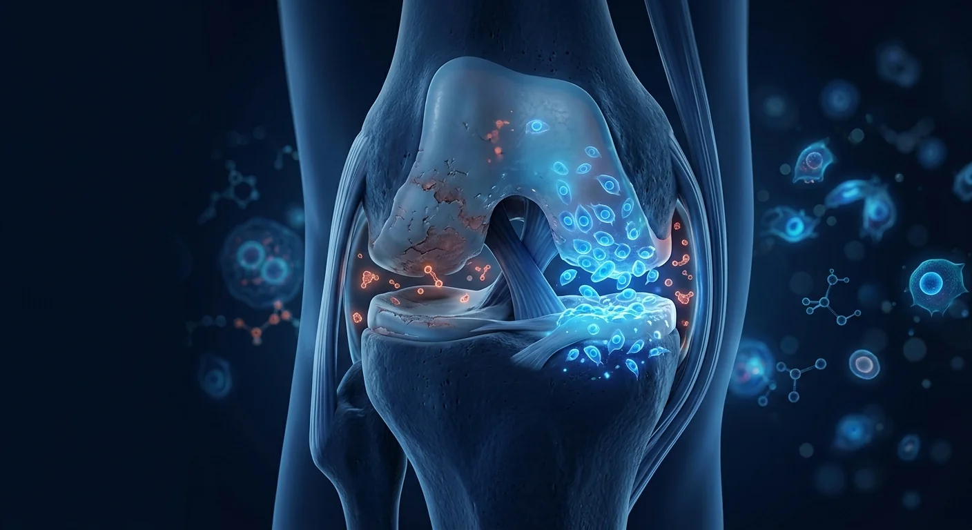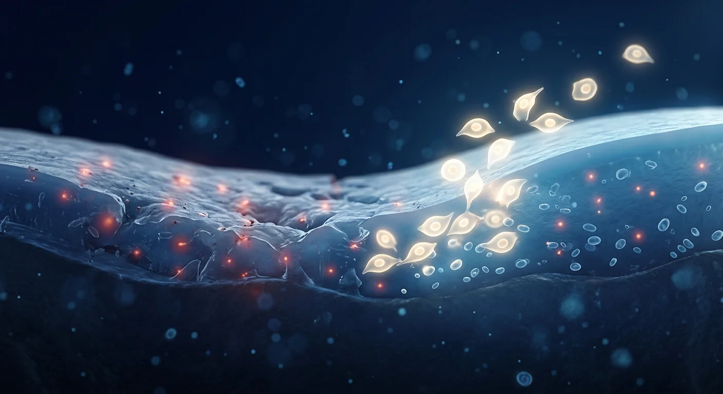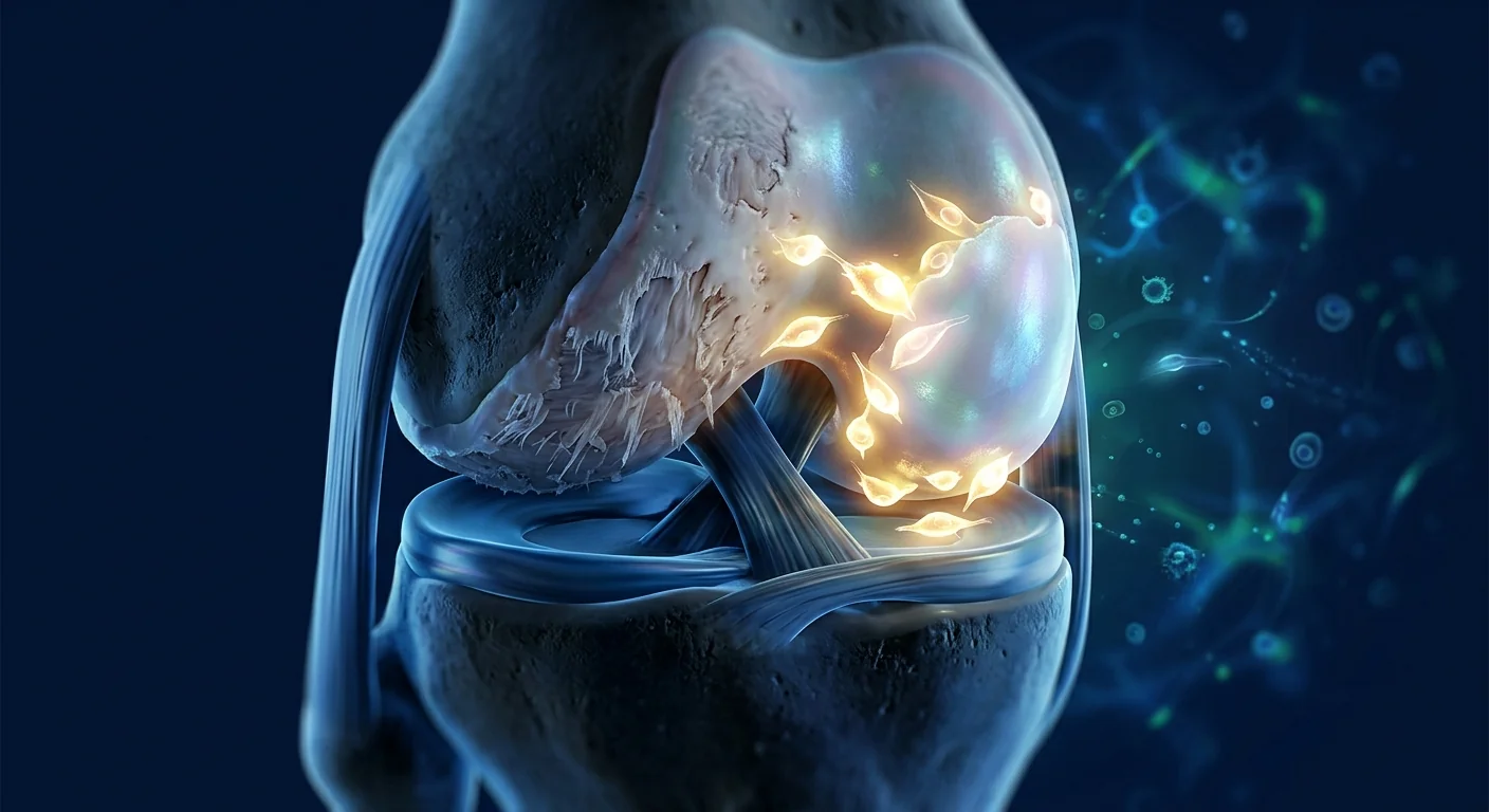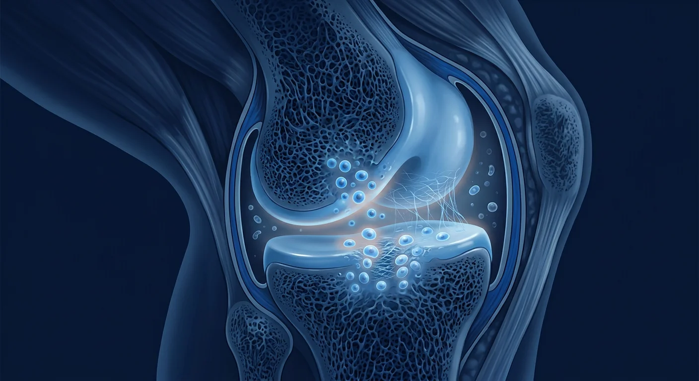
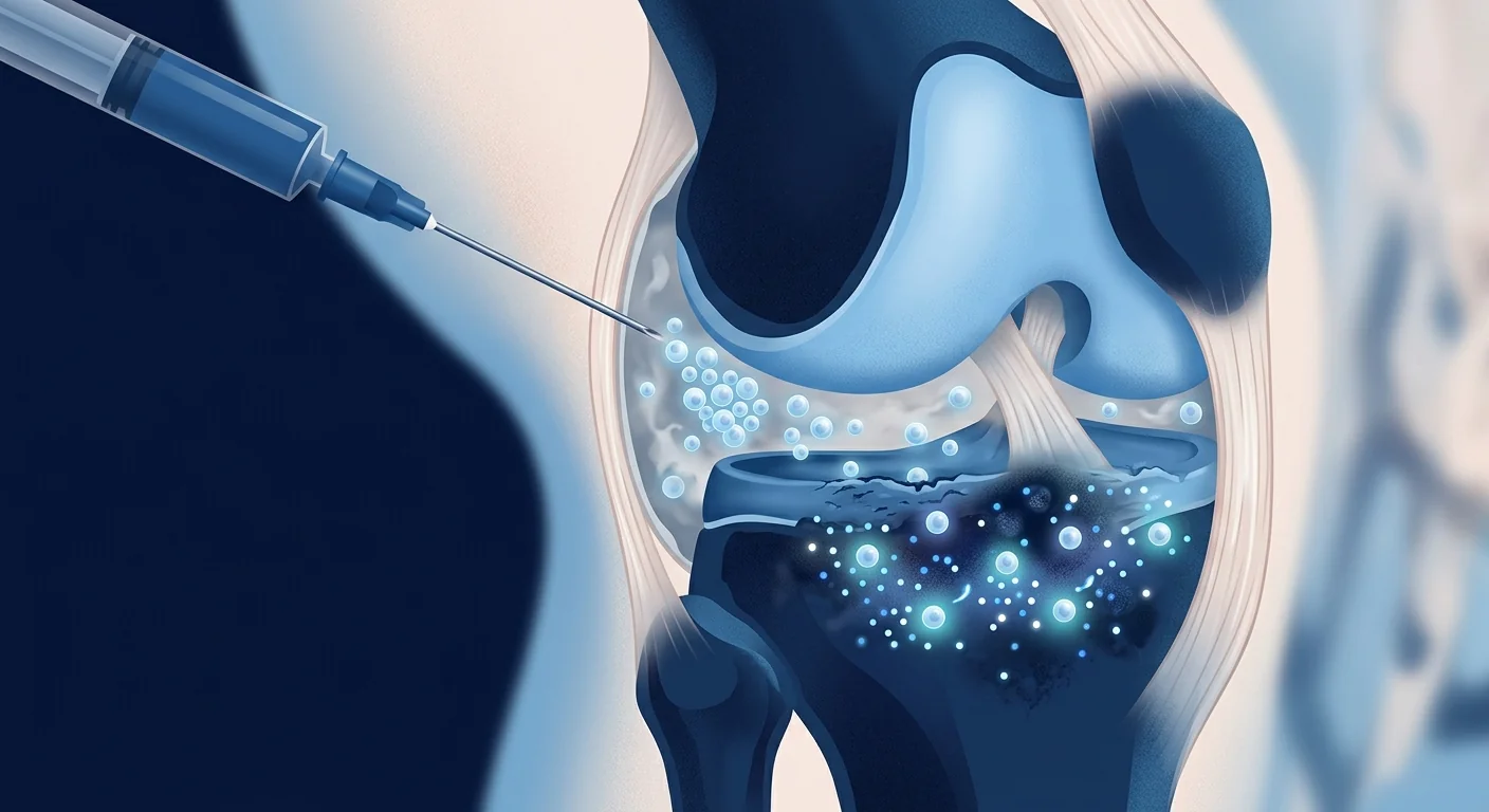
Stem cell therapy for knee osteoarthritis means injecting mesenchymal stem cells (MSCs) directly into the knee joint — under imaging or clinical guidance — with the goal of calming the low-grade inflammation that drives the disease and supporting the joint's own capacity to maintain its cartilage. Unlike a knee replacement, it is a biologic intervention aimed at joints that still have cartilage worth preserving; unlike hyaluronic acid, it attempts to address the biology of the joint rather than lubricate it.
Why the knee. Osteoarthritis is the leading cause of chronic knee pain in adults over 60, and the knee is by far the most commonly affected joint — so the MSC evidence base is richest for the knee. If a condition has the most trials, the knee does.
The honest frame. As of 2026, no MSC product is approved by the FDA or EMA as standard-of-care treatment for osteoarthritis. The responsible description is advanced investigational therapy with a growing and largely consistent evidence base — not a cure. Anything less honest is marketing, and we explain below how to tell the difference.
What this article covers. What is actually happening inside an osteoarthritic knee, why MSCs are a rational candidate, what the randomised trials show on pain and function, what the structural imaging data suggest, how to evaluate a provider, and what the evidence does not yet support.
What is knee osteoarthritis — and why "wear and tear" is an incomplete story
Knee osteoarthritis is a whole-joint disease in which the articular cartilage progressively loses its structure, the underlying bone remodels, and the synovial lining becomes inflamed. The "wear and tear" framing is incomplete because OA is now understood to have a significant inflammatory and metabolic component: the disease starts at the cellular level, not at the mechanical one [1]. Age, obesity, prior knee injury (including ligament tears and meniscal loss), and genetic factors all tilt the balance toward degeneration.
The consequence is a familiar one: a knee that aches with stairs, swells after activity, and progressively loses range of motion. For decades, the treatment ladder was pain relief, physiotherapy, and — when the joint space has narrowed to bone-on-bone — total knee arthroplasty. MSC therapy sits on that ladder as a new rung: a biological intervention that may slow progression and reduce symptoms in joints that have not yet reached the surgical endpoint.
What happens inside an osteoarthritic knee
Three processes run in parallel, and a rational cell therapy has to address all three of them.
Cartilage failure begins with chondrocyte senescence. Cartilage is maintained by resident cells — chondrocytes — that continuously synthesise and remodel the collagen and proteoglycan matrix. In OA, these cells become senescent: they stop dividing, accumulate damaged organelles, and start secreting matrix-degrading enzymes (notably MMP-13 and ADAMTS-5) and pro-inflammatory signals that accelerate their own damage and that of their neighbours [2]. The cartilage surface fibrillates, fissures, and eventually delaminates.
Inflammation is compartmentalised, not systemic. Synovial macrophages release IL-1β, TNF-α, and IL-6 into the joint space. This inflammation is real but largely confined to the joint — which is precisely why a locally delivered therapy makes sense, and why systemic immunosuppression does not. Cytokine-driven sensitisation of peripheral nociceptors also explains a large share of the pain that patients report [3].
The subchondral bone remodelling. Abnormal bone remodelling beneath the cartilage produces sclerotic plates and osteophytes. It contributes to pain and to the characteristic narrowing visible on X-ray. Structural change lags symptom change by months to years, which matters when interpreting trial data.
Why mesenchymal stem cells are a candidate for knee osteoarthritis
The rationale for MSCs maps directly onto the three processes above. MSCs do not need to become cartilage to be useful; their primary value in the joint is paracrine — the signals they secrete into the joint space.
Immunomodulation. MSCs suppress the pro-inflammatory (M1) phenotype of synovial macrophages and shift them toward a tissue-remodelling (M2) phenotype, lowering the intra-articular levels of IL-1β and TNF-α [4]. This is the mechanism with the most consistent preclinical support, and it matches the compartmentalised nature of OA inflammation.
Chondroprotection and trophic support. MSCs secrete growth factors — TGF-β, IGF-1, BMPs — that support the survival and matrix-producing activity of the patient's own chondrocytes. In co-culture experiments, MSCs reduce chondrocyte apoptosis and slow the loss of proteoglycan from cartilage explants exposed to inflammatory stimuli [5].
Direct matrix contribution. Although MSCs are not expected to regenerate a pristine articular surface de novo, they can differentiate toward chondrocyte-like cells and contribute to repair tissue, particularly when delivered alongside a scaffold or microfracture procedure [6].
Delivery is almost always intra-articular — injected directly into the joint space. Local delivery concentrates the cells where they are needed, keeps systemic exposure low, and is consistent with the favourable safety profile observed across the trials.
What the clinical evidence actually shows
The knee OA MSC literature is the largest of any MSC indication — multiple randomised controlled trials, several meta-analyses, and consistent signals. A recent systematic review and meta-analysis of RCTs concluded that intra-articular MSC injection is associated with statistically significant and clinically meaningful reductions in pain, as measured by the Visual Analogue Scale (VAS) and the WOMAC pain subscale, and with function improvements that persist for 12 to 24 months in many studies [7].
Several individual trials define the evidence. In a 2015 randomised controlled trial, intra-articular allogeneic bone-marrow MSCs produced significantly better pain and cartilage-quality scores (measured by T2 mapping on MRI) than hyaluronic acid at 12 months in 30 patients [8]. A larger multicentre phase I/II trial randomised patients to two MSC doses or hyaluronic acid; the higher dose produced significant WOMAC and VAS improvements at 12 months compared with control [9]. A 2019 randomised phase I/II trial of umbilical-cord MSCs — repeated dosing versus a single dose versus hyaluronic acid — found the repeated-dose group superior on pain and function at 12 months, and MRI suggested a disease-modifying effect: less progression of cartilage loss in the MSC-treated knees [10].
Cell sources vary across the literature — bone marrow, umbilical cord, adipose tissue, placenta — with umbilical-cord-derived MSCs producing some of the strongest signals in recent trials because they are younger, more proliferative, and avoid the morbidity of a bone-marrow harvest [11].
The honest headline
No MSC product is approved by the FDA or EMA for osteoarthritis as of 2026. The evidence is considerably stronger than for many other MSC indications — multiple RCTs with consistent pain and function signals — but it has not reached the level of regulatory approval. Any clinic promising guaranteed cartilage regrowth or complete pain resolution is misrepresenting the data.
How outcomes are measured — and what MSCs actually move
Knowing the endpoints tells you what a trial can and cannot claim. The patient-reported gold standard is the WOMAC index (pain, stiffness, function across 24 items); the VAS pain score (0–100 mm) is a simpler single-dimension readout. Structurally, MRI with T2 mapping quantifies cartilage composition, while plain radiographs track joint-space narrowing — with the caveat that structural change lags symptoms by months to years.
Across the RCTs, the pattern is consistent: MSC groups show greater pain reduction than hyaluronic acid or saline controls by 6–12 months, with mean VAS improvements of roughly 20–40 mm and WOMAC improvements of 15–25 points — above the thresholds considered clinically meaningful. The structural data are more preliminary: several trials report slower cartilage volume loss or improved T2 relaxation times on MRI, but the effects are modest and not universal [12]. The strongest structural signals come from repeated dosing rather than a single injection [10], and combination approaches (MSCs with platelet-rich plasma or scaffolds) remain under investigation [13].
What the evidence supports — and what it does not
A fair reading supports three conclusions. Intra-articular MSC therapy for knee OA has a well-established short- and medium-term safety record, with adverse events generally limited to transient post-injection pain and swelling. It consistently reduces pain and improves function in controlled trials, with effect sizes that are clinically meaningful and appear durable for 12–24 months. And there are preliminary structural signals suggesting possible disease modification — signals that require confirmation in larger, longer trials with MRI-based primary endpoints.
Equally important is what the evidence does not yet support:
- It does not establish reversal of established OA. Grade 4 (bone-on-bone) joints with complete cartilage loss do not regenerate a new cartilage surface. The strongest trial signals come from moderate OA (Kellgren-Lawrence grades 2–3), where cartilage is still present.
- It does not confirm durability beyond 2–3 years. Most trials follow patients 12–24 months; longer follow-up data are sparse.
- It does not define a single optimal protocol. Cell source, dose, and frequency vary widely across studies, and head-to-head comparisons are limited.
- It does not show superiority over total knee arthroplasty for end-stage disease. When conservative measures are exhausted, arthroplasty remains the definitive option.
MSC therapy is best understood as a bridge — a biologic intervention that may extend the functional life of a joint and defer arthroplasty — not as a replacement for weight management, physiotherapy, and appropriate analgesia.
How to evaluate a provider before you commit
The diligence is straightforward, and the same questions protect against over-promise in any medical domain. Ask:
- What cell type and source? Umbilical-cord MSCs, bone-marrow MSCs, and adipose stromal tissue are biologically different products with different evidence bases.
- Can the provider cite published data for their specific protocol? General references to "the field" are not the same as data for the exact cells, dose, and schedule they will use.
- What outcome measures do they use? A provider who tracks WOMAC and VAS scores before and after treatment operates with a rigour that is absent from clinics offering only testimonials.
- What quality data exist for the cells? Identity testing against the ISCT criteria (CD73+, CD90+, CD105+, CD45-), viability above 95% at delivery, and sterility release before use are the baseline for any serious cell therapy.
Be deeply sceptical of guarantees, of claims that bone-on-bone knees will regenerate cartilage, and of any suggestion that MSC therapy replaces established conservative care.
The VELAR perspective
At VELAR Center, knee osteoarthritis is one of the conditions we treat most frequently — and precisely because it is common, we hold ourselves to a higher evidence bar, not a lower one. We use umbilical-cord-derived MSCs manufactured under cGMP conditions with ISCT-compliant characterisation and third-party sterility release, we deliver the cells fresh — never frozen — and we track structured outcomes (WOMAC, VAS, and where indicated, follow-up imaging). We position MSC therapy as a bridge that may extend the functional life of a joint and defer arthroplasty, not as a miracle that undoes decades of degeneration. Every consultation begins with an honest conversation about what the evidence says, what it does not say, and whether — given your specific imaging and clinical status — a biologic approach is a rational next step or whether the data suggest you would be better served by other options.
Frequently Asked Questions
How much does stem cell therapy for knee osteoarthritis cost?
Costs vary substantially by provider, cell source, and dosing schedule — single-injection programs are at the low end, repeated-dose programs higher. Because the evidence is investigational, MSC therapy for OA is typically not covered by insurance, and prices in Thailand commonly range from several thousand to several tens of thousands of USD. Any legitimate provider will quote a fixed, itemised price before treatment begins.
Does MSC therapy actually regrow cartilage?
Current evidence does not support full cartilage regeneration in established osteoarthritis. Trials show modest structural signals — slower cartilage volume loss and improved T2 mapping in some studies — but the primary, consistent effect is reduction in pain and improvement in function. Claims of complete cartilage regrowth are not supported by the published data.
How long do the effects last?
Most randomised trials report meaningful pain and function improvements at 12 months, with durability signals to 24 months in several studies. Data beyond 2–3 years are limited, and repeated dosing appears to be associated with stronger and more durable effects than a single injection.
Is the procedure safe?
Intra-articular MSC injection has a favourable short- and medium-term safety profile across the published trials. Reported adverse events are generally limited to transient post-injection pain, swelling, or stiffness. As with any cell therapy, sterility and identity testing of the cells, and a clean injection technique, are what make that profile what it is.
Who is a good candidate for MSC therapy for knee osteoarthritis?
The strongest evidence comes from patients with moderate OA (Kellgren-Lawrence grades 2–3) — enough cartilage remaining for a biologic intervention to have something to support. Patients with severe, bone-on-bone disease, uncontrolled inflammatory conditions, or active joint infection are generally not candidates. Imaging and a clinical assessment determine candidacy for any individual patient.
Limitations of this review
MSC therapy for knee osteoarthritis remains investigational. The published evidence — while consistent on symptoms — comes from relatively small trials, with heterogeneous cell sources, doses, and follow-up durations. Structural endpoints are preliminary, and long-term durability and optimal dosing are not yet established. This article summarises published research for educational purposes and does not constitute medical advice; treatment decisions should be made with a qualified clinician after imaging and clinical assessment.
References
- Wang D, Alkhatib H, Lalevée M, et al. Osteoarthritis: burden of disease and management. Rheumatology International. 2014;34(12):1783-1789. doi:10.1007/s00394-014-2395-8 ↩
- Lehman AM, Kishida CF, Martin JA, et al. Chondrocyte senescence: a target for the treatment of osteoarthritis. Current Opinion in Biotechnology. 2016;37:59-64. doi:10.1016/j.copbio.2016.01.005 ↩
- Buckley EL, Fuchs PK, Kraus VB, Loeser RF. The role of inflammation in osteoarthritis pain. Seminars in Arthritis and Rheumatism. 2015;45(1):85-88. doi:10.1016/j.semarthrit.2014.12.007 ↩
- Muller J, Roehrich S, Riehn D, et al. Mesenchymal stem cell therapy for inflammatory and degenerative diseases. Journal of Internal Medicine. 2021;289(6):649-669. doi:10.1111/joim.13149 ↩
- Kusabuchi H, Nakagawa K, Nakashima M, et al. Human mesenchymal stem cells ameliorate immune-mediated inflammation and tissue destruction in rheumatoid arthritis. Arthritis & Rheumatism. 2007;56(11):3473-3482. doi:10.1002/art.22873 ↩
- Wakitani S, Saito M, Mabuchi K, et al. Tissue engineering: current progress and clinical applications. Journal of Orthopaedic Science. 2004;9(4):411-424. doi:10.1007/s00776-004-0755-z ↩
- Cao Y, Ou Y, Sheng W, et al. Efficacy and safety of mesenchymal stem cells in knee osteoarthritis: a systematic review and meta-analysis of randomized controlled trials. Stem Cell Research & Therapy. 2025;16(1):122. doi:10.1186/s13287-025-04252-2 ↩
- Vega A, Martín-Ferrero MA, Del Canto F, et al. Treatment of knee osteoarthritis with allogeneic bone marrow mesenchymal stem cells: a randomized controlled trial. Transplantation. 2015;99(8):1681-1690. doi:10.1097/TP.0000000000000678 ↩
- Lamo-Espinosa JM, Mora G, Blanco JF, et al. Intra-articular injection of two different doses of autologous bone marrow mesenchymal stem cells versus hyaluronic acid in the treatment of knee osteoarthritis: multicenter randomized controlled clinical trial (phase I/II). Journal of Translational Medicine. 2016;14:246. doi:10.1186/s12967-016-0998-2 ↩
- Matas J, Orrego M, Amenabar D, et al. Umbilical cord-derived mesenchymal stromal cells (MSCs) for knee osteoarthritis: repeated MSC dosing is superior to a single MSC dose and to hyaluronic acid in a controlled randomized phase I/II trial. Stem Cells Translational Medicine. 2019;8(3):215-224. doi:10.1002/sctm.18-0053 ↩
- Freitag J, Bates D, Boyd R, et al. Mesenchymal stem cell therapy in the treatment of osteoarthritis: reparative pathways, safety and efficacy — a review. BMC Musculoskeletal Disorders. 2016;17:230. doi:10.1186/s12891-016-1085-9 ↩
- Kim YS, Choi YJ, Lee SW, et al. Assessment of clinical and MRI outcomes after mesenchymal stem cell implantation in patients with knee osteoarthritis: a prospective study. Osteoarthritis and Cartilage. 2016;24(2):237-245. doi:10.1016/j.joca.2015.08.009 ↩
- Bastos R, Mathias M, Andrade R, et al. Intra-articular injection of culture-expanded mesenchymal stem cells with or without addition of platelet-rich plasma is effective in decreasing pain and symptoms in knee osteoarthritis: a controlled, double-blind clinical trial. Knee Surgery, Sports Traumatology, Arthroscopy. 2020;28(7):1989-1999. doi:10.1007/s00167-019-05732-8 ↩
- Jo CH, Lee YG, Shin WH, et al. Intra-articular injection of mesenchymal stem cells for the treatment of osteoarthritis of the knee: a proof-of-concept clinical trial. Stem Cells. 2014;32(5):1254-1266. doi:10.1002/stem.1634 ↩
膝关节骨关节炎的干细胞治疗,是指将间充质干细胞(MSC)直接注射到膝关节内——在影像或临床引导下进行——目的是平息驱动该疾病的低度炎症,并支持关节自身维持软骨的能力。与膝关节置换不同,这是一种生物干预,针对的仍是尚有值得保留的软骨的关节;与透明质酸不同,它试图解决关节的生物学问题,而不是仅仅润滑它。
为什么是膝盖。骨关节炎是60岁以上成人慢性膝痛的首要原因,而膝关节是受累最多的关节——因此MSC的证据基础在膝关节上最丰富。哪个病种的试验最多,就是膝关节。
诚实的定位。截至2026年,没有任何MSC产品被FDA或EMA批准为骨关节炎的标准治疗。负责任的表述是证据基础不断扩大的先进研究性疗法——而非治愈手段。任何不够诚实的宣传都只是营销,下文将解释如何辨别。
本文内容。骨关节炎膝关节内实际发生了什么、为什么MSC是合理的候选、随机试验在疼痛和功能上显示了什么、结构影像数据提示了什么、如何评估医疗机构,以及证据目前尚未支持什么。
什么是膝关节骨关节炎——为什么"磨损"是不完整的故事
膝关节骨关节炎是一种全关节疾病:关节软骨逐渐失去结构,其下的骨发生重塑,滑膜内层出现炎症。"磨损"框架之所以不完整,是因为OA目前被认为具有显著的炎症和代谢成分:疾病始于细胞层面,而非机械层面 [1]。年龄、肥胖、既往膝部损伤(包括韧带撕裂和半月板缺失)以及遗传因素都会使天平倒向退变一侧。
后果是熟悉的:上下楼梯时疼痛的膝盖、活动后肿胀、逐步丧失活动范围。几十年来,治疗阶梯是止痛、物理治疗——当关节间隙狭窄到骨对骨时——进行全膝关节置换。MSC治疗在这条阶梯上是一个新的层级:一种可能延缓进展、减轻症状的生物干预,适用于尚未到达手术终点的关节。
骨关节炎膝关节内发生了什么
三个过程并行发生,而理性的细胞疗法必须同时应对这三者。
软骨失败始于软骨细胞衰老。软骨由驻留细胞——软骨细胞——维持,它们持续合成和重塑胶原与蛋白聚糖基质。在OA中,这些细胞变得衰老:停止分裂、积累受损细胞器,并开始分泌基质降解酶(尤其是MMP-13和ADAMTS-5)以及加速自身和周围细胞损伤的促炎信号 [2]。软骨表面纤维化、开裂,最终剥脱。
炎症是局部的,而非全身性的。滑膜巨噬细胞向关节腔释放IL-1β、TNF-α和IL-6。这种炎症是真实存在的,但基本局限于关节内——这正是局部给药治疗合理、而全身免疫抑制不合理的原因。细胞因子驱动的周围伤害感受器敏化也解释了患者所报告疼痛的很大一部分 [3]。
软骨下骨重塑。软骨下异常骨重塑形成硬化板与骨赘,它贡献了疼痛,也是X线上特征性狭窄的表现。结构变化滞后于症状变化数月至数年,这在解读试验数据时很重要。
为什么间充质干细胞是膝关节骨关节炎的候选
MSC的理论与上述三个过程直接对应。MSC不需要变成软骨才有用;它们在关节内的主要价值是旁分泌的——它们分泌到关节腔中的信号。
免疫调节。MSC抑制滑膜巨噬细胞的促炎(M1)表型,使其转向组织重塑(M2)表型,降低关节内IL-1β和TNF-α水平 [4]。这是临床前支持最一致的机制,也与OA炎症的局限性特征相符。
软骨保护与营养支持。MSC分泌生长因子——TGF-β、IGF-1、BMPs——支持患者自身软骨细胞的存活与基质产生活性。共培养实验中,MSC减少软骨细胞凋亡,并减缓炎症刺激下软骨外植体蛋白聚糖的丢失 [5]。
直接基质贡献。虽然不期望MSC从头再生出完美的关节面,但它们可向软骨样细胞分化并贡献修复组织,尤其是与支架或微骨折术联合给药时 [6]。
给药几乎总是关节内——直接注射到关节腔。局部给药将细胞集中在需要的地方、降低全身暴露,并与试验中观察到的良好安全性特征一致。
临床证据实际显示了什么
膝关节OA的MSC文献是任何MSC适应症中最庞大的——多项随机对照试验、多个荟萃分析,以及一致的信号。近期一项RCT系统评价与荟萃分析得出结论:关节内MSC注射与疼痛的统计学显著且具有临床意义的减轻相关(以视觉模拟量表VAS和WOMAC疼痛分量表测量),且功能改善在许多研究中持续12至24个月 [7]。
几项个体试验定义了证据。一项2015年的随机对照试验中,关节内同种异体骨髓MSC在30名患者中产生了比透明质酸显著更好的疼痛与软骨质量评分(以MRI T2映射测量),随访12个月 [8]。一项更大的多中心I/II期试验将患者随机分配到两种MSC剂量或透明质酸;高剂量组在12个月时产生显著的WOMAC与VAS改善 [9]。一项2019年脐带MSC随机I/II期试验——重复给药对比单次给药对比透明质酸——发现重复给药组在12个月时疼痛与功能更优,且MRI提示疾病修饰效应:MSC治疗的膝关节软骨丢失进展更少 [10]。
文献中的细胞来源各异——骨髓、脐带、脂肪、胎盘——其中脐带来源MSC在近期试验中产生了一些最强信号,因为它们更年轻、增殖能力更强,且避免了骨髓采集的不适 [11]。
诚实的结论
截至2026年,没有任何MSC产品被FDA或EMA批准用于骨关节炎。证据比许多其他MSC适应症强得多——多项RCT有一致的疼痛与功能信号——但尚未达到监管批准的水平。任何承诺保证软骨再生或完全消除疼痛的机构都在歪曲数据。
结果如何衡量——以及MSC实际改变了什么
了解终点能告诉你一项试验能声称什么。患者报告的金标准是WOMAC指数(24个项目涵盖疼痛、僵硬、功能);VAS疼痛评分(0–100 mm)是更简单的单一维度读数。结构上,MRI T2映射可量化软骨成分,平片则跟踪关节间隙狭窄——但结构变化滞后症状数月至数年。
各RCT中模式一致:MSC组在6–12个月时显示比透明质酸或盐水对照更大的疼痛减轻,平均VAS改善约20–40 mm,WOMAC改善15–25分——均超过被认为具有临床意义的阈值。结构数据更初步:若干试验报告MRI上软骨体积丢失减慢或T2弛豫时间改善,但效应温和且并非普遍 [12]。最强的结构信号来自重复给药而非单次注射 [10],联合方案(MSC联合富血小板血浆或支架)仍在研究中 [13]。
证据支持什么——又不支持什么
公允的解读支持三点结论。膝关节OA的关节内MSC治疗具有确立良好的短期与中期安全记录,不良事件通常限于短暂的注射后疼痛与肿胀。在对照试验中它持续减轻疼痛并改善功能,效应量具有临床意义且似乎持续12–24个月。此外存在初步的结构信号,提示可能的疾病修饰——这些信号需要在以MRI为主要终点的更大、更长期试验中确认。
同样重要的是证据目前尚未支持的:
- 未确立逆转已确立的OA。4级(骨对骨)关节完全丧失软骨,不会再生出新的软骨面。最强的试验信号来自中度OA(Kellgren-Lawrence 2–3级),此时仍有软骨存在。
- 未确认超过2–3年的持久性。大多数试验随访12–24个月,更长期的数据稀疏。
- 未确定单一最佳方案。细胞来源、剂量、频率在各研究中差异很大,头对头比较有限。
- 未显示对终末期疾病优于全膝关节置换。当保守措施耗尽时,关节置换仍是确定性选择。
MSC治疗最好被理解为一种桥梁——可能延长关节功能寿命、推迟关节置换的生物干预——而非体重管理、物理治疗与适当镇痛的替代品。
如何在你决定之前评估医疗机构
审慎是直接的,同样的问题在任何医疗领域都能防范过度承诺。要问:
- 何种细胞类型与来源?脐带MSC、骨髓MSC与脂肪基质组织是生物学上不同的产品,证据基础各异。
- 机构能否为其具体方案引用已发表的数据?对"该领域"的一般性引用,与其将使用的确切细胞、剂量和方案的数据不是一回事。
- 他们使用什么结果测量?治疗前后跟踪WOMAC与VAS评分的机构,其严谨程度是仅提供患者感言的机构所不具备的。
- 细胞存在什么质量数据?按ISCT标准(CD73+、CD90+、CD105+、CD45-)进行身份检测、交付时活性高于95%、使用前无菌放行,是任何严肃细胞治疗的基线。
对保证、对骨对骨膝盖将再生软骨的说法、以及对MSC治疗可替代已确立保守治疗的任何暗示,都要深度怀疑。
VELAR 的观点
在VELAR中心,膝关节骨关节炎是我们最常治疗的病症之一——正因为它常见,我们对自身施加更高的证据标准而非更低。我们使用在cGMP条件下制造、具有ISCT合规鉴定与第三方无菌放行的脐带来源MSC,细胞新鲜交付——从不冷冻——并跟踪结构化结果(WOMAC、VAS,以及适当时的随访影像)。我们将MSC治疗定位为可能延长关节功能寿命、推迟关节置换的桥梁,而非消除数十年退化的奇迹。每次咨询都以关于证据说了什么、没说什么的诚实对话开始,以及根据您的具体影像与临床状态,生物方法是否是合理的下一步,还是数据表明其他选择可能更适合您。
常见问题
膝关节骨关节炎的干细胞治疗费用是多少?
费用因机构、细胞来源与给药方案差异很大——单次注射方案处于低端,重复给药方案更高。由于证据仍属研究性,OA的MSC治疗通常不在保险覆盖范围内,泰国价格通常从数千到数万美元不等。任何正规机构都会在治疗前提供固定、逐项列明的报价。
MSC治疗真的能让软骨再生吗?
目前证据不支持已确立骨关节炎的完全软骨再生。试验显示温和的结构信号——部分研究中软骨体积丢失减慢、T2映射改善——但主要且一致的效应是疼痛减轻与功能改善。完全软骨再生的说法未获已发表数据支持。
效果能持续多久?
大多数随机试验报告12个月时有意义的疼痛与功能改善,若干研究中持久性信号达24个月。超过2–3年的数据有限,且重复给药似乎与比单次注射更强、更持久的效应相关。
这个程序安全吗?
已发表试验中,关节内MSC注射具有良好的短期与中期安全性特征。报告的不良事件通常限于短暂的注射后疼痛、肿胀或僵硬。与任何细胞治疗一样,细胞的无菌与身份检测,以及无菌的注射技术,决定了这一安全性特征。
谁适合膝关节骨关节炎的MSC治疗?
最强证据来自中度OA(Kellgren-Lawrence 2–3级)患者——仍有足够软骨供生物干预支持。重度骨对骨病变、未控制的炎症性疾病或活动性关节感染者通常不适合。影像与临床评估决定任何个体患者的适宜性。
本综述的局限性
膝关节OA的MSC治疗仍属研究性。已发表证据——在症状上一致——来自规模相对较小的试验,细胞来源、剂量与随访时长异质。结构终点是初步的,长期持久性与最佳剂量尚未确立。本文总结已发表研究用于教育目的,不构成医疗建议;治疗决定应在影像与临床评估后与合格医生共同做出。
参考文献
- Wang D, Alkhatib H, Lalevée M, et al. Osteoarthritis: burden of disease and management. Rheumatology International. 2014;34(12):1783-1789. doi:10.1007/s00394-014-2395-8 ↩
- Lehman AM, Kishida CF, Martin JA, et al. Chondrocyte senescence: a target for the treatment of osteoarthritis. Current Opinion in Biotechnology. 2016;37:59-64. doi:10.1016/j.copbio.2016.01.005 ↩
- Buckley EL, Fuchs PK, Kraus VB, Loeser RF. The role of inflammation in osteoarthritis pain. Seminars in Arthritis and Rheumatism. 2015;45(1):85-88. doi:10.1016/j.semarthrit.2014.12.007 ↩
- Muller J, Roehrich S, Riehn D, et al. Mesenchymal stem cell therapy for inflammatory and degenerative diseases. Journal of Internal Medicine. 2021;289(6):649-669. doi:10.1111/joim.13149 ↩
- Kusabuchi H, Nakagawa K, Nakashima M, et al. Human mesenchymal stem cells ameliorate immune-mediated inflammation and tissue destruction in rheumatoid arthritis. Arthritis & Rheumatism. 2007;56(11):3473-3482. doi:10.1002/art.22873 ↩
- Wakitani S, Saito M, Mabuchi K, et al. Tissue engineering: current progress and clinical applications. Journal of Orthopaedic Science. 2004;9(4):411-424. doi:10.1007/s00776-004-0755-z ↩
- Cao Y, Ou Y, Sheng W, et al. Efficacy and safety of mesenchymal stem cells in knee osteoarthritis: a systematic review and meta-analysis of randomized controlled trials. Stem Cell Research & Therapy. 2025;16(1):122. doi:10.1186/s13287-025-04252-2 ↩
- Vega A, Martín-Ferrero MA, Del Canto F, et al. Treatment of knee osteoarthritis with allogeneic bone marrow mesenchymal stem cells: a randomized controlled trial. Transplantation. 2015;99(8):1681-1690. doi:10.1097/TP.0000000000000678 ↩
- Lamo-Espinosa JM, Mora G, Blanco JF, et al. Intra-articular injection of two different doses of autologous bone marrow mesenchymal stem cells versus hyaluronic acid in the treatment of knee osteoarthritis: multicenter randomized controlled clinical trial (phase I/II). Journal of Translational Medicine. 2016;14:246. doi:10.1186/s12967-016-0998-2 ↩
- Matas J, Orrego M, Amenabar D, et al. Umbilical cord-derived mesenchymal stromal cells (MSCs) for knee osteoarthritis: repeated MSC dosing is superior to a single MSC dose and to hyaluronic acid in a controlled randomized phase I/II trial. Stem Cells Translational Medicine. 2019;8(3):215-224. doi:10.1002/sctm.18-0053 ↩
- Freitag J, Bates D, Boyd R, et al. Mesenchymal stem cell therapy in the treatment of osteoarthritis: reparative pathways, safety and efficacy — a review. BMC Musculoskeletal Disorders. 2016;17:230. doi:10.1186/s12891-016-1085-9 ↩
- Kim YS, Choi YJ, Lee SW, et al. Assessment of clinical and MRI outcomes after mesenchymal stem cell implantation in patients with knee osteoarthritis: a prospective study. Osteoarthritis and Cartilage. 2016;24(2):237-245. doi:10.1016/j.joca.2015.08.009 ↩
- Bastos R, Mathias M, Andrade R, et al. Intra-articular injection of culture-expanded mesenchymal stem cells with or without addition of platelet-rich plasma is effective in decreasing pain and symptoms in knee osteoarthritis: a controlled, double-blind clinical trial. Knee Surgery, Sports Traumatology, Arthroscopy. 2020;28(7):1989-1999. doi:10.1007/s00167-019-05732-8 ↩
- Jo CH, Lee YG, Shin WH, et al. Intra-articular injection of mesenchymal stem cells for the treatment of osteoarthritis of the knee: a proof-of-concept clinical trial. Stem Cells. 2014;32(5):1254-1266. doi:10.1002/stem.1634 ↩
يعني علاج فصال العظم (التآكل) في الركبة بالخلية الجذعية حقن خلايا جذعية وسите (MSCs) مباشرةً في جوف المفصل الركبي — تحت إرشاد التصوير أو التقدير السريري — بهدف تهدئة الالتهاب منخفض الدرجة الذي يقود المرض ودعم قدرة المفصل على الحفاظ على غضروفه. وعلى خلاف استبدال الركبة، فهو تدخّل بيولوجي يستهدف مفاصل لا يزال فيها غضروف يستحق الحفظ؛ وعلى خلاف حمض الهيالورونيك، فهو يحاول معالجة بيولوجيا المفصل لا مجرد تزليقه.
لماذا الركبة؟ فصال العظم هو السبب الأول لألم الركبة المزمن لدى البالغين فوق سن الستين، والركبة هي المفصل الأكثر تضررًا بكثير — ولذلك قاعدة الأدلة حول الخلايا الجذعية الوسيطة أغنى فيما يخص الركبة. إن كان هناك استطباب تمتلك تجاربه العشوائية المضبوطة العدد الأكبر، فهو الركبة.
الإطار الصريح. حتى عام 2026، لا يوجد أي منتج للخلايا الجذعية الوسيطة معتمد من إدارة الغذاء والدواء الأمريكية (FDA) أو وكالة الأدوية الأوروبية (EMA) كعلاج قياسي لفصال العظم. الوصف المسؤول هو علاج تجريبي متقدم بقاعدة أدلة متنامية ومتسقة في الغالب — لا علاج شافٍ. وكل ما دون ذلك من وعود هو تسويق، وسنشرح أدناه كيف تفرّق بينهما.
ما يغطيه هذا المقال. ما يحدث فعلًا داخل ركبة مصابة بفصال العظم، ولماذا الخلايا الجذعية الوسيطة مرشحة عقلية، وما الذي تظهره التجارب العشوائية المضبوطة من ألم ووظيفة، وما الذي تقترحه بيانات التصوير البنيوي، وكيف تقيّم مزوّد العلاج، وما الذي لا تدعمه الأدلة بعد.
ما هو فصال العظم في الركبة — ولماذا "التآكل" قصة ناقصة
فصال العظم في الركبة مرض يشمل المفصل بأكمله: يفقد الغضروف المفصلي بنيته تدريجيًا، ويُعاد تشكيل العظم التحت-غضروفي، وتصبح البطانة الزليلية ملتهبة. إن الإطار القائم على "التآكل" ناقص لأن فصال العظم يُفهم اليوم على أن له مكوّنًا التهابيًا واستقلابيًا كبيرًا: فالمرض يبدأ على مستوى الخلية لا على مستوى الميكانيكا [1]. والعمر والسمنة والإصابات الركبية السابقة (بما فيها تمزق الأربطة وفقدان القرص الغضروفي) والعوامل الوراثية كلها تميل الكفة نحو التنكس.
والنتيجة مألوفة: ركبة تؤلم مع صعود الدرج، وتورم بعد النشاط، وفقدان تدريجي لنطاق الحركة. ولعقود، كان سلّم العلاج هو تخفيف الألم والعلاج الطبيعي — وحين يضيق جوف المفصل إلى حد العظم على العظم — استبدال الركبة كليًا. ويستقر علاج الخلايا الجذعية الوسيطة في هذا السلّم كحلمة جديدة: تدخّل بيولوجي قد يبطئ التقدم ويقلل الأعراض في المفاصل التي لم تبلغ بعد نقطة النهاية الجراحية.
ما الذي يحدث داخل ركبة مصابة بفصال العظم
ثلاث عمليات تجري بالتوازي، ويجب أن يتصدى علاج الخلية العقلية لكلّها.
فشل الغضروف يبدأ بشيوخة الخلايا الغضروفية. الغضروف تحافظ عليه خلايا مقيمة — الخلايا الغضروفية — تصنع وتعيد تشكيل نسيج الكولاجين والبروتيوغليكان باستمرار. وفي فصال العظم تصبح هذه الخلايا شائخة: تتوقف عن الانقسام وتتراكم فيها عضيات تالفة، وتبدأ بإفراز إنزيمات محلّلة للنسيج (خصوصًا MMP-13 وADAMTS-5) وإشارات التهابية تسرّع ضررها هي وضرر جيرانها [2]. يتشقق سطح الغضروف ويتشقق ثم يتقشر.
الالتهاب مقصور على الحيّز وليس جهازيًا. البلاعم الزليلية تفرز IL-1β وTNF-α وIL-6 في جوف المفصل. هذا الالتهاب حقيقي لكنه محصور في المفصل غالبًا — وهذا بالذات يجعل العلاج المُعطى موضعيًا معقولًا بينما لا يكون المثبط المناعي الجهازي كذلك. وتحسيس مستقبلات الألم الطرفية الذي تقوده السيتوكينات يفسّر أيضًا حصة كبيرة من الألم الذي يبلغ به المرضى [3].
إعادة تشكيل العظم تحت الغضروفي. إعادة تشكيل العظم غير الطبيعية تحت الغضروف تنتج صفائح متصلبة ونوابت عظمية؛ وهي تساهم في الألم وفي التضيّق المميّظ الظاهر على الأشعة السينية. ويتأخر التغيّر البنيوي عن التغيّرات العرضية بأشهر إلى سنوات، وهذا مهم عند تفسير بيانات التجارب.
لماذا الخلايا الجذعية الوسيطة مرشحة لفصال العظم في الركبة
الأساس المنطقي للخلايا الجذعية الوسيطة يطابق العمليات الثلاث أعلاه مباشرةً. الخلايا الجذعية لا تحتاج أن تصبح غضروفًا لتفيد؛ قيمتها الأساسية في المفصل هي جانب-خلوية — الإشارات التي تفرزها في جوف المفصل.
تعديل المناعة. تكبح الخلايا الجذعية نمط البلاعم الزليلية المحرض على الالتهاب (M1) وتحوّله نحو نمط إعادة تشكيل النسيج (M2)، ما يخفض مستواي IL-1β وTNF-α داخل المفصل [4]. وهذا هو الآلية الأكثر دعمًا قبل السرير، وهو يطابق طبيعة التهاب فصال العظم المقصورة على الحيّز.
حماية الغضروف ودعم غذائي. تفرز الخلايا الجذعية عوامل نمو — TGF-β وIGF-1 وBMPs — تدعم بقاء خلايا الغضروف الخاصة بالمريض ونشاطها في إنتاج النسيج. وفي تجارب الزرع المشترك، تقلل الخلايا الجذعية استماتة الخلايا الغضروفية وتبطئ فقدان البروتيوغليكان من رقاقات الغضروف المعرّضة لمنبهات التهابية [5].
مساهمة مباشرة في النسيج. وإن لم يُتوقع من الخلايا الجذعية إيجاد سطح مفصلي سليم من العدم، فإنها تستطيع التمايز نحو خلايا شبيهة بالغضروفية والإسهام في نسيج الإصلاح، خصوصًا عند توصيلها مع دعامة أو إجراء الكسور الدقيقة [6].
التوصيل يكون دائمًا تقريبًا داخل المفصل — حقنًا مباشرًا في جوف المفصل. يركّز التوصيل الموضعي الخلايا حيث الحاجة، ويحافظ على التعرض الجهازي منخفضًا، وهو متسق مع ملمح الأمان المواتي الملاحظ عبر التجارب.
ماذا تُظهر الأدلة السريرية فعلًا
أدبيات فصال العظم في الركبة بالخلايا الجذعية هي الأكبر بين كل استطابات الخلايا الجذعية — تجارب عشوائية مضبوطة متعددة، ومراجعات تحليلية تلوية عدة، وإشارات متسقة. خلصت مراجعة منهجية وتحليل تلوي حديثان للتجارب العشوائية المضبوطة إلى أن حقن الخلايا الجذعية داخل المفصل يرتبط بانخفاض في الألم ذي دلالة إحصائية وأهمية سريرية، مقيسًا بمقياس التمثيل البصري (VAS) ومقياس WOMAC الفرعي للألم، وبالتحسّن في الوظيفة الذي يستمر من 12 إلى 24 شهرًا في كثير من الدراسات [7].
وتحدّد تجارب فردية عدة وجهات الأدلة. في تجربة عشوائية مضبوطة عام 2015، حقن الخلايا الجذعية الوسيطة الخيفية من نخاع العظم داخل المفصل أنتج درجات ألم وجودة غضروف أفضل بكثير (مقيسةً بتخطيط T2 بالرنين المغناطيسي) من حمض الهيالورونيك عند 12 شهرًا لدى 30 مريضًا [8]. وفي تجربة أكبر متعددة المراكز من المرحلة I/II عُدّي المرضى إلى جرعتين من الخلايا الجذعية أو حمض الهيالورونيك؛ فأنجزت الجرعة الأعلى تحسّنات ذات دلالة في WOMAC وVAS عند 12 شهرًا مقارنةً بالشاهد [9]. وفي 2019، وجد في تجربة عشوائية مضبوطة من المرحلة I/II لخلايا وسите من الحبل السري — جرعة متكررة مقابل جرعة واحدة مقابل حمض الهيالورونيك — أن مجموعة الجرعة المتكررة كانت الأفضل في الألم والوظيفة عند 12 شهرًا، وأشار الرنين المغناطيسي إلى تأثير معدّل للمرض: تقدّم أقل في فقدان الغضروف في الركَب المعالَجة بالخلايا الجذعية [10].
تتباين مصادر الخلايا في الأدبيات — نخاع العظم، الحبل السري، النسيج الدهني، المشيمة — مع إنتاج خلايا الحبل السري بعضًا من أقوى الإشارات في التجارب الحديثة لأنّها أصغر سنًا وأكثر انقسامًا، وتجنب إحداث الضرر من جمع نخاع العظم [11].
الخلاصة الصريحة
لا يوجد أي منتج للخلايا الجذعية الوسيطة معتمد من إدارة الغذاء والدواء الأمريكية (FDA) أو وكالة الأدوية الأوروبية (EMA) لفصال العظم حتى 2026. الأدلة أقوى بكثير من العديد من الاستطابات الأخرى للخلايا الجذعية — تجارب عشوائية مضبوطة متعددة بإشارات ألم ووظيفة متسقة — لكنها لم تبلغ مستوى الاعتماد التنظيمي بعد. وأي عيادة تعد بتجدد الغضروف المضمون أو بزوال الألم الكامل فهي تُشوّه البيانات.
كيف تُقاس النتائج — وما الذي تحرّكه الخلايا الجذعية فعلًا
معرفة نقاط النهاية تخبرك بما تستطيع التجربة أن تدّعيه. معيار المريض المبلغ عنه الذهبي هو مؤشر WOMAC (الألم والتيبّس والوظيفة عبر 24 بندًا)؛ ودرجة VAS للألم (0–100 مم) قراءة أحادية الأبعاد أبسط. أما بنيويًا، فيمكن لتخطيط T2 بالرنين المغناطيسي أن يكمّم تركيب الغضروف، بينما تتتبع الأشعة السينية العادية تضيّق جوف المفصل — مع التحذير من أن التغيّر البنيوي يتأخر عن العرض بأشهر إلى سنوات.
عبر التجارب العشوائية المضبوطة، النمط متسق: تُظهر المجموعات المعالجة بالخلايا الجذعية انخفاضًا أكبر في الألم من مقياسي حمض الهيالورونيك أو المحلول الملحي بحلول 6–12 شهرًا، بتحسّنات متوسطة في VAS تقارب 20–40 مم وتحسّنات في WOMAC بواقع 15–25 نقطة — كلاهما فوق العتبات التي تُعتبر ذات أهمية سريرية. أما البيانات البنيوية فهي أكثر أولوية: فقد أبلغت تجارب عدة عن فقدان أبطأ لحجم الغضروف أو تحسّن أزمنة الاسترخاء T2 على الرنين المغناطيسي، لكن التأثيرات متواضعة وليست عالمية [12]. وتأتي أقوى الإشارات البنيوية من الجرعات المتكررة لا الحقنة الواحدة [10]، وتبقى الأساليب المشتركة (الخلايا الجذعية مع البلازما الغنية بالصفيحات أو الدعائم) قيد الدراسة [13].
ما الذي تدعمه الأدلة — وما لا تدعمه
قراءة عادلة تدعم ثلاثة استنتاجات. إن علاج الخلايا الجذعية الوسيطة داخل المفصل لفصال العظم في الركبة يمتلك سجل أمان مثبتًا جيدًا قصير ومتوسط المدى، والحوادث غير المرغوبة محدودة غالبًا بألم وتورم طائفيين بعد الحقن. إنه يقلل الألم باستمرار ويحسّن الوظيفة في التجارب المضبوطة، بمقاييس تأثير ذات أهمية سريرية ويبدو أنها تدوم 12–24 شهرًا. وهناك إشارات بنيوية أولية تقترح تعديلًا محتملًا للمرض — إشارات تحتاج إلى تأكيد في تجارب أكبر وأطول بنهايات رئيسية قائمة على الرنين المغناطيسي.
ومما لا تقل أهمية، ما لا تدعمه الأدلة بعد:
- لا تثبت عكس فصال العظم الراسخ. مفاصل الدرجة 4 (عظم على عظم) التي فقدت كامل الغضروف لا تجدّد سطح غضروف جديدًا. وتأتي أقوى إشارات التجارب من فصال العظم المعتدل (درجات Kellgren-Lawrence 2–3) حيث لا يزال الغضروف حاضرًا.
- لا تؤكد دوامًا يتجاوز 2–3 سنوات. تتبع معظم التجارب المرضى 12–24 شهرًا؛ وبيانات المتابعة الأطول متناثرة.
- لا تحدد بروتوكولًا واحدًا مثاليًا. يتفاوت مصدر الخلية والجرعة والتكرير تفاوتًا واسعًا بين الدراسات، ومقارنات الرأس-مقابل-الرأس محدودة.
- لا تظهر تفوقًا على استبدال الركبة الكلي في الأمراض المتقدمة. حين تنفد الإجراءات التحفظية، يبقى استبدال المفصل الخيار المحدد.
يُفهم علاج الخلايا الجذعية الوسيطة على أفضل وجه كـجسر — تدخّل بيولوجي قد يطيل العمر الوظيفي للمفصل ويؤجل استبدال المفصل — لا كعوض عن إدارة الوزن والعلاج الطبيعي والتسكين المناسب.
كيف تقيّم مزوّدًا قبل أن تلتزم
الاجتهاد مباشر، والأسئلة نفسها تحمي من المبالغة في الوعد في أي ميدان طبي. اسأل:
- ما نوع الخلية ومصدرها؟ خلايا الحبل السري، وخلايا نخاع العظم، ونسيج الدهون الوسيطي هي منتجات مختلفة بيولوجيًا بقواعد أدلة مختلفة.
- هل يستطيع المزوّد أن يستشهد ببيانات منشورة لبروتوكوله المحدد؟ المرجعيات العامة "للحقل" ليست هي بيانات الخلايا والجرعة والجدول الدقيق الذي سيستخدمونه.
- ما مقاييس النتائج التي يستخدمونها؟ مزوّد يتتبع درجات WOMAC وVAS قبل العلاج وبعده يعمل بدقة لا تمتلكها العيادات التي تقدّم مجرد شهادات المرضى.
- ما بيانات الجودة الموجودة للخلايا؟ اختبار الهوية وفق معايير ISCT (CD73+، CD90+، CD105+، CD45-)، وحيوية أعلى من 95% عند التسليم، وإطلاق خلو من الجراثيم قبل الاستخدام — خط الأساس لأي علاج خلوي جاد.
كن شاكًّا بعمق من الضمانات، ومن الادعاءات بأن الركَب العظم على العظم ستجدّد الغضروف، ومن أي إرشاد بأن علاج الخلايا الجذعية الوسيطة يستبدل الرعاية التحفظية الراسخة.
رؤية VELAR
في مركز VELAR، فصال العظم في الركبة واحد من أكثر الحالات التي نعالجها — وبالضبط لأنّه شائع، نحمل أنفسنا إلى معيار أدلة أعلى لا أدنى. نستخدم خلايا وسите مستخرجة من الحبل السري مصنّعة وفق ظروف cGMP مع ترشيح متوافق مع ISCT وإطلاق خلو من الجراثيم من طرف ثالث، ونقدّم الخلايا طازجة — لا مجمدة أبدًا — ونعتمد نتائج مبنية (WOMAC وVAS، والتصوير المتتبّع حيث يُشار). ونحن نضع علاج الخلايا الجذعية الوسيطة كجسر قد يطيل العمر الوظيفي للمفصل ويؤجل استبدال المفصل، لا كعجيبة تعكس عقودًا من التنكس. تبدأ كل استشارة بمحادثة صادقة عمّا تقوله الأدلة وما لا تقوله، وهل — في ضوء تصويرك وحالتك السريرية المحددة — نهج بيولوجي خطوة منطقية أم أن البيانات تشير إلى أنك ستُخدَم بخيارات أخرى.
الأسئلة الشائعة
كم يكلّف علاج الخلايا الجذعية لفصال العظم في الركبة؟
يتفاوت التكلفة تفاوتًا كبيرًا حسب المزوّد ومصدر الخلية وجدول الجرعات — برامج الحقنة الواحدة عند الطرف الأدنى وبرامج الجرعات المتكررة أعلى. وبما أن الأدلة لا تزال تجريبية، يكون علاج الخلايا الجذعية الوسيطة لفصال العظم عادةً غير مغطى بالتأمين، وأسعار تايلاند تتراوح عادةً من بضع آلاف إلى بضع عشرات من الآلاف من الدولارات الأمريكية. وسيقدّم أي مزوّد مشروع سعرًا ثابتًا مفصّلًا البنود قبل بدء العلاج.
هل يعالج العلاج بالخلية الجذعية الغضروف فعلًا؟
لا تدعم الأدلة الحالية تجدّد الغضروف الكامل في فصال العظم الراسخ. تُظهر التجارب إشارات بنيوية متواضعة — فقدان أبطأ لحجم الغضروف وتحسّن تخطيط T2 في بعض الدراسات — لكن التأثير الرئيسي والمتسق هو تقليل الألم وتحسين الوظيفة. ولا تدعم البيانات المنشورة ادعاءات تجدّد الغضروف الكامل.
كم تدوم التأثيرات؟
تبلغ معظم التجارب العشوائية المضبوطة بتحسّنات ذات معنى في الألم والوظيفة عند 12 شهرًا، مع إشارات دوام حتى 24 شهرًا في دراسات عدة. البيانات التي تتجاوز 2–3 سنوات محدودة، ويبدو أن الجرعات المتكررة ترتبط بتأثيرات أقوى وأدوم من الحقنة الواحدة.
هل الإجراء آمن؟
يتمتع حقن الخلايا الجذعية الوسيطة داخل المفصل بملمح أمان قصير ومتوسط المدى مواتٍ عبر التجارب المنشورة. الحوادث غير المرغوبة المبلغ عنها محدودة غالبًا بألم أو تورم أو تيبّس طائفيين بعد الحقن. وكما في أي علاج خلوي، اختبار خلو الخلايا من الجراثيم واختبار هويتها، وتقنية الحقن النظيفة، هي ما يجعل ملمح الأمان ما هو عليه.
من مرشح جيد لعلاج الخلايا الجذعية لفصال العظم في الركبة؟
تأتي أقوى الأدلة من المرضى الذين لديهم فصال عظم معتدل (درجات Kellgren-Lawrence 2–3) — بقدر كافٍ من الغضروف المتبقي ليجد له تدخّل بيولوجي ما يدعمه. المرضى ذوو المرض الشديد (عظم على عظم)، أو الحالات الالتهابية غير المضبوطة، أو التهاب المفصل النشط هم عموماً غير مرشحين. التصوير والتقييم السريري يحددان ملاءمة أي مريض فردي.
قيود هذه المراجعة
لا يزال علاج فصال العظم في الركبة بالخلية الجذعية تجريبياً. الأدلة المنشورة — وإن كانت متسقة في الأعراض — تأتي من تجارب صغيرة نسبيًا، بمصادر خلوية وجرعات ومدد متابعة متباينة. نقاط النهاية البنيوية أولية، والدوام طويل المدى والجرعة المثلى غير معرّفة بعد. يُلخّص هذا المقال أبحاثًا منشورة لأغراض تعليمية ولا يشكل نصيحة طبية؛ يجب اتخاذ قرارات العلاج مع طبيب مؤهل بعد التصوير والتقييم السريري.
المراجع
- Wang D, Alkhatib H, Lalevée M, et al. Osteoarthritis: burden of disease and management. Rheumatology International. 2014;34(12):1783-1789. doi:10.1007/s00394-014-2395-8 ↩
- Lehman AM, Kishida CF, Martin JA, et al. Chondrocyte senescence: a target for the treatment of osteoarthritis. Current Opinion in Biotechnology. 2016;37:59-64. doi:10.1016/j.copbio.2016.01.005 ↩
- Buckley EL, Fuchs PK, Kraus VB, Loeser RF. The role of inflammation in osteoarthritis pain. Seminars in Arthritis and Rheumatism. 2015;45(1):85-88. doi:10.1016/j.semarthrit.2014.12.007 ↩
- Muller J, Roehrich S, Riehn D, et al. Mesenchymal stem cell therapy for inflammatory and degenerative diseases. Journal of Internal Medicine. 2021;289(6):649-669. doi:10.1111/joim.13149 ↩
- Kusabuchi H, Nakagawa K, Nakashima M, et al. Human mesenchymal stem cells ameliorate immune-mediated inflammation and tissue destruction in rheumatoid arthritis. Arthritis & Rheumatism. 2007;56(11):3473-3482. doi:10.1002/art.22873 ↩
- Wakitani S, Saito M, Mabuchi K, et al. Tissue engineering: current progress and clinical applications. Journal of Orthopaedic Science. 2004;9(4):411-424. doi:10.1007/s00776-004-0755-z ↩
- Cao Y, Ou Y, Sheng W, et al. Efficacy and safety of mesenchymal stem cells in knee osteoarthritis: a systematic review and meta-analysis of randomized controlled trials. Stem Cell Research & Therapy. 2025;16(1):122. doi:10.1186/s13287-025-04252-2 ↩
- Vega A, Martín-Ferrero MA, Del Canto F, et al. Treatment of knee osteoarthritis with allogeneic bone marrow mesenchymal stem cells: a randomized controlled trial. Transplantation. 2015;99(8):1681-1690. doi:10.1097/TP.0000000000000678 ↩
- Lamo-Espinosa JM, Mora G, Blanco JF, et al. Intra-articular injection of two different doses of autologous bone marrow mesenchymal stem cells versus hyaluronic acid in the treatment of knee osteoarthritis: multicenter randomized controlled clinical trial (phase I/II). Journal of Translational Medicine. 2016;14:246. doi:10.1186/s12967-016-0998-2 ↩
- Matas J, Orrego M, Amenabar D, et al. Umbilical cord-derived mesenchymal stromal cells (MSCs) for knee osteoarthritis: repeated MSC dosing is superior to a single MSC dose and to hyaluronic acid in a controlled randomized phase I/II trial. Stem Cells Translational Medicine. 2019;8(3):215-224. doi:10.1002/sctm.18-0053 ↩
- Freitag J, Bates D, Boyd R, et al. Mesenchymal stem cell therapy in the treatment of osteoarthritis: reparative pathways, safety and efficacy — a review. BMC Musculoskeletal Disorders. 2016;17:230. doi:10.1186/s12891-016-1085-9 ↩
- Kim YS, Choi YJ, Lee SW, et al. Assessment of clinical and MRI outcomes after mesenchymal stem cell implantation in patients with knee osteoarthritis: a prospective study. Osteoarthritis and Cartilage. 2016;24(2):237-245. doi:10.1016/j.joca.2015.08.009 ↩
- Bastos R, Mathias M, Andrade R, et al. Intra-articular injection of culture-expanded mesenchymal stem cells with or without addition of platelet-rich plasma is effective in decreasing pain and symptoms in knee osteoarthritis: a controlled, double-blind clinical trial. Knee Surgery, Sports Traumatology, Arthroscopy. 2020;28(7):1989-1999. doi:10.1007/s00167-019-05732-8 ↩
- Jo CH, Lee YG, Shin WH, et al. Intra-articular injection of mesenchymal stem cells for the treatment of osteoarthritis of the knee: a proof-of-concept clinical trial. Stem Cells. 2014;32(5):1254-1266. doi:10.1002/stem.1634 ↩