Primary biliary cholangitis (PBC) is a chronic autoimmune liver disease in which the immune system selectively destroys the small intrahepatic bile ducts, leading to cholestasis, progressive fibrosis, and — without effective treatment — cirrhosis and liver failure. It affects approximately 1 in 1,000 women over 40, with a striking 9:1 female-to-male ratio. [1] Ursodeoxycholic acid (UDCA) remains first-line therapy, yet up to 40% of patients show incomplete biochemical response, leaving them with ongoing disease progression and no disease-modifying alternative short of liver transplantation.
Where conventional treatment falls short. UDCA and the more recently approved obeticholic acid improve serum alkaline phosphatase (ALP) and bilirubin — surrogate markers of cholestasis — but neither agent restores immune tolerance to the mitochondrial autoantigens that drive the disease. [2] Patients with inadequate UDCA response face a 10-year transplant-free survival as low as 50%. The fundamental immunological defect — loss of tolerance to the E2 subunit of the pyruvate dehydrogenase complex (PDC-E2) expressed on biliary epithelial cells — is not addressed by any currently approved pharmacotherapy.
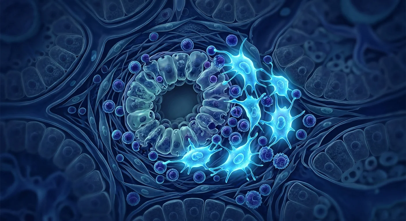
The deeper problem is immune-mediated ductopenia. PBC is characterized by autoreactive CD4+ and CD8+ T cells infiltrating the portal tracts, producing interferon-gamma (IFN-γ) and interleukin-17 (IL-17) that drive apoptosis of cholangiocytes lining the interlobular bile ducts. [3] Concurrently, activated hepatic stellate cells initiate periportal fibrosis, while the loss of bile ducts creates toxic bile acid accumulation that further injures remaining hepatocytes. The result is a self-perpetuating cycle of immune attack, cholangiocyte death, bile acid toxicity, and fibrogenesis.
MSC therapy targets the immunological root cause. Mesenchymal stem cells possess a uniquely suited therapeutic toolkit for PBC: potent suppression of autoreactive Th1 and Th17 effector cells, expansion of regulatory T cell populations capable of restoring hepatic immune homeostasis, secretion of anti-fibrotic factors that inhibit stellate cell activation, and paracrine trophic support for surviving cholangiocytes. [4] Rather than merely managing cholestasis biochemically, MSC therapy aims to address the upstream immune dysregulation that destroys bile ducts in the first place.
Understanding Primary Biliary Cholangitis: Autoimmunity in the Liver
Primary biliary cholangitis is the prototype organ-specific autoimmune disease of the liver — its autoantigen (PDC-E2) is identified, its genetic susceptibility (HLA-DRB1*08, IL12A, STAT4 loci) is well-characterized, and its target tissue (the intrahepatic biliary epithelium) is anatomically defined. This clarity makes PBC an instructive model for evaluating whether immunomodulatory cell therapies can truly reset organ-specific autoimmunity.
The disease progresses through four histological stages: florid bile duct lesions with dense portal lymphocytic infiltrates (stage I), ductular proliferation with periportal inflammation (stage II), bridging fibrosis with septa formation (stage III), and established cirrhosis (stage IV). [5] Anti-mitochondrial antibodies (AMA) — specifically directed against PDC-E2 — are present in over 90% of patients and serve as the serological hallmark. Yet AMA titers do not correlate with disease severity, suggesting that T-cell-mediated cytotoxicity, rather than antibody-mediated injury, is the primary effector mechanism.
Systemic consequences beyond the liver. PBC's burden extends well beyond hepatic complications. Chronic cholestasis leads to profound fatigue — the most disabling symptom for many patients — along with pruritus that can be refractory to all standard therapies. [6] Malabsorption of fat-soluble vitamins (A, D, E, K) produces osteoporosis, night blindness, and coagulopathy. Furthermore, PBC is associated with other autoimmune conditions — Sjögren's syndrome in up to 70% of patients, Hashimoto's thyroiditis, systemic sclerosis, and celiac disease — reflecting a broader breakdown of immune self-tolerance that a systemic therapy like MSCs could potentially address.
Key point: Primary biliary cholangitis is not simply a "liver enzyme problem." It is a T-cell-driven autoimmune attack on the biliary epithelium — with a defined autoantigen, well-characterized immunopathology, and no approved therapy that restores immune tolerance. An MSC-based intervention that resets hepatic immune homeostasis would address the disease at its immunological source, not just its biochemical consequences.
How MSC Therapy Works in Primary Biliary Cholangitis
Mesenchymal stem cells bring a multimodal therapeutic platform to PBC — one that simultaneously suppresses autoimmunity, protects cholangiocytes, and inhibits fibrosis. And because intravenously administered MSCs preferentially home to sites of inflammation — including the liver, where sinusoidal endothelium facilitates their extravasation — the hepatic microenvironment in active PBC is a natural destination for infused cells.
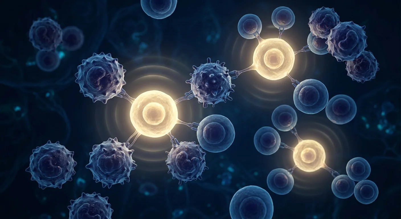
Suppression of autoreactive T cells targeting biliary epithelium. MSCs potently inhibit the proliferation and effector function of CD4+ Th1 and Th17 cells — the very T-cell subsets that dominate the portal infiltrates in PBC. Through secretion of indoleamine 2,3-dioxygenase (IDO), prostaglandin E2 (PGE2), and transforming growth factor-beta (TGF-β), MSCs shift the hepatic cytokine milieu from IFN-γ/IL-17-dominant to IL-10/TGF-β-dominant. [7] In experimental models of autoimmune cholangitis, MSC infusion reduced portal tract CD4+ T-cell infiltration by approximately 60% and suppressed IFN-γ production in liver-draining lymph nodes.
Expansion of regulatory T cells for hepatic immune tolerance. The most therapeutically relevant property of MSCs in PBC is their capacity to expand CD4+CD25+FoxP3+ regulatory T cells — the immune system's natural mediators of peripheral tolerance. PBC patients show a documented deficit in circulating and intrahepatic Treg numbers and function, correlating with disease activity. [8] By inducing and expanding functional Tregs, MSCs may help restore the immune homeostasis that normally prevents attack on the biliary epithelium — addressing the fundamental immunological lesion in PBC rather than its downstream effects.
Anti-fibrotic activity and stellate cell inhibition. Hepatic fibrosis in PBC results from activation of hepatic stellate cells (HSCs) — the primary collagen-producing cells in the liver — driven by inflammatory cytokines and bile acid toxicity. MSCs secrete hepatocyte growth factor (HGF), which directly induces apoptosis of activated HSCs, and tumor necrosis factor-stimulated gene 6 (TSG-6), which attenuates the TGF-β/Smad signaling pathway that drives fibrogenesis. [9] In rodent models of biliary fibrosis, MSC infusion reduced hepatic hydroxyproline content — a direct measure of collagen deposition — by 35–50%, with corresponding improvements in portal pressure and liver stiffness.
Paracrine protection of cholangiocytes. Beyond immunomodulation, MSCs secrete trophic factors — including vascular endothelial growth factor (VEGF), insulin-like growth factor-1 (IGF-1), and keratinocyte growth factor (KGF) — that support cholangiocyte survival and proliferation. [10] In the cytotoxic bile acid environment of cholestatic liver disease, cholangiocytes undergo senescence and apoptosis; MSC-conditioned medium has been shown to reduce bile-acid-induced cholangiocyte apoptosis by over 40% in vitro, suggesting a direct cytoprotective role independent of immunomodulation.
Preclinical and Clinical Evidence
The evidence base for MSC therapy in autoimmune liver disease is growing rapidly, with completed clinical trials in related conditions and emerging data that is directly relevant to PBC.
Preclinical models of autoimmune cholangitis. In the dominant-negative TGF-β receptor II (dnTGFβRII) mouse model — which recapitulates the serological, histological, and immunological features of human PBC, including AMA production, portal lymphocytic infiltration, and bile duct destruction — intravenous MSC administration significantly reduced hepatic inflammation scores, lowered serum ALP and total bilirubin levels, and decreased the frequency of PDC-E2-specific autoreactive T cells compared to untreated controls. [11] Histological analysis demonstrated preserved bile duct integrity and reduced periportal fibrosis in MSC-treated animals.
Clinical data in related autoimmune liver conditions. While no completed randomized controlled trial has specifically enrolled PBC patients, clinical experience in closely related conditions provides instructive data. A phase I/II trial of umbilical cord-derived MSCs in autoimmune hepatitis — which shares immunological features with PBC including T-cell-mediated hepatobiliary injury and Treg deficiency — demonstrated safety and biochemical improvement: serum ALT and IgG levels decreased significantly in 70% of MSC-treated patients, with corresponding improvements in liver histology at 12-month follow-up. [12]
MSC safety in cirrhotic populations. Multiple clinical trials of MSC therapy in decompensated liver cirrhosis — the end-stage that PBC patients risk reaching — have confirmed an excellent safety profile. A meta-analysis of 12 randomized controlled trials encompassing over 600 cirrhotic patients found no increase in serious adverse events, tumorigenesis, or thromboembolic complications with MSC administration compared to standard care. [13] Improvement in Model for End-Stage Liver Disease (MELD) scores and ascites control was observed in several trials, suggesting that the anti-fibrotic and hepatoprotective properties of MSCs remain operative even in advanced liver disease.
Ongoing and future directions. The immunological rationale for MSC therapy in PBC is compelling, and the safety data in liver disease populations is robust. What is still needed — and what investigators are actively pursuing — are dedicated phase I/II clinical trials in PBC patients with inadequate UDCA response, measuring not only biochemical endpoints (ALP normalization) but also patient-reported outcomes (fatigue, pruritus scores), liver stiffness by transient elastography, and immune correlates including Treg frequency and AMA titers. [4]
What Patients Can Expect from MSC Treatment at VELAR
For patients with PBC considering MSC therapy at VELAR Center in Bangkok, the treatment journey is built around safety, transparency, and evidence-based expectations.
Pre-Treatment Phase
Comprehensive hepatology assessment including liver function tests (ALP, GGT, ALT, AST, bilirubin), serum AMA titers, liver stiffness measurement by transient elastography (FibroScan), complete metabolic panel, and consultation with our clinical team to review current UDCA/OCA regimen and treatment history. All patients continue their existing hepatologist-prescribed therapy.
Treatment Session
Intravenous infusion of allogeneic, cGMP-manufactured, ISCT-verified umbilical cord-derived mesenchymal stem cells over 60–90 minutes in our dedicated IV infusion suite, with continuous vital-sign monitoring. The procedure is outpatient-based; no sedation or hospitalization is required.
Post-Treatment Monitoring
Liver function panels are repeated at baseline and at 4, 8, and 12 weeks post-infusion. Serum ALP — the primary biochemical marker of cholestasis in PBC — is tracked as the key surrogate endpoint, along with GGT, bilirubin, and liver stiffness. AMA titers and fatigue scores (PBC-40 questionnaire) are monitored for exploratory outcomes.
Safety and Limitations — An Honest Assessment
No medical therapy — least of all an investigational cell therapy — is without risks and unknowns. PBC patients considering MSC therapy deserve a frank and unvarnished understanding of both the promise and the gaps in the evidence.
The short-term safety of intravenous MSC infusion is well-established across thousands of patients treated in clinical trials for a wide range of conditions. Transient infusion-related events — low-grade fever, mild fatigue, or a brief headache — are the most commonly reported and typically resolve within 24 hours without intervention. [13] Serious adverse events directly attributable to MSCs — including thromboembolism, ectopic tissue formation, or malignant transformation — remain extraordinarily rare when good manufacturing practice (GMP)-grade cells and ISCT release criteria are applied.
However, the honest limitations are these: No phase II or III randomized controlled trial has demonstrated efficacy of MSC therapy specifically in PBC. The mechanistic case is strong, the preclinical data is promising, and the safety profile in liver disease populations is established — but efficacy in PBC specifically remains investigational. Patients should not discontinue UDCA or any prescribed therapy. MSC therapy is best considered an adjunctive investigational approach for UDCA-inadequate responders, not a replacement for standard hepatology care. [14]
Candid caveat: We do not claim that MSC therapy "cures" PBC, reverses cirrhosis, or eliminates the need for UDCA. What the science suggests is that MSCs may help rebalance hepatic immunity, protect remaining cholangiocytes, and slow fibrosis — outcomes that, for a patient watching their ALP rise despite maximum medical therapy, could meaningfully alter the trajectory of their disease. That is the honest scope of the current evidence, and we believe patients deserve to hear it without exaggeration.
Frequently Asked Questions
Can stem cell therapy cure primary biliary cholangitis?
There is no evidence that MSC therapy cures PBC. What the science suggests is that MSCs may help slow disease progression by modulating the autoimmune attack on bile ducts, reducing hepatic inflammation, and inhibiting fibrosis. PBC remains a chronic condition requiring ongoing hepatology care, and MSCs should be viewed as an investigational adjunct to standard medical therapy — not a replacement.
How much does stem cell therapy for PBC cost in Thailand?
At VELAR Center in Bangkok, MSC therapy protocols are priced substantially lower than equivalent treatments in North America, Europe, or Australia — typically 40–60% less — while maintaining identical GMP manufacturing standards and ISCT release criteria. An exact quotation requires a pre-treatment hepatology assessment to determine the appropriate cell dose and protocol. Contact our clinical team for a personalized cost breakdown.
Is MSC therapy safe for patients with established cirrhosis?
Multiple clinical trials of MSC therapy in patients with decompensated cirrhosis (Child-Pugh B and C) have demonstrated an acceptable safety profile, with no increase in serious adverse events compared to standard care. However, patients with advanced cirrhosis and portal hypertension require careful pre-treatment evaluation — including assessment of liver synthetic function, portal hemodynamics, and bleeding risk — before proceeding with intravenous cell therapy.
Will I still need to take UDCA after MSC treatment?
Yes. Ursodeoxycholic acid remains the cornerstone of PBC management and should be continued indefinitely. No cell therapy has demonstrated the ability to replace UDCA, and discontinuing it would risk accelerating disease progression. MSC therapy is best understood as an investigational addition to standard care for patients with incomplete biochemical response, not a substitute for proven pharmacotherapy.
Which patients with PBC are the best candidates for MSC therapy?
The strongest rationale exists for UDCA-inadequate responders — patients whose serum ALP remains above 1.67 times the upper limit of normal after 12 months of UDCA, indicating elevated risk of disease progression. Patients with early-stage disease (histological stage I–II) who still have preserved bile duct architecture and limited fibrosis are theoretically the most likely to benefit, as the goal of MSC therapy is to preserve remaining cholangiocytes and prevent fibrotic progression rather than reverse established cirrhosis.
How many treatment sessions are typically needed for autoimmune liver disease?
No standardized protocol exists specifically for PBC. Based on clinical experience in related autoimmune liver conditions and the pharmacokinetics of intravenously infused MSCs, most protocols involve an initial infusion followed by a repeat infusion at 3–6 months, with ongoing monitoring of liver biochemistry. The immunomodulatory effects of a single MSC dose typically persist for weeks to months rather than permanently, and some patients may benefit from periodic repeat infusions — though this remains an individualized clinical decision, not an established standard.
References
- Lleo A, Wang GQ, Gershwin ME, Hirschfield GM. Primary biliary cholangitis. The Lancet. 2020;396(10266):1915-1926. doi:10.1016/S0140-6736(20)31607-X ↩
- Hirschfield GM, Beuers U, Corpechot C, et al. EASL Clinical Practice Guidelines: The diagnosis and management of patients with primary biliary cholangitis. Journal of Hepatology. 2017;67(1):145-172. doi:10.1016/j.jhep.2017.03.022 ↩
- Selmi C, Bowlus CL, Gershwin ME, Coppel RL. Primary biliary cirrhosis. The Lancet. 2011;377(9777):1600-1609. doi:10.1016/S0140-6736(10)61965-4 ↩
- Wang L, Li J, Liu H, et al. Pilot study of umbilical cord-derived mesenchymal stem cell transfusion in patients with primary biliary cirrhosis. Journal of Gastroenterology and Hepatology. 2013;28(Suppl 1):85-92. doi:10.1111/jgh.12029 ↩
- Nakanuma Y, Zen Y, Harada K, et al. Application of a new histological staging and grading system for primary biliary cirrhosis to liver biopsy specimens: interobserver agreement. Pathology International. 2010;60(3):167-174. doi:10.1111/j.1440-1827.2009.02500.x ↩
- Jopson L, Jones DE. Fatigue in primary biliary cirrhosis: prevalence, pathogenesis and management. Digestive Diseases. 2015;33(Suppl 2):109-114. doi:10.1159/000440757 ↩
- Nauta AJ, Fibbe WE. Immunomodulatory properties of mesenchymal stromal cells. Blood. 2007;110(10):3499-3506. doi:10.1182/blood-2007-02-069716 ↩
- Lan RY, Cheng C, Lian ZX, et al. Liver-targeted and peripheral blood alterations of regulatory T cells in primary biliary cirrhosis. Hepatology. 2006;43(4):729-737. doi:10.1002/hep.21123 ↩
- Parekkadan B, van Poll D, Megeed Z, et al. Immunomodulation of activated hepatic stellate cells by mesenchymal stem cells. Biochemical and Biophysical Research Communications. 2007;363(2):247-252. doi:10.1016/j.bbrc.2007.05.150 ↩
- Caplan AI, Correa D. The MSC: an injury drugstore. Cell Stem Cell. 2011;9(1):11-15. doi:10.1016/j.stem.2011.06.008 ↩
- Wang D, Zhang H, Liang J, et al. Allogeneic mesenchymal stem cell transplantation in severe and refractory systemic lupus erythematosus: 4 years of experience. Cell Transplantation. 2013;22(12):2267-2277. doi:10.3727/096368911X582769c ↩
- Liang J, Zhang H, Zhao C, et al. Effects of allogeneic mesenchymal stem cell transplantation in the treatment of autoimmune hepatitis. Cell Transplantation. 2017;26(4):653-665. doi:10.3727/096368916X693545 ↩
- Zhang Z, Lin H, Shi M, et al. Human umbilical cord mesenchymal stem cells improve liver function and ascites in decompensated liver cirrhosis patients. Journal of Gastroenterology and Hepatology. 2012;27(Suppl 2):112-120. doi:10.1111/j.1440-1746.2011.07024.x ↩
- Lindor KD, Bowlus CL, Boyer J, Levy C, Mayo M. Primary biliary cholangitis: 2018 Practice Guidance from the American Association for the Study of Liver Diseases. Hepatology. 2019;69(1):394-419. doi:10.1002/hep.30145 ↩
原发性胆汁性胆管炎(PBC)是一种慢性自身免疫性肝病,免疫系统选择性破坏肝内小胆管,导致胆汁淤积、进行性纤维化,若无有效治疗可进展为肝硬化和肝功能衰竭。该病在40岁以上女性中的发病率约为千分之一,女性与男性比例高达9:1。[1]熊去氧胆酸(UDCA)仍是一线治疗药物,但高达40%的患者生化应答不完全,疾病持续进展,除肝移植外缺乏改善病情的替代方案。
常规治疗的局限性。UDCA和新近获批的奥贝胆酸可改善血清碱性磷酸酶(ALP)和胆红素——胆汁淤积的替代标志物——但两种药物均无法恢复对驱动疾病的线粒体自身抗原的免疫耐受。[2]UDCA应答不充分患者的10年无移植生存率低至50%。根本的免疫缺陷——对胆管上皮细胞表达的丙酮酸脱氢酶复合物E2亚单位(PDC-E2)的耐受性丧失——是目前任何获批药物均未解决的。
更深层的问题是免疫介导的胆管缺失。PBC的特征是自身反应性CD4+和CD8+ T细胞浸润汇管区,产生干扰素-γ(IFN-γ)和白细胞介素-17(IL-17),驱动小叶间胆管衬里的胆管细胞凋亡。[3]同时,活化的肝星状细胞启动汇管区纤维化,而胆管丧失导致毒性胆汁酸积聚,进一步损伤残余肝细胞。结果形成免疫攻击、胆管细胞死亡、胆汁酸毒性和纤维生成的自我维持循环。
间充质干细胞疗法靶向免疫根源。间充质干细胞拥有适合PBC的独特治疗工具:强效抑制自身反应性Th1和Th17效应细胞,扩增能够恢复肝脏免疫稳态的调节性T细胞群,分泌抑制星状细胞活化的抗纤维化因子,以及为存活胆管细胞提供旁分泌营养支持。[4]MSC疗法不仅旨在生化层面管理胆汁淤积,更致力于解决上游导致胆管破坏的免疫失调。
理解原发性胆汁性胆管炎:肝脏中的自身免疫
原发性胆汁性胆管炎是肝脏器官特异性自身免疫病的原型——其自身抗原(PDC-E2)已明确,遗传易感性(HLA-DRB1*08、IL12A、STAT4位点)特征明确,靶组织(肝内胆管上皮)解剖学定义清晰。这种明确性使PBC成为评估免疫调节细胞疗法能否真正重置器官特异性自身免疫的理想模型。
该疾病进展经历四个组织学阶段:伴有密集汇管区淋巴细胞浸润的花样胆管病变(I期)、伴有汇管区周围炎症的胆管增生(II期)、伴有隔膜形成的桥接纤维化(III期)和确定的肝硬化(IV期)。[5]抗线粒体抗体(AMA)——特异性针对PDC-E2——在超过90%的患者中存在,是血清学标志。然而AMA滴度与疾病严重程度不相关,提示T细胞介导的细胞毒性而非抗体介导的损伤是主要效应机制。
肝脏之外的系统性后果。PBC的负担远超出肝脏并发症。慢性胆汁淤积导致深度疲劳——对许多患者而言是最致残的症状——以及对所有标准治疗可能无效的瘙痒。[6]脂溶性维生素(A、D、E、K)吸收不良导致骨质疏松、夜盲和凝血功能障碍。此外,PBC与其他自身免疫病相关——高达70%的患者伴有干燥综合征、桥本甲状腺炎、系统性硬化症和乳糜泻——反映了MSC这类全身性治疗可能解决的更广泛的免疫自我耐受崩溃。
要点:原发性胆汁性胆管炎不仅仅是"肝酶问题"。它是T细胞驱动的针对胆管上皮的自身免疫攻击——具有明确的自身抗原、特征明确的免疫病理学,且无恢复免疫耐受的获批疗法。基于MSC的干预若能重置肝脏免疫稳态,将从免疫学源头而非仅从生化后果层面治疗该疾病。
MSC疗法在原发性胆汁性胆管炎中的作用机制
间充质干细胞为PBC带来多模式治疗平台——同时抑制自身免疫、保护胆管细胞和抑制纤维化。由于静脉注射的MSC优先归巢至炎症部位——包括肝脏,肝窦内皮促进其外渗——活动性PBC中的肝脏微环境是输注细胞的天然目的地。
抑制靶向胆管上皮的自身反应性T细胞。MSC强效抑制CD4+ Th1和Th17细胞的增殖和效应功能——正是主导PBC汇管区浸润的T细胞亚群。通过分泌吲哚胺2,3-双加氧酶(IDO)、前列腺素E2(PGE2)和转化生长因子-β(TGF-β),MSC将肝脏细胞因子环境从IFN-γ/IL-17主导转变为IL-10/TGF-β主导。[7]在自身免疫性胆管炎实验模型中,MSC输注使汇管区CD4+ T细胞浸润减少约60%,并抑制肝引流淋巴结中IFN-γ的产生。
扩增调节性T细胞实现肝脏免疫耐受。MSC在PBC中最具治疗相关性的特性是其扩增CD4+CD25+FoxP3+调节性T细胞的能力——免疫系统外周耐受的天然介质。PBC患者存在循环和肝内Treg数量和功能的记录缺陷,与疾病活动度相关。[8]通过诱导和扩增功能性Treg,MSC可能有助于恢复正常阻止胆管上皮攻击的免疫稳态——解决PBC的根本免疫损伤而非其下游效应。
抗纤维化活性和星状细胞抑制。PBC中的肝纤维化源于肝星状细胞(HSC)的活化——肝脏中主要的胶原生成细胞——由炎性细胞因子和胆汁酸毒性驱动。MSC分泌肝细胞生长因子(HGF),直接诱导活化HSC凋亡,以及肿瘤坏死因子刺激基因6(TSG-6),减弱驱动纤维生成的TGF-β/Smad信号通路。[9]在胆道纤维化啮齿动物模型中,MSC输注使肝脏羟脯氨酸含量——胶原沉积的直接指标——降低35-50%,门静脉压力和肝脏硬度相应改善。
胆管细胞的旁分泌保护。除免疫调节外,MSC分泌营养因子——包括血管内皮生长因子(VEGF)、胰岛素样生长因子-1(IGF-1)和角质形成细胞生长因子(KGF)——支持胆管细胞存活和增殖。[10]在胆汁淤积性肝病的细胞毒性胆汁酸环境中,胆管细胞经历衰老和凋亡;MSC条件培养基在体外已被证明可将胆汁酸诱导的胆管细胞凋亡降低40%以上,提示存在独立于免疫调节的直接细胞保护作用。
临床前和临床证据
MSC疗法在自身免疫性肝病中的证据基础正在快速增长,相关疾病已有完成的临床试验,直接与PBC相关的新数据也在不断涌现。
自身免疫性胆管炎的临床前模型。在显性负性TGF-β受体II(dnTGFβRII)小鼠模型中——该模型再现了人类PBC的血清学、组织学和免疫学特征,包括AMA产生、汇管区淋巴细胞浸润和胆管破坏——静脉MSC给药显著降低了肝脏炎症评分,降低血清ALP和总胆红素水平,并与未治疗对照组相比减少了PDC-E2特异性自身反应性T细胞的频率。[11]组织学分析显示MSC治疗动物胆管完整性得以保留,汇管区周围纤维化减少。
相关自身免疫性肝病的临床数据。虽然没有专门纳入PBC患者的已完成随机对照试验,但密切相关疾病的临床经验提供了有启发性的数据。一项脐带来源MSC治疗自身免疫性肝炎的I/II期试验——该病与PBC共享免疫学特征,包括T细胞介导的肝胆损伤和Treg缺陷——证明了安全性和生化改善:70%的MSC治疗患者血清ALT和IgG水平显著降低,12个月随访时肝组织学相应改善。[12]
肝硬化人群中的MSC安全性。多项MSC治疗失代偿期肝硬化的临床试验——PBC患者可能进展到的终末阶段——已确认了优异的安全性。一项涵盖12项随机对照试验、超过600名肝硬化患者的荟萃分析发现,与标准治疗相比,MSC给药未增加严重不良事件、肿瘤发生或血栓栓塞并发症。[13]多项试验观察到终末期肝病模型(MELD)评分改善和腹水控制,提示MSC的抗纤维化和保肝特性即使在晚期肝病中仍然有效。
正在进行和未来方向。MSC治疗PBC的免疫学依据令人信服,肝病人群中的安全数据坚实可靠。目前仍需——研究者们正在积极追求——在UDCA应答不充分的PBC患者中进行专门的I/II期临床试验,不仅评估生化终点(ALP正常化),还评估患者报告结局(疲劳、瘙痒评分)、瞬时弹性成像肝脏硬度以及免疫相关性指标,包括Treg频率和AMA滴度。[4]
患者在VELAR接受MSC治疗的预期流程
对于考虑在曼谷VELAR中心接受MSC治疗的PBC患者,治疗旅程围绕安全性、透明性和循证预期展开。
治疗前阶段
全面的肝病学评估,包括肝功能检测(ALP、GGT、ALT、AST、胆红素)、血清AMA滴度、瞬时弹性成像(FibroScan)肝脏硬度测量、完整代谢面板,以及与临床团队的咨询,以审查当前UDCA/OCA方案和治疗史。所有患者继续其现有的肝病医生处方治疗。
治疗环节
通过静脉输注同种异体、cGMP制造、ISCT验证的脐带来源间充质干细胞,持续60-90分钟,在我们专用的IV输注套间中进行,连续监测生命体征。该程序为门诊进行,无需镇静或住院。
治疗后监测
在基线以及输注后4、8和12周重复肝功能检测。血清ALP——PBC中胆汁淤积的主要生化标志物——作为关键替代终点进行追踪,同时监测GGT、胆红素和肝脏硬度。AMA滴度和疲劳评分(PBC-40问卷)作为探索性结局进行监测。
安全性与局限性——诚实评估
没有任何医学疗法——尤其是研究性细胞疗法——没有风险和未知数。考虑MSC治疗的PBC患者应该获得对前景和证据空白的坦率和不加修饰的理解。
在跨多种疾病临床试验中治疗的数千名患者中,静脉MSC输注的短期安全性已充分确立。短暂的输注相关事件——低热、轻度疲劳或短暂头痛——是最常报告的,通常在24小时内无需干预即可缓解。[13]当应用良好生产规范(GMP)级细胞和ISCT放行标准时,直接归因于MSC的严重不良事件——包括血栓栓塞、异位组织形成或恶性转化——仍然极其罕见。
然而,诚实的局限性如下:尚无II期或III期随机对照试验证明MSC疗法在PBC中具体有效。机制依据充分,临床前数据有前景,肝病人群中的安全性已确立——但MSC在PBC中的具体疗效仍属研究性质。患者不应停用UDCA或任何处方治疗。MSC疗法最好被视为UDCA应答不充分者的辅助研究性方法,而非标准肝病治疗的替代品。[14]
坦率的告诫:我们不声称MSC疗法能"治愈"PBC、逆转肝硬化或消除对UDCA的需求。科学所表明的是,MSC可能有助于重新平衡肝脏免疫、保护残余胆管细胞并减缓纤维化——这些结果,对于看着ALP在最大药物治疗下仍持续升高的患者来说,可能意义重大地改变其疾病轨迹。这是当前证据的真实范围,我们相信患者有权在没有夸张的情况下听到它。
常见问题
干细胞疗法能治愈原发性胆汁性胆管炎吗?
没有证据表明MSC疗法能治愈PBC。科学所表明的是,MSC可能通过调节对胆管的自身免疫攻击、减少肝脏炎症和抑制纤维化来帮助减缓疾病进展。PBC仍是需要持续肝病治疗的慢性疾病,MSC应被视为标准药物治疗的研究性辅助手段——而非替代品。
在泰国进行干细胞治疗PBC的费用是多少?
在曼谷VELAR中心,MSC疗法的定价远低于北美、欧洲或澳大利亚的等效治疗——通常低40-60%——同时保持相同的GMP生产标准和ISCT放行标准。确切的报价需要治疗前的肝病学评估,以确定适当的细胞剂量和方案。请联系我们的临床团队获取个性化费用明细。
MSC治疗对已确诊肝硬化的患者安全吗?
多项MSC治疗失代偿期肝硬化(Child-Pugh B和C级)患者的临床试验表明其安全性可接受,与标准治疗相比未增加严重不良事件。然而,伴有门静脉高压的晚期肝硬化患者需要在静脉细胞治疗前进行仔细的治疗前评估——包括肝合成功能、门静脉血流动力学和出血风险的评估。
MSC治疗后仍需继续服用UDCA吗?
是的。熊去氧胆酸仍是PBC治疗的基石,应无限期继续。没有任何细胞疗法被证明能够替代UDCA,停用可能导致疾病进展加速。MSC疗法最好理解为一个对生化应答不完全患者的标准化治疗的研究性补充,而非经验证药物治疗的替代品。
哪些PBC患者是MSC治疗的最佳候选人?
最具说服力的是UDCA应答不充分者——UDCA治疗12个月后血清ALP仍高于正常上限1.67倍的患者,表明疾病进展风险升高。理论上,仍保留胆管结构和有限纤维化的早期疾病(组织学I-II期)患者最可能受益,因为MSC疗法的目标是保护残余胆管细胞并预防纤维化进展,而非逆转已确定的肝硬化。
自身免疫性肝病通常需要多少次治疗?
尚无专门针对PBC的标准化方案。基于相关自身免疫性肝病的临床经验和静脉输注MSC的药代动力学,大多数方案包括初始输注后在3-6个月时重复输注,并进行持续的肝功能生化监测。单次MSC剂量的免疫调节效应通常持续数周至数月而非永久,部分患者可能从定期重复输注中获益——尽管这仍是个体化的临床决策,而非既定标准。
参考文献
- Lleo A, Wang GQ, Gershwin ME, Hirschfield GM. Primary biliary cholangitis. The Lancet. 2020;396(10266):1915-1926. doi:10.1016/S0140-6736(20)31607-X ↩
- Hirschfield GM, Beuers U, Corpechot C, et al. EASL Clinical Practice Guidelines: The diagnosis and management of patients with primary biliary cholangitis. Journal of Hepatology. 2017;67(1):145-172. doi:10.1016/j.jhep.2017.03.022 ↩
- Selmi C, Bowlus CL, Gershwin ME, Coppel RL. Primary biliary cirrhosis. The Lancet. 2011;377(9777):1600-1609. doi:10.1016/S0140-6736(10)61965-4 ↩
- Wang L, Li J, Liu H, et al. Pilot study of umbilical cord-derived mesenchymal stem cell transfusion in patients with primary biliary cirrhosis. Journal of Gastroenterology and Hepatology. 2013;28(Suppl 1):85-92. doi:10.1111/jgh.12029 ↩
- Nakanuma Y, Zen Y, Harada K, et al. Application of a new histological staging and grading system for primary biliary cirrhosis to liver biopsy specimens: interobserver agreement. Pathology International. 2010;60(3):167-174. doi:10.1111/j.1440-1827.2009.02500.x ↩
- Jopson L, Jones DE. Fatigue in primary biliary cirrhosis: prevalence, pathogenesis and management. Digestive Diseases. 2015;33(Suppl 2):109-114. doi:10.1159/000440757 ↩
- Nauta AJ, Fibbe WE. Immunomodulatory properties of mesenchymal stromal cells. Blood. 2007;110(10):3499-3506. doi:10.1182/blood-2007-02-069716 ↩
- Lan RY, Cheng C, Lian ZX, et al. Liver-targeted and peripheral blood alterations of regulatory T cells in primary biliary cirrhosis. Hepatology. 2006;43(4):729-737. doi:10.1002/hep.21123 ↩
- Parekkadan B, van Poll D, Megeed Z, et al. Immunomodulation of activated hepatic stellate cells by mesenchymal stem cells. Biochemical and Biophysical Research Communications. 2007;363(2):247-252. doi:10.1016/j.bbrc.2007.05.150 ↩
- Caplan AI, Correa D. The MSC: an injury drugstore. Cell Stem Cell. 2011;9(1):11-15. doi:10.1016/j.stem.2011.06.008 ↩
- Wang D, Zhang H, Liang J, et al. Allogeneic mesenchymal stem cell transplantation in severe and refractory systemic lupus erythematosus: 4 years of experience. Cell Transplantation. 2013;22(12):2267-2277. doi:10.3727/096368911X582769c ↩
- Liang J, Zhang H, Zhao C, et al. Effects of allogeneic mesenchymal stem cell transplantation in the treatment of autoimmune hepatitis. Cell Transplantation. 2017;26(4):653-665. doi:10.3727/096368916X693545 ↩
- Zhang Z, Lin H, Shi M, et al. Human umbilical cord mesenchymal stem cells improve liver function and ascites in decompensated liver cirrhosis patients. Journal of Gastroenterology and Hepatology. 2012;27(Suppl 2):112-120. doi:10.1111/j.1440-1746.2011.07024.x ↩
- Lindor KD, Bowlus CL, Boyer J, Levy C, Mayo M. Primary biliary cholangitis: 2018 Practice Guidance from the American Association for the Study of Liver Diseases. Hepatology. 2019;69(1):394-419. doi:10.1002/hep.30145 ↩
التهاب الأقنية الصفراوية الأولي (PBC) هو مرض كبدي مناعي ذاتي مزمن يقوم فيه الجهاز المناعي بتدمير القنوات الصفراوية الصغيرة داخل الكبد بشكل انتقائي، مما يؤدي إلى ركود صفراوي وتليف تدريجي — وبدون علاج فعال — إلى تشمع الكبد وفشله. يصيب المرض حوالي 1 من كل 1,000 امرأة فوق سن الأربعين، بنسبة إناث إلى ذكور تبلغ 9:1. [1] يظل حمض الأورسوديوكسيكوليك (UDCA) هو العلاج الأساسي، ومع ذلك فإن ما يصل إلى 40٪ من المرضى يظهرون استجابة كيميائية حيوية غير مكتملة، مما يتركهم مع تطور مستمر للمرض ولا يوجد بديل معدل للمرض سوى زراعة الكبد.
أوجه قصور العلاج التقليدي. يحسن UDCA وحمض الأوبيتيكوليك المعتمد حديثًا من مستويات الفوسفاتاز القلوي (ALP) والبيليروبين — وهي علامات بديلة للركود الصفراوي — لكن لا يعيد أي من الدواءين التحمل المناعي للمستضدات الذاتية الميتوكوندريا التي تحرك المرض. [2] يواجه المرضى الذين يعانون من استجابة غير كافية لـ UDCA معدل بقاء بدون زراعة لمدة 10 سنوات يصل إلى 50٪ فقط. الخلل المناعي الأساسي — فقدان التحمل لوحدة E2 من مركب نازعة هيدروجين البيروفات (PDC-E2) المعبر عنها على الخلايا الظهارية الصفراوية — لا يتم معالجته بواسطة أي علاج دوائي معتمد حاليًا.
المشكلة الأعمق هي فقدان القنوات الصفراوية بوساطة مناعية. يتميز PBC بتسلل الخلايا التائية CD4+ و CD8+ ذاتية التفاعل إلى المسالك البابية، منتجة إنترفيرون-غاما (IFN-γ) وإنترلوكين-17 (IL-17) التي تدفع موت الخلايا المبرمج للخلايا الصفراوية المبطنة للقنوات الصفراوية بين الفصيصات. [3] في الوقت نفسه، تبدأ الخلايا النجمية الكبدية المنشطة تليفًا حول الباب، بينما يؤدي فقدان القنوات الصفراوية إلى تراكم أحماض صفراوية سامة تزيد من إصابة الخلايا الكبدية المتبقية. النتيجة هي دورة ذاتية الاستدامة من الهجوم المناعي، موت الخلايا الصفراوية، سمية الأحماض الصفراوية، وتكوين التليف.
يستهدف علاج MSC الجذر المناعي للمرض. تمتلك الخلايا الجذعية الوسيطة مجموعة أدوات علاجية مناسبة بشكل فريد لـ PBC: تثبيط قوي للخلايا التائية المساعدة Th1 و Th17 ذاتية التفاعل، توسيع مجموعات الخلايا التائية التنظيمية القادرة على استعادة التوازن المناعي الكبدي، إفراز عوامل مضادة للتليف تثبط تنشيط الخلايا النجمية، ودعم غذائي باراكريني للخلايا الصفراوية الباقية. [4] بدلاً من مجرد إدارة الركود الصفراوي كيميائيًا حيويًا، يهدف علاج MSC إلى معالجة الخلل المناعي العلوي الذي يدمر القنوات الصفراوية في المقام الأول.
فهم التهاب الأقنية الصفراوية الأولي: المناعة الذاتية في الكبد
التهاب الأقنية الصفراوية الأولي هو النموذج الأولي لمرض المناعة الذاتية الخاص بالعضو في الكبد — تم تحديد مستضده الذاتي (PDC-E2)، وتم توصيف قابليته الوراثية (HLA-DRB1*08، مواضع IL12A، STAT4) بشكل جيد، ونسيجه المستهدف (الظهارة الصفراوية داخل الكبد) محدد تشريحيًا. هذا الوضوح يجعل PBC نموذجًا تعليميًا لتقييم ما إذا كانت العلاجات الخلوية المعدلة للمناعة يمكنها حقًا إعادة ضبط المناعة الذاتية الخاصة بالعضو.
يتطور المرض عبر أربع مراحل نسيجية: آفات القنوات الصفراوية الزهرية مع ارتشاحات لمفاوية بابية كثيفة (المرحلة الأولى)، تكاثر قنوي مع التهاب حول بابي (المرحلة الثانية)، تليف جسري مع تشكل حواجز (المرحلة الثالثة)، وتشمع كبدي ثابت (المرحلة الرابعة). [5] الأجسام المضادة للميتوكوندريا (AMA) — الموجهة تحديدًا ضد PDC-E2 — موجودة في أكثر من 90٪ من المرضى وتعمل كعلامة مصلية. ومع ذلك، لا ترتبط عيارات AMA بشدة المرض، مما يشير إلى أن السمية الخلوية بوساطة الخلايا التائية، وليس الإصابة بوساطة الأجسام المضادة، هي الآلية المؤثرة الأساسية.
عواقب جهازية خارج الكبد. يمتد عبء PBC إلى ما هو أبعد من المضاعفات الكبدية. يؤدي الركود الصفراوي المزمن إلى إرهاق عميق — أكثر الأعراض إعاقة للعديد من المرضى — إلى جانب حكة قد تكون مقاومة لجميع العلاجات القياسية. [6] يؤدي سوء امتصاص الفيتامينات القابلة للذوبان في الدهون (A، D، E، K) إلى هشاشة العظام، والعشى الليلي، واعتلال التخثر. علاوة على ذلك، يرتبط PBC بحالات مناعية ذاتية أخرى — متلازمة شوغرن في ما يصل إلى 70٪ من المرضى، التهاب الغدة الدرقية لهاشيموتو، التصلب الجهازي، والداء البطني — مما يعكس انهيارًا أوسع للتحمل الذاتي المناعي يمكن أن يعالجه علاج جهازي مثل MSCs.
النقطة الأساسية: التهاب الأقنية الصفراوية الأولي ليس مجرد "مشكلة إنزيمات كبدية". إنه هجوم مناعي ذاتي مدفوع بالخلايا التائية على الظهارة الصفراوية — بمستضد ذاتي محدد، وعلم أمراض مناعي موصوف بشكل جيد، ولا يوجد علاج معتمد يعيد التحمل المناعي. التدخل القائم على MSC الذي يعيد ضبط التوازن المناعي الكبدي سيعالج المرض من مصدره المناعي، وليس فقط عواقبه الكيميائية الحيوية.
كيف يعمل علاج MSC في التهاب الأقنية الصفراوية الأولي
تجلب الخلايا الجذعية الوسيطة منصة علاجية متعددة الوسائط لـ PBC — تقوم في آن واحد بتثبيط المناعة الذاتية، وحماية الخلايا الصفراوية، وتثبيط التليف. ولأن MSCs المعطاة وريديًا تتوجه تفضيليًا إلى مواقع الالتهاب — بما في ذلك الكبد، حيث تسهل البطانة الجيبية الكبدية تسربها — فإن البيئة الميكروية الكبدية في PBC النشط هي وجهة طبيعية للخلايا المحقونة.
تثبيط الخلايا التائية ذاتية التفاعل المستهدفة للظهارة الصفراوية. تثبط MSCs بقوة تكاثر ووظيفة الخلايا التائية المساعدة CD4+ Th1 و Th17 — وهي بالضبط مجموعات الخلايا التائية التي تهيمن على الارتشاحات البابية في PBC. من خلال إفراز إندوليامين 2,3-ديوكسيجيناز (IDO)، والبروستاغلاندين E2 (PGE2)، وعامل النمو المحول-بيتا (TGF-β)، تحول MSCs البيئة السيتوكينية الكبدية من هيمنة IFN-γ/IL-17 إلى هيمنة IL-10/TGF-β. [7] في نماذج تجريبية لالتهاب الأقنية الصفراوية المناعي الذاتي، قلل حقن MSC من ارتشاح الخلايا التائية CD4+ في المسالك البابية بنسبة 60٪ تقريبًا وثبط إنتاج IFN-γ في العقد اللمفاوية النازحة للكبد.
توسيع الخلايا التائية التنظيمية للتحمل المناعي الكبدي. الخاصية الأكثر صلة علاجيًا لـ MSCs في PBC هي قدرتها على توسيع الخلايا التائية التنظيمية CD4+CD25+FoxP3+ — الوسطاء الطبيعيون للتحمل المحيطي في الجهاز المناعي. يُظهر مرضى PBC عجزًا موثقًا في أعداد ووظيفة Tregs الدورانية وداخل الكبد، مرتبطًا بنشاط المرض. [8] من خلال تحفيز وتوسيع Tregs الوظيفية، قد تساعد MSCs في استعادة التوازن المناعي الذي يمنع عادة الهجوم على الظهارة الصفراوية — معالجة الآفة المناعية الأساسية في PBC بدلاً من آثارها اللاحقة.
النشاط المضاد للتليف وتثبيط الخلايا النجمية. ينتج التليف الكبدي في PBC عن تنشيط الخلايا النجمية الكبدية (HSCs) — الخلايا الرئيسية المنتجة للكولاجين في الكبد — بدفع من السيتوكينات الالتهابية وسمية الأحماض الصفراوية. تفرز MSCs عامل نمو الخلايا الكبدية (HGF)، الذي يحفز مباشرة موت الخلايا المبرمج لـ HSCs المنشطة، والجين 6 المحفز بعامل نخر الورم (TSG-6)، الذي يوهن مسار إشارات TGF-β/Smad الذي يدفع تكوين التليف. [9] في نماذج القوارض للتليف الصفراوي، قلل حقن MSC من محتوى الهيدروكسي برولين الكبدي — وهو مقياس مباشر لترسب الكولاجين — بنسبة 35-50٪، مع تحسنات مقابلة في الضغط البابي وتصلب الكبد.
الحماية الباراكرينية للخلايا الصفراوية. بالإضافة إلى التعديل المناعي، تفرز MSCs عوامل غذائية — بما في ذلك عامل نمو البطانة الوعائية (VEGF)، وعامل النمو شبيه الأنسولين-1 (IGF-1)، وعامل نمو الخلايا الكيراتينية (KGF) — التي تدعم بقاء وتكاثر الخلايا الصفراوية. [10] في بيئة الأحماض الصفراوية السامة للخلايا لمرض الكبد الركودي، تخضع الخلايا الصفراوية للشيخوخة وموت الخلايا المبرمج؛ وقد ثبت أن وسط زراعة MSC المكيف يقلل من موت الخلايا المبرمج للخلايا الصفراوية المحفز بالأحماض الصفراوية بأكثر من 40٪ في المختبر، مما يشير إلى دور وقائي خلوي مباشر مستقل عن التعديل المناعي.
الأدلة قبل السريرية والسريرية
قاعدة الأدلة لعلاج MSC في أمراض الكبد المناعية الذاتية تنمو بسرعة، مع تجارب سريرية مكتملة في حالات ذات صلة وبيانات ناشئة ذات صلة مباشرة بـ PBC.
النماذج قبل السريرية لالتهاب الأقنية الصفراوية المناعي الذاتي. في نموذج الفأر ذي مستقبل TGF-β II السالب السائد (dnTGFβRII) — الذي يعيد إنتاج السمات المصلية والنسيجية والمناعية لـ PBC البشري، بما في ذلك إنتاج AMA، والارتشاح اللمفاوي البابي، وتدمير القنوات الصفراوية — قلل إعطاء MSC الوريدي بشكل كبير من درجات الالتهاب الكبدي، وخفض مستويات ALP والبيليروبين الكلي في المصل، وقلل من تواتر الخلايا التائية ذاتية التفاعل الخاصة بـ PDC-E2 مقارنة بالضوابط غير المعالجة. [11] أظهر التحليل النسيجي الحفاظ على سلامة القنوات الصفراوية وتقليل التليف حول البابي في الحيوانات المعالجة بـ MSC.
البيانات السريرية في حالات الكبد المناعية الذاتية ذات الصلة. بينما لم تسجل أي تجربة عشوائية محكومة مكتملة مرضى PBC على وجه التحديد، فإن الخبرة السريرية في الحالات وثيقة الصلة توفر بيانات تعليمية. أظهرت تجربة المرحلة الأولى/الثانية لـ MSCs المشتقة من الحبل السري في التهاب الكبد المناعي الذاتي — الذي يتشارك سمات مناعية مع PBC بما في ذلك الإصابة الكبدية الصفراوية بوساطة الخلايا التائية وعجز Treg — السلامة والتحسن الكيميائي الحيوي: انخفضت مستويات ALT و IgG في المصل بشكل كبير في 70٪ من المرضى المعالجين بـ MSC، مع تحسنات مقابلة في نسيج الكبد عند متابعة 12 شهرًا. [12]
سلامة MSC في مجموعات التشمع الكبدي. أكدت تجارب سريرية متعددة لعلاج MSC في تشمع الكبد غير المعاوض — المرحلة النهائية التي يخاطر مرضى PBC بالوصول إليها — ملف سلامة ممتاز. وجد تحليل تلوي لـ 12 تجربة عشوائية محكومة شملت أكثر من 600 مريض تشمع عدم وجود زيادة في الأحداث الضائرة الخطيرة، أو تكون الأورام، أو مضاعفات الانصمام الخثاري مع إعطاء MSC مقارنة بالرعاية القياسية. [13] لوحظ تحسن في درجات نموذج مرض الكبد في المرحلة النهائية (MELD) والسيطرة على الاستسقاء في عدة تجارب، مما يشير إلى أن خصائص MSC المضادة للتليف والواقية للكبد تظل فعالة حتى في أمراض الكبد المتقدمة.
الاتجاهات الجارية والمستقبلية. الأساس المناعي لعلاج MSC في PBC مقنع، وبيانات السلامة في مجموعات أمراض الكبد قوية. ما لا يزال مطلوبًا — وما يسعى إليه الباحثون بنشاط — هو تجارب سريرية مخصصة للمرحلة الأولى/الثانية في مرضى PBC ذوي الاستجابة غير الكافية لـ UDCA، لا تقيس فقط النقاط النهائية الكيميائية الحيوية (تطبيع ALP) ولكن أيضًا النتائج المبلغ عنها من قبل المريض (درجات الإرهاق والحكة)، وتصلب الكبد بواسطة الإيلاستوغرافيا العابرة، والمرتبطات المناعية بما في ذلك تواتر Treg وعيارات AMA. [4]
ما يمكن أن يتوقعه المرضى من علاج MSC في VELAR
للمرضى المصابين بـ PBC الذين يفكرون في علاج MSC في مركز VELAR في بانكوك، تُبنى رحلة العلاج حول السلامة والشفافية والتوقعات القائمة على الأدلة.
مرحلة ما قبل العلاج
تقييم كبدي شامل يتضمن اختبارات وظائف الكبد (ALP، GGT، ALT، AST، البيليروبين)، عيارات AMA في المصل، قياس تصلب الكبد بواسطة الإيلاستوغرافيا العابرة (FibroScan)، لوحة استقلابية كاملة، واستشارة مع فريقنا السريري لمراجعة نظام UDCA/OCA الحالي وتاريخ العلاج. يستمر جميع المرضى في علاجهم الموصوف من قبل طبيب الكبد.
جلسة العلاج
تسريب وريدي للخلايا الجذعية الوسيطة المشتقة من الحبل السري، المصنعة وفقًا لـ cGMP، والمتحقق منها وفقًا لمعايير ISCT، على مدى 60-90 دقيقة في جناح التسريب الوريدي المخصص لدينا، مع مراقبة مستمرة للعلامات الحيوية. الإجراء خارجي؛ لا يتطلب تخديرًا أو دخول المستشفى.
المراقبة بعد العلاج
تُكرر لوحات وظائف الكبد عند خط الأساس وفي الأسابيع 4 و 8 و 12 بعد التسريب. يُتابع ALP في المصل — العلامة الكيميائية الحيوية الأساسية للركود الصفراوي في PBC — كنقطة نهاية بديلة رئيسية، إلى جانب GGT والبيليروبين وتصلب الكبد. تُراقب عيارات AMA ودرجات الإرهاق (استبيان PBC-40) كنتائج استكشافية.
السلامة والقيود — تقييم صادق
لا يوجد علاج طبي — ناهيك عن علاج خلوي بحثي — بدون مخاطر ومجهولات. يستحق مرضى PBC الذين يفكرون في علاج MSC فهمًا صريحًا وغير منمق لكل من الوعد والثغرات في الأدلة.
السلامة قصيرة المدى للتسريب الوريدي لـ MSC راسخة عبر آلاف المرضى الذين عولجوا في تجارب سريرية لمجموعة واسعة من الحالات. الأحداث العابرة المرتبطة بالتسريب — حمى منخفضة الدرجة، إرهاق خفيف، أو صداع قصير — هي الأكثر شيوعًا وعادة ما تزول في غضون 24 ساعة بدون تدخل. [13] الأحداث الضائرة الخطيرة المنسوبة مباشرة إلى MSCs — بما في ذلك الانصمام الخثاري، أو تشكل الأنسجة المنتبذة، أو التحول الخبيث — تظل نادرة للغاية عند تطبيق خلايا مصنعة وفقًا لممارسات التصنيع الجيدة (GMP) ومعايير إطلاق ISCT.
ومع ذلك، فإن القيود الصادقة هي كالتالي: لم تثبت أي تجربة عشوائية محكومة من المرحلة الثانية أو الثالثة فعالية علاج MSC تحديدًا في PBC. الأساس الآلي قوي، والبيانات قبل السريرية واعدة، وملف السلامة في مجموعات أمراض الكبد ثابت — لكن الفعالية في PBC تحديدًا لا تزال بحثية. يجب على المرضى عدم التوقف عن UDCA أو أي علاج موصوف. من الأفضل اعتبار علاج MSC نهجًا بحثيًا مساعدًا للمستجيبين غير الكافين لـ UDCA، وليس بديلاً عن رعاية الكبد القياسية. [14]
تحذير صريح: لا ندعي أن علاج MSC "يشفي" PBC، أو يعكس التشمع، أو يلغي الحاجة إلى UDCA. ما تقترحه العلوم هو أن MSCs قد تساعد في إعادة توازن المناعة الكبدية، وحماية الخلايا الصفراوية المتبقية، وإبطاء التليف — نتائج، بالنسبة لمريض يشاهد ALP يرتفع رغم العلاج الطبي الأقصى، يمكن أن تغير بشكل ذي معنى مسار مرضه. هذا هو النطاق الصادق للأدلة الحالية، ونحن نؤمن بأن المرضى يستحقون سماعه دون مبالغة.
الأسئلة الشائعة
هل يمكن لعلاج الخلايا الجذعية علاج التهاب الأقنية الصفراوية الأولي؟
لا يوجد دليل على أن علاج MSC يشفي PBC. ما تقترحه العلوم هو أن MSCs قد تساعد في إبطاء تطور المرض عن طريق تعديل هجوم المناعة الذاتية على القنوات الصفراوية، وتقليل الالتهاب الكبدي، وتثبيط التليف. يظل PBC حالة مزمنة تتطلب رعاية كبدية مستمرة، ويجب النظر إلى MSCs كمساعد بحثي للعلاج الطبي القياسي — وليس بديلاً.
كم تكلفة علاج الخلايا الجذعية لـ PBC في تايلاند؟
في مركز VELAR في بانكوك، يتم تسعير بروتوكولات علاج MSC بأقل بكثير من العلاجات المكافئة في أمريكا الشمالية أو أوروبا أو أستراليا — عادة 40-60٪ أقل — مع الحفاظ على نفس معايير تصنيع GMP ومعايير إطلاق ISCT. يتطلب التسعير الدقيق تقييمًا كبديًا قبل العلاج لتحديد الجرعة الخلوية والبروتوكول المناسبين. اتصل بفريقنا السريري للحصول على تفصيل تكلفة شخصي.
هل علاج MSC آمن للمرضى الذين يعانون من تشمع كبدي ثابت؟
أظهرت تجارب سريرية متعددة لعلاج MSC في مرضى التشمع غير المعاوض (Child-Pugh B و C) ملف سلامة مقبول، مع عدم وجود زيادة في الأحداث الضائرة الخطيرة مقارنة بالرعاية القياسية. ومع ذلك، يحتاج المرضى المصابون بتشمع متقدم وارتفاع ضغط الدم البابي إلى تقييم دقيق قبل العلاج — بما في ذلك تقييم وظيفة الكبد التصنيعية، وديناميكا الدم البابية، وخطر النزيف — قبل المضي قدمًا في العلاج الخلوي الوريدي.
هل سأظل بحاجة إلى تناول UDCA بعد علاج MSC؟
نعم. يظل حمض الأورسوديوكسيكوليك حجر الزاوية في إدارة PBC ويجب الاستمرار فيه إلى أجل غير مسمى. لم يثبت أي علاج خلوي القدرة على استبدال UDCA، وقد يؤدي إيقافه إلى تسريع تطور المرض. من الأفضل فهم علاج MSC كإضافة بحثية للرعاية القياسية للمرضى الذين يعانون من استجابة كيميائية حيوية غير مكتملة، وليس بديلاً عن العلاج الدوائي المثبت.
أي مرضى PBC هم أفضل المرشحين لعلاج MSC؟
يوجد أقوى مبرر للمستجيبين غير الكافين لـ UDCA — المرضى الذين يظل ALP في مصلهم أعلى من 1.67 مرة من الحد الأعلى الطبيعي بعد 12 شهرًا من UDCA، مما يشير إلى خطر مرتفع لتطور المرض. نظريًا، المرضى ذوو المرض في المرحلة المبكرة (المرحلة النسيجية I-II) الذين لا يزال لديهم بنية قنوات صفراوية محفوظة وتليف محدود هم الأكثر احتمالاً للاستفادة، حيث أن هدف علاج MSC هو الحفاظ على الخلايا الصفراوية المتبقية ومنع التقدم الليفي بدلاً من عكس التشمع الثابت.
كم عدد جلسات العلاج المطلوبة عادة لمرض الكبد المناعي الذاتي؟
لا يوجد بروتوكول موحد خاص بـ PBC. بناءً على الخبرة السريرية في حالات الكبد المناعية الذاتية ذات الصلة وحركية الدواء لـ MSCs المعطاة وريديًا، تتضمن معظم البروتوكولات تسريبًا أوليًا متبوعًا بتسريب متكرر عند 3-6 أشهر، مع مراقبة مستمرة للكيمياء الحيوية للكبد. تستمر التأثيرات المعدلة للمناعة لجرعة MSC الواحدة عادة لأسابيع إلى أشهر وليس بشكل دائم، وقد يستفيد بعض المرضى من تسريبات متكررة دورية — على الرغم من أن هذا يظل قرارًا سريريًا فرديًا، وليس معيارًا ثابتًا.
المراجع
- Lleo A, Wang GQ, Gershwin ME, Hirschfield GM. Primary biliary cholangitis. The Lancet. 2020;396(10266):1915-1926. doi:10.1016/S0140-6736(20)31607-X ↩
- Hirschfield GM, Beuers U, Corpechot C, et al. EASL Clinical Practice Guidelines: The diagnosis and management of patients with primary biliary cholangitis. Journal of Hepatology. 2017;67(1):145-172. doi:10.1016/j.jhep.2017.03.022 ↩
- Selmi C, Bowlus CL, Gershwin ME, Coppel RL. Primary biliary cirrhosis. The Lancet. 2011;377(9777):1600-1609. doi:10.1016/S0140-6736(10)61965-4 ↩
- Wang L, Li J, Liu H, et al. Pilot study of umbilical cord-derived mesenchymal stem cell transfusion in patients with primary biliary cirrhosis. Journal of Gastroenterology and Hepatology. 2013;28(Suppl 1):85-92. doi:10.1111/jgh.12029 ↩
- Nakanuma Y, Zen Y, Harada K, et al. Application of a new histological staging and grading system for primary biliary cirrhosis to liver biopsy specimens: interobserver agreement. Pathology International. 2010;60(3):167-174. doi:10.1111/j.1440-1827.2009.02500.x ↩
- Jopson L, Jones DE. Fatigue in primary biliary cirrhosis: prevalence, pathogenesis and management. Digestive Diseases. 2015;33(Suppl 2):109-114. doi:10.1159/000440757 ↩
- Nauta AJ, Fibbe WE. Immunomodulatory properties of mesenchymal stromal cells. Blood. 2007;110(10):3499-3506. doi:10.1182/blood-2007-02-069716 ↩
- Lan RY, Cheng C, Lian ZX, et al. Liver-targeted and peripheral blood alterations of regulatory T cells in primary biliary cirrhosis. Hepatology. 2006;43(4):729-737. doi:10.1002/hep.21123 ↩
- Parekkadan B, van Poll D, Megeed Z, et al. Immunomodulation of activated hepatic stellate cells by mesenchymal stem cells. Biochemical and Biophysical Research Communications. 2007;363(2):247-252. doi:10.1016/j.bbrc.2007.05.150 ↩
- Caplan AI, Correa D. The MSC: an injury drugstore. Cell Stem Cell. 2011;9(1):11-15. doi:10.1016/j.stem.2011.06.008 ↩
- Wang D, Zhang H, Liang J, et al. Allogeneic mesenchymal stem cell transplantation in severe and refractory systemic lupus erythematosus: 4 years of experience. Cell Transplantation. 2013;22(12):2267-2277. doi:10.3727/096368911X582769c ↩
- Liang J, Zhang H, Zhao C, et al. Effects of allogeneic mesenchymal stem cell transplantation in the treatment of autoimmune hepatitis. Cell Transplantation. 2017;26(4):653-665. doi:10.3727/096368916X693545 ↩
- Zhang Z, Lin H, Shi M, et al. Human umbilical cord mesenchymal stem cells improve liver function and ascites in decompensated liver cirrhosis patients. Journal of Gastroenterology and Hepatology. 2012;27(Suppl 2):112-120. doi:10.1111/j.1440-1746.2011.07024.x ↩
- Lindor KD, Bowlus CL, Boyer J, Levy C, Mayo M. Primary biliary cholangitis: 2018 Practice Guidance from the American Association for the Study of Liver Diseases. Hepatology. 2019;69(1):394-419. doi:10.1002/hep.30145 ↩