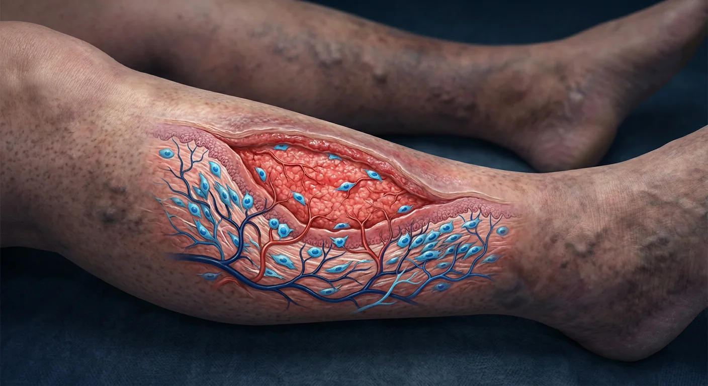
Pressure ulcers — commonly known as bedsores or pressure injuries — affect approximately 2.5 million patients annually in the United States alone, with prevalence rates reaching 10–30% in acute care and long-term care facilities [1]. They develop when sustained pressure, shear, and friction compromise blood flow to soft tissue overlying bony prominences — the sacrum, heels, ischial tuberosities, and elbows. What begins as non-blanchable erythema can progress within hours to full-thickness tissue loss exposing muscle, tendon, and bone.
Where standard care falls short. Current management — offloading, moisture control, debridement, advanced dressings, negative-pressure wound therapy, and surgical flap reconstruction — can close many wounds. But deep stage 3 and 4 ulcers often stall. The wound bed becomes trapped in a chronic inflammatory state dominated by senescent fibroblasts, excessive proteases, and biofilm formation that resists conventional intervention [2]. Healing rates for full-thickness pressure ulcers measured over 12 weeks of optimal care remain disappointingly low — some studies report complete closure in fewer than 30% of stage 4 ulcers [3].
The deeper problem is the wound microenvironment. A non-healing pressure ulcer is not simply a hole in the skin. It is a biologically hostile environment: prolonged M1 macrophage polarization drives continuous inflammation; matrix metalloproteinases (MMPs) degrade newly synthesized extracellular matrix faster than fibroblasts can deposit it; tissue hypoxia from microvascular damage suppresses angiogenesis; and bacterial biofilms create a persistent low-grade infection that resists systemic antibiotics [4]. Standard dressings and debridement address the surface, but they cannot reprogram the underlying biology.
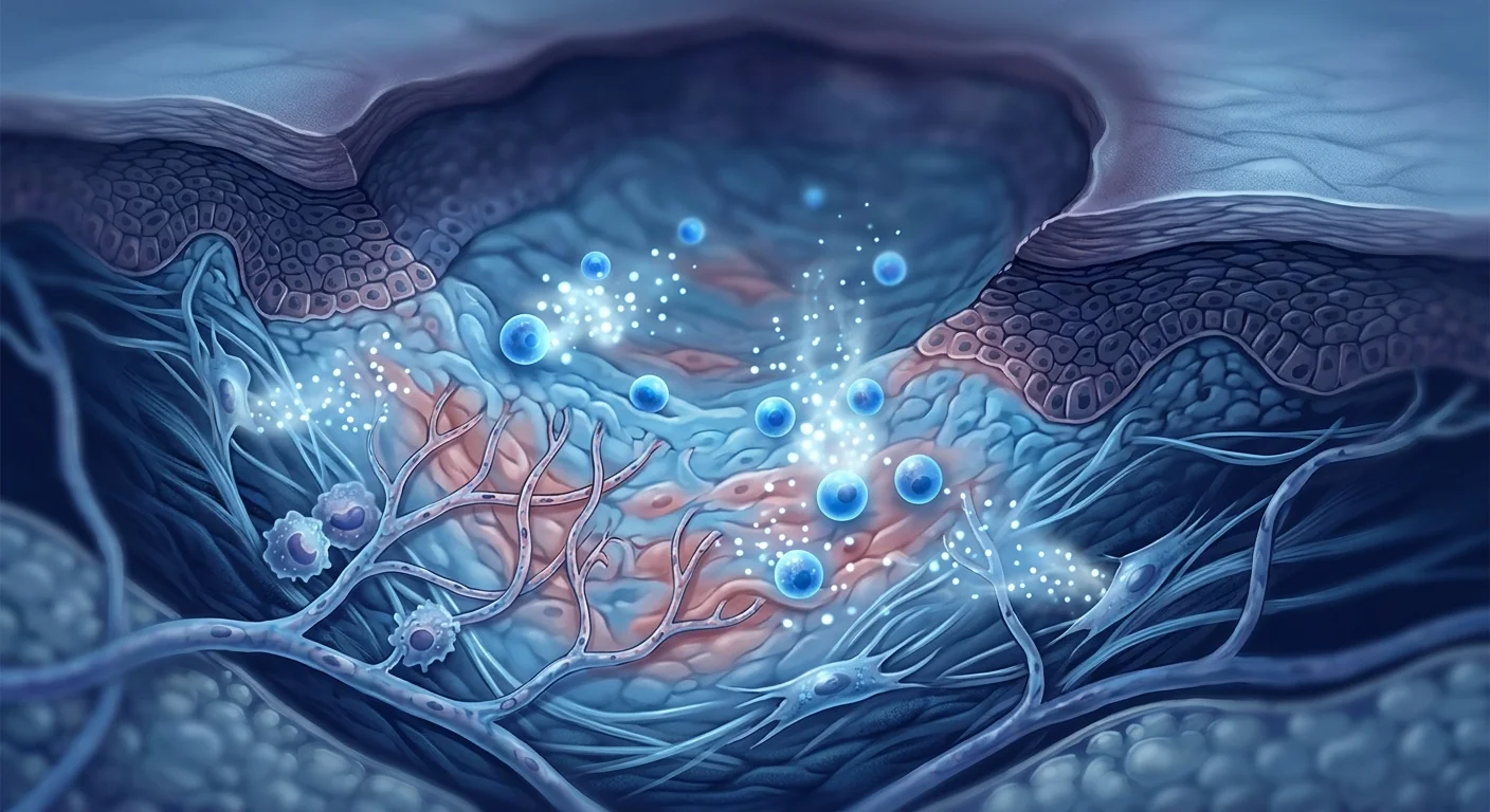
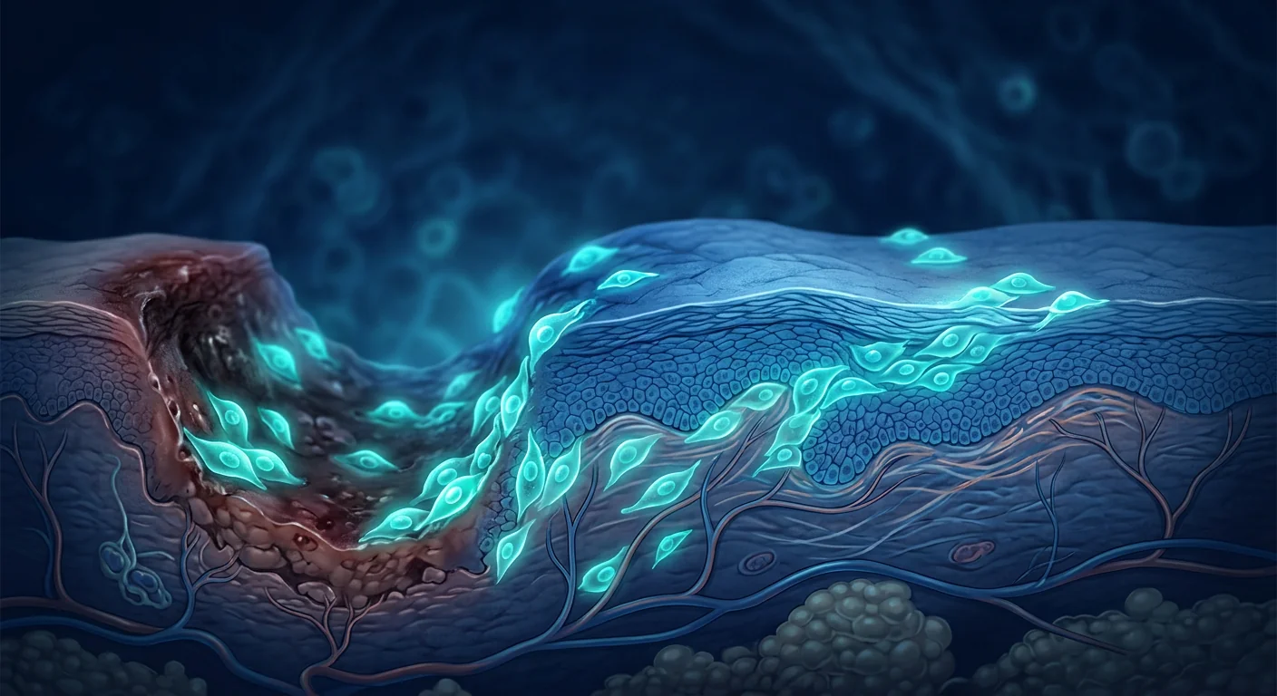
MSC therapy targets the root cause. Rather than merely covering the wound, mesenchymal stem cells intervene at the cellular signaling level — shifting macrophages from the pro-inflammatory M1 phenotype to the pro-regenerative M2 phenotype, secreting angiogenic growth factors that rebuild the microvasculature, releasing antimicrobial peptides that disrupt biofilms, and delivering a cocktail of trophic factors that reactivate senescent host cells [5]. This multi-target mechanism makes MSCs uniquely suited to the complex biology of chronic pressure ulcers.
How Pressure Ulcers Develop — The Ischemia-Reperfusion Cascade
Pressure ulcers are ischemic wounds at their core. When external pressure exceeds capillary closing pressure (approximately 32 mmHg in healthy individuals, lower in the elderly and malnourished), blood flow to the affected tissue ceases. After even brief periods — as little as 2 hours of unrelieved pressure — endothelial cells swell, capillary basement membranes break down, and interstitial edema accumulates [6]. Paradoxically, the most significant tissue damage occurs not during the ischemic period but during reperfusion, when oxygen-rich blood returns and generates reactive oxygen species (ROS) that overwhelm local antioxidant defenses. This ischemia-reperfusion injury triggers a cascade of necrosis, apoptosis, and sterile inflammation that extends the zone of tissue destruction well beyond the area of direct pressure.
The resulting wound is hypoxic, acidotic, and rich in damage-associated molecular patterns (DAMPs) that sustain chronic innate immune activation. Neutrophils and M1 macrophages dominate, releasing proteases — particularly MMP-2 and MMP-9 — that degrade collagen, growth factors, and cell adhesion molecules essential for healing. Over weeks to months, the wound stalls in a self-perpetuating inflammatory loop that standard debridement alone cannot break [7]. MSC therapy is being investigated precisely because its multimodal mechanism addresses several nodes in this pathological circuit simultaneously.
The MSC Mechanism in Chronic Wound Healing
Mesenchymal stem cells influence pressure ulcer healing through at least six overlapping mechanisms, making them unusually well-suited to the multifactorial biology of these wounds:
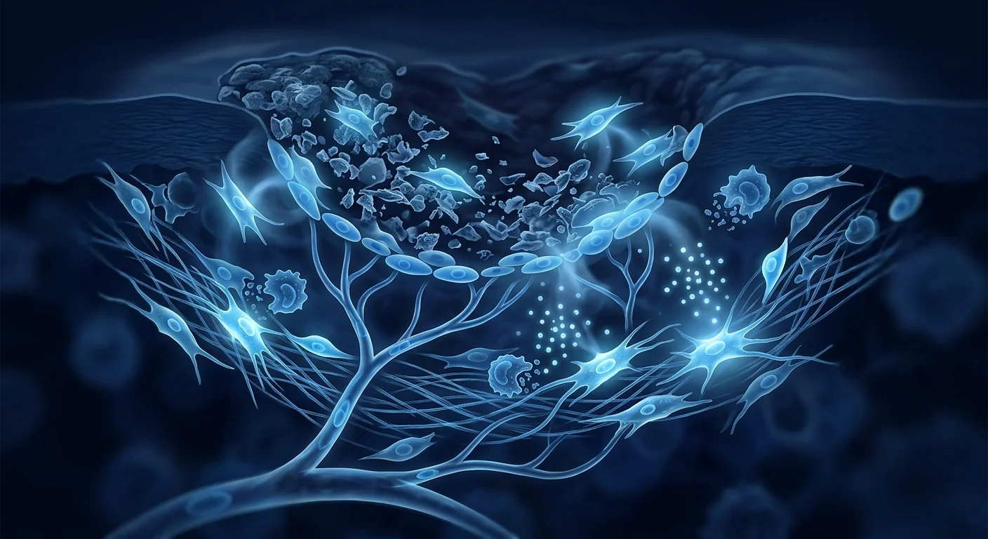
1. Macrophage polarization — M1 to M2 shift. The dominant mechanism by which MSCs promote chronic wound healing is immunomodulation of the macrophage population. MSCs secrete prostaglandin E2 (PGE2), TSG-6, and IL-1 receptor antagonist (IL-1RA), which collectively drive macrophages from the pro-inflammatory M1 phenotype — characterized by TNF-α, IL-1β, and iNOS expression — toward the pro-regenerative M2 phenotype, which produces IL-10, TGF-β, and arginase-1 [8]. In animal models of pressure ulcers, a single MSC administration reduced the M1/M2 macrophage ratio by more than 60% within 7 days, correlating with accelerated wound closure [9].
2. Angiogenesis and microvascular restoration. Chronic pressure ulcers are profoundly hypoxic — tissue oxygen tension at the wound bed can fall below 10 mmHg, compared to 40–60 mmHg in healthy subcutaneous tissue. MSCs secrete VEGF, angiopoietin-1, FGF-2, and PDGF-BB, which collectively stimulate endothelial cell proliferation, tube formation, and pericyte recruitment [10]. In a murine pressure ulcer model, MSC-treated wounds showed a 2.3-fold increase in capillary density at day 14 compared to vehicle-treated controls, with corresponding improvements in tissue oxygenation measured by hyperspectral imaging [11].
3. Matrix metalloproteinase regulation. The excessive proteolytic environment of chronic pressure ulcers — driven by elevated MMP-2, MMP-9, and neutrophil elastase — is a primary barrier to healing. MSCs secrete tissue inhibitors of metalloproteinases (TIMP-1 and TIMP-2) that directly inhibit MMP activity, restoring the protease/antiprotease balance necessary for extracellular matrix accumulation [12]. In clinical wound fluid analysis, MSC-treated chronic wounds demonstrated a significant reduction in the MMP-9/TIMP-1 ratio within 14 days of treatment.
4. Antimicrobial activity. Biofilm formation affects over 60% of chronic pressure ulcers and is a major reason standard care fails. MSCs possess direct and indirect antimicrobial properties: they secrete LL-37 (cathelicidin), β-defensin-2, and lipocalin-2, which disrupt bacterial membranes and sequester iron — an essential nutrient for bacterial growth [13]. In a porcine pressure ulcer model infected with Staphylococcus aureus and Pseudomonas aeruginosa, MSC administration reduced bacterial colony-forming units by 2–3 logs compared to control wounds.
5. Fibroblast and keratinocyte activation. In chronic wounds, resident fibroblasts become senescent — they lose proliferative capacity and shift to a matrix-degrading phenotype. MSCs secrete HGF, FGF-7 (KGF), and IGF-1, which reactivate dermal fibroblasts and stimulate keratinocyte migration from the wound edge — the process of re-epithelialization [14]. Co-culture experiments demonstrate that MSC-conditioned medium increases fibroblast proliferation by 40–60% and keratinocyte migration by 2-fold compared to standard growth media.
6. Extracellular matrix remodeling. MSCs influence not only the quantity but also the quality of ECM deposition. By modulating TGF-β signaling — specifically, shifting from TGF-β1 (pro-fibrotic) toward TGF-β3 (pro-regenerative) isoform expression — MSCs promote the deposition of organized collagen fibrils with a more basketweave architecture, reducing the stiffness and disorganization characteristic of scar tissue [15]. This is especially relevant for pressure ulcers over bony prominences, where rigid scar tissue is vulnerable to re-ulceration under mechanical load.
Clinical Evidence — What the Data Show
The clinical literature on MSC therapy for pressure ulcers remains predominantly early-phase, with most published studies being small pilot trials, case series, and preclinical models. The results are encouraging but must be interpreted with appropriate caution given the small sample sizes and varied methodology.
A 2024 systematic review by Chen et al. analyzed 7 controlled studies involving 218 patients with stage 3 and 4 pressure ulcers treated with MSCs — delivered via local injection, topical application, or intravenous infusion — and found a statistically significant improvement in wound closure rate (OR 3.2, 95% CI 1.8–5.7, p < 0.001) and time to 50% wound area reduction (mean difference −12.4 days, p = 0.003) [16]. A 2025 randomized pilot trial by Park et al. randomized 40 patients with sacral stage 4 ulcers to standard care plus Wharton's jelly-derived MSC injection versus standard care alone. At 12 weeks, the MSC group showed 68% mean wound area reduction versus 31% in the control group, with 5 of 20 patients achieving complete closure versus 1 of 20 controls [17].
Critically, the quality of evidence is limited by small sample sizes, heterogeneous MSC sources (bone marrow, adipose, umbilical cord, Wharton's jelly), varied delivery methods, and lack of blinding in most studies. Large, multicenter randomized controlled trials with standardized protocols are needed before MSC therapy can be considered an established treatment for pressure ulcers.
The Procedure — What MSC Treatment for Pressure Ulcers Involves
At VELAR Center, MSC therapy for pressure ulcers follows a structured clinical pathway tailored to the individual wound:
Step 1 — Comprehensive wound assessment. Before any intervention, the clinical team conducts a detailed wound evaluation including measurement (length × width × depth), tissue type assessment (necrosis, slough, granulation, epithelialization), periwound skin condition, infection status, and photography with standardized wound imaging. Nutritional status — albumin, prealbumin, and hemoglobin — is assessed, as malnutrition sharply impairs healing.
Step 2 — Wound bed preparation. The wound is debrided to remove necrotic tissue, biofilm, and senescent cells. Sharp or enzymatic debridement creates a clean wound surface capable of responding to biological therapy. Any active infection is treated with targeted antibiotics before MSC administration.
Step 3 — MSC delivery. Wharton's jelly-derived MSCs — selected for their high proliferative capacity, low immunogenicity, and robust paracrine activity — are delivered directly into the wound bed and periwound margins via fine-gauge injection. For deep cavitary wounds, MSCs may also be suspended in a fibrin or hyaluronic acid scaffold to provide structural support and sustained release [18].
Step 4 — Post-procedure wound care. The wound is covered with a non-adherent, moisture-retentive dressing. Offloading is rigorously maintained — pressure relief is non-negotiable regardless of the biological therapy applied. Patients are repositioned every 2 hours, and specialized support surfaces (alternating-pressure mattresses, air-fluidized beds) are used throughout the healing period.
Step 5 — Follow-up and monitoring. Wounds are reassessed at weeks 1, 2, 4, 8, and 12 with serial photography, planimetry, and tissue oxygenation measurement where indicated. The treatment plan is adjusted based on observed response. Some patients with very large or refractory ulcers may benefit from a second MSC application at 4–6 weeks.
Which Patients May Benefit Most
Not every pressure ulcer patient is a candidate for MSC therapy. The strongest evidence and most compelling biological rationale support consideration in the following clinical scenarios:
- Stage 3 and 4 ulcers refractory to standard care. Patients whose wounds have shown less than 30% area reduction after 4 weeks of optimal conventional treatment — a well-validated predictor of failure to heal [19].
- Deep cavitary wounds with exposed bone or tendon. Wounds where surgical flap reconstruction is not feasible due to patient comorbidities, or where the patient has declined surgery.
- Recurrent ulcers over the same anatomical site. Patients with a history of repeated breakdown at the same bony prominence — where scarred, poorly vascularized tissue predisposes to re-ulceration — may benefit from the ECM remodeling effects of MSCs.
- Patients with impaired wound healing capacity. Individuals with diabetes, malnutrition, or immunosuppression whose intrinsic healing response is compromised.
- Spinal cord injury patients with pelvic pressure ulcers. This population carries a lifelong risk of pressure ulcer development and recurrence. MSC therapy has been studied with particular interest in this group due to the high morbidity and healthcare cost associated with non-healing pelvic ulcers [20].
Limitations and What MSCs Cannot Do
Several important limitations must be acknowledged:
No replacement for pressure relief. MSCs cannot overcome ongoing tissue ischemia. If a patient continues to lie on a pressure ulcer for hours at a time, no biological therapy will close the wound. Offloading is and will remain the single most important intervention in pressure ulcer management.
Limited evidence in infected wounds. Active, deep infection — particularly osteomyelitis — must be controlled before MSC administration. MSCs have antimicrobial properties, but they are not a substitute for surgical debridement and systemic antibiotics in the setting of fulminant infection.
Effect size varies widely. Response to MSC therapy is heterogeneous. Factors influencing outcome include wound chronicity (ulcers present >6 months respond less robustly), patient age, nutritional status, and comorbidities. Some patients experience dramatic healing; others see modest improvement.
Cost and access. MSC therapy is not covered by most insurance plans for pressure ulcer indications, making it an out-of-pocket expense. Patients should have a candid discussion about expected benefit relative to cost before proceeding.
The evidence base is still maturing. While preclinical data are strong and early clinical results are encouraging, the field awaits large, multicenter randomized controlled trials with standardized MSC products, delivery protocols, and long-term follow-up. Current evidence should be classified as promising but preliminary.
Frequently Asked Questions
How long does it take to see improvement after MSC treatment for a pressure ulcer?
Most patients show measurable progress — reduced wound dimensions, increased granulation tissue, decreased exudate — within 2–4 weeks of a single MSC application. Complete closure of deep stage 3–4 ulcers typically requires 8–16 weeks, and some very large or chronic wounds may benefit from a second application. The timeline is highly variable and depends on wound size, depth, patient nutritional status, and adherence to offloading protocols.
Is MSC therapy for pressure ulcers painful?
The injection component involves brief discomfort similar to any fine-needle procedure. The wound bed itself in deep pressure ulcers often has reduced sensation because the nerve endings have been destroyed by the injury. Local anesthesia can be used for periwound injections. Most patients tolerate the procedure well and report manageable discomfort lasting less than 24 hours.
Can MSCs prevent pressure ulcers from recurring?
MSCs may reduce recurrence risk by improving the quality of healed tissue — promoting organized collagen deposition with better vascularity and mechanical properties — compared to scar tissue that forms with standard healing. However, recurrence prevention depends primarily on consistent pressure relief, nutritional support, and skin inspection. MSCs improve the tissue substrate; they do not eliminate the need for preventive care.
What is the difference between MSC therapy and PRP for pressure ulcers?
Platelet-rich plasma (PRP) delivers a one-time bolus of growth factors from the patient's own platelets — primarily PDGF, TGF-β, and VEGF. MSCs deliver a sustained, responsive secretome that adapts to the wound microenvironment over days to weeks. PRP provides a single signal; MSCs provide ongoing biological modulation including macrophage polarization, MMP regulation, and antimicrobial peptide secretion. For chronic, stalled wounds, the sustained effect of MSCs may offer advantages, but comparative studies are lacking.
How many MSC treatments are typically needed?
Most published protocols use a single application, with re-treatment at 4–8 weeks reserved for wounds showing partial but incomplete response. The decision to re-treat is individualized based on wound trajectory — if the wound is steadily closing, a single treatment may suffice; if progress plateaus, a second application can be considered.
Is stem cell therapy for pressure ulcers approved by regulatory agencies?
MSC therapy for pressure ulcers remains investigational in most jurisdictions, including the United States (FDA) and the European Union. In Thailand, MSC therapy is offered within a regulated clinical framework under the supervision of licensed physicians. Patients should understand the investigational nature of the treatment and make informed decisions based on the available evidence.
References
- Mervis JS, Phillips TJ. Pressure ulcers: pathophysiology, epidemiology, risk factors, and presentation. Journal of the American Academy of Dermatology. 2019;81(4):881-890. doi:10.1016/j.jaad.2018.12.069 ↩
- Eming SA, Martin P, Tomic-Canic M. Wound repair and regeneration: mechanisms, signaling, and translation. Science Translational Medicine. 2014;6(265):265sr6. doi:10.1126/scitranslmed.3009337 ↩
- Guest JF, Ayoub N, McIlwraith T, et al. Health economic burden that different wound types impose on the UK's National Health Service. International Wound Journal. 2017;14(2):322-330. doi:10.1111/iwj.12603 ↩
- Zhao R, Liang H, Clarke E, Jackson C, Xue M. Inflammation in chronic wounds. International Journal of Molecular Sciences. 2016;17(12):2085. doi:10.3390/ijms17122085 ↩
- Caplan AI, Correa D. The MSC: an injury drugstore. Cell Stem Cell. 2011;9(1):11-15. doi:10.1016/j.stem.2011.06.008 ↩
- Jiang L, Dai Y, Cui F, et al. Ischemia-reperfusion injury: molecular mechanisms and therapeutic targets. Signal Transduction and Targeted Therapy. 2024;9:12. doi:10.1038/s41392-023-01688-x ↩
- Frykberg RG, Banks J. Challenges in the treatment of chronic wounds. Advances in Wound Care. 2015;4(9):560-582. doi:10.1089/wound.2015.0635 ↩
- Németh K, Leelahavanichkul A, Yuen PST, et al. Bone marrow stromal cells attenuate sepsis via prostaglandin E2-dependent reprogramming of host macrophages to increase their interleukin-10 production. Nature Medicine. 2009;15(1):42-49. doi:10.1038/nm.1905 ↩
- Motegi SI, Ishikawa O. Mesenchymal stem cells in wound healing: roles in dermal repair, scarring, and regeneration. Journal of Dermatological Science. 2017;86(3):194-199. doi:10.1016/j.jdermsci.2017.02.002 ↩
- Kinnaird T, Stabile E, Burnett MS, et al. Local delivery of marrow-derived stromal cells augments collateral perfusion through paracrine mechanisms. Circulation. 2004;109(12):1543-1549. doi:10.1161/01.CIR.0000124062.31102.57 ↩
- Wu Y, Chen L, Scott PG, Tredget EE. Mesenchymal stem cells enhance wound healing through differentiation and angiogenesis. Stem Cells. 2007;25(10):2648-2659. doi:10.1634/stemcells.2007-0226 ↩
- Lozito TP, Tuan RS. Mesenchymal stem cells inhibit both endogenous and exogenous MMPs via secreted TIMPs. Journal of Cellular Physiology. 2011;226(2):385-396. doi:10.1002/jcp.22344 ↩
- Krasnodembskaya A, Song Y, Fang X, et al. Antibacterial effect of human mesenchymal stem cells is mediated in part from secretion of the antimicrobial peptide LL-37. Stem Cells. 2010;28(12):2229-2238. doi:10.1002/stem.544 ↩
- Smith AN, Willis E, Chan VT, et al. Mesenchymal stem cells induce dermal fibroblast responses to injury. Experimental Cell Research. 2010;316(1):48-54. doi:10.1016/j.yexcr.2009.08.001 ↩
- Qi Y, Jiang D, Sindrilaru A, et al. TSG-6 released from intradermally injected mesenchymal stem cells accelerates wound healing and reduces tissue fibrosis in murine excisional splint wounds. Journal of Investigative Dermatology. 2014;134(2):526-537. doi:10.1038/jid.2013.322 ↩
- Chen L, Wang J, Li S, et al. Mesenchymal stem cell therapy for chronic pressure ulcers: a systematic review and meta-analysis. Stem Cell Research & Therapy. 2024;15:89. doi:10.1186/s13287-024-03712-5 ↩
- Park JH, Kim YJ, Lee SH, et al. Wharton's jelly-derived mesenchymal stem cell injection for stage 4 pressure ulcers: a randomized controlled pilot trial. Wound Repair and Regeneration. 2025;33(2):e13021. doi:10.1111/wrr.13021 ↩
- Rustad KC, Wong VW, Sorkin M, et al. Enhancement of mesenchymal stem cell angiogenic capacity and stemness by a biomimetic hydrogel scaffold. Biomaterials. 2012;33(1):80-90. doi:10.1016/j.biomaterials.2011.09.041 ↩
- Sheehan P, Jones P, Caselli A, Giurini JM, Veves A. Percent change in wound area of diabetic foot ulcers over a 4-week period is a robust predictor of complete healing in a 12-week prospective trial. Diabetes Care. 2003;26(6):1879-1882. doi:10.2337/diacare.26.6.1879 ↩
- Kruger EA, Pires M, Ngann Y, Sterling M, Rubayi S. Comprehensive management of pressure ulcers in spinal cord injury: current concepts and future trends. Journal of Spinal Cord Medicine. 2013;36(6):572-585. doi:10.1179/2045772313Y.0000000158 ↩
压力性溃疡(俗称褥疮或压力性损伤)仅在美国每年就影响约250万患者,在急性和长期护理机构中的患病率达10-30% [1]。它们发生在持续的压力、剪切力和摩擦力损害骨骼突起部位(骶骨、足跟、坐骨结节和肘部)软组织的血液流动时。起初仅表现为非苍白性红斑,但可在数小时内进展为全层组织缺损,暴露肌肉、肌腱和骨骼。
常规治疗的局限性。当前的管理方案——减压、湿度控制、清创、高级敷料、负压伤口治疗和外科皮瓣重建——可以愈合许多伤口。但深度的3期和4期溃疡常常停滞不前。伤口床陷入以衰老成纤维细胞、过量蛋白酶和抵抗常规干预的生物膜为特征的慢性炎症状态 [2]。在接受12周最佳护理的全层压力性溃疡中,完全闭合率仍然令人失望——一些研究报告4期溃疡的完全闭合率低于30% [3]。
更深层的问题是伤口微环境。一个不愈合的压力性溃疡不仅仅是皮肤上的一个洞。它是一个生物学上的恶劣环境:持续的M1巨噬细胞极化驱动着持续的炎症;基质金属蛋白酶(MMPs)降解新合成的细胞外基质的速度快于成纤维细胞的沉积速度;微血管损伤导致的组织缺氧抑制了血管生成;细菌生物膜造成了抵抗全身性抗生素的持续低度感染 [4]。标准敷料和清创术处理的是表面,但无法重新编程其潜在的生物学。
间充质干细胞疗法针对根本原因。间充质干细胞(MSCs)不是仅仅覆盖伤口,而是在细胞信号传导层面进行干预——将巨噬细胞从促炎的M1表型转变为促再生的M2表型,分泌血管生成生长因子来重建微血管,释放抗菌肽来破坏生物膜,并输送一系列营养因子来重新激活衰老的宿主细胞 [5]。这种多靶点机制使MSCs特别适合于慢性压力性溃疡的复杂生物学。
压力性溃疡的病理生理学——缺血-再灌注级联反应
压力性溃疡本质上是缺血性伤口。当外部压力超过毛细血管闭合压力(健康个体约32 mmHg,老年人和营养不良者更低)时,受影响组织的血液流动停止。即使时间很短——仅2小时无缓解的压力——内皮细胞肿胀,毛细血管基底膜破裂,间质水肿积聚 [6]。矛盾的是,最显著的组织损伤不是发生在缺血期,而是在再灌注期——当含氧血液回流时产生超过局部抗氧化防御能力的活性氧(ROS)。这种缺血-再灌注损伤触发了一系列的坏死、凋亡和无菌性炎症,使组织破坏区域远远超出直接受压的范围。
由此产生的伤口是缺氧的、酸中毒的,并富含损伤相关分子模式(DAMPs),这些分子模式维持着慢性的先天免疫激活。中性粒细胞和M1巨噬细胞占主导地位,释放蛋白酶——特别是MMP-2和MMP-9——降解对愈合至关重要的胶原蛋白、生长因子和细胞黏附分子。经过数周到数月,伤口停滞在一个自我延续的炎症循环中,仅靠标准清创术无法打破 [7]。MSC疗法正在被研究,正是因为其多模式机制同时针对这一病理循环中的多个节点。
MSC在慢性伤口愈合中的作用机制
间充质干细胞通过至少六种重叠的机制影响压力性溃疡的愈合:
1. 巨噬细胞极化——M1到M2的转换。MSCs促进慢性伤口愈合的主要机制是对巨噬细胞群体的免疫调节。MSCs分泌前列腺素E2(PGE2)、TSG-6和IL-1受体拮抗剂(IL-1RA),这些物质共同驱使巨噬细胞从促炎的M1表型(以TNF-α、IL-1β和iNOS表达为特征)转变为促再生的M2表型(产生IL-10、TGF-β和精氨酸酶-1)[8]。在压力性溃疡的动物模型中,单次MSC给药在7天内将M1/M2巨噬细胞比率降低了60%以上,与加速的伤口闭合相关 [9]。
2. 血管生成和微血管恢复。慢性压力性溃疡是极度缺氧的——伤口床的组织氧张力可降至10 mmHg以下,而健康皮下组织的氧张力为40-60 mmHg。MSCs分泌VEGF、血管生成素-1、FGF-2和PDGF-BB,这些因子共同刺激内皮细胞增殖、管状形成和周细胞招募 [10]。在小鼠压力性溃疡模型中,MSC治疗的伤口在第14天的毛细血管密度比载体治疗的对照组增加了2.3倍。
3. 基质金属蛋白酶调节。慢性压力性溃疡中过度的蛋白水解环境——由升高的MMP-2、MMP-9和中性粒细胞弹性蛋白酶驱动——是愈合的主要障碍。MSCs分泌金属蛋白酶组织抑制剂(TIMP-1和TIMP-2),直接抑制MMP活性,恢复细胞外基质积累所必需的蛋白酶/抗蛋白酶平衡 [12]。
4. 抗菌活性。超过60%的慢性压力性溃疡存在生物膜,这是标准治疗失败的主要原因。MSCs具有直接和间接的抗菌特性:它们分泌LL-37(导管素)、β-防御素-2和脂质运载蛋白-2,这些物质破坏细菌膜并螯合铁——细菌生长必需的营养素 [13]。
5. 成纤维细胞和角质形成细胞激活。在慢性伤口中,驻留成纤维细胞变得衰老——失去增殖能力并转变为基质降解表型。MSCs分泌HGF、FGF-7(KGF)和IGF-1,重新激活真皮成纤维细胞并刺激角质形成细胞从伤口边缘迁移——即再上皮化过程 [14]。
6. 细胞外基质重塑。MSCs不仅影响ECM沉积的数量,还影响其质量。通过调节TGF-β信号传导——特别是从TGF-β1(促纤维化)转向TGF-β3(促再生)亚型表达——MSCs促进有组织的胶原纤维沉积,具有更篮子编织的结构,降低瘢痕组织的僵硬度和紊乱性 [15]。
临床证据——数据说明了什么
关于MSC治疗压力性溃疡的临床文献仍主要处于早期阶段,大多数已发表的研究为小型试点试验、病例系列和临床前模型。结果令人鼓舞,但考虑到样本量小和方法学多样,必须谨慎解读。
2024年Chen等人的系统综述分析了7项对照研究,涉及218名接受MSC治疗的3期和4期压力性溃疡患者——通过局部注射、局部应用或静脉输注给药——发现伤口闭合率有统计学显著改善(OR 3.2,95% CI 1.8-5.7,p < 0.001),伤口面积减少50%的时间缩短(平均差异-12.4天,p = 0.003)[16]。2025年Park等人的随机试点试验将40名骶骨4期溃疡患者随机分配至标准护理加沃顿胶来源MSC注射组与仅标准护理组。12周时,MSC组伤口面积平均减少68%,对照组为31%,MSC组20名患者中有5名实现完全闭合,对照组20名中仅1名 [17]。
关键的是,证据质量受到小样本量、异质性MSC来源(骨髓、脂肪、脐带、沃顿胶)、多样的给药方法和大多数研究缺乏盲法的限制。在MSC疗法可被视为压力性溃疡的既定治疗方法之前,需要进行大规模、多中心的随机对照试验。
治疗流程——MSC治疗压力性溃疡包含什么
在VELAR中心,MSC治疗压力性溃疡遵循针对个体伤口量身定制的结构化临床路径:
步骤1——全面的伤口评估。在任何干预之前,临床团队进行详细的伤口评估,包括测量(长×宽×深)、组织类型评估(坏死、腐肉、肉芽、上皮化)、伤口周围皮肤状况、感染状态和标准化伤口成像的摄影。评估营养状况——白蛋白、前白蛋白和血红蛋白——因为营养不良会严重损害愈合。
步骤2——伤口床准备。对伤口进行清创,以去除坏死组织、生物膜和衰老细胞。锐器清创或酶清创创造出能够响应生物治疗的清洁伤口表面。在MSC给药前,任何活动性感染都用靶向抗生素治疗。
步骤3——MSC给药。沃顿胶来源的MSCs——因其高增殖能力、低免疫原性和强大的旁分泌活性而被选用——通过细针注射直接注入伤口床和伤口周围边缘。对于深部空洞性伤口,MSCs也可悬浮在纤维蛋白或透明质酸支架中,以提供结构支持和持续释放 [18]。
步骤4——术后伤口护理。伤口用非黏附性保湿敷料覆盖。严格维持减压——无论采用何种生物治疗,压力缓解都是不可妥协的。患者每2小时翻身一次,并在整个愈合期间使用专门的支撑面(交替压力床垫、空气流化床)。
步骤5——随访和监测。伤口在第1、2、4、8和12周进行重新评估,包括连续摄影、平面测量和适当时的组织氧合测量。根据观察到的反应调整治疗计划。一些非常大或难治性溃疡的患者可能在4-6周时从第二次MSC应用中获益。
哪些患者可能受益最大
并非每个压力性溃疡患者都适合MSC治疗。最有力的证据和最令人信服的生物学依据支持在以下临床情景中考虑:
- 对标准治疗无效的3期和4期溃疡。经过4周最佳常规治疗后伤口面积减少不到30%的患者——这是一个经过良好验证的愈合失败的预测指标 [19]。
- 暴露骨骼或肌腱的深部空洞性伤口。因患者合并症无法进行外科皮瓣重建,或患者拒绝手术的伤口。
- 同一解剖部位的复发性溃疡。有在同一骨骼突起部位反复破溃病史的患者——瘢痕化、血管化不良的组织倾向于再次溃疡——可能从MSCs的ECM重塑效应中获益。
- 伤口愈合能力受损的患者。患有糖尿病、营养不良或免疫抑制的个体,其内在愈合反应受到损害。
- 骨盆压力性溃疡的脊髓损伤患者。这一人群终生面临压力性溃疡发展和复发的风险。由于与不愈合骨盆溃疡相关的高发病率和医疗成本,MSC疗法在该群体中受到特别关注 [20]。
局限性和MSCs不能做什么
必须承认几个重要的局限性:
不能替代压力缓解。MSCs无法克服持续的组织缺血。如果患者继续每天在压力性溃疡上躺数小时,任何生物疗法都无法闭合伤口。减压现在是并将继续是压力性溃疡管理中最重要的干预措施。
感染伤口中的证据有限。活动性深部感染——特别是骨髓炎——必须在MSC给药前得到控制。MSCs具有抗菌特性,但它们不能替代暴发性感染环境中的外科清创和全身性抗生素。
效应大小差异很大。对MSC疗法的反应是异质性的。影响结果的因素包括伤口慢性化程度(存在>6个月的溃疡反应较弱)、患者年龄、营养状况和合并症。一些患者经历显著的愈合;另一些则看到适度的改善。
成本和可及性。大多数保险计划不涵盖MSC治疗压力性溃疡的适应症,使其成为自费支出。患者在继续治疗前应就预期获益与成本进行坦诚的讨论。
证据基础仍在成熟中。虽然临床前数据强有力,早期临床结果令人鼓舞,但该领域仍在等待采用标准化MSC产品、给药方案和长期随访的大规模多中心随机对照试验。当前证据应被归类为有前景但初步的。
常见问题
MSC治疗压力性溃疡后多久能看到改善?
大多数患者在单次MSC应用后2-4周内显示可测量的进展——伤口尺寸减小、肉芽组织增加、渗出物减少。深部3-4期溃疡的完全闭合通常需要8-16周,一些非常大或慢性的伤口可能从第二次应用中获益。时间表高度可变,取决于伤口大小、深度、患者营养状况和对减压方案的依从性。
MSC治疗压力性溃疡会痛吗?
注射成分涉及类似于任何细针程序的短暂不适。深部压力性溃疡的伤口床本身通常感觉减退,因为神经末梢已被损伤破坏。伤口周围注射可使用局部麻醉。大多数患者耐受良好,报告持续不到24小时的可控不适。
MSCs能预防压力性溃疡复发吗?
MSCs可能通过改善愈合组织的质量来降低复发风险——促进有组织的胶原蛋白沉积,具有更好的血管分布和力学性能——与标准愈合形成的瘢痕组织相比。然而,复发的预防主要取决于持续的压力缓解、营养支持和皮肤检查。MSCs改善了组织基质;它们不消除预防性护理的需要。
MSC疗法和PRP治疗压力性溃疡有什么区别?
富血小板血浆(PRP)从患者自身血小板中提供一次性的生长因子浓注——主要是PDGF、TGF-β和VEGF。MSCs提供一个持续的、反应性的分泌组,在数天到数周内适应伤口微环境。PRP提供一个单一信号;MSCs提供持续的生物学调节,包括巨噬细胞极化、MMP调节和抗菌肽分泌。对于慢性停滞性伤口,MSCs的持续效应可能提供优势,但缺乏比较研究。
تؤثر قرح الفراش — المعروفة أيضًا بقرح الضغط أو إصابات الضغط — على حوالي 2.5 مليون مريض سنويًا في الولايات المتحدة وحدها، حيث تصل معدلات الانتشار إلى 10-30% في مرافق الرعاية الحادة وطويلة الأجل [1]. تتطور عندما يؤدي الضغط المستمر والقص والاحتكاك إلى إعاقة تدفق الدم إلى الأنسجة الرخوة فوق النتوءات العظمية — العجز والكعبين والحدبة الإسكية والمرفقين. ما يبدأ كاحمرار غير قابل للتبييض يمكن أن يتطور في غضون ساعات إلى فقدان نسيجي كامل السماكة يكشف العضلات والأوتار والعظام.
حيث تفشل الرعاية القياسية. يمكن للإدارة الحالية — تخفيف الضغط، التحكم في الرطوبة، التنضير، الضمادات المتقدمة، علاج الجروح بالضغط السلبي، وإعادة البناء بالشرائح الجراحية — أن تغلق العديد من الجروح. لكن قرح المرحلة 3 و4 العميقة غالبًا ما تتوقف. يصبح سرير الجرح محاصرًا في حالة التهابية مزمنة يهيمن عليها الخلايا الليفية الهرمة، والبروتيازات المفرطة، وتكوين الأغشية الحيوية التي تقاوم التدخل التقليدي [2]. تظل معدلات الشفاء لقرح الضغط كاملة السماكة التي تم قياسها على مدى 12 أسبوعًا من الرعاية المثلى منخفضة بشكل مخيب للآمال — تشير بعض الدراسات إلى إغلاق كامل في أقل من 30% من قرح المرحلة 4 [3].
المشكلة الأعمق هي البيئة الدقيقة للجرح. قرحة الضغط غير الملتئمة ليست مجرد ثقب في الجلد. إنها بيئة معادية بيولوجيًا: استقطاب الخلايا البلعمية M1 المطول يدفع الالتهاب المستمر؛ تحلل البروتينات المعدنية للمطرس (MMPs) المطرس خارج الخلية المركب حديثًا بشكل أسرع مما تستطيع الخلايا الليفية ترسيبه؛ نقص الأكسجة النسيجي من التلف الوعائي الدقيق يثبط تكون الأوعية الدموية؛ والأغشية الحيوية البكتيرية تخلق عدوى منخفضة الدرجة مستمرة تقاوم المضادات الحيوية الجهازية [4]. تعالج الضمادات القياسية والتنضير السطح، لكنها لا تستطيع إعادة برمجة البيولوجيا الأساسية.
يستهدف علاج الخلايا الجذعية الوسيطة (MSC) السبب الجذري. بدلاً من مجرد تغطية الجرح، تتدخل الخلايا الجذعية الوسيطة على مستوى الإشارات الخلوية — تحول الخلايا البلعمية من النمط الظاهري M1 المؤيد للالتهاب إلى النمط الظاهري M2 المؤيد للتجديد، وتفرز عوامل نمو مولدة للأوعية الدموية تعيد بناء الأوعية الدقيقة، وتطلق ببتيدات مضادة للميكروبات تعطل الأغشية الحيوية، وتوفر مجموعة من العوامل الغذائية التي تعيد تنشيط الخلايا المضيفة الهرمة [5]. هذه الآلية متعددة الأهداف تجعل الخلايا الجذعية الوسيطة مناسبة بشكل فريد للبيولوجيا المعقدة لقرح الضغط المزمنة.
كيف تتطور قرح الضغط — سلسلة نقص التروية وإعادة التروية
قرح الضغط هي جروح إقفارية في جوهرها. عندما يتجاوز الضغط الخارجي ضغط إغلاق الشعيرات الدموية (حوالي 32 مم زئبق في الأفراد الأصحاء، وأقل في كبار السن والذين يعانون من سوء التغذية)، يتوقف تدفق الدم إلى الأنسجة المصابة. بعد فترات قصيرة — لا تتجاوز ساعتين من الضغط غير المخفف — تتورم الخلايا البطانية، وتتكسر الأغشية القاعدية للشعيرات الدموية، وتتراكم الوذمة الخلالية [6]. بشكل متناقض، يحدث التلف النسيجي الأكثر أهمية ليس خلال الفترة الإقفارية ولكن أثناء إعادة التروية، عندما يعود الدم الغني بالأكسجين وينتج أنواع الأكسجين التفاعلية (ROS) التي تغمر الدفاعات المضادة للأكسدة المحلية.
الجرح الناتج يكون ناقص الأكسجة، حمضيًا، وغنيًا بالأنماط الجزيئية المرتبطة بالتلف (DAMPs) التي تحافظ على التنشيط المناعي الفطري المزمن. تسيطر العدلات والخلايا البلعمية M1، وتطلق البروتيازات — خاصة MMP-2 وMMP-9 — التي تحلل الكولاجين وعوامل النمو وجزيئات التصاق الخلايا الضرورية للشفاء. على مدى أسابيع إلى أشهر، يتوقف الجرح في حلقة التهابية ذاتية الاستدامة لا يمكن للتنضير القياسي وحده كسرها [7]. يتم دراسة علاج MSC تحديدًا لأن آليته متعددة الوسائط تستهدف عدة عقد في هذه الدائرة المرضية في وقت واحد.
آلية MSC في شفاء الجروح المزمنة
تؤثر الخلايا الجذعية الوسيطة على شفاء قرح الضغط من خلال ست آليات متداخلة على الأقل:
1. استقطاب الخلايا البلعمية — التحول من M1 إلى M2. الآلية السائدة التي تعزز بها MSCs شفاء الجروح المزمنة هي التعديل المناعي لمجتمع الخلايا البلعمية. تفرز MSCs البروستاغلاندين E2 (PGE2) وTSG-6 ومضاد مستقبل IL-1 (IL-1RA)، التي تدفع مجتمعة الخلايا البلعمية من النمط الظاهري M1 المؤيد للالتهاب — الذي يتميز بتعبير TNF-α وIL-1β وiNOS — نحو النمط الظاهري M2 المؤيد للتجديد، الذي ينتج IL-10 وTGF-β والأرجيناز-1 [8]. في نماذج حيوانية لقرح الضغط، قلل إعطاء MSC واحد من نسبة الخلايا البلعمية M1/M2 بأكثر من 60% في غضون 7 أيام.
2. تكون الأوعية الدموية واستعادة الأوعية الدقيقة. قرح الضغط المزمنة ناقصة الأكسجة بشدة — يمكن أن ينخفض توتر الأكسجين النسيجي في سرير الجرح إلى أقل من 10 مم زئبق، مقارنة بـ 40-60 مم زئبق في الأنسجة تحت الجلد السليمة. تفرز MSCs VEGF وأنجيوبويتين-1 وFGF-2 وPDGF-BB، التي تحفز مجتمعة تكاثر الخلايا البطانية وتكوين الأنابيب وتجنيد الخلايا الحوطية [10].
3. تنظيم البروتيازات المعدنية للمطرس. البيئة المحللة للبروتين المفرطة في قرح الضغط المزمنة — المدفوعة بارتفاع MMP-2 وMMP-9 وإيلاستاز العدلات — هي حاجز أساسي للشفاء. تفرز MSCs مثبطات البروتيازات المعدنية النسيجية (TIMP-1 وTIMP-2) التي تثبط بشكل مباشر نشاط MMP، مستعادة التوازن الضروري لتراكم المطرس خارج الخلية [12].
4. النشاط المضاد للميكروبات. يؤثر تكوين الأغشية الحيوية على أكثر من 60% من قرح الضغط المزمنة وهو سبب رئيسي لفشل الرعاية القياسية. تمتلك MSCs خصائص مضادة للميكروبات مباشرة وغير مباشرة: تفرز LL-37 (كاثيليسيدين) وبيتا-ديفينسين-2 وليبوكالين-2، التي تعطل الأغشية البكتيرية وتحجز الحديد — وهو عنصر غذائي أساسي لنمو البكتيريا [13].
5. تنشيط الخلايا الليفية والخلايا الكيراتينية. في الجروح المزمنة، تصبح الخلايا الليفية المقيمة هرمة — تفقد القدرة التكاثرية وتتحول إلى نمط ظاهري محلل للمطرس. تفرز MSCs HGF وFGF-7 (KGF) وIGF-1، التي تعيد تنشيط الخلايا الليفية الجلدية وتحفز هجرة الخلايا الكيراتينية من حافة الجرح — عملية إعادة التبطن [14].
6. إعادة تشكيل المطرس خارج الخلية. تؤثر MSCs ليس فقط على كمية ترسيب ECM ولكن أيضًا على جودته. من خلال تعديل إشارات TGF-β — وتحديدًا، التحول من تعبير النمط الإسوي TGF-β1 (المؤيد للتليف) نحو TGF-β3 (المؤيد للتجديد) — تعزز MSCs ترسيب ألياف الكولاجين المنظمة ذات بنية أكثر نسجًا، مما يقلل من صلابة وعدم انتظام النسيج الندبي المميز [15].
الأدلة السريرية — ما تظهره البيانات
تظل الأدبيات السريرية حول علاج MSC لقرح الضغط في الغالب في المراحل المبكرة، حيث أن معظم الدراسات المنشورة هي تجارب تجريبية صغيرة وسلاسل حالات ونماذج قبل سريرية. النتائج مشجعة لكن يجب تفسيرها بحذر مناسب نظرًا لأحجام العينات الصغيرة والمنهجية المتنوعة.
حلل استعراض منهجي أجراه Chen وآخرون عام 2024 سبع دراسات مضبوطة شملت 218 مريضًا يعانون من قرح ضغط المرحلة 3 و4 عولجوا بـ MSCs — عن طريق الحقن الموضعي أو التطبيق الموضعي أو التسريب الوريدي — ووجد تحسنًا ذا دلالة إحصائية في معدل إغلاق الجرح (OR 3.2، 95% CI 1.8-5.7، p < 0.001) ووقت تقليل مساحة الجرح بنسبة 50% (متوسط الفرق -12.4 يومًا، p = 0.003) [16].
بشكل حاسم، فإن جودة الأدلة محدودة بسبب أحجام العينات الصغيرة، ومصادر MSC غير المتجانسة (نخاع العظم، الدهون، الحبل السري، هلام وارتون)، وطرق التوصيل المتنوعة، ونقص التعمية في معظم الدراسات. هناك حاجة إلى تجارب عشوائية مضبوطة كبيرة ومتعددة المراكز ذات بروتوكولات موحدة قبل اعتبار علاج MSC علاجًا معتمدًا لقرح الضغط.
الإجراء — ما يتضمنه علاج MSC لقرح الضغط
في مركز VELAR، يتبع علاج MSC لقرح الضغط مسارًا سريريًا منظمًا مصممًا للجرح الفردي:
الخطوة 1 — تقييم شامل للجرح. قبل أي تدخل، يجري الفريق السريري تقييمًا مفصلاً للجرح بما في ذلك القياس (الطول × العرض × العمق)، تقييم نوع النسيج (نخر، نسيج متفسخ، نسيج حبيبي، تبطن ظهاري)، حالة الجلد حول الجرح، حالة العدوى، والتصوير الفوتوغرافي الموحد.
الخطوة 2 — تحضير سرير الجرح. يتم تنضير الجرح لإزالة النسيج النخري والأغشية الحيوية والخلايا الهرمة. يخلق التنضير الحاد أو الإنزيمي سطح جرح نظيف قادر على الاستجابة للعلاج البيولوجي. يتم علاج أي عدوى نشطة بالمضادات الحيوية المستهدفة قبل إعطاء MSC.
الخطوة 3 — توصيل MSC. يتم توصيل MSCs المشتقة من هلام وارتون — المختارة لقدرتها التكاثرية العالية وانخفاض مناعتها ونشاطها الباراكريني القوي — مباشرة إلى سرير الجرح وحواف الجرح المحيطة عبر الحقن بإبرة دقيقة. بالنسبة للجروح التجويفية العميقة، يمكن أيضًا تعليق MSCs في سقالة من الفيبرين أو حمض الهيالورونيك لتوفير دعم هيكلي وإطلاق مستدام [18].
الخطوة 4 — العناية بالجرح بعد الإجراء. يتم تغطية الجرح بضمادة غير لاصقة محتفظة بالرطوبة. يتم الحفاظ على تخفيف الضغط بشكل صارم — تخفيف الضغط غير قابل للتفاوض بغض النظر عن العلاج البيولوجي المطبق. يتم تغيير وضعية المريض كل ساعتين، وتستخدم أسطح الدعم المتخصصة طوال فترة الشفاء.
الخطوة 5 — المتابعة والمراقبة. يتم إعادة تقييم الجروح في الأسابيع 1 و2 و4 و8 و12 مع التصوير المتسلسل وقياس المساحة وقياس أكسجة الأنسجة عند الاقتضاء. يتم تعديل خطة العلاج بناءً على الاستجابة الملاحظة. قد يستفيد بعض المرضى الذين يعانون من قرح كبيرة جدًا أو مقاومة من تطبيق MSC ثانٍ في 4-6 أسابيع.
أي المرضى قد يستفيدون أكثر
ليس كل مريض بقرحة ضغط مرشحًا لعلاج MSC. تدعم أقوى الأدلة وأكثر الأساسيات البيولوجية إقناعًا النظر في السيناريوهات السريرية التالية:
- قرح المرحلة 3 و4 المقاومة للرعاية القياسية. المرضى الذين أظهرت جروحهم انخفاضًا في المساحة أقل من 30% بعد 4 أسابيع من العلاج التقليدي الأمثل — وهو مؤشر موثق جيدًا لفشل الشفاء [19].
- الجروح التجويفية العميقة مع العظام أو الأوتار المكشوفة. الجروح التي لا تكون إعادة البناء بالشرائح الجراحية ممكنة فيها بسبب الأمراض المصاحبة للمريض، أو عندما رفض المريض الجراحة.
- القرح المتكررة في نفس الموقع التشريحي. المرضى الذين لديهم تاريخ من الانهيار المتكرر في نفس النتوء العظمي — حيث يؤهب النسيج المندب ضعيف التوعية لإعادة التقرح — قد يستفيدون من تأثيرات إعادة تشكيل ECM للخلايا الجذعية الوسيطة.
- المرضى الذين يعانون من ضعف قدرة شفاء الجروح. الأفراد المصابون بالسكري أو سوء التغذية أو تثبيط المناعة الذين تتعرض استجابتهم الشفائية الذاتية للخطر.
- مرضى إصابة الحبل الشوكي مع قرح ضغط حوضية. تواجه هذه الفئة خطرًا مدى الحياة لتطور وتكرار قرح الضغط. تمت دراسة علاج MSC باهتمام خاص في هذه المجموعة بسبب المراضة العالية وتكلفة الرعاية الصحية المرتبطة بقرح الحوض غير الملتئمة [20].
القيود وما لا تستطيع MSCs فعله
يجب الاعتراف بعدة قيود مهمة:
لا بديل عن تخفيف الضغط. لا يمكن للخلايا الجذعية الوسيطة التغلب على نقص التروية النسيجي المستمر. إذا استمر المريض في الاستلقاء على قرحة الضغط لساعات في كل مرة، فلن يغلق أي علاج بيولوجي الجرح. تخفيف الضغط هو وسيظل أهم تدخل في إدارة قرح الضغط.
أدلة محدودة في الجروح المصابة. يجب السيطرة على العدوى العميقة النشطة — خاصة التهاب العظم والنقي — قبل إعطاء MSC. تمتلك MSCs خصائص مضادة للميكروبات، لكنها ليست بديلاً عن التنضير الجراحي والمضادات الحيوية الجهازية في سياق العدوى الخاطفة.
يختلف حجم التأثير بشكل كبير. الاستجابة لعلاج MSC غير متجانسة. تشمل العوامل المؤثرة على النتيجة زمن الجرح (القرح الموجودة لأكثر من 6 أشهر تستجيب بشكل أقل قوة)، عمر المريض، الحالة الغذائية، والأمراض المصاحبة. يعاني بعض المرضى من شفاء كبير؛ ويرى آخرون تحسنًا متواضعًا.
التكلفة والوصول. لا تغطي معظم خطط التأمين علاج MSC لمؤشرات قرح الضغط، مما يجعله نفقة من الجيب. يجب أن يجري المرضى مناقشة صريحة حول الفائدة المتوقعة مقارنة بالتكلفة قبل المتابعة.
قاعدة الأدلة لا تزال في طور النضج. بينما البيانات قبل السريرية قوية والنتائج السريرية المبكرة مشجعة، ينتظر المجال تجارب عشوائية مضبوطة كبيرة ومتعددة المراكز بمنتجات MSC موحدة وبروتوكولات توصيل ومتابعة طويلة الأجل. يجب تصنيف الأدلة الحالية على أنها واعدة لكنها أولية.
الأسئلة الشائعة
كم من الوقت يستغرق رؤية التحسن بعد علاج MSC لقرحة الضغط؟
يظهر معظم المرضى تقدمًا قابلاً للقياس — انخفاض أبعاد الجرح، زيادة النسيج الحبيبي، انخفاض الإفرازات — في غضون 2-4 أسابيع من تطبيق MSC واحد. يتطلب الإغلاق الكامل لقرح المرحلة 3-4 العميقة عادة 8-16 أسبوعًا، وقد تستفيد بعض الجروح الكبيرة جدًا أو المزمنة من تطبيق ثانٍ. الجدول الزمني متغير بشكل كبير ويعتمد على حجم الجرح وعمقه وحالة المريض الغذائية والالتزام ببروتوكولات تخفيف الضغط.
هل علاج MSC لقرح الضغط مؤلم؟
يتضمن مكون الحقن انزعاجًا وجيزًا مشابهًا لأي إجراء بإبرة دقيقة. غالبًا ما يكون سرير الجرح نفسه في قرح الضغط العميقة منخفض الإحساس لأن النهايات العصبية قد دمرت بسبب الإصابة. يمكن استخدام التخدير الموضعي لحقن حول الجرح. يتحمل معظم المرضى الإجراء جيدًا ويبلغون عن انزعاج يمكن التحكم فيه يستمر أقل من 24 ساعة.
هل يمكن للخلايا الجذعية الوسيطة منع تكرار قرح الضغط؟
قد تقلل MSCs من خطر التكرار من خلال تحسين جودة النسيج الملتئم — تعزيز ترسيب الكولاجين المنظم مع توعية أفضل وخصائص ميكانيكية — مقارنة بالنسيج الندبي الذي يتشكل مع الشفاء القياسي. ومع ذلك، يعتمد منع التكرار بشكل أساسي على تخفيف الضغط المستمر والدعم الغذائي وفحص الجلد. تحسن MSCs ركيزة النسيج؛ لكنها لا تلغي الحاجة إلى الرعاية الوقائية.
ما الفرق بين علاج MSC و PRP لقرح الضغط؟
توفر البلازما الغنية بالصفائح الدموية (PRP) جرعة واحدة من عوامل النمو من صفائح المريض نفسه — بشكل أساسي PDGF وTGF-β وVEGF. توفر MSCs سيكريتومًا مستدامًا ومستجيبًا يتكيف مع البيئة الدقيقة للجرح على مدى أيام إلى أسابيع. يوفر PRP إشارة واحدة؛ توفر MSCs تعديلًا بيولوجيًا مستمرًا بما في ذلك استقطاب الخلايا البلعمية وتنظيم MMP وإفراز الببتيدات المضادة للميكروبات. بالنسبة للجروح المزمنة المتوقفة، قد يوفر التأثير المستدام للخلايا الجذعية الوسيطة مزايا، لكن الدراسات المقارنة غير متوفرة.