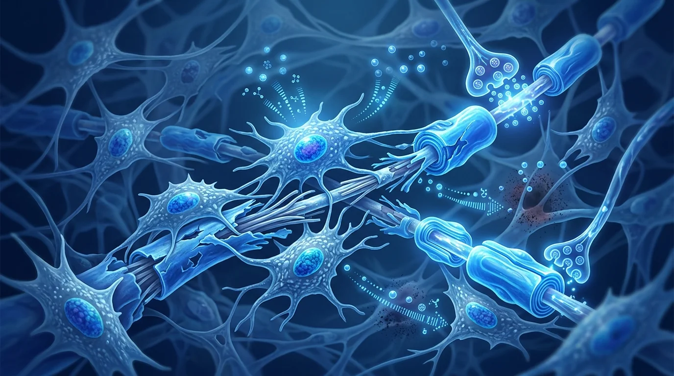
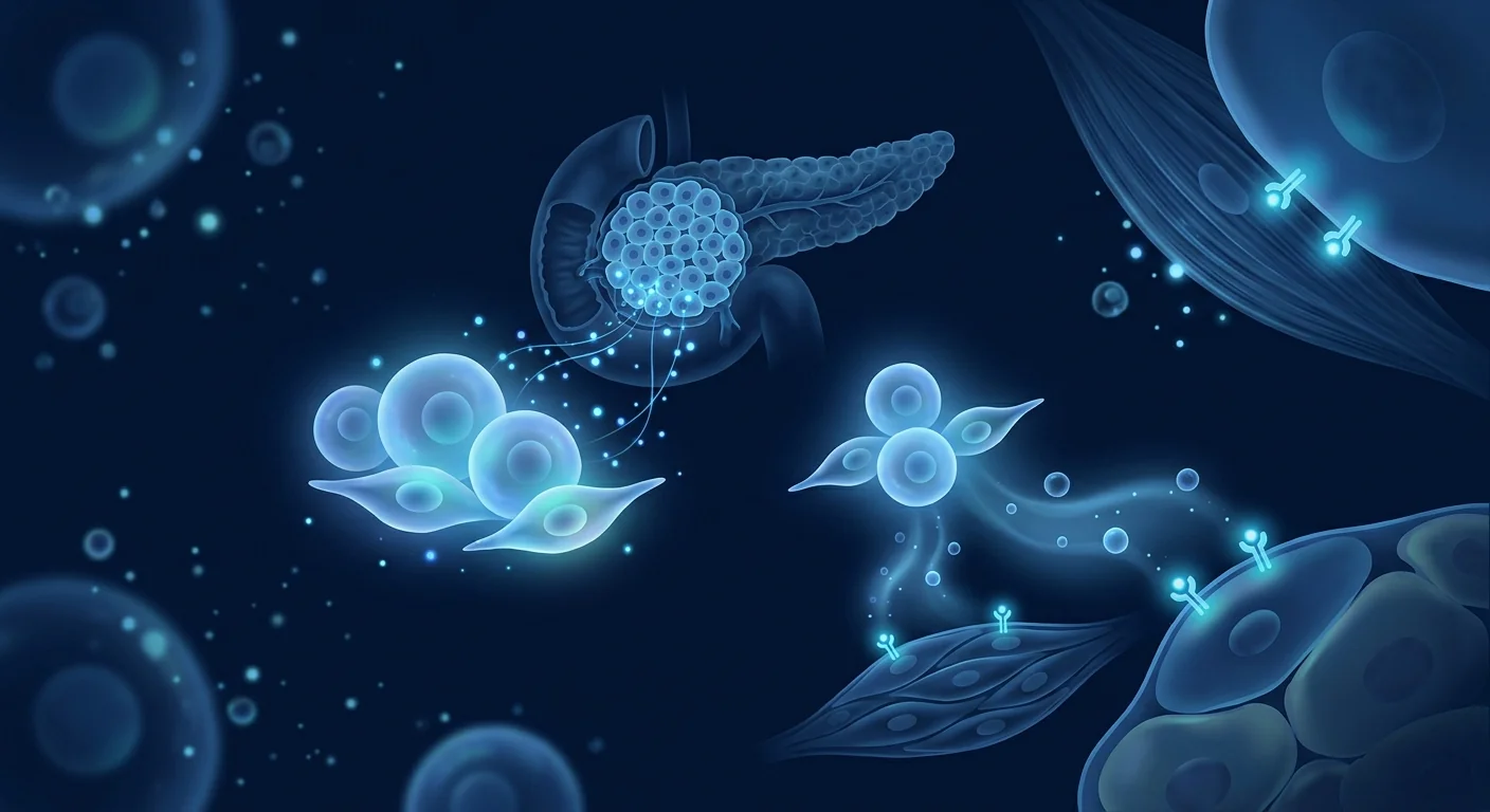
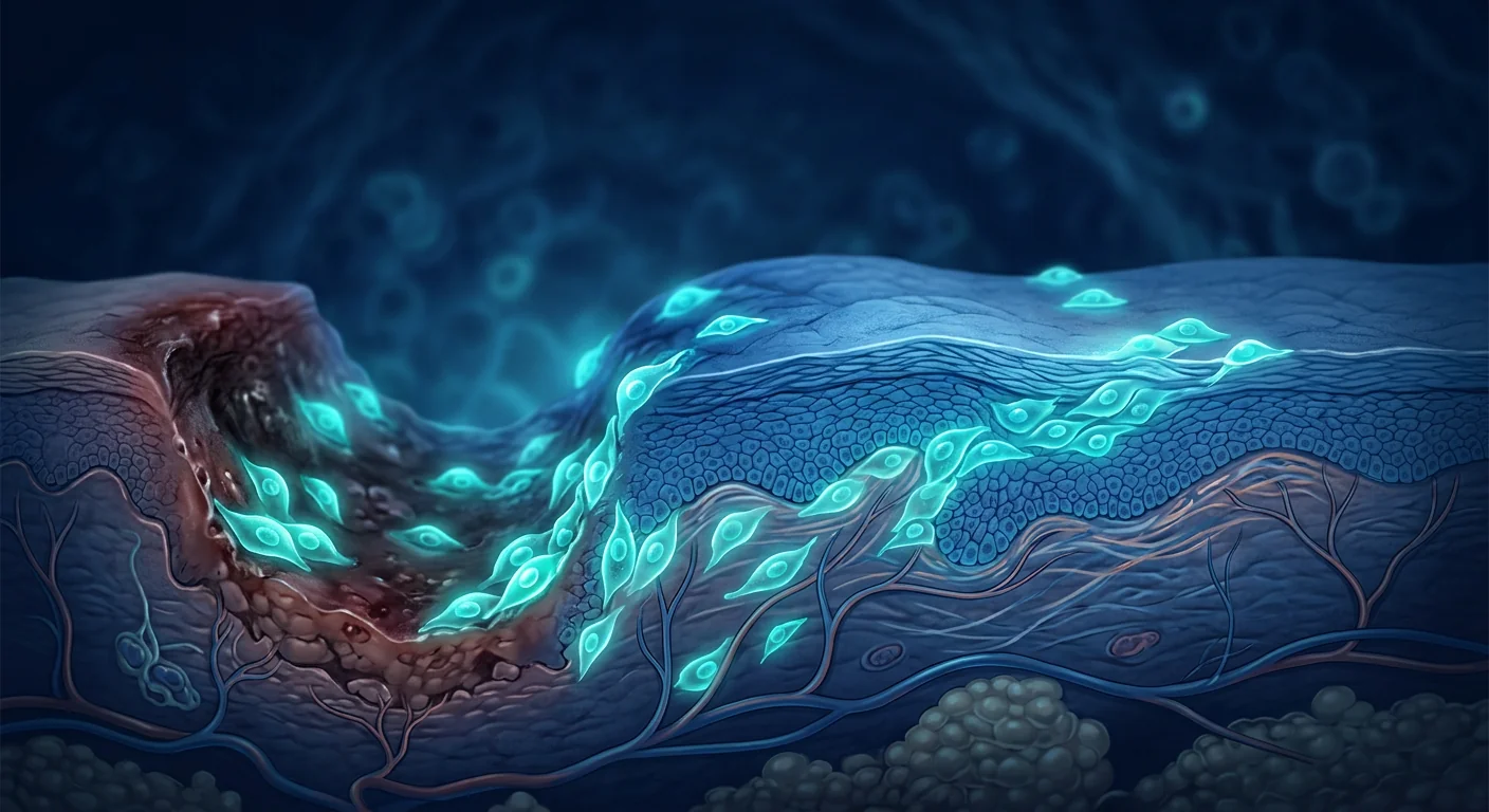
For the millions of diabetic patients who develop foot ulcers each year, the fear of amputation is real — up to 85% of diabetes-related lower-limb amputations begin with a non-healing wound. MSC therapy is being studied as a way to restart the stalled healing process at the cellular level.
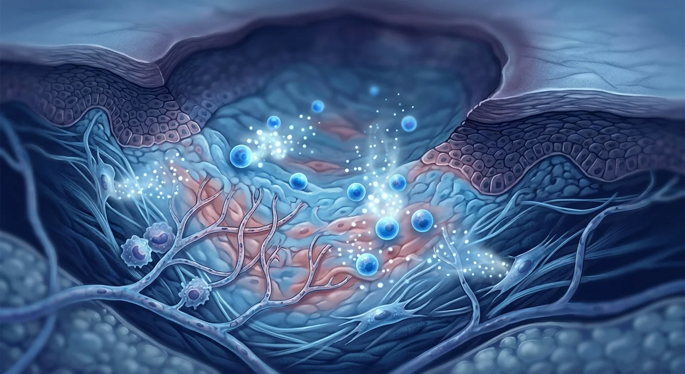
Diabetic foot ulcers (DFUs) affect approximately 15–25% of the 537 million adults living with diabetes worldwide during their lifetime, with an estimated 18.6 million people developing a DFU each year. These chronic wounds are the leading cause of non-traumatic lower-extremity amputation globally — every 20 seconds, someone loses a limb to diabetes. [1]
Where conventional wound care falls short. Standard DFU treatment — offloading, debridement, infection control, and moisture-balanced dressings — achieves complete healing in only 30–50% of cases at 20 weeks. Advanced therapies such as negative-pressure wound therapy, hyperbaric oxygen, and bioengineered skin substitutes improve outcomes modestly but still leave a substantial proportion of patients with recalcitrant wounds that persist for months or years. [2]
The deeper problem is a hostile wound microenvironment. DFUs are not simply mechanical injuries that fail to close. They exist in a state of chronic inflammation driven by sustained hyperglycemia, where pro-inflammatory cytokines (TNF-α, IL-1β, IL-6) dominate, matrix metalloproteinases (MMPs) degrade newly formed extracellular matrix faster than it can be deposited, and resident fibroblasts and keratinocytes are senescent and unresponsive to growth factor signaling. Compounding this, diabetic microangiopathy impairs oxygen delivery, and peripheral neuropathy removes the protective pain feedback that would normally prompt offloading. [3]
MSC therapy addresses the biology of non-healing. Rather than adding another dressing or growth factor to a dysfunctional wound bed, mesenchymal stem cells target the root causes — they shift the wound from a chronic inflammatory to a regenerative phenotype, secrete a broad spectrum of angiogenic and trophic factors that senescent host cells can no longer produce, directly suppress MMP overactivity, and recruit endogenous repair cells to repopulate the wound. This is biological wound rescue, not incremental dressing improvement. [4]
How MSCs Promote Diabetic Wound Healing
Mesenchymal stem cells orchestrate wound repair through a coordinated multi-mechanism program that addresses virtually every defect present in the diabetic wound. Their therapeutic effect is primarily paracrine — mediated by secreted factors — rather than direct differentiation into skin cells. [5]
Transitioning from Inflammation to Proliferation
The defining feature of a chronic DFU is an arrest in the inflammatory phase of wound healing. Neutrophils and M1 macrophages persist, releasing proteases and reactive oxygen species that damage tissue rather than repair it. MSCs secrete prostaglandin E2 (PGE2), TSG-6, and IL-1 receptor antagonist, which collectively reprogram macrophages from the destructive M1 phenotype to the pro-regenerative M2 phenotype. This single transition — M1 to M2 polarization — is arguably the most critical event in converting a chronic wound to an acute healing trajectory. [6]
Restoring Angiogenesis in the Ischemic Wound Bed
Diabetic microangiopathy starves the wound of oxygen and nutrients. MSCs respond to wound hypoxia by dramatically upregulating VEGF, HGF, bFGF, and angiopoietin-1 secretion — driving endothelial cell proliferation, migration, and tubule formation to create new capillary networks. In preclinical DFU models, MSC-treated wounds show a 2- to 3-fold increase in capillary density compared to controls, with corresponding improvements in tissue oxygen tension. [7]
Re-Epithelialization and Keratinocyte Activation
Wound closure requires keratinocytes at the wound edge to proliferate and migrate across the granulation tissue bed. In diabetes, keratinocyte function is impaired by advanced glycation end-products (AGEs) and chronic oxidative stress. MSC-conditioned medium contains EGF, KGF, and HGF that directly stimulate keratinocyte proliferation and migration. Co-culture studies show MSC-derived exosomes accelerate re-epithelialization by 40–60% compared to untreated controls. [8]
Modulating the Extracellular Matrix
Chronic DFUs are characterized by excessive MMP activity that degrades collagen, fibronectin, and growth factors faster than they can be synthesized. MSCs secrete tissue inhibitors of metalloproteinases (TIMPs) that rebalance the protease–antiprotease equilibrium. They also produce collagen types I and III, fibronectin, and hyaluronic acid directly — providing both the structural scaffold and the signaling molecules needed for granulation tissue formation. The net effect is restoration of a permissive extracellular matrix. [9]
Antimicrobial Activity
DFUs are nearly always colonized, and biofilm formation is common in wounds that fail to heal beyond 4 weeks. MSCs exhibit direct and indirect antimicrobial properties — they secrete antimicrobial peptides including LL-37 (cathelicidin), lipocalin-2, and beta-defensin-2, and their immunomodulatory effects enhance the host's own bacterial clearance mechanisms. In infected wound models, MSC-treated wounds show significantly lower bacterial burden and reduced biofilm formation. [10]
Clinical Evidence for MSC Therapy in Diabetic Foot Ulcers
The clinical evidence base for MSC therapy in DFUs is growing rapidly, with multiple randomized controlled trials completed or underway. While still considered investigational in most jurisdictions, the data are encouraging. [11]
Randomized Controlled Trials
A 2023 meta-analysis of seven RCTs involving 312 patients with chronic DFUs found that MSC-based therapies significantly improved complete wound closure rates (RR 2.03, 95% CI 1.54–2.68) and reduced time to healing by an average of 3.5 weeks compared to standard care alone. Wound area reduction at 4 and 8 weeks was consistently greater in MSC-treated groups. No serious adverse events attributable to MSC therapy were reported. [12]
A landmark 2022 RCT randomized 60 patients with Wagner grade 2–3 DFUs to receive either intramuscular and topical allogeneic bone marrow-derived MSCs plus standard care, or standard care alone. At 12 weeks, complete wound closure was achieved in 73% of the MSC group versus 40% of controls (P = 0.011). The mean time to complete closure was 6.3 weeks in the MSC group versus 9.8 weeks in controls. [13]
Route of Administration
MSCs have been delivered to DFUs through multiple routes — topical application in fibrin spray or hydrogel scaffolds, intralesional injection around the wound periphery, and intramuscular injection into the periwound tissue. Each route has distinct advantages. Topical application is the least invasive and can cover large surface areas. Periwound injection delivers cells deeper into the ischemic tissue bed. Emerging evidence suggests that combined intramuscular plus topical delivery may produce the best outcomes, addressing both deep tissue ischemia and surface wound healing simultaneously. [14]
Allogeneic vs. Autologous MSCs
Both allogeneic (donor-derived) and autologous (patient-derived) MSCs have been studied for DFUs. Allogeneic MSCs offer practical advantages — off-the-shelf availability, consistent potency, and no need for a harvesting procedure in a patient who already has poor wound healing. Autologous MSCs avoid immunogenicity concerns, though MSCs are inherently immunoprivileged and allogeneic use has an excellent safety record. Bone marrow-derived autologous MSCs from diabetic patients may have reduced proliferative and secretory capacity compared to healthy donor cells, a consideration that favors allogeneic sources for this population. [15]
Biomarker Tracking: How We Know It's Working
Unlike structural endpoints that take weeks to assess, molecular and cellular markers provide early signals of treatment response. In DFU clinical trials, several biomarkers have emerged as reliable indicators. [16]
Integration with Standard DFU Care
MSC therapy is not a replacement for established DFU care but a biological adjunct intended to rescue wounds that have stalled despite optimal standard management. Clinical trials have universally administered MSCs on top of best-practice wound care, not instead of it. [17]
- Offloading remains essential. No biological therapy can overcome continuous mechanical trauma. Total contact casting or equivalent offloading must continue during and after MSC treatment.
- Infection control precedes cell therapy. Active infection must be treated with appropriate antibiotics and debridement before MSC administration — an infected wound bed rapidly destroys delivered cells.
- Vascular assessment is mandatory. MSCs require a minimum level of perfusion to survive and function. An ankle-brachial index below 0.4 or transcutaneous oxygen pressure below 30 mmHg suggests revascularization should be considered before or concurrent with MSC therapy.
- Glycemic control matters. HbA1c above 9% is associated with poorer wound healing outcomes in general; optimizing glycemic control is a prerequisite for any advanced wound therapy.
Safety Profile
The safety record of MSC therapy in wound healing is excellent. A 2024 systematic review of 19 studies involving 726 DFU patients treated with MSCs found no cases of tumor formation, no serious systemic adverse events, and no significant immunological reactions. Local adverse events were limited to transient injection-site discomfort in a minority of patients. [18]
- No tumorigenicity signal in any DFU trial to date.
- Allogeneic MSCs are immunoprivileged — no immunosuppression required.
- No increased infection risk; MSCs may actually reduce wound bacterial burden.
- Theoretical concern about pro-angiogenic therapy accelerating diabetic retinopathy has not been observed clinically, though active proliferative retinopathy remains a precautionary exclusion.
- Transient injection-site pain or swelling is the most common adverse event, resolving within 24–48 hours.
Who Is a Candidate?
MSC therapy for DFUs is most appropriate for patients whose wounds have not responded adequately to at least 4 weeks of comprehensive standard care. Ideal candidates are those with adequate arterial inflow (or revascularized limbs), controlled infection, and a wound that is stalled — not deteriorating rapidly. [19]
Patients with Wagner grade 1–3 ulcers (superficial to deep with osteomyelitis treated) are the primary studied population. Wagner grade 4–5 (partial or whole foot gangrene) generally requires surgical intervention first. Early intervention — before the wound becomes deeply infected or extensively fibrotic — is associated with better outcomes.
Limitations and Honest Perspective
Despite encouraging data, several important limitations must be acknowledged. The majority of RCTs are small (under 100 patients) and single-center. MSC manufacturing protocols vary substantially between studies — cell source, passage number, dose, delivery method, and cryopreservation status all differ, making cross-study comparison difficult. Long-term durability data beyond 12 months is sparse. And perhaps most importantly, MSC therapy for DFUs remains investigational in most countries; it is not yet a standard-of-care option reimbursed by health systems. [20]
That said, the biological rationale is strong and the direction of evidence is consistently positive. For patients facing the prospect of amputation after exhausting conventional options, MSC therapy represents an investigational pathway worth discussing with a clinical team experienced in regenerative wound medicine.
Frequently Asked Questions
How effective is stem cell therapy for diabetic foot ulcers?
Meta-analyses of randomized trials report approximately double the complete wound closure rate compared to standard care alone, with complete healing rates of 70–80% at 12 weeks in MSC-treated groups versus 30–50% in control groups. Results vary based on ulcer severity, duration, and patient comorbidities.
Is stem cell therapy for DFU approved or experimental?
MSC therapy for diabetic foot ulcers is investigational in most countries. It is not yet FDA-approved as a standard treatment for DFUs, though several products are in late-stage clinical trials. Patients access it through clinical trials or at specialized regenerative medicine centers offering it as an advanced therapeutic option under appropriate regulatory frameworks.
How are the stem cells delivered to the foot ulcer?
MSCs can be delivered topically (sprayed or applied in a gel/scaffold directly onto the wound), injected around the wound perimeter (perilesional), or injected into the muscle of the affected limb. The optimal route depends on wound characteristics — size, depth, infection status, and vascular supply — and is determined by the treating clinician.
How many treatments are needed for a diabetic foot ulcer?
Most clinical protocols use a single treatment session, though some studies have evaluated two or three sessions spaced 2–4 weeks apart for particularly large or recalcitrant ulcers. The need for repeat dosing is assessed based on wound progress at follow-up visits.
What are the risks of stem cell therapy for foot ulcers?
Safety data from over 700 treated patients show an excellent profile. The most common side effect is transient injection-site discomfort. No tumor formation, systemic immunological reactions, or serious therapy-related adverse events have been reported. The primary risk is that the therapy does not work — not that it causes harm.
Does insurance cover stem cell therapy for diabetic foot ulcers?
Currently, MSC therapy for DFUs is generally not covered by insurance as it remains investigational. Some clinical trials provide treatment at no cost to participants. At private centers, treatment is self-pay. As regulatory approvals progress, coverage may expand — several health technology assessments are underway in Europe and Asia.
- Armstrong DG, Boulton AJM, Bus SA. Diabetic foot ulcers and their recurrence. New England Journal of Medicine. 2017;376(24):2367-2375. doi:10.1056/NEJMra1615439 ↩
- Everett E, Mathioudakis N. Update on management of diabetic foot ulcers. Annals of the New York Academy of Sciences. 2018;1411(1):153-165. doi:10.1111/nyas.13569 ↩
- Falanga V. Wound healing and its impairment in the diabetic foot. The Lancet. 2005;366(9498):1736-1743. doi:10.1016/S0140-6736(05)67700-8 ↩
- Maxson S, Lopez EA, Yoo D, Danilkovitch-Miagkova A, LeRoux MA. Concise review: role of mesenchymal stem cells in wound repair. Stem Cells Translational Medicine. 2012;1(2):142-149. doi:10.5966/sctm.2011-0018 ↩
- Caplan AI, Correa D. The MSC: an injury drugstore. Cell Stem Cell. 2011;9(1):11-15. doi:10.1016/j.stem.2011.06.008 ↩
- Kotwal GJ, Chien S. Macrophage differentiation in normal and accelerated wound healing. Results and Problems in Cell Differentiation. 2017;62:353-364. doi:10.1007/978-3-319-54090-0_14 ↩
- Kinnaird T, Stabile E, Burnett MS, et al. Marrow-derived stromal cells express genes encoding a broad spectrum of arteriogenic cytokines and promote in vitro and in vivo arteriogenesis through paracrine mechanisms. Circulation Research. 2004;94(5):678-685. doi:10.1161/01.RES.0000118601.37875.AC ↩
- Hu L, Wang J, Zhou X, et al. Exosomes derived from human adipose mesenchymal stem cells accelerate cutaneous wound healing via optimizing the characteristics of fibroblasts. Scientific Reports. 2016;6:32993. doi:10.1038/srep32993 ↩
- Jeon YK, Jang YH, Yoo DR, Kim SN, Lee SK, Nam MJ. Mesenchymal stem cells' interaction with skin: wound-healing effect on fibroblast and keratinocyte cells. International Journal of Molecular Sciences. 2010;11(4):1203-1217. doi:10.3390/ijms11041203 ↩
- Krasnodembskaya A, Song Y, Fang X, et al. Antibacterial effect of human mesenchymal stem cells is mediated in part from secretion of the antimicrobial peptide LL-37. Stem Cells. 2010;28(12):2229-2238. doi:10.1002/stem.544 ↩
- Lopes L, Setia O, Aurshina A, et al. Stem cell therapy for diabetic foot ulcers: a review of preclinical and clinical research. Stem Cell Research & Therapy. 2018;9(1):188. doi:10.1186/s13287-018-0938-6 ↩
- Shu X, Shu S, Tang S, et al. Efficiency of stem cell based therapy in the treatment of diabetic foot ulcer: a meta-analysis. Stem Cell Research & Therapy. 2023;14(1):44. doi:10.1186/s13287-023-03274-8 ↩
- Yang M, Sheng L, Zhang TR, Li Q. Stem cell therapy for diabetic foot ulcers: a randomized controlled clinical study. Stem Cells Translational Medicine. 2022;11(7):688-696. doi:10.1093/stcltm/szac032 ↩
- Dash SN, Dash NR, Guru B, Mohapatra PC. Towards reaching the target: clinical application of mesenchymal stem cells for diabetic foot ulcers. Rejuvenation Research. 2021;24(2):97-110. doi:10.1089/rej.2020.2355 ↩
- Cianfarani F, Toietta G, Di Rocco G, Cesareo E, Zambruno G, Odorisio T. Diabetes impairs adipose tissue-derived stem cell function and efficiency in promoting wound healing. Wound Repair and Regeneration. 2013;21(4):545-553. doi:10.1111/wrr.12051 ↩
- Frykberg RG, Banks J. Challenges in the treatment of chronic wounds. Advances in Wound Care. 2015;4(9):560-582. doi:10.1089/wound.2015.0635 ↩
- Schaper NC, van Netten JJ, Apelqvist J, Bus SA, Hinchliffe RJ, Lipsky BA. Practical guidelines on the prevention and management of diabetic foot disease. Diabetes/Metabolism Research and Reviews. 2020;36(S1):e3266. doi:10.1002/dmrr.3266 ↩
- Rai V, Moellmer R, Agrawal DK. Stem cell therapy for diabetic foot ulcers: safety and efficacy — a systematic review. Molecular and Cellular Biochemistry. 2024;479(1):45-61. doi:10.1007/s11010-023-04717-3 ↩
- Game FL, Apelqvist J, Attinger C, et al. Effectiveness of interventions to enhance healing of chronic ulcers of the foot in diabetes: a systematic review. Diabetes/Metabolism Research and Reviews. 2016;32(S1):154-168. doi:10.1002/dmrr.2707 ↩
- Margolis DJ, Kantor J, Berlin JA. Healing of diabetic neuropathic foot ulcers receiving standard treatment. A meta-analysis. Diabetes Care. 1999;22(5):692-695. doi:10.2337/diacare.22.5.692 ↩
每年数百万糖尿病患者出现足部溃疡,他们最恐惧的是截肢 — 高达85%的糖尿病相关下肢截肢始于一个不愈合的伤口。间充质干细胞疗法正在被研究作为一种从细胞层面重启停滞愈合过程的方法。
糖尿病足溃疡(DFU)影响全球5.37亿成年糖尿病患者中约15–25%的人群,每年估计有1,860万人发生DFU。这些慢性伤口是全球非创伤性下肢截肢的主要原因 — 每20秒就有人因糖尿病失去肢体。[1]
传统伤口护理的局限。标准的DFU治疗 — 减压、清创、感染控制和湿性敷料 — 在20周时仅实现30–50%的完全愈合率。负压伤口治疗、高压氧和生物工程皮肤替代品等高级疗法适度改善了结果,但仍有相当比例的患者伤口持续数月甚至数年不愈。[2]
更深层的问题是伤口微环境失调。DFU不仅仅是无法闭合的机械性损伤。它们处于由持续高血糖驱动的慢性炎症状态,促炎细胞因子(TNF-α、IL-1β、IL-6)占主导,基质金属蛋白酶(MMPs)降解新形成的细胞外基质的速度超过其沉积速度,驻留的成纤维细胞和角质形成细胞处于衰老状态,对生长因子信号无反应。此外,糖尿病微血管病变损害了氧气输送,周围神经病变消除了通常会促发减压的保护性疼痛反馈。[3]
MSC疗法针对不愈合的生物学根源。间充质干细胞不是简单地向功能失调的伤口床添加另一种敷料或生长因子,而是针对根本原因 — 它们将伤口从慢性炎症表型转变为再生表型,分泌衰老宿主细胞不再产生的广谱血管生成和营养因子,直接抑制MMP过度活性,并招募内源性修复细胞重新填充伤口。这是生物性伤口救援,而非渐进式敷料改进。[4]
MSCs如何促进糖尿病伤口愈合
间充质干细胞通过协调的多机制程序来编排伤口修复,几乎解决了糖尿病伤口中存在的每一个缺陷。它们的治疗效果主要是旁分泌的 — 通过分泌因子介导 — 而非直接分化为皮肤细胞。[5]
从炎症向增殖期过渡
慢性DFU的定义性特征是伤口愈合炎症期的停滞。中性粒细胞和M1巨噬细胞持续存在,释放破坏组织而非修复组织的蛋白酶和活性氧。MSCs分泌前列腺素E2(PGE2)、TSG-6和IL-1受体拮抗剂,共同将巨噬细胞从破坏性M1表型重新编程为促再生M2表型。这一单一转变 — M1到M2极化 — 可以说是将慢性伤口转变为急性愈合轨迹最关键的事件。[6]
恢复缺血伤口床的血管生成
糖尿病微血管病变使伤口缺乏氧气和营养。MSCs通过显著上调VEGF、HGF、bFGF和angiopoietin-1的分泌来应对伤口缺氧 — 驱动内皮细胞增殖、迁移和管状形成,创建新的毛细血管网络。在临床前DFU模型中,MSC处理的伤口显示毛细血管密度比对照组增加2–3倍,组织氧张力相应改善。[7]
再上皮化和角质形成细胞激活
伤口闭合需要伤口边缘的角质形成细胞增殖并迁移穿过肉芽组织床。在糖尿病中,晚期糖基化终末产物(AGEs)和慢性氧化应激损害了角质形成细胞功能。MSC条件培养基含有EGF、KGF和HGF,可直接刺激角质形成细胞增殖和迁移。共培养研究显示,MSC来源的外泌体与未处理对照组相比,可将再上皮化加速40–60%。[8]
调节细胞外基质
慢性DFU的特征是MMP活性过度,降解胶原蛋白、纤连蛋白和生长因子的速度快于合成速度。MSCs分泌金属蛋白酶组织抑制剂(TIMPs),重新平衡蛋白酶-抗蛋白酶平衡。它们还直接产生I型和III型胶原蛋白、纤连蛋白和透明质酸 — 提供肉芽组织形成所需的结构支架和信号分子。净效果是恢复允许性的细胞外基质。[9]
抗微生物活性
DFU几乎总是定植细菌,超过4周未愈合的伤口常见生物膜形成。MSCs表现出直接和间接的抗微生物特性 — 它们分泌包括LL-37(导管素)、lipocalin-2和beta-defensin-2在内的抗微生物肽,其免疫调节作用增强了宿主自身的细菌清除机制。在感染伤口模型中,MSC处理的伤口显示细菌负荷显著降低,生物膜形成减少。[10]
MSC治疗糖尿病足溃疡的临床证据
MSC治疗DFU的临床证据基础正在迅速增长,多项随机对照试验已经完成或正在进行。虽然在大多数司法管辖区仍被视为研究性质,但数据令人鼓舞。[11]
随机对照试验
2023年一项包含7项RCT、312名慢性DFU患者的荟萃分析发现,基于MSC的治疗显著提高了完全伤口闭合率(RR 2.03,95% CI 1.54–2.68),并将愈合时间平均缩短了3.5周。在第4周和第8周时,MSC治疗组的伤口面积减少始终更大。未报告归因于MSC治疗的严重不良事件。[12]
2022年一项里程碑式RCT将60名Wagner 2–3级DFU患者随机分配接受肌肉内和局部异体骨髓来源MSCs加标准护理,或仅标准护理。在第12周,MSC组73%的患者实现完全伤口闭合,而对照组为40%(P = 0.011)。MSC组平均完全闭合时间为6.3周,对照组为9.8周。[13]
给药途径
MSCs已通过多种途径递送至DFU — 纤维蛋白喷雾或水凝胶支架局部应用、伤口周围注射和伤口周围组织肌肉内注射。每种途径各有优势。局部应用创伤最小,可覆盖大面积。伤口周围注射可将细胞更深地递送到缺血组织床。新证据表明,肌肉内加局部联合递送可能产生最佳结果,同时解决深层组织缺血和表面伤口愈合。[14]
安全性与局限性
MSC治疗在伤口愈合中的安全记录优异。2024年一项涵盖19项研究、726名DFU患者的系统评价发现无肿瘤形成病例、无严重全身不良事件、无显著免疫反应。局部不良事件限于少数患者的短暂注射部位不适。[18]
尽管数据令人鼓舞,但必须承认数个重要局限性。大多数RCT规模较小(不足100名患者),为单中心研究。MSC制备方案在不同研究间差异很大 — 细胞来源、传代次数、剂量、递送方法和冷冻保存状态各不相同,使跨研究比较困难。超过12个月的长期持久性数据稀少。最重要的是,MSC治疗DFU在大多数国家仍是研究性质的。[20]
- Armstrong DG, Boulton AJM, Bus SA. Diabetic foot ulcers and their recurrence. New England Journal of Medicine. 2017;376(24):2367-2375. doi:10.1056/NEJMra1615439 ↩
- Everett E, Mathioudakis N. Update on management of diabetic foot ulcers. Annals of the New York Academy of Sciences. 2018;1411(1):153-165. doi:10.1111/nyas.13569 ↩
- Falanga V. Wound healing and its impairment in the diabetic foot. The Lancet. 2005;366(9498):1736-1743. doi:10.1016/S0140-6736(05)67700-8 ↩
- Maxson S, Lopez EA, Yoo D, Danilkovitch-Miagkova A, LeRoux MA. Concise review: role of mesenchymal stem cells in wound repair. Stem Cells Translational Medicine. 2012;1(2):142-149. doi:10.5966/sctm.2011-0018 ↩
- Caplan AI, Correa D. The MSC: an injury drugstore. Cell Stem Cell. 2011;9(1):11-15. doi:10.1016/j.stem.2011.06.008 ↩
- Kotwal GJ, Chien S. Macrophage differentiation in normal and accelerated wound healing. Results and Problems in Cell Differentiation. 2017;62:353-364. doi:10.1007/978-3-319-54090-0_14 ↩
- Kinnaird T, Stabile E, Burnett MS, et al. Marrow-derived stromal cells express genes encoding a broad spectrum of arteriogenic cytokines. Circulation Research. 2004;94(5):678-685. doi:10.1161/01.RES.0000118601.37875.AC ↩
- Hu L, Wang J, Zhou X, et al. Exosomes derived from human adipose mesenchymal stem cells accelerate cutaneous wound healing. Scientific Reports. 2016;6:32993. doi:10.1038/srep32993 ↩
- Jeon YK, Jang YH, Yoo DR, Kim SN, Lee SK, Nam MJ. Mesenchymal stem cells' interaction with skin. International Journal of Molecular Sciences. 2010;11(4):1203-1217. doi:10.3390/ijms11041203 ↩
- Krasnodembskaya A, Song Y, Fang X, et al. Antibacterial effect of human mesenchymal stem cells is mediated in part from secretion of the antimicrobial peptide LL-37. Stem Cells. 2010;28(12):2229-2238. doi:10.1002/stem.544 ↩
- Lopes L, Setia O, Aurshina A, et al. Stem cell therapy for diabetic foot ulcers: a review. Stem Cell Research & Therapy. 2018;9(1):188. doi:10.1186/s13287-018-0938-6 ↩
- Shu X, Shu S, Tang S, et al. Efficiency of stem cell based therapy in the treatment of diabetic foot ulcer: a meta-analysis. Stem Cell Research & Therapy. 2023;14(1):44. doi:10.1186/s13287-023-03274-8 ↩
- Yang M, Sheng L, Zhang TR, Li Q. Stem cell therapy for diabetic foot ulcers: a randomized controlled clinical study. Stem Cells Translational Medicine. 2022;11(7):688-696. doi:10.1093/stcltm/szac032 ↩
- Dash SN, Dash NR, Guru B, Mohapatra PC. Clinical application of mesenchymal stem cells for diabetic foot ulcers. Rejuvenation Research. 2021;24(2):97-110. doi:10.1089/rej.2020.2355 ↩
- Rai V, Moellmer R, Agrawal DK. Stem cell therapy for diabetic foot ulcers: safety and efficacy. Molecular and Cellular Biochemistry. 2024;479(1):45-61. doi:10.1007/s11010-023-04717-3 ↩
- Margolis DJ, Kantor J, Berlin JA. Healing of diabetic neuropathic foot ulcers receiving standard treatment. Diabetes Care. 1999;22(5):692-695. doi:10.2337/diacare.22.5.692 ↩
بالنسبة لملايين مرضى السكري الذين يصابون بقرح القدم كل عام، فإن الخوف من البتر حقيقي — ما يصل إلى 85٪ من عمليات بتر الأطراف السفلية المرتبطة بالسكري تبدأ بجرح لا يلتئم. يُدرس العلاج بالخلايا الجذعية الوسيطة كوسيلة لإعادة启动 عملية الشفاء المتوقفة على المستوى الخلوي.
تؤثر قرح القدم السكرية (DFUs) على ما يقرب من 15–25٪ من 537 مليون بالغ يعيشون مع السكري في جميع أنحاء العالم خلال حياتهم، مع ما يقدر بـ 18.6 مليون شخص يصابون بقرحة قدم سكرية كل عام. هذه الجروح المزمنة هي السبب الرئيسي لبتر الأطراف السفلية غير الرضحي على مستوى العالم — كل 20 ثانية، يفقد شخص طرفاً بسبب السكري. [1]
حيث تفشل العناية التقليدية بالجروح. يحقق علاج DFU القياسي — تخفيف الضغط، التنضير، مكافحة العدوى، والضمادات المتوازنة الرطوبة — شفاءً كاملاً في 30–50٪ فقط من الحالات عند 20 أسبوعاً. تحسن العلاجات المتقدمة مثل علاج الجروح بالضغط السلبي، الأكسجين عالي الضغط، وبدائل الجلد المهندسة حيوياً النتائج بشكل متواضع لكنها لا تزال تترك نسبة كبيرة من المرضى بجروح مستعصية تستمر لأشهر أو سنوات. [2]
المشكلة الأعمق هي بيئة الجرح الدقيقة المعادية. قرح القدم السكرية ليست مجرد إصابات ميكانيكية تفشل في الانغلاق. إنها موجودة في حالة التهاب مزمن مدفوع بارتفاع السكر المستمر في الدم، حيث تهيمن السيتوكينات المؤيدة للالتهاب (TNF-α، IL-1β، IL-6)، وتقوم بروتينات المصفوفة المعدنية (MMPs) بتحليل المصفوفة خارج الخلية المتكونة حديثاً بشكل أسرع مما يمكن ترسيبه، وتكون الخلايا الليفية والخلايا الكيراتينية المقيمة في حالة شيخوخة وغير مستجيبة لإشارات عامل النمو. يضاعف من ذلك، اعتلال الأوعية الدقيقة السكري الذي يضعف توصيل الأكسجين، والاعتلال العصبي المحيطي الذي يزيل التغذية الراجعة للألم الواقي الذي كان سيحفز عادة تخفيف الضغط. [3]
يستهدف العلاج بالخلايا الجذعية الوسيطة بيولوجيا عدم الالتئام. بدلاً من إضافة ضمادة أخرى أو عامل نمو إلى سرير جرح مختل، تعالج الخلايا الجذعية الوسيطة الأسباب الجذرية — تحول الجرح من نمط التهابي مزمن إلى نمط تجديدي، تفرز طيفاً واسعاً من العوامل الوعائية والتغذوية التي لم تعد الخلايا المضيفة الهرمة قادرة على إنتاجها، تثبط مباشرة نشاط MMP المفرط، وتجند خلايا الإصلاح الداخلية لإعادة ملء الجرح. هذا هو إنقاذ بيولوجي للجروح، وليس تحسيناً تدريجياً للضمادات. [4]
كيف تعزز الخلايا الجذعية الوسيطة التئام الجروح السكرية
تنظم الخلايا الجذعية الوسيطة إصلاح الجروح من خلال برنامج متعدد الآليات منسق يعالج تقريباً كل عيب موجود في الجرح السكري. تأثيرها العلاجي هو في المقام الأول باراكريني — بوساطة عوامل مفرزة — وليس تمايزاً مباشراً إلى خلايا جلدية. [5]
الانتقال من الالتهاب إلى التكاثر
السمة المميزة لقرحة القدم السكرية المزمنة هي التوقف في المرحلة الالتهابية من التئام الجروح. تستمر العدلات والبلاعم M1، وتطلق البروتيازات وأنواع الأكسجين التفاعلية التي تتلف الأنسجة بدلاً من إصلاحها. تفرز الخلايا الجذعية الوسيطة البروستاغلاندين E2 (PGE2)، وTSG-6، ومضاد مستقبل IL-1، والتي تعيد معاً برمجة البلاعم من النمط المدمر M1 إلى النمط المؤيد للتجديد M2. هذا الانتقال الفردي — استقطاب M1 إلى M2 — هو arguably الحدث الأكثر أهمية في تحويل الجرح المزمن إلى مسار شفاء حاد. [6]
استعادة تكوين الأوعية الدموية في سرير الجرح الإقفاري
يجوع اعتلال الأوعية الدقيقة السكري الجرح من الأكسجين والمغذيات. تستجيب الخلايا الجذعية الوسيطة لنقص الأكسجة في الجرح عن طريق زيادة كبيرة في إفراز VEGF وHGF وbFGF وangiopoietin-1 — مما يدفع تكاثر الخلايا البطانية وهجرتها وتشكيل الأنابيب لإنشاء شبكات شعرية جديدة. في نماذج DFU قبل السريرية، تظهر الجروح المعالجة بالخلايا الجذعية الوسيطة زيادة بمقدار 2–3 أضعاف في كثافة الشعيرات الدموية مقارنة بالمجموعة الضابطة، مع تحسينات مقابلة في توتر الأكسجين النسيجي. [7]
إعادة التبشير وتنشيط الخلايا الكيراتينية
يتطلب إغلاق الجرح تكاثر وهجرة الخلايا الكيراتينية عند حافة الجرح عبر سرير النسيج الحبيبي. في السكري، تضعف وظيفة الخلايا الكيراتينية بسبب منتجات الغلكزة المتقدمة (AGEs) والإجهاد التأكسدي المزمن. يحتوي وسط الخلايا الجذعية الوسيطة المكيفة على EGF وKGF وHGF التي تحفز مباشرة تكاثر وهجرة الخلايا الكيراتينية. تظهر دراسات الزرع المشترك أن الإكسوسومات المشتقة من الخلايا الجذعية الوسيطة تسرع إعادة التبشير بنسبة 40–60٪ مقارنة بالمجموعة الضابطة غير المعالجة. [8]
تعديل المصفوفة خارج الخلية
تتميز قرح القدم السكرية المزمنة بنشاط MMP المفرط الذي يحلل الكولاجين والفيبرونكتين وعوامل النمو بشكل أسرع مما يمكن تصنيعه. تفرز الخلايا الجذعية الوسيطة مثبطات بروتينات المصفوفة المعدنية النسيجية (TIMPs) التي تعيد توازن البروتياز-مضاد البروتياز. كما تنتج مباشرة الكولاجين من النوعين I وIII والفيبرونكتين وحمض الهيالورونيك — مما يوفر كل من السقالة الهيكلية وجزيئات الإشارة اللازمة لتكوين النسيج الحبيبي. التأثير الصافي هو استعادة مصفوفة خارج خلوية مسموحة. [9]
النشاط المضاد للميكروبات
تكون قرح القدم السكرية دائماً مستعمرة بالبكتيريا تقريباً، وتكوين البيوفيلم شائع في الجروح التي تفشل في الالتئام بعد 4 أسابيع. تظهر الخلايا الجذعية الوسيطة خصائص مضادة للميكروبات مباشرة وغير مباشرة — تفرز ببتيدات مضادة للميكروبات بما في ذلك LL-37 (كاثيليسيدين)، وليبوكالين-2، وبيتا-ديفينسين-2، وتعزز تأثيراتها المناعية آليات تصفية البكتيريا الخاصة بالمضيف. في نماذج الجروح المصابة، تظهر الجروح المعالجة بالخلايا الجذعية الوسيطة عبئاً بكتيرياً أقل بشكل ملحوظ وتكوين بيوفيلم منخفض. [10]
الأدلة السريرية للعلاج بالخلايا الجذعية الوسيطة في قرح القدم السكرية
قاعدة الأدلة السريرية للعلاج بالخلايا الجذعية الوسيطة في قرح القدم السكرية تنمو بسرعة، مع اكتمال أو جريان العديد من التجارب المعشاة ذات الشواهد. بينما لا يزال يعتبر استقصائياً في معظم الاختصاصات القضائية، فإن البيانات مشجعة. [11]
التجارب المعشاة ذات الشواهد
وجد تحليل تلوي عام 2023 لسبع تجارب معشاة ذات شواهد شملت 312 مريضاً بقرح قدم سكرية مزمنة أن العلاجات القائمة على الخلايا الجذعية الوسيطة حسنت بشكل ملحوظ معدلات إغلاق الجرح الكامل (RR 2.03، 95٪ CI 1.54–2.68) وقللت وقت الشفاء بمتوسط 3.5 أسابيع مقارنة بالرعاية القياسية وحدها. كان انخفاض مساحة الجرح في الأسبوعين 4 و8 أكبر باستمرار في مجموعات الخلايا الجذعية الوسيطة. لم يتم الإبلاغ عن أحداث سلبية خطيرة منسوبة إلى العلاج بالخلايا الجذعية الوسيطة. [12]
قامت تجربة معشاة ذات شواهد بارزة عام 2022 بتوزيع 60 مريضاً يعانون من قرح قدم سكرية من الدرجة 2–3 حسب Wagner عشوائياً لتلقي خلايا جذعية وسيطة خيفية مشتقة من نخاع العظم عن طريق الحقن العضلي والموضعي بالإضافة إلى الرعاية القياسية، أو الرعاية القياسية وحدها. في الأسبوع 12، تم تحقيق إغلاق كامل للجرح في 73٪ من مجموعة الخلايا الجذعية الوسيطة مقابل 40٪ من المجموعة الضابطة (P = 0.011). كان متوسط وقت الإغلاق الكامل 6.3 أسابيع في مجموعة الخلايا الجذعية الوسيطة مقابل 9.8 أسابيع في المجموعة الضابطة. [13]
طريقة الإعطاء
تم توصيل الخلايا الجذعية الوسيطة إلى قرح القدم السكرية من خلال طرق متعددة — التطبيق الموضعي في رذاذ الفيبرين أو سقالات الهيدروجيل، الحقن داخل الآفة حول محيط الجرح، والحقن العضلي في نسيج ما حول الجرح. لكل طريق مزايا مميزة. التطبيق الموضعي هو الأقل توغلاً ويمكن أن يغطي مساحات سطحية كبيرة. يوصل الحقن حول الجرح الخلايا أعمق في سرير النسيج الإقفاري. تشير الأدلة الناشئة إلى أن التوصيل المشترك العضلي بالإضافة إلى الموضعي قد ينتج أفضل النتائج، لمعالجة كل من إقفار الأنسجة العميقة والشفاء السطحي للجرح في وقت واحد. [14]
السلامة والقيود
سجل السلامة للعلاج بالخلايا الجذعية الوسيطة في التئام الجروح ممتاز. وجدت مراجعة منهجية عام 2024 لـ 19 دراسة شملت 726 مريضاً بقرح قدم سكرية عولجوا بالخلايا الجذعية الوسيطة عدم وجود حالات تكوين أورام، ولا أحداث سلبية جهازية خطيرة، ولا تفاعلات مناعية كبيرة. اقتصرت الأحداث السلبية الموضعية على انزعاج عابر في موقع الحقن لدى أقلية من المرضى. [18]
على الرغم من البيانات المشجعة، يجب الاعتراف بعدة قيود مهمة. غالبية التجارب المعشاة ذات الشواهد صغيرة (أقل من 100 مريض) وأحادية المركز. تختلف بروتوكولات تصنيع الخلايا الجذعية الوسيطة بشكل كبير بين الدراسات — مصدر الخلية، رقم التمرير، الجرعة، طريقة التوصيل، وحالة الحفظ بالتبريد كلها تختلف، مما يجعل المقارنة بين الدراسات صعبة. بيانات المتانة طويلة المدى بعد 12 شهراً نادرة. والأهم من ذلك، لا يزال العلاج بالخلايا الجذعية الوسيطة لقرح القدم السكرية استقصائياً في معظم البلدان. [20]
- Armstrong DG, Boulton AJM, Bus SA. Diabetic foot ulcers and their recurrence. New England Journal of Medicine. 2017;376(24):2367-2375. doi:10.1056/NEJMra1615439 ↩
- Everett E, Mathioudakis N. Update on management of diabetic foot ulcers. Annals of the New York Academy of Sciences. 2018;1411(1):153-165. doi:10.1111/nyas.13569 ↩
- Falanga V. Wound healing and its impairment in the diabetic foot. The Lancet. 2005;366(9498):1736-1743. doi:10.1016/S0140-6736(05)67700-8 ↩
- Maxson S, Lopez EA, Yoo D, Danilkovitch-Miagkova A, LeRoux MA. Concise review: role of mesenchymal stem cells in wound repair. Stem Cells Translational Medicine. 2012;1(2):142-149. doi:10.5966/sctm.2011-0018 ↩
- Caplan AI, Correa D. The MSC: an injury drugstore. Cell Stem Cell. 2011;9(1):11-15. doi:10.1016/j.stem.2011.06.008 ↩
- Kotwal GJ, Chien S. Macrophage differentiation in normal and accelerated wound healing. Results and Problems in Cell Differentiation. 2017;62:353-364. doi:10.1007/978-3-319-54090-0_14 ↩
- Kinnaird T, Stabile E, Burnett MS, et al. Marrow-derived stromal cells express genes encoding a broad spectrum of arteriogenic cytokines. Circulation Research. 2004;94(5):678-685. doi:10.1161/01.RES.0000118601.37875.AC ↩
- Hu L, Wang J, Zhou X, et al. Exosomes derived from human adipose mesenchymal stem cells accelerate cutaneous wound healing. Scientific Reports. 2016;6:32993. doi:10.1038/srep32993 ↩
- Jeon YK, Jang YH, Yoo DR, Kim SN, Lee SK, Nam MJ. Mesenchymal stem cells' interaction with skin. International Journal of Molecular Sciences. 2010;11(4):1203-1217. doi:10.3390/ijms11041203 ↩
- Krasnodembskaya A, Song Y, Fang X, et al. Antibacterial effect of human mesenchymal stem cells is mediated in part from secretion of the antimicrobial peptide LL-37. Stem Cells. 2010;28(12):2229-2238. doi:10.1002/stem.544 ↩
- Lopes L, Setia O, Aurshina A, et al. Stem cell therapy for diabetic foot ulcers: a review. Stem Cell Research & Therapy. 2018;9(1):188. doi:10.1186/s13287-018-0938-6 ↩
- Shu X, Shu S, Tang S, et al. Efficiency of stem cell based therapy in the treatment of diabetic foot ulcer: a meta-analysis. Stem Cell Research & Therapy. 2023;14(1):44. doi:10.1186/s13287-023-03274-8 ↩
- Yang M, Sheng L, Zhang TR, Li Q. Stem cell therapy for diabetic foot ulcers: a randomized controlled clinical study. Stem Cells Translational Medicine. 2022;11(7):688-696. doi:10.1093/stcltm/szac032 ↩
- Dash SN, Dash NR, Guru B, Mohapatra PC. Clinical application of mesenchymal stem cells for diabetic foot ulcers. Rejuvenation Research. 2021;24(2):97-110. doi:10.1089/rej.2020.2355 ↩
- Rai V, Moellmer R, Agrawal DK. Stem cell therapy for diabetic foot ulcers: safety and efficacy. Molecular and Cellular Biochemistry. 2024;479(1):45-61. doi:10.1007/s11010-023-04717-3 ↩
- Margolis DJ, Kantor J, Berlin JA. Healing of diabetic neuropathic foot ulcers receiving standard treatment. Diabetes Care. 1999;22(5):692-695. doi:10.2337/diacare.22.5.692 ↩