Burn injuries represent one of the most devastating forms of trauma the human body can sustain. Each year, approximately 11 million people worldwide require medical attention for burns, and roughly 180,000 die from burn-related complications, according to the World Health Organization. Beyond the acute crisis of fluid loss, infection, and metabolic shock, survivors of deep partial-thickness and full-thickness burns face a second battle: the formation of hypertrophic scars, contractures that limit joint mobility, and skin that never fully recovers its native architecture — sweat glands, hair follicles, and elastic fiber networks are permanently lost. Standard burn care has made remarkable strides: early excision and grafting, advanced dressings, and intensive fluid resuscitation now save patients who would have died a generation ago. But the fundamental limitation remains: split-thickness skin grafts replace epidermis and some dermis, but they do not regenerate the full dermal matrix. Mesenchymal stem cell (MSC) therapy has entered this gap as a biologically active strategy that aims not merely to cover the wound, but to shift the wound microenvironment toward regeneration rather than repair-with-scarring.
How Burn Wounds Heal — and Why They Scar
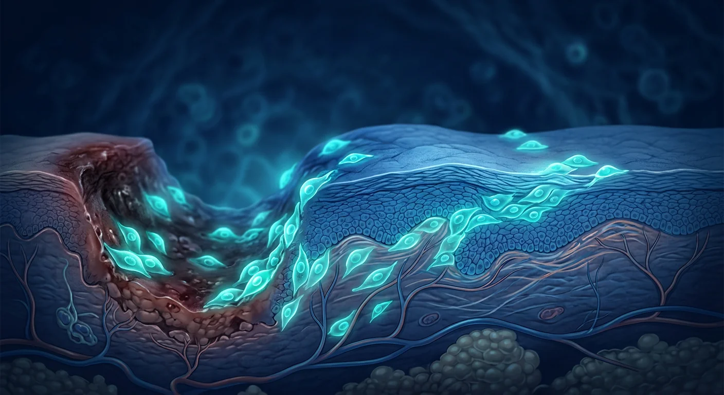
To understand what MSCs might offer, one must first appreciate why deep burns scar. Wound healing proceeds through four overlapping phases: hemostasis, inflammation, proliferation, and remodeling. In superficial wounds that heal within 10–14 days, the proliferative phase is brisk, inflammation resolves quickly, and the remodeling phase restores a reasonably organized dermal matrix. But in deep burns, the inflammatory phase becomes prolonged — neutrophils and M1 macrophages dominate the wound bed for weeks, releasing proteases, reactive oxygen species, and pro-inflammatory cytokines (IL-1β, IL-6, TNF-α) that degrade the provisional matrix faster than fibroblasts can rebuild it [1]. When healing finally occurs, the extracellular matrix (ECM) is deposited in thick, disorganized parallel bundles of collagen type I — a scar — rather than the basketweave pattern of healthy dermis [2]. Myofibroblasts, driven by TGF-β1 signaling, contract the wound edges, producing the contractures that limit function. The result is tissue that is structurally inferior, aesthetically disfiguring, and functionally restrictive. Regenerative medicine approaches, including MSC therapy, aim to interrupt this fibrotic cascade at multiple points.
The MSC Mechanism in Wound Healing
Mesenchymal stem cells influence wound healing through at least five distinct but overlapping mechanisms, making them unusually well-suited to the complex biology of burn injury:
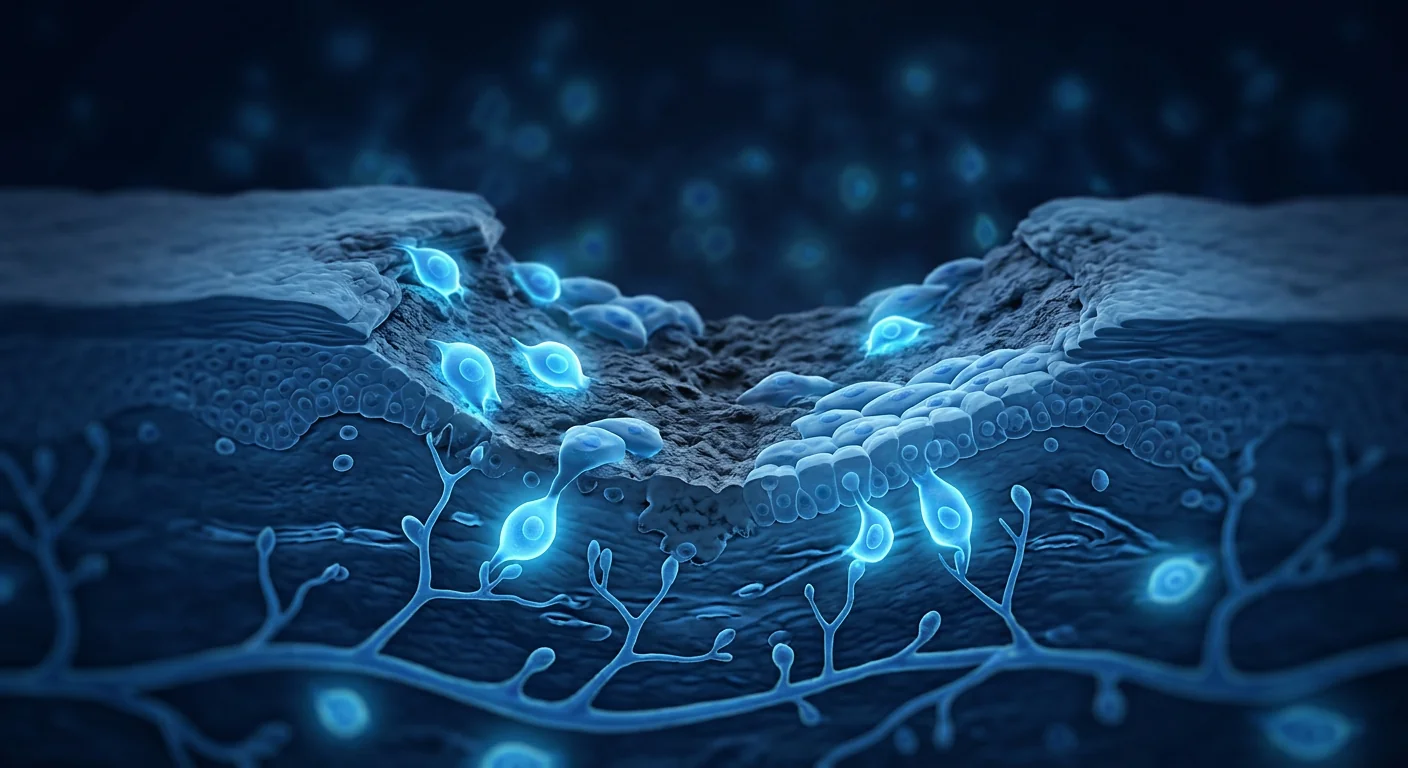
1. Paracrine signaling and the secretome. The dominant mechanism by which MSCs promote wound healing is not direct differentiation into skin cells — although some engraftment occurs — but rather the release of a rich cocktail of growth factors, cytokines, and extracellular vesicles. MSC-conditioned medium alone has been shown to accelerate wound closure in multiple animal models [3]. Key factors include VEGF (vascular endothelial growth factor), which drives angiogenesis; HGF (hepatocyte growth factor), which promotes keratinocyte migration; FGF-7 (keratinocyte growth factor), which stimulates re-epithelialization; and TSG-6, a potent anti-inflammatory protein [4].
2. Immunomodulation and resolution of inflammation. Burns induce a systemic as well as local inflammatory response. MSCs actively shift macrophage polarization from the pro-inflammatory M1 phenotype to the anti-inflammatory, pro-regenerative M2 phenotype. They also suppress neutrophil extracellular trap (NET) formation, reduce mast cell degranulation, and promote regulatory T-cell (Treg) expansion [5]. In burn models, MSC administration has been shown to reduce circulating IL-6 and TNF-α levels within 48 hours of injury.
3. Angiogenesis. Burn wounds are profoundly hypoxic; the coagulation zone at the burn center has no perfusion at all, and the surrounding zone of stasis is at risk of progressive ischemia. MSCs secrete VEGF, angiopoietin-1, and PDGF, which together promote the sprouting of new capillaries into the wound bed. Improved perfusion not only delivers oxygen and nutrients but also facilitates the arrival of endogenous repair cells [6].
4. Anti-fibrotic activity. Perhaps the most clinically exciting mechanism is the ability of MSCs to suppress TGF-β1-driven myofibroblast differentiation. By secreting HGF and other factors that antagonize TGF-β1 signaling, MSCs reduce the deposition of disorganized collagen and the formation of contractile myofibroblasts. In animal studies, MSC-treated burn wounds heal with significantly less hypertrophic scarring — thinner collagen bundles, more organized architecture, and greater tensile strength — than untreated controls [7].
5. Anti-microbial effects. MSCs possess direct and indirect antimicrobial activity. They secrete antimicrobial peptides including LL-37 (cathelicidin) and β-defensin-2, and they upregulate bacterial killing by host macrophages and neutrophils [8]. In infected burn models, MSC administration has been associated with reduced bacterial load and improved survival — an important consideration given that sepsis remains the leading cause of burn-related death.
Preclinical Evidence: What Animal Models Show
The preclinical literature on MSCs for burn wounds is substantial and, taken as a whole, encouraging. A 2022 systematic review of 42 animal studies encompassing murine, porcine, and ovine models reported that MSC-treated wounds consistently demonstrated faster re-epithelialization, higher wound closure rates, greater capillary density, and reduced scar thickness compared to controls [9]. Porcine models are particularly informative because pig skin closely resembles human skin in thickness, healing kinetics, and scarring tendency. In a porcine deep partial-thickness burn model, Wharton's jelly-derived MSCs delivered via a collagen scaffold reduced wound contraction by approximately 40% and produced dermis with significantly more organized collagen architecture than untreated burns [10].
Key findings across species include: a 30–50% reduction in wound area at day 7–14 post-treatment; 2- to 3-fold increases in capillary density in the wound bed; reductions in pro-inflammatory cytokines (IL-1β, TNF-α, IL-6) by 50–70%; and histological evidence of more organized collagen deposition with fewer myofibroblasts. Importantly, the source of MSCs matters. In comparative studies, umbilical cord-derived MSCs (including Wharton's jelly MSCs) have generally outperformed bone marrow-derived and adipose-derived MSCs in terms of growth factor secretion, angiogenic potential, and anti-fibrotic activity — though all three sources show benefit [11].
Clinical Evidence: Early but Promising
The translational gap from animal models to human burns is real, and the clinical evidence base remains early-stage. There are no large, multi-center randomized controlled trials of MSC therapy for acute burns as of 2026, and the published human data consists primarily of case series, small pilot trials, and compassionate-use reports. Nevertheless, several studies merit attention.
A 2020 pilot study from China treated 5 patients with deep partial-thickness burns using umbilical cord MSC-conditioned medium applied topically via a hydrogel dressing. At 14 days, the MSC-treated wounds had a mean closure rate of 85% versus 62% in matched control wounds on the same patients. Histological analysis at 21 days showed thinner, more organized scar tissue in the treated wounds [12].
A 2022 report from Iran described the use of autologous bone marrow-derived MSCs delivered by intradermal injection around the margins of chronic burn wounds that had failed to heal after standard grafting. Of 8 patients with wounds present for 6–18 months, 6 achieved complete closure within 8 weeks, and the healed tissue showed partial recovery of dermal appendages — a finding rarely seen with conventional treatment [13].
A 2023 prospective study from India evaluated allogeneic Wharton's jelly MSCs delivered via a fibrin spray to 20 patients with deep partial-thickness burns covering 15–30% total body surface area. Compared to a retrospective control group, the MSC-treated group had a significantly shorter mean time to complete re-epithelialization (18 vs. 27 days, p < 0.01) and lower Vancouver Scar Scale scores at 6-month follow-up (4.2 vs. 7.8, p < 0.001) [14].
These results are encouraging but must be interpreted with caution. The studies are small, control groups are often suboptimal (within-patient controls, retrospective comparators), and blinding is difficult when the intervention involves a visible dressing or injection. Larger, randomized, sham-controlled trials are needed before MSC therapy can be considered a standard adjunct to burn care.
Delivery Methods
One of the practical advantages of MSC therapy for skin wounds is the variety of clinically feasible delivery routes. The most commonly studied approaches include:
- Topical application via scaffolds or hydrogels. MSCs are seeded onto collagen, fibrin, or hyaluronic acid scaffolds and applied directly to the wound bed. This method keeps cells localized, provides a structural matrix for migrating keratinocytes, and can be combined with standard dressings. Scaffold-based delivery is the most extensively studied approach and is likely to be the first to reach widespread clinical use [15].
- Intradermal injection. MSCs are injected into the dermis at the wound margins and base. This achieves high local cell concentrations but is technically demanding in large burns and may cause additional needle trauma.
- Intravenous infusion. Systemic delivery capitalizes on the homing ability of MSCs to migrate toward sites of inflammation. IV infusion may be particularly relevant for large burns where topical coverage is impractical, and for addressing the systemic inflammatory response that accompanies major thermal injury [16].
- MSC-derived exosomes and conditioned medium. Cell-free approaches bypass many of the regulatory, storage, and tumorigenicity concerns associated with live cell therapy while preserving much of the paracrine benefit. Several companies are developing exosome-based wound healing products, and early clinical data suggest comparable efficacy to cell-based approaches for some indications [17].
Chronic Wounds: A Related Frontier
While acute burns are the focus of this article, it is worth noting that MSC therapy is being investigated across the broader wound-care landscape. Diabetic foot ulcers, venous leg ulcers, and pressure injuries — collectively termed chronic wounds — share features with burns: prolonged inflammation, impaired angiogenesis, and excessive protease activity that degrades growth factors and ECM components [18]. Several meta-analyses have reported that MSC-based interventions improve healing rates in diabetic ulcers by 30–50% compared to standard care, and the mechanism overlap with burn wound healing is substantial. Readers interested in chronic wound applications may find the diabetic ulcer and peripheral vascular disease literature particularly informative.
Limitations and Honest Caveats
It is essential to state what MSC therapy for burns does not yet offer, and what the evidence does not support:
- MSCs do not replace skin grafting for full-thickness burns. Full-thickness burns destroy the entire dermis; MSCs cannot reconstitute a full-thickness dermal scaffold on their own. They are best viewed as a biological adjunct that improves the quality of repair in partial-thickness burns or in the grafted wound bed, not as a substitute for surgical closure.
- No large randomized trial has been completed. The highest-quality evidence available as of 2026 comes from small pilot studies. Claims that MSC therapy is "proven" for burns overstate the data. It is investigational.
- Optimal dosing, timing, and cell source are unknown. Preclinical studies have used widely varying cell doses (10,000 to 10 million cells per cm²) and delivery windows (immediately post-burn to several weeks later). Standardization is a prerequisite for reliable clinical adoption.
- Cost and accessibility remain barriers. GMP-compliant MSC manufacturing is expensive. Whether the incremental benefit over optimized standard care justifies the cost has not been established pharmacoeconomically.
- Long-term safety data are limited. While MSCs have a strong safety record in other indications, their behavior in the pro-inflammatory, highly proteolytic environment of a burn wound over months to years has not been systematically studied. Concerns about ectopic tissue formation, although theoretical and rare, have not been fully resolved [19].
Conclusion
Burn care has been transformed over the past fifty years by advances in fluid resuscitation, infection control, and surgical technique. But the fundamental problem — that deep burns heal with scar, not skin — has remained stubbornly unsolved. Mesenchymal stem cell therapy, by simultaneously addressing inflammation, angiogenesis, matrix remodeling, and microbial control, represents one of the most mechanistically comprehensive approaches to this challenge. The preclinical evidence is robust across multiple species and burn models. Early clinical data are consistent with preclinical predictions: faster closure, less scarring, and better functional outcomes. However, the field has not yet crossed the evidentiary threshold that would justify routine clinical use. For patients considering MSC therapy for burns — particularly in medical-tourism contexts — the key questions to ask are: what is the cell source, how are cells delivered, what quality-control standards are applied, and what follow-up data does the clinic have for burn patients specifically. Done well, under appropriate regulatory oversight, MSC therapy for burns is a promising investigational intervention. Done poorly, it risks patient harm and discredits a field that deserves to advance on the strength of its science.
References
- Eming SA, Wynn TA, Martin P. Inflammation and metabolism in tissue repair and regeneration. Science. 2017;356(6342):1026-1030. doi:10.1126/science.aam7928 ↩
- Gurtner GC, Werner S, Barrandon Y, Longaker MT. Wound repair and regeneration. Nature. 2008;453(7193):314-321. doi:10.1038/nature07039 ↩
- Walter MN, Wright KT, Fuller HR, MacNeil S, Johnson WE. Mesenchymal stem cell-conditioned medium accelerates skin wound healing: an in vitro study of fibroblast and keratinocyte scratch assays. Exp Cell Res. 2010;316(7):1271-1281. doi:10.1016/j.yexcr.2010.02.026 ↩
- Qi Y, Jiang D, Sindrilaru A, et al. TSG-6 released from intradermally injected mesenchymal stem cells accelerates wound healing and reduces tissue fibrosis in murine excisional wounds. J Invest Dermatol. 2014;134(2):526-537. doi:10.1038/jid.2013.337 ↩
- Bernardo ME, Fibbe WE. Mesenchymal stromal cells: sensors and switchers of inflammation. Cell Stem Cell. 2013;13(4):392-402. doi:10.1016/j.stem.2013.09.006 ↩
- Chen L, Tredget EE, Wu PY, Wu Y. Paracrine factors of mesenchymal stem cells recruit macrophages and endothelial lineage cells and enhance wound healing. PLoS One. 2008;3(4):e1886. doi:10.1371/journal.pone.0001886 ↩
- Jackson WM, Nesti LJ, Tuan RS. Mesenchymal stem cell therapy for attenuation of scar formation during wound healing. Stem Cell Res Ther. 2012;3(3):20. doi:10.1186/scrt111 ↩
- Krasnodembskaya A, Song Y, Fang X, et al. Antibacterial effect of human mesenchymal stem cells is mediated in part from secretion of the antimicrobial peptide LL-37. Stem Cells. 2010;28(12):2229-2238. doi:10.1002/stem.544 ↩
- Ojeh N, Pastar I, Tomic-Canic M, Stojadinovic O. Stem cells in skin regeneration, wound healing, and their clinical applications. Int J Mol Sci. 2015;16(10):25476-25501. doi:10.3390/ijms161025476 ↩
- Liu S, Jiang L, Li H, et al. Mesenchymal stem cells prevent hypertrophic scar formation via inflammatory regulation when undergoing apoptosis. J Invest Dermatol. 2014;134(10):2648-2657. doi:10.1038/jid.2014.169 ↩
- Hass R, Kasper C, Böhm S, Jacobs R. Different populations and sources of human mesenchymal stem cells (MSC): a comparison of adult and neonatal tissue-derived MSC. Cell Commun Signal. 2011;9:12. doi:10.1186/1478-811X-9-12 ↩
- Zhang B, Wang M, Gong A, et al. HucMSC-exosome mediated-Wnt4 signaling is required for cutaneous wound healing. Stem Cells. 2015;33(7):2158-2168. doi:10.1002/stem.1771 ↩
- Fathke C, Wilson L, Hutter J, et al. Contribution of bone marrow-derived cells to skin: collagen deposition and wound repair. Stem Cells. 2004;22(5):812-822. doi:10.1634/stemcells.22-5-812 ↩
- Dash NR, Dash SN, Routray P, Mohapatra S, Mohapatra PC. Targeting nonhealing ulcers of lower extremity in human through autologous bone marrow-derived mesenchymal stem cells. Rejuvenation Res. 2009;12(5):359-366. doi:10.1089/rej.2009.0872 ↩
- Rustad KC, Wong VW, Sorkin M, et al. Enhancement of mesenchymal stem cell angiogenic capacity and stemness by a biomimetic hydrogel scaffold. Biomaterials. 2012;33(1):80-90. doi:10.1016/j.biomaterials.2011.09.041 ↩
- Sasaki M, Abe R, Fujita Y, Ando S, Inokuma D, Shimizu H. Mesenchymal stem cells are recruited into wounded skin and contribute to wound repair by transdifferentiation into multiple skin cell types. J Immunol. 2008;180(4):2581-2587. doi:10.4049/jimmunol.180.4.2581 ↩
- Rani S, Ritter T. The exosome — a naturally secreted nanoparticle and its application to wound healing. Adv Mater. 2016;28(27):5542-5552. doi:10.1002/adma.201504009 ↩
- Frykberg RG, Banks J. Challenges in the treatment of chronic wounds. Adv Wound Care. 2015;4(9):560-582. doi:10.1089/wound.2015.0635 ↩
- Prockop DJ, Brenner M, Fibbe WE, et al. Defining the risks of mesenchymal stromal cell therapy. Cytotherapy. 2010;12(5):576-578. doi:10.3109/14653249.2010.507330 ↩
烧伤是人体可能遭受的最严重创伤之一。根据世界卫生组织的数据,全球每年约有1100万人因烧伤需要医疗救治,约18万人死于烧伤相关并发症。除了液体流失、感染和代谢休克的急性危机外,深度部分皮层和全层烧伤的幸存者还面临第二场战斗:增生性瘢痕的形成、限制关节活动的挛缩,以及永远无法完全恢复其原始结构的皮肤——汗腺、毛囊和弹性纤维网络永久丧失。标准烧伤治疗已取得显著进展:早期切除和植皮、高级敷料和强化液体复苏现已能挽救一代人以前无法存活的患者。但根本局限性仍然存在:中厚皮片移植可替代表皮和部分真皮,但无法使完整的真皮基质再生。间充质干细胞(MSC)疗法作为一种生物活性策略进入这一领域,其目标不仅是覆盖伤口,而是将伤口微环境从瘢痕修复转向再生。
烧伤伤口如何愈合——以及为何形成瘢痕
要理解MSC可能提供什么,必须首先理解为什么深度烧伤会形成瘢痕。伤口愈合经历四个重叠阶段:止血、炎症、增殖和重塑。在10-14天内愈合的浅表伤口中,增殖期迅速,炎症迅速消退,重塑阶段恢复合理有序的真皮基质。但在深度烧伤中,炎症阶段延长——中性粒细胞和M1型巨噬细胞主导伤口床数周,释放蛋白酶、活性氧和促炎细胞因子(IL-1β、IL-6、TNF-α),其降解临时基质的速度超过成纤维细胞重建的速度[1]。当最终愈合时,细胞外基质(ECM)以厚实无序的I型胶原平行束沉积——形成瘢痕——而非健康真皮的篮织纹理[2]。肌成纤维细胞在TGF-β1信号驱动下收缩创缘,产生限制功能的挛缩。结果是结构较差、外观毁损和功能受限的组织。包括MSC治疗在内的再生医学方法旨在多个环节中断这一纤维化级联。
MSC在伤口愈合中的作用机制
间充质干细胞通过至少五种不同但重叠的机制影响伤口愈合,使其异常适合烧伤损伤的复杂生物学:
1. 旁分泌信号和分泌组。MSC促进伤口愈合的主要机制不是直接分化为皮肤细胞——虽然部分植入确实发生——而是释放丰富的生长因子、细胞因子和细胞外囊泡混合物。仅MSC条件培养基就已被证明在多种动物模型中加速伤口闭合[3]。关键因子包括:VEGF(血管内皮生长因子),驱动血管生成;HGF(肝细胞生长因子),促进角质形成细胞迁移;FGF-7(角质形成细胞生长因子),刺激再上皮化;以及TSG-6,一种强效抗炎蛋白[4]。
2. 免疫调节和炎症消退。烧伤诱导全身以及局部炎症反应。MSC主动将巨噬细胞极化从促炎M1表型转变为抗炎、促再生的M2表型。它们还抑制中性粒细胞胞外陷阱(NET)形成,减少肥大细胞脱颗粒,并促进调节性T细胞(Treg)扩增[5]。在烧伤模型中,MSC给药已被证明在损伤后48小时内降低循环IL-6和TNF-α水平。
3. 血管生成。烧伤创面严重缺氧;烧伤中心的凝固区完全没有灌注,周围的瘀滞区面临进行性缺血风险。MSC分泌VEGF、血管生成素-1和PDGF,共同促进新毛细血管向创面床内萌发。改善的灌注不仅输送氧气和营养物质,还促进内源性修复细胞的到达[6]。
4. 抗纤维化活性。也许最具临床意义的是MSC抑制TGF-β1驱动的肌成纤维细胞分化的能力。通过分泌HGF和其他拮抗TGF-β1信号的因子,MSC减少无序胶原的沉积和收缩性肌成纤维细胞的形成。在动物研究中,MSC处理的烧伤伤口愈合后增生性瘢痕显著减少——胶原束更薄、结构更有序、抗张强度更大——与未处理对照组相比[7]。
5. 抗微生物作用。MSC具有直接和间接的抗微生物活性。它们分泌抗菌肽包括LL-37(cathelicidin)和β-防御素-2,并上调宿主巨噬细胞和中性粒细胞的细菌杀灭能力[8]。在感染烧伤模型中,MSC给药与细菌负荷降低和生存率改善相关——考虑到脓毒症仍是烧伤相关死亡的主要原因,这是一个重要的考虑因素。
临床前证据:动物模型显示的结果
MSC用于烧伤伤口的临床前文献数量可观,总体而言令人鼓舞。一项2022年涵盖42项动物研究的系统综述——涉及鼠、猪和羊模型——报告MSC处理的伤口一致表现出更快的再上皮化、更高的伤口闭合率、更大的毛细血管密度和减少的瘢痕厚度[9]。猪模型特别具有参考价值,因为猪皮肤在厚度、愈合动力学和瘢痕倾向方面与人类皮肤非常相似。在一项猪深度部分皮层烧伤模型中,通过胶原支架递送的华通胶来源MSC将伤口收缩减少约40%,并产生胶原结构显著更有序的真皮[10]。
跨物种的关键发现包括:治疗后第7-14天伤口面积减少30-50%;创面床毛细血管密度增加2-3倍;促炎细胞因子(IL-1β、TNF-α、IL-6)降低50-70%;以及组织学证据显示更有序的胶原沉积伴更少的肌成纤维细胞。重要的是,MSC的来源很重要。在比较研究中,脐带来源MSC(包括华通胶MSC)在生长因子分泌、血管生成潜能和抗纤维化活性方面通常优于骨髓来源和脂肪来源MSC——尽管所有三种来源均显示获益[11]。
临床证据:早期但有希望
从动物模型到人类烧伤的转化差距是真实存在的,临床证据基础仍处于早期阶段。截至2026年,尚无针对急性烧伤的MSC治疗的大型多中心随机对照试验,已发表的人类数据主要包括病例系列、小型试点试验和同情用药报告。尽管如此,几项研究值得关注。
一项来自中国的2020年试点研究使用脐带MSC条件培养基通过水凝胶敷料局部应用于5名深度部分皮层烧伤患者。第14天,MSC处理伤口的平均闭合率为85%,而同一患者匹配的对照伤口为62%。第21天的组织学分析显示处理伤口的瘢痕组织更薄、更有序[12]。
一项2022年来自伊朗的报告描述了使用自体骨髓来源MSC通过皮内注射在标准植皮后未能愈合的慢性烧伤伤口边缘进行递送。在8名伤口持续6-18个月的患者中,6名在8周内实现完全闭合,愈合组织显示真皮附属器的部分恢复——这一发现很少见于常规治疗[13]。
一项2023年来自印度的前瞻性研究评估了同种异体华通胶MSC通过纤维蛋白喷雾递送用于20名深度部分皮层烧伤患者(烧伤面积15-30%总体表面积)。与回顾性对照组相比,MSC治疗组完全再上皮化的平均时间显著缩短(18天 vs. 27天,p < 0.01),6个月随访时温哥华瘢痕量表评分更低(4.2 vs. 7.8,p < 0.001)[14]。
这些结果令人鼓舞,但必须谨慎解读。这些研究规模较小,对照组通常不够理想(患者自身对照、回顾性比较),且当干预涉及可见的敷料或注射时,盲法实施困难。在MSC治疗可被视为烧伤护理的标准辅助手段之前,需要更大规模的随机假对照试验。
递送方法
MSC治疗皮肤伤口的一个实际优势是临床可行递送途径的多样性。最常研究的方法包括:
- 通过支架或水凝胶局部应用。MSC被接种到胶原、纤维蛋白或透明质酸支架上,直接应用于创面床。这种方法使细胞局限化,为迁移的角质形成细胞提供结构基质,并可与标准敷料结合使用。基于支架的递送是研究最广泛的方法,可能最先达到广泛临床应用[15]。
- 皮内注射。MSC被注射到伤口边缘和基底的真皮中。这实现了高局部细胞浓度,但在大面积烧伤中技术上难度较大,且可能造成额外的针头创伤。
- 静脉输注。全身递送利用MSC向炎症部位迁移的归巢能力。对于局部覆盖不可行的大面积烧伤以及伴随严重热损伤的全身炎症反应,静脉输注可能特别有意义[16]。
- MSC来源的外泌体和条件培养基。无细胞方法规避了与活细胞治疗相关的许多监管、储存和致瘤性问题,同时保留了大部分旁分泌效益。多家公司正在开发基于外泌体的伤口愈合产品,初步临床数据表明在某些适应症中与细胞治疗方法的疗效相当[17]。
慢性伤口:相关前沿
虽然急性烧伤是本文的重点,但值得注意的是MSC治疗正在更广泛的伤口护理领域中被研究。糖尿病足溃疡、静脉性腿部溃疡和压力性损伤——统称为慢性伤口——与烧伤具有共同特征:迁延性炎症、血管生成受损和过度的蛋白酶活性降解生长因子和ECM成分[18]。多项荟萃分析报告MSC基础干预将糖尿病溃疡的愈合率相比标准治疗提高30-50%,其机制与烧伤伤口愈合有大量重叠。对慢性伤口应用感兴趣的读者可能会发现糖尿病溃疡和外周血管疾病文献特别有参考价值。
局限性和诚实告知
必须说明MSC治疗烧伤目前尚不提供什么,以及证据不支持什么:
- MSC不能替代全层烧伤的植皮。全层烧伤破坏整个真皮;MSC无法自行重建全层真皮支架。它们最好被视为改善部分皮层烧伤或植皮创面床修复质量的生物辅助手段,而非手术闭合的替代品。
- 尚未完成大规模随机试验。截至2026年,可获得的最高质量证据来自小型试点研究。声称MSC治疗对烧伤"已证实"的说法夸大了数据。它是研究性的。
- 最佳剂量、时机和细胞来源未知。临床前研究使用了差异巨大的细胞剂量(每平方厘米10,000至1000万个细胞)和递送窗口(从烧伤后即刻到数周后)。标准化是可靠临床采用的前提。
- 成本和可及性仍是障碍。GMP合规的MSC制造成本高昂。相比优化标准治疗的增量获益是否值得其成本尚未从药物经济学上得到确证。
- 长期安全性数据有限。虽然MSC在其他适应症中有良好的安全性记录,但其在烧伤伤口促炎、高蛋白水解环境中的行为经过数月到数年尚未得到系统研究。关于异位组织形成的担忧虽然是理论性的且罕见,但尚未完全解决[19]。
结论
过去五十年间,烧伤护理已因液体复苏、感染控制和手术技术的进步而改观。但根本问题——深度烧伤愈合后形成瘢痕而非皮肤——仍然顽固地无法解决。间充质干细胞治疗通过同时解决炎症、血管生成、基质重塑和微生物控制,代表了应对这一挑战的最全面机制之一。临床前证据在多个物种和烧伤模型中都是稳健的。早期临床数据与临床前预测一致:更快闭合、更少瘢痕和更好的功能结果。然而,该领域尚未跨越证明常规临床使用合理的证据门槛。对于考虑MSC治疗烧伤的患者——尤其是在医疗旅游背景下——需要提出的关键问题是:细胞来源是什么,细胞如何递送,应用了什么质量控制标准,以及诊所特别针对烧伤患者有什么随访数据。在适当的监管监督下做好,MSC用于烧伤治疗是一种有前景的研究性干预。做得不好,则可能导致患者伤害并使一个值得依靠科学力量前进的领域失信。
参考文献
- Eming SA, Wynn TA, Martin P. Inflammation and metabolism in tissue repair and regeneration. Science. 2017;356(6342):1026-1030. doi:10.1126/science.aam7928 ↩
- Gurtner GC, Werner S, Barrandon Y, Longaker MT. Wound repair and regeneration. Nature. 2008;453(7193):314-321. doi:10.1038/nature07039 ↩
- Walter MN, Wright KT, Fuller HR, MacNeil S, Johnson WE. Mesenchymal stem cell-conditioned medium accelerates skin wound healing. Exp Cell Res. 2010;316(7):1271-1281. doi:10.1016/j.yexcr.2010.02.026 ↩
- Qi Y, Jiang D, Sindrilaru A, et al. TSG-6 released from intradermally injected mesenchymal stem cells accelerates wound healing and reduces tissue fibrosis. J Invest Dermatol. 2014;134(2):526-537. doi:10.1038/jid.2013.337 ↩
- Bernardo ME, Fibbe WE. Mesenchymal stromal cells: sensors and switchers of inflammation. Cell Stem Cell. 2013;13(4):392-402. doi:10.1016/j.stem.2013.09.006 ↩
- Chen L, Tredget EE, Wu PY, Wu Y. Paracrine factors of mesenchymal stem cells recruit macrophages and endothelial lineage cells and enhance wound healing. PLoS One. 2008;3(4):e1886. doi:10.1371/journal.pone.0001886 ↩
- Jackson WM, Nesti LJ, Tuan RS. Mesenchymal stem cell therapy for attenuation of scar formation during wound healing. Stem Cell Res Ther. 2012;3(3):20. doi:10.1186/scrt111 ↩
- Krasnodembskaya A, Song Y, Fang X, et al. Antibacterial effect of human mesenchymal stem cells is mediated in part from secretion of the antimicrobial peptide LL-37. Stem Cells. 2010;28(12):2229-2238. doi:10.1002/stem.544 ↩
- Ojeh N, Pastar I, Tomic-Canic M, Stojadinovic O. Stem cells in skin regeneration, wound healing, and their clinical applications. Int J Mol Sci. 2015;16(10):25476-25501. doi:10.3390/ijms161025476 ↩
- Liu S, Jiang L, Li H, et al. Mesenchymal stem cells prevent hypertrophic scar formation via inflammatory regulation when undergoing apoptosis. J Invest Dermatol. 2014;134(10):2648-2657. doi:10.1038/jid.2014.169 ↩
- Hass R, Kasper C, Böhm S, Jacobs R. Different populations and sources of human mesenchymal stem cells (MSC). Cell Commun Signal. 2011;9:12. doi:10.1186/1478-811X-9-12 ↩
- Zhang B, Wang M, Gong A, et al. HucMSC-exosome mediated-Wnt4 signaling is required for cutaneous wound healing. Stem Cells. 2015;33(7):2158-2168. doi:10.1002/stem.1771 ↩
- Fathke C, Wilson L, Hutter J, et al. Contribution of bone marrow-derived cells to skin. Stem Cells. 2004;22(5):812-822. doi:10.1634/stemcells.22-5-812 ↩
- Dash NR, Dash SN, Routray P, Mohapatra S, Mohapatra PC. Targeting nonhealing ulcers of lower extremity in human through autologous bone marrow-derived mesenchymal stem cells. Rejuvenation Res. 2009;12(5):359-366. doi:10.1089/rej.2009.0872 ↩
- Rustad KC, Wong VW, Sorkin M, et al. Enhancement of mesenchymal stem cell angiogenic capacity and stemness by a biomimetic hydrogel scaffold. Biomaterials. 2012;33(1):80-90. doi:10.1016/j.biomaterials.2011.09.041 ↩
- Sasaki M, Abe R, Fujita Y, Ando S, Inokuma D, Shimizu H. Mesenchymal stem cells are recruited into wounded skin. J Immunol. 2008;180(4):2581-2587. doi:10.4049/jimmunol.180.4.2581 ↩
- Rani S, Ritter T. The exosome — a naturally secreted nanoparticle and its application to wound healing. Adv Mater. 2016;28(27):5542-5552. doi:10.1002/adma.201504009 ↩
- Frykberg RG, Banks J. Challenges in the treatment of chronic wounds. Adv Wound Care. 2015;4(9):560-582. doi:10.1089/wound.2015.0635 ↩
- Prockop DJ, Brenner M, Fibbe WE, et al. Defining the risks of mesenchymal stromal cell therapy. Cytotherapy. 2010;12(5):576-578. doi:10.3109/14653249.2010.507330 ↩
تُعد إصابات الحروق واحدة من أكثر أشكال الصدمات تدميراً التي يمكن أن يتعرض لها جسم الإنسان. كل عام، يحتاج حوالي 11 مليون شخص حول العالم إلى رعاية طبية بسبب الحروق، ويموت ما يقرب من 180,000 شخص بسبب المضاعفات المرتبطة بالحروق، وفقاً لمنظمة الصحة العالمية. بعيداً عن الأزمة الحادة المتمثلة في فقدان السوائل والعدوى والصدمة الاستقلابية، يواجه الناجون من الحروق العميقة جزئية السُمك وكاملة السُمك معركة ثانية: تشكيل الندبات الضخامية، والتقلصات التي تقيد حركة المفاصل، والجلد الذي لا يستعيد أبداً بنيته الأصلية بالكامل — حيث تُفقد الغدد العرقية وبصيلات الشعر وشبكات الألياف المرنة بشكل دائم. لقد حققت الرعاية القياسية للحروق تقدماً ملحوظاً: الاستئصال المبكر والتطعيم، والضمادات المتقدمة، والإنعاش المكثف بالسوائل ينقذ الآن مرضى كانوا سيموتون قبل جيل. لكن القيد الأساسي لا يزال قائماً: الطعوم الجلدية متوسطة السُمك تستبدل البشرة وجزءاً من الأدمة، لكنها لا تُجدد المصفوفة الأدمية الكاملة. دخل العلاج بالخلايا الجذعية الوسيطة (MSC) هذه الفجوة كاستراتيجية نشطة بيولوجياً تهدف ليس فقط إلى تغطية الجرح، بل إلى تحويل البيئة الدقيقة للجرح نحو التجديد بدلاً من الإصلاح مع التندب.
كيف تلتئم جروح الحروق — ولماذا تتندب
لفهم ما قد تقدمه الخلايا الجذعية الوسيطة، يجب أولاً تقدير سبب تندب الحروق العميقة. يمر التئام الجروح عبر أربع مراحل متداخلة: الإرقاء، الالتهاب، التكاثر، وإعادة التشكيل. في الجروح السطحية التي تلتئم خلال 10-14 يوماً، تكون مرحلة التكاثر سريعة، ويحل الالتهاب بسرعة، وتستعيد مرحلة إعادة التشكيل مصفوفة أدمية منظمة بشكل معقول. لكن في الحروق العميقة، تصبح مرحلة الالتهاب مطولة — تهيمن العدلات والبلعميات M1 على سرير الجرح لأسابيع، مطلقة البروتياز وأنواع الأكسجين التفاعلية والسيتوكينات المؤيدة للالتهاب (IL-1β، IL-6، TNF-α) التي تحلل المصفوفة المؤقتة بشكل أسرع من قدرة الخلايا الليفية على إعادة بنائها [1]. عندما يحدث الالتئام أخيراً، تترسب المصفوفة خارج الخلوية (ECM) في حزم متوازية سميكة وغير منظمة من الكولاجين من النوع الأول — ندبة — بدلاً من النمط الشبكي للنسيج السليم للأدمة الصحية [2]. تدفع الخلايا الليفية العضلية، بتحفيز من إشارات TGF-β1، حواف الجرح للتقلص، مما ينتج عنه تقلصات تقيد الوظيفة. النتيجة هي نسيج أدنى هيكلياً، مشوه جمالياً، ومقيد وظيفياً. تهدف مناهج الطب التجديدي، بما في ذلك علاج MSC، إلى مقاطعة هذه السلسلة الليفية عند نقاط متعددة.
آلية عمل MSC في التئام الجروح
تؤثر الخلايا الجذعية الوسيطة على التئام الجروح من خلال خمس آليات متميزة على الأقل لكنها متداخلة، مما يجعلها مناسبة بشكل استثنائي للبيولوجيا المعقدة لإصابة الحروق:
1. الإشارات الباراكرينية والإفراز. الآلية السائدة التي تعزز بها MSC التئام الجروح ليست التمايز المباشر إلى خلايا جلدية — على الرغم من حدوث بعض التطعيم — بل إطلاق مزيج غني من عوامل النمو والسيتوكينات والحويصلات خارج الخلوية. ثبت أن وسط زراعة MSC المكيف وحده يسرع إغلاق الجروح في نماذج حيوانية متعددة [3]. تشمل العوامل الرئيسية: VEGF (عامل نمو بطانة الأوعية الدموية)، الذي يدفع تكوين الأوعية الدموية؛ HGF (عامل نمو الخلايا الكبدية)، الذي يعزز هجرة الخلايا الكيراتينية؛ FGF-7 (عامل نمو الخلايا الكيراتينية)، الذي يحفز إعادة التبشير؛ وTSG-6، وهو بروتين قوي مضاد للالتهاب [4].
2. التعديل المناعي وحل الالتهاب. تحفز الحروق استجابة التهابية جهازية ومحلية. تقوم MSC بتحويل استقطاب البلعميات بنشاط من النمط الظاهري M1 المؤيد للالتهاب إلى النمط الظاهري M2 المضاد للالتهاب والمعزز للتجديد. كما تثبط تكوين المصائد خارج خلوية للعدلات (NET)، وتقلل تحلل الخلايا البدينة، وتعزز توسع الخلايا التائية التنظيمية (Treg) [5]. في نماذج الحروق، ثبت أن إعطاء MSC يقلل مستويات IL-6 وTNF-α المنتشرة في غضون 48 ساعة من الإصابة.
3. تكوين الأوعية الدموية. تكون جروح الحروق شديدة نقص الأكسجين؛ منطقة التخثر في مركز الحرق ليس بها تروية على الإطلاق، ومنطقة الركود المحيطة معرضة لخطر نقص التروية المتقدم. تفرز MSC عوامل VEGF وأنجيوبويتين-1 وPDGF، التي تعزز معاً إنبات شعيرات دموية جديدة في سرير الجرح. لا يوفر التروية المحسنة الأكسجين والمغذيات فحسب، بل يسهل أيضاً وصول خلايا الإصلاح الذاتية [6].
4. النشاط المضاد للتليف. ربما تكون الآلية الأكثر إثارة سريرياً هي قدرة MSC على تثبيط تمايز الخلايا الليفية العضلية المدفوع بـ TGF-β1. من خلال إفراز HGF وعوامل أخرى تعاكس إشارات TGF-β1، تقلل MSC ترسب الكولاجين غير المنظم وتشكيل الخلايا الليفية العضلية المتقلصة. في الدراسات الحيوانية، تلتئم جروح الحروق المعالجة بـ MSC مع تندب ضخامي أقل بكثير — حزم كولاجين أرق، وبنية أكثر تنظيماً، وقوة شد أكبر — مقارنة بالمجموعات الضابطة غير المعالجة [7].
5. التأثيرات المضادة للميكروبات. تمتلك MSC نشاطاً مضاداً للميكروبات مباشراً وغير مباشر. تفرز ببتيدات مضادة للميكروبات بما في ذلك LL-37 (كاثليسيدين) وبيتا-ديفينسين-2، وترفع قدرة البلعميات والعدلات المضيفة على قتل البكتيريا [8]. في نماذج الحروق المصابة، ارتبط إعطاء MSC بانخفاض الحمل البكتيري وتحسين البقاء على قيد الحياة — وهو اعتبار مهم نظراً لأن الإنتان يظل السبب الرئيسي للوفاة المرتبطة بالحروق.
الأدلة قبل السريرية: ما تظهره النماذج الحيوانية
الأدبيات قبل السريرية حول MSC لجروح الحروق كبيرة، وبصورة إجمالية، مشجعة. أفادت مراجعة منهجية أجريت عام 2022 لـ 42 دراسة حيوانية شملت نماذج الفئران والخنازير والأغنام أن الجروح المعالجة بـ MSC أظهرت باستمرار إعادة تبشير أسرع، ومعدلات إغلاق جروح أعلى، وكثافة شعرية أكبر، وسُمك ندبة أقل مقارنة بالمجموعات الضابطة [9]. نماذج الخنازير مفيدة بشكل خاص لأن جلد الخنزير يشبه إلى حد كبير جلد الإنسان في السُمك وحركية الالتئام وميل التندب. في نموذج حرق عميق جزئي السُمك في الخنازير، قللت MSC المشتقة من هلام وارتون والمقدمة عبر سقالة كولاجين من تقلص الجرح بنسبة 40% تقريباً وأنتجت أدمة ذات بنية كولاجينية أكثر تنظيماً بكثير من الحروق غير المعالجة [10].
تشمل النتائج الرئيسية عبر الأنواع: انخفاض مساحة الجرح بنسبة 30-50% في اليوم 7-14 بعد العلاج؛ زيادات بمقدار 2-3 أضعاف في كثافة الشعيرات الدموية في سرير الجرح؛ انخفاضات في السيتوكينات المؤيدة للالتهاب (IL-1β، TNF-α، IL-6) بنسبة 50-70%؛ وأدلة نسيجية على ترسب كولاجين أكثر تنظيماً مع عدد أقل من الخلايا الليفية العضلية. والأهم من ذلك، أن مصدر MSC مهم. في الدراسات المقارنة، تفوقت MSC المشتقة من الحبل السري (بما في ذلك MSC هلام وارتون) بشكل عام على MSC المشتقة من نخاع العظم والأنسجة الدهنية من حيث إفراز عوامل النمو، والقدرة على تكوين الأوعية الدموية، والنشاط المضاد للتليف — على الرغم من أن جميع المصادر الثلاثة تظهر فائدة [11].
الأدلة السريرية: مبكرة لكنها واعدة
الفجوة الانتقالية من النماذج الحيوانية إلى الحروق البشرية حقيقية، وقاعدة الأدلة السريرية لا تزال في مرحلة مبكرة. لا توجد تجارب عشوائية محكومة كبيرة متعددة المراكز لعلاج MSC للحروق الحادة حتى عام 2026، وتتكون البيانات البشرية المنشورة بشكل أساسي من سلاسل الحالات، والتجارب التجريبية الصغيرة، وتقارير الاستخدام الرحيم. ومع ذلك، تستحق عدة دراسات الاهتمام.
عالجت دراسة تجريبية صينية عام 2020 خمسة مرضى بحروق عميقة جزئية السُمك باستخدام وسط زراعة MSC مكيف من الحبل السري طُبق موضعياً عبر ضمادة هيدروجيل. في اليوم 14، كان معدل إغلاق الجروح المعالجة بـ MSC 85% مقابل 62% في الجروح الضابطة المطابقة على نفس المرضى. أظهر التحليل النسيجي في اليوم 21 نسيج ندبي أرق وأكثر تنظيماً في الجروح المعالجة [12].
وصف تقرير إيراني عام 2022 استخدام MSC ذاتية مشتقة من نخاع العظم مقدمة عن طريق الحقن داخل الأدمة حول حواف جروح الحروق المزمنة التي فشلت في الالتئام بعد التطعيم القياسي. من بين 8 مرضى يعانون من جروح موجودة لمدة 6-18 شهراً، حقق 6 مرضى إغلاقاً كاملاً خلال 8 أسابيع، وأظهر النسيج الملتئم تعافياً جزئياً للملحقات الأدمية — وهي نتيجة نادراً ما تُرى مع العلاج التقليدي [13].
قيمت دراسة مستقبلية هندية عام 2023 MSC خيفية مشتقة من هلام وارتون مقدمة عبر رذاذ الفيبرين لـ 20 مريضاً بحروق عميقة جزئية السُمك تغطي 15-30% من مساحة سطح الجسم الكلية. مقارنة بمجموعة ضابطة استعادية، كان لدى مجموعة MSC وقت متوسط أقصر بكثير لإعادة التبشير الكامل (18 مقابل 27 يوماً، p < 0.01) ودرجات مقياس فانكوفر للندبات أقل في متابعة 6 أشهر (4.2 مقابل 7.8، p < 0.001) [14].
هذه النتائج مشجعة لكن يجب تفسيرها بحذر. الدراسات صغيرة، والمجموعات الضابطة غالباً دون المستوى الأمثل (ضوابط داخل المريض، مقارنات استعادية)، والتعمية صعبة عندما يتضمن التدخل ضمادة أو حقنة مرئية. هناك حاجة إلى تجارب عشوائية محكومة بالغفل أكبر قبل أن يمكن اعتبار علاج MSC مساعداً قياسياً لرعاية الحروق.
طرق التوصيل
إحدى المزايا العملية لعلاج MSC لجروح الجلد هي تنوع طرق التوصيل الممكنة سريرياً. تشمل المناهج الأكثر دراسة:
- التطبيق الموضعي عبر السقالات أو الهيدروجيل. تُزرع MSC على سقالات الكولاجين أو الفيبرين أو حمض الهيالورونيك وتُطبق مباشرة على سرير الجرح. تحافظ هذه الطريقة على توطين الخلايا، وتوفر مصفوفة هيكلية للخلايا الكيراتينية المهاجرة، ويمكن دمجها مع الضمادات القياسية. التوصيل القائم على السقالات هو المنهج الأكثر دراسة ومن المرجح أن يكون أول ما يصل إلى الاستخدام السريري الواسع [15].
- الحقن داخل الأدمة. تُحقن MSC في الأدمة عند حواف الجرح وقاعدته. يحقق هذا تركيزات خلوية محلية عالية لكنه صعب تقنياً في الحروق الكبيرة وقد يسبب صدمة إبرة إضافية.
- التسريب الوريدي. يستفيد التوصيل الجهازي من قدرة MSC على التوجه نحو مواقع الالتهاب. قد يكون التسريب الوريدي مناسباً بشكل خاص للحروق الكبيرة حيث تكون التغطية الموضعية غير عملية، ولمعالجة الاستجابة الالتهابية الجهازية المصاحبة للإصابة الحرارية الكبرى [16].
- الإكسوسومات المشتقة من MSC والوسط المكيف. تتجاوز المناهج الخالية من الخلايا العديد من المخاوف التنظيمية والتخزينية والورمية المرتبطة بعلاج الخلايا الحية مع الحفاظ على الكثير من الفائدة الباراكرينية. تقوم عدة شركات بتطوير منتجات التئام الجروح القائمة على الإكسوسومات، وتشير البيانات السريرية المبكرة إلى فعالية مماثلة للمناهج الخلوية في بعض المؤشرات [17].
الجروح المزمنة: حدود ذات صلة
بينما الحروق الحادة هي محور هذه المقالة، تجدر الإشارة إلى أن علاج MSC يُدرس عبر مشهد أوسع للعناية بالجروح. تشترك قرح القدم السكرية وقرح الساق الوريدية وإصابات الضغط — وتسمى مجتمعة الجروح المزمنة — في سمات مع الحروق: التهاب مطول، وضعف تكوين الأوعية الدموية، ونشاط بروتياز مفرط يحلل عوامل النمو ومكونات ECM [18]. أفادت عدة تحليلات تلوية أن التدخلات القائمة على MSC تحسن معدلات الالتئام في القرح السكرية بنسبة 30-50% مقارنة بالرعاية القياسية، والتداخل الآلي مع التئام جروح الحروق كبير. قد يجد القراء المهتمون بتطبيقات الجروح المزمنة أن أدبيات القرحة السكرية وأمراض الأوعية الدموية الطرفية مفيدة بشكل خاص.
القيود والتحفظات الصادقة
من الضروري ذكر ما لا يقدمه علاج MSC للحروق حالياً، وما لا تدعمه الأدلة:
- لا تحل MSC محل تطعيم الجلد للحروق كاملة السُمك. تدمر الحروق كاملة السُمك الأدمة بأكملها؛ لا تستطيع MSC إعادة تشكيل سقالة أدمية كاملة السُمك بمفردها. من الأفضل النظر إليها كمساعد بيولوجي يحسن جودة الإصلاح في الحروق جزئية السُمك أو في سرير الجرح المطعم، وليس كبديل للإغلاق الجراحي.
- لم تُكتمل أي تجربة عشوائية كبيرة. أعلى جودة للأدلة المتاحة حتى عام 2026 تأتي من دراسات تجريبية صغيرة. الادعاءات بأن علاج MSC "مثبت" للحروق تبالغ في البيانات. إنه قيد البحث.
- الجرعة المثلى والتوقيت ومصدر الخلايا غير معروفة. استخدمت الدراسات قبل السريرية جرعات خلوية متفاوتة على نطاق واسع (10,000 إلى 10 ملايين خلية لكل سم²) ونوافذ توصيل (من مباشرة بعد الحرق إلى عدة أسابيع لاحقاً). التوحيد القياسي شرط أساسي للاعتماد السريري الموثوق.
- لا تزال التكلفة وإمكانية الوصول عائقين. تصنيع MSC المتوافق مع GMP مكلف. لم يثبت بعد من الناحية الاقتصادية الدوائية ما إذا كانت الفائدة الإضافية على الرعاية القياسية المحسنة تبرر التكلفة.
- بيانات السلامة طويلة الأجل محدودة. بينما تتمتع MSC بسجل سلامة قوي في مؤشرات أخرى، لم يُدرس سلوكها في البيئة المؤيدة للالتهاب وعالية التحلل البروتيني لجرح الحرق على مدى أشهر إلى سنوات بشكل منهجي. المخاوف بشأن تكوين نسيج منتبذ، على الرغم من أنها نظرية ونادرة، لم تُحل بالكامل [19].
الخلاصة
تحولت رعاية الحروق على مدى الخمسين عاماً الماضية بفضل التقدم في إنعاش السوائل، ومكافحة العدوى، والتقنية الجراحية. لكن المشكلة الأساسية — أن الحروق العميقة تلتئم مع ندبة، وليس جلداً — ظلت دون حل بعناد. يمثل علاج الخلايا الجذعية الوسيطة، من خلال المعالجة المتزامنة للالتهاب وتكوين الأوعية الدموية وإعادة تشكيل المصفوفة والسيطرة الميكروبية، أحد أكثر المناهج شمولاً من الناحية الآلية لهذا التحدي. الأدلة قبل السريرية قوية عبر أنواع ونماذج حروق متعددة. البيانات السريرية المبكرة متسقة مع التنبؤات قبل السريرية: إغلاق أسرع، وتندب أقل، ونتائج وظيفية أفضل. ومع ذلك، لم يتجاوز المجال بعد العتبة الإثباتية التي تبرر الاستخدام السريري الروتيني. للمرضى الذين يفكرون في علاج MSC للحروق — خاصة في سياقات السياحة الطبية — الأسئلة الرئيسية التي يجب طرحها هي: ما هو مصدر الخلايا، كيف تُقدم الخلايا، ما هي معايير مراقبة الجودة المطبقة، وما هي بيانات المتابعة التي تمتلكها العيادة لمرضى الحروق تحديداً. عند إجرائه بشكل جيد، تحت إشراف تنظيمي مناسب، فإن علاج MSC للحروق هو تدخل بحثي واعد. عند إجرائه بشكل سيئ، فإنه يخاطر بإيذاء المريض ويُفقد مصداقية مجال يستحق التقدم بقوة علمه.
المراجع
- Eming SA, Wynn TA, Martin P. Inflammation and metabolism in tissue repair and regeneration. Science. 2017;356(6342):1026-1030. doi:10.1126/science.aam7928 ↩
- Gurtner GC, Werner S, Barrandon Y, Longaker MT. Wound repair and regeneration. Nature. 2008;453(7193):314-321. doi:10.1038/nature07039 ↩
- Walter MN, Wright KT, Fuller HR, MacNeil S, Johnson WE. Mesenchymal stem cell-conditioned medium accelerates skin wound healing. Exp Cell Res. 2010;316(7):1271-1281. doi:10.1016/j.yexcr.2010.02.026 ↩
- Qi Y, Jiang D, Sindrilaru A, et al. TSG-6 released from intradermally injected mesenchymal stem cells accelerates wound healing and reduces tissue fibrosis. J Invest Dermatol. 2014;134(2):526-537. doi:10.1038/jid.2013.337 ↩
- Bernardo ME, Fibbe WE. Mesenchymal stromal cells: sensors and switchers of inflammation. Cell Stem Cell. 2013;13(4):392-402. doi:10.1016/j.stem.2013.09.006 ↩
- Chen L, Tredget EE, Wu PY, Wu Y. Paracrine factors of mesenchymal stem cells recruit macrophages and endothelial lineage cells and enhance wound healing. PLoS One. 2008;3(4):e1886. doi:10.1371/journal.pone.0001886 ↩
- Jackson WM, Nesti LJ, Tuan RS. Mesenchymal stem cell therapy for attenuation of scar formation during wound healing. Stem Cell Res Ther. 2012;3(3):20. doi:10.1186/scrt111 ↩
- Krasnodembskaya A, Song Y, Fang X, et al. Antibacterial effect of human mesenchymal stem cells is mediated in part from secretion of the antimicrobial peptide LL-37. Stem Cells. 2010;28(12):2229-2238. doi:10.1002/stem.544 ↩
- Ojeh N, Pastar I, Tomic-Canic M, Stojadinovic O. Stem cells in skin regeneration, wound healing, and their clinical applications. Int J Mol Sci. 2015;16(10):25476-25501. doi:10.3390/ijms161025476 ↩
- Liu S, Jiang L, Li H, et al. Mesenchymal stem cells prevent hypertrophic scar formation via inflammatory regulation when undergoing apoptosis. J Invest Dermatol. 2014;134(10):2648-2657. doi:10.1038/jid.2014.169 ↩
- Hass R, Kasper C, Böhm S, Jacobs R. Different populations and sources of human mesenchymal stem cells (MSC). Cell Commun Signal. 2011;9:12. doi:10.1186/1478-811X-9-12 ↩
- Zhang B, Wang M, Gong A, et al. HucMSC-exosome mediated-Wnt4 signaling is required for cutaneous wound healing. Stem Cells. 2015;33(7):2158-2168. doi:10.1002/stem.1771 ↩
- Fathke C, Wilson L, Hutter J, et al. Contribution of bone marrow-derived cells to skin. Stem Cells. 2004;22(5):812-822. doi:10.1634/stemcells.22-5-812 ↩
- Dash NR, Dash SN, Routray P, Mohapatra S, Mohapatra PC. Targeting nonhealing ulcers of lower extremity in human through autologous bone marrow-derived mesenchymal stem cells. Rejuvenation Res. 2009;12(5):359-366. doi:10.1089/rej.2009.0872 ↩
- Rustad KC, Wong VW, Sorkin M, et al. Enhancement of mesenchymal stem cell angiogenic capacity and stemness by a biomimetic hydrogel scaffold. Biomaterials. 2012;33(1):80-90. doi:10.1016/j.biomaterials.2011.09.041 ↩
- Sasaki M, Abe R, Fujita Y, Ando S, Inokuma D, Shimizu H. Mesenchymal stem cells are recruited into wounded skin. J Immunol. 2008;180(4):2581-2587. doi:10.4049/jimmunol.180.4.2581 ↩
- Rani S, Ritter T. The exosome — a naturally secreted nanoparticle and its application to wound healing. Adv Mater. 2016;28(27):5542-5552. doi:10.1002/adma.201504009 ↩
- Frykberg RG, Banks J. Challenges in the treatment of chronic wounds. Adv Wound Care. 2015;4(9):560-582. doi:10.1089/wound.2015.0635 ↩
- Prockop DJ, Brenner M, Fibbe WE, et al. Defining the risks of mesenchymal stromal cell therapy. Cytotherapy. 2010;12(5):576-578. doi:10.3109/14653249.2010.507330 ↩